NEUROLOGICAL FOUNDATIONS OF COGNITIVE NEUROSCIENCE - PART 2 pot
Bạn đang xem bản rút gọn của tài liệu. Xem và tải ngay bản đầy đủ của tài liệu tại đây (546.72 KB, 30 trang )
of the object itself or to axes intrinsic to the
environment.
Attention and Intention
Attention and intention are tightly linked. The
extent to which perception and actions are coordi-
nated in the formation and sustenance of spatial rep-
resentations is remarkable. The actions themselves,
whether they are eye movements, head movements,
or limb movements in space, are also related to
notions of different kinds of reference frames.
Attention and Perception
Attention and perception may not be as distinct
as is often thought. Processing of relatively early
stages of perception seems to be modulated by
attention, although the precise boundaries between
the two remain to be worked out.
Unresolved Issues
Despite this convergence of ideas, I would like to
mention some issues that in my view warrant further
consideration. Some questions involve research in
neglect directly and others involve the relationship
of findings in neglect and other approaches.
Contralesional Hyperorientation in Neglect
Why do patients with right brain damage sometimes
“hyperorient” into contralesional space, rather than
neglect contralesional space? We are used to think-
ing of neglect as the tendency to orient toward or
act in ipsilesional space. However, in some cases
patients seem to be drawn contralesionally. The
most robust of these contralesional productive
behaviors is the crossover phenomenon, in which
patients bisect short lines (usually less than 4cm)
to the left of the midline. However, there are other
dramatic instances of contralesional hyperorienta-
tion (Chatterjee, 1998). Some patients bisect long
lines in contralesional space (Adair, Chatterjee,
Schwartz, & Heilman, 1998a; Kwon & Heilman,
1991). Some patients will point into contralesional
space when asked to indicate the environmental
midline (Chokron & Bartolomeo, 1998). What has
happened to left-sided representations or to motor
systems directed contralesionally to produce this
paradoxical behavior?
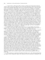
Memory, Attention, and Representation
How does memory interact with attention to affect
online processing of stimuli in neglect? Functional
imaging studies and neurophysiological studies
suggest that there is considerable overlap between
circuits dedicated to spatial attention and spatial
working memory. Monkey lesion studies indicate
an important role for spatial memories in online
processing (Gaffan & Hornak, 1997). We recently
reported that memory traces of contralesional
stimuli might have a disproportionate influence
on online representations in patients with neglect
(Chatterjee et al., 2000). A conceptual framework
that relates spatial memory and attention in influ-
encing online perception remains to be articulated.
Frontal and Parietal Differences
How different are the roles of the frontal and pari-
etal cortices in spatial attention? The notion that
parietal neglect is attentional and frontal neglect
is intentional has great appeal. Unfortunately, the
empirical evidence for such a clear dichotomy is
mixed at best. It is not even clear that these
distinctions make conceptual sense, since what
has been called “attentional neglect” involves eye
movements and what has been called “intentional
neglect” involves limb movements. Single-cell neu-
rophysiological studies suggest that neurons within
both parietal and frontal cortices mediate spatial
actions. It may be the case that the actions are more
clearly segregated in the frontal cortex than in the
parietal cortex. However, it is not clear that one
should expect clean behavioral dissociations from
lesions to the frontal and parietal cortices. Perhaps
eye and limb movements may be coded within the
same array of neurons, as suggested by Andersen
and colleagues (Andersen, 1995a) and Pouget and
Anjan Chatterjee 18
Sejnowski (1997) for the coding of visual reference
frames. If that were the case, it is not clear how
lesions would bias behavior toward different forms
of neglect. Furthermore, the ways in which frontal
and parietal areas interact based on their intercon-
nections is not well understood. In humans, damage
to the posterior superior longitudinal fasciculus and
the inferior frontal fasciculus is associated with
more severe and long-lasting neglect. Similarly in
monkeys, transection of the white matter underly-
ing the parietal cortex is also associated with greater
neglect.
Distinctions within the Parietal Cortex
What are the roles of different regions within the
posterior parietotemporal lobes? Lesion studies in
humans suggest that damage to the inferior parietal
lobule or the superior temporal gyrus produces the
most consistent and profound disorder of spatial
attention and representation. Lesion studies in
humans suggest that damage to the inferior parietal
lobule or superior temporal gyrus produces the most
consistent and profound disorder of spatial attention
and representation. By contrast, functional imaging
studies activate more dersal regions within the
intraparietal sulcus and the superior parietal sulcus
most consistently. Why this discrepancy? Per-
haps the greater dorsal involvement in functional
imaging studies is related to the design of the
studies, which emphasize shifts of visual attention.
Perhaps experimental probes emphasizing the
integration of both “what” and “where” information
would be more likely to involve the inferior parietal
cortex. Recent functional imaging data suggest that
the temporal-parietal junction may be preferentially
activated when subjects detect targets, rather than
simply attend to locations (Corbetta et al., 2000).
Monkey lesion studies may not be able to resolve
the discrepancy for two reasons. As mentioned
below, the appropriate anatomical monkey–human
homologs are not clear, and neglectlike symptoms
occur only transiently following parietal lesions in
monkeys.
Monkey and Human Homologs
What are the appropriate anatomical homologs
between humans and monkeys? Human lesion
studies focus on the inferior parietal lobule. It is not
clear that an analogous structure exists in monkeys
(Watson et al., 1994). Both human functional imag-
ing studies and monkey neurophysiology emphasize
the role of the intraparietal sulcus. However, it is not
clear that these two structures are homologous
across species.
In summary, we know a great deal about spatial
attention and representation. Across the varied dis-
ciplines there is a remarkable convergence of the
kinds of questions being asked and solutions being
proposed. However, many questions remain. Acom-
prehensive and coherent understanding of spatial
attention and representation is more likely with
the recognition of insights gleaned from different
methods.
Acknowledgments
This work was supported by National Institutes & Health
grout RO1 NS37539. I would like to thank Lisa Santer for
her critical reading of early drafts of this chapter.
References
Adair, J., Chatterjee, A., Schwartz, R., & Heilman, K.
(1998a). Ipsilateral neglect: Reversal of bias or exagger-
ated cross-over phenomenon? Cortex, 34, 147–153.
Adair, J. C., Na, D. L., Schwartz, R. L., & Heilman, K. M.
(1998b). Analysis of primary and secondary influences on
spatial neglect. Brain and Cognition, 37, 351–367.
Albert, M. L. (1973). A simple test of visual neglect.
Neurology, 23, 658–664.
Andersen, R. A. (1995a). Coordinate transformation and
motor planning in parietal cortex. In M. S. Gazzaniga
(Ed.), The cognitive neurosciences (pp. 519–532).
Cambridge, MA: MIT Press.
Andersen, R. A. (1995b). Encoding of intention and spatial
location in the posterior parietal cortex. Cerebral Cortex,
5, 457–469.
Neglect 19
Andersen, R. A., Bracewell, R. M., Barash, S., Gnadt, J.
W., & Fogassi, L. (1990). Eye position effects on visual,
memory, and saccade-related activity in areas LIP and 7a
of macaque. Journal of Neuroscience, 10, 1176–1196.
Andersen, R. A., Essick, G. K., & Siegel, R. M. (1985).
Encoding of spatial locations by posterior parietal
neurons. Science, 230, 456–458.
Anderson, B. (1993). Spared awareness for the left side
of internal visual images in patients with left-sided extra-
personal neglect. Neurology, 43, 213–216.
Anderson, B. (1996). A mathematical model of line
bisection behaviour in neglect. Brain, 119, 841–850.
Barcelo, F., Suwazono, S., & Knight, R. (2000). Prefrontal
modulation of visual processing in humans. Nature
Neuroscience, 3, 399–403.
Behrmann, M., Moscovitch, M., Black, S. E., & Mozer,
M. (1994). Object-centered neglect in patients with uni-
lateral neglect: Effects of left-right coordinates of objects.
Journal of Cognitive Neuroscience, 6, 1–16.
Bender, M. B., & Furlow, C. T. (1945). Phenomenon
of visual extinction and homonomous fields and psycho-
logical principles involved. Archives of Neurology and
Psychiatry, 53, 29–33.
Beschin, N., & Robertson, I. H. (1997). Personal versus
extrapersonal neglect: A group study of their dissociation
using a reliable clinical test. Cortex, 33, 379–384.
Binder, J., Marshall, R., Lazar, R., Benjamin, J., & Mohr,
J. (1992). Distinct syndromes of hemineglect. Archives of
Neurology, 49, 1187–1194.
Bisiach, E. (1993). Mental representation in unilateral
neglect and related disorders: The twentieth Bartlett
Memorial lecture. Quarterly Journal of Experimental
Psychology, 46A, 435–461.
Bisiach, E., Bulgarelli, C., Sterzi, R., & Vallar, G. (1983).
Line bisection and cognitive plasticity of unilateral neglect
of space. Brain and Cognition, 2, 32–38.
Bisiach, E., Geminiani, G., Berti, A., & Rusconi, M. L.
(1990). Perceptual and premotor factors of unilateral
neglect. Neurology, 40, 1278–1281.
Bisiach, E., & Luzzatti, C. (1978). Unilateral neglect of
representational space. Cortex, 14, 129–133.
Bisiach, E., Luzzatti, C., & Perani, D. (1979). Unilateral
neglect, representational schema and consciousness.
Brain, 102, 609–618.
Bisiach, E., Perani, D., Vallar, G., & Berti, A. (1986).
Unilateral neglect: Personal and extrapersonal. Neuro-
psychologia, 24, 759–767.
Bisiach, E., Ricci, R., Lualdi, M., & Colombo, M. R.
(1998a). Perceptual and response bias in unilateral
neglect: Two modified versions of the Milner landmark
task. Brain and Cognition, 37, 369–386.
Bisiach, E., Ricci, R., & Modona, M. N. (1998b). Visual
awareness and anisometry of space representation in
unilateral neglect: A panoramic investigation by means
of a line extension task. Consciousness and Cognition, 7,
327–355.
Bisiach, E., & Rusconi, M. L. (1990). Breakdown of
perceptual awareness in unilateral neglect. Cortex, 26,
643–649.
Bisiach, E., Tegnér, R., Làdavas, E., Rusconi, M. L.,
Mijovic, D., & Hjaltason, H. (1995). Dissociation of
ophthalmokinetic and melokinetic attention in unilateral
neglect. Cerebral Cortex, 5, 439–447.
Brain, W. R. (1941). Visual disorientation with special
reference to lesions of the right hemisphere. Brain, 64,
224–272.
Brefczynski, J. A., & DeYoe, E. A. (1999). A physiologi-
cal correlate of the “spotlight” of visual attention. Nature
Neuroscience, 2, 370–374.
Brotchie, P. R., Anderson, R. A., Snyder, L. H., &
Goodman, S. J. (1995). Head position signals used by
parietal neurons to encode locations of visual stimuli.
Nature, 375, 232–235.
Burcham, K. J., Corwin, J. V., Stoll, M. L., & Reep, R. L.
(1997). Disconnection of medial agranular and posterior
patietal cortex produces multimodal neglect in rats.
Behavioral Brain Research, 90, 187–197.
Butter, C. M., Evans, J., Kirsch, N., & Kewman, D.
(1989). Altitudinal neglect following traumatic brain
injury. Cortex, 25, 135–146.
Caminiti, R., Ferraina, S., & Johnson, P. (1996). The
source of visual information to the primate frontal lobe: A
novel role for the superior parietal lobule. Cerebral
Cortex, 6, 319–328.
Cappa, S., Sterzi, R., Guiseppe, V., & Bisiach, E. (1987).
Remission of hemineglect and anosagnosia during
vestibular stimulation. Neuropsychologia, 25, 775–782.
Chatterjee, A. (1994). Picturing unilateral spatial neglect:
Viewer versus object centred reference frames. Journal of
Neurology, Neurosurgery and Psychiatry, 57, 1236–1240.
Chatterjee, A. (1995). Cross over, completion and confab-
ulation in unilateral spatial neglect. Brain, 118, 455–465.
Chatterjee, A. (1998). Motor minds and mental models in
neglect. Brain and Cognition, 37, 339–349.
Anjan Chatterjee 20
Chatterjee, A., Dajani, B. M., & Gage, R. J. (1994a).
Psychophysical constraints on behavior in unilateral
spatial neglect. Neuropsychiatry, Neuropsychology and
Behavioral Neurology, 7, 267–274.
Chatterjee, A., & Mennemeier, M. (1996). Anosognosia
for hemiplegia: Patient retrospections. Cognitive Neuro-
psychiatry, 1, 221–237.
Chatterjee, A., Mennemeier, M., & Heilman, K. M.
(1992a). Search patterns and neglect: A case study.
Neuropsychologia, 30, 657–672.
Chatterjee, A., Mennemeier, M., & Heilman, K. M.
(1992b). A stimulus-response relationship in unilateral
neglect: The power function. Neuropsychologia, 30,
1101–1108.
Chatterjee, A., Mennemeier, M., & Heilman, K. M.
(1994b). The psychophysical power law and unilateral
spatial neglect. Brain and Cognition, 25, 92–107.
Chatterjee, A., Ricci, R., & Calhoun, J. (2000). Weighing
the evidence for cross over in neglect. Neuropsychologia,
38, 1390–1397.
Chatterjee, A., & Thompson, K. A. (1998). Weigh(t)ing for
awareness. Brain and Cognition, 37, 477–490.
Chatterjee, A., Thompson, K. A., & Ricci, R. (1999).
Quantitative analysis of cancellation tasks in neglect.
Cortex, 35, 253–262.
Chawla, D., Rees, G., & Friston, K. (1999). The physio-
logical basis of attentional modulation in extrastriate
visual areas. Nature Neuroscience, 2, 671–676.
Chokron, S., & Bartolomeo, P. (1998). Position of the
egocentric reference and directional movements in right
brain-damaged patients. Brain and Cognition, 46, 34–38.
Colby, C. L. (1998). Action-oriented spatial reference
frames in cortex. Neuron, 20, 15–24.
Colby, C., & Duhamel, J R. (1991). Heterogeneity of
extrastriate visual areas and multiple parietal areas in the
macaque monkey. Neuropsychologia, 29, 497–515.
Colby, C. L., Duhamel, J R., & Goldberg, M. E. (1993).
Ventral intraparietal area of the macaque: Anatomic
location and visual response properties. Journal of
Neurophysiology, 69, 902–914.
Colby, C. L., & Goldberg, G. E. (1999). Space and atten-
tion in parietal cortex. Annual Review of Neuroscience, 23,
319–349.
Corbetta, M. (1998). Frontoparietal cortical networks for
directing atention and the eye to visual locations: Identi-
cal, independent, or overlapping neural systems. Proceed-
ings of the National Academy of Sciences U.S.A., 95,
831–838.
Corbetta, M., Kincade, J. M., Ollinger, J. M., McAvoy, M.
P., & Shulman, G. M. (2000). Voluntary orienting is dis-
sociated from target detection in human posterior parietal
cortex. Nature Neuroscience, 3, 292–296.
Corbetta, M., Miezen, F. M., Shulman, G. L., & Peterson,
S. E. (1993). A PET study of visuospatial attention.
Journal of Neuroscience 11, 1202–1226.
Corwin, J. V., Kanter, S., Watson, R. T., Heilman, K. M.,
Valenstein, E., & Hashimoto, A. (1986). Apomorphine has
a therapeutic effect on neglect produced by unilateral dor-
somedial prefrontal cortex lesions in rats. Experimental
Neurology, 36, 683–698.
Corwin, J. V., & Reep, R. L. (1998). Rodent posterior pari-
etal cortex as a component of a cortical mediating directed
spatial attention. Psychobiology, 26, 87–102.
Coslett, H. B. (1997). Neglect in vision and visual
imagery: A double dissociation. Brain, 120, 1163–1171.
Coslett, H. B. (1998). Evidence for a disturbance of
the body schema in neglect. Brain and Cognition, 37,
529–544.
Coslett, H. B., Bowers, D., Fitzpatrick, E., Haws, B., &
Heilman, K. M. (1990). Directional hypokinesia and
hemispatial inattention in neglect. Brain, 113, 475–486.
Coull, J. T., & Frith, C. D. (1998). Differential activation
of right superior parietal cortex and intraparietal sulcus by
spatial and nonspatial attention. Neuroimage, 8, 176–187.
Critchley, M. (1974). Misoplegia or hatred of hemiplegia.
Mt. Sinai Journal of Medicine, 41, 82–87.
di Pellegrino, G., Basso, G., & Frassinetti, F. (1998).
Visual extinction as a spatio-temporal disorder of selective
attention. Neuroreport, 9, 835–839.
Doricchi, F., Guariglia, C., Paolucci, S., & Pizzamiglio, L.
(1993). Disturbance of the rapid eye movements (REM)
of REM sleep in patients with unilateral attentional
neglect: Clue for the understanding of the functional
meaning of REMs. Electroencephalography and Clinical
Neurophysiolog, 87, 105–116.
Driver, J., Baylis, G., & Rafal, R. (1992). Preserved figure-
ground segregation and symmetry perception in visual
neglect. Nature, 360, 73–75.
Driver, J., & Halligan, P. W. (1991). Can visual neglect
operate in object-centered coordinates? An affirmative
single-case study. Cognitive Neuropsychology, 8, 475–
496.
Neglect 21
Driver, J., & Spence, C. (1998). Cross-modal links in
spatial attention. Philosophical Transactions of the Royal
Society of London, Sen. B, 353, 1319–1331.
Duel, R. (1987). Neural dysfunction during hemineglect
after cortical damage in two monkey models. In M.
Jeannerod (Ed.), Neurophysiological and neuropsycholog-
ical aspects of spatial neglect (pp. 315–334). Amsterdam:
Elsevier.
Duhamel, J., Colby, C. L., & Goldberg, M. E. (1992). The
updating of representation of visual space in parietal
cortex by intended eye movements. Science, 255, 90–92.
Duhamel, J R., Colby, C. L., & Goldberg, M. E. (1998).
Ventral intraparietal area of the macaque: Confluent visual
and somatic response properties. Journal of Neurophysi-
ology, 79, 126–136.
Farah, M. J., Brun, J. L., Wong, A. B., Wallace, M. A., &
Carpenter, P. A. (1990). Frames of reference for allocating
attention to space: Evidence from the neglect syndrome.
Neuropsychologia, 28, 335–347.
Fechner, G. T. (1899). Elemente der Psychophysik, Vol. II.
(H. E. Leipzig: Breitkopfund Härtel.
Feinberg, T., Haber, L., & Stacy, C. (1990). Ipsilateral
extinction in the hemineglect syndrome. Archives of
Neurology, 47, 802–804.
Fink, G. R., Dolan, R. J., Halligan, P. W., Marshall, J. C.,
& Frith, C. D. (1997). Space-based and object-based
visual attention: Shared and specific neural domains.
Brain, 120, 2013–2028.
Fleet, W. S., Valenstein, E., Watson, R. T., & Heilman, K.
M. (1987). Dopamine agonist therapy for neglect in
humans. Neurology, 37, 1765–1770.
Fogassi, L., Gallese, L., Fadiga, L., Luppino, G., Matelli,
M., & Rizzolatti, G. (1996). Coding of peripersonal space
in inferior premotor cortex (area F4). Journal of Neuro-
physiology, 76, 141–157.
Gaffan, D., & Hornak, J. (1997). Visual neglect in the
monkey: Representation and disconnection. Brain, 120,
1647–1657.
Gainotti, G., Messerli, P., & Tissot, R. (1972). Qualitative
analysis of unilateral and spatial neglect in relation to
laterality of cerebral lesions. Journal of Neurology,
Neurosurgery and Psychiatry, 35, 545–550.
Gandhi, S. P., Heeger, D. J., & Boynton, G. M. (1999).
Spatial attention affects brain activity in human primary
visual cortex. Proceedings of the National Academy of
Sciences U.S.A., 96, 3314–3319.
Geminiani, G., Bottini, G., & Sterzi, R. (1998). Dopamin-
ergic stimulation in unilateral neglect. Journal of Neurol-
ogy, Neurosurgery and Psychiatry, 65, 344–347.
Gentilucci, M., Fogassi, L., Luppino, G., Matelli, M.,
Camarda, R., & Rizzolatti, G. (1988). Functional organi-
zation of inferior area 6 in the macaque monkey: I.
Somatotopy and the control of proximal movements.
Experimental Brain Research, 71, 475–490.
Gitelman, D. R., Alpert, N. M., Kosslyn, S., Daffner, K.,
Scinto, L., Thompson, W., & Mesulam, M M. (1996).
Functional imaging of human right hemispheric activation
for exploratory movements. Annals of Neurology, 39,
174–179.
Gitelman, D., Nobre, A., Parish, T., LaBar, K., Kim,
Y H., Meyer, J., & Mesulam, M M. (1999). Large-scale
distributed network for covert spatial attention: Further
anatomical delineation based on stringent behavioral and
cognitive controls. Brain, 122, 1093–1106.
Graziano, M. S. A., & Gross, C. G. (1995). The represen-
tation of extrapersonal space: A possible role for bimodal,
visual-tactile neurons. In M. S. Gazzaniga (Ed.), The cog-
nitive neurosciences (pp. 1021–1034). Cambridge, MA:
MIT Press.
Graziano, M. S. A., Yap, G. S., & Gross, C. G. (1994).
Coding of visual space by premotor neurons. Science, 266,
1054–1056.
Gross, C. G., & Graziano, M. S. A. (1995). Multiple
representations of space in the brain. Neuroscientist, 1,
43–50.
Halligan, P. W., & Marshall, J. C. (1988). How long is a
piece of string? A study of line bisection in a case of visual
neglect. Cortex, 24, 321–328.
Halligan, P. W., & Marshall, J. C. (1992). Left
visuo-spatial neglect: A meaningless entity? Cortex, 28,
525–535.
Halligan, P. W., & Marshall, J. C. (1994). Spatial neglect:
Position papers on theory and practice. Hillsdale, NJ:
Lawrence Erlbaum Associates.
Halligan, P. W., & Marshall, J. C. (1998). Visuo-spatial
neglect: The ultimate deconstruction. Brain and Cogni-
tion, 37, 419–438.
Halligan, P. W., Marshall, J. C., & Wade, D. T. (1992). Left
on the right: Allochiria in a case of left visuo-spatial
neglect. Journal of Neurology, Neurosurgery and Psychi-
atry, 55, 717–719.
Heilman, K. M. (1979). Neglect and related disorders. In
K. M. H. a. E. Valenstein (Ed.), Clinical neuropsychology
(pp. 268–307). New York: Oxford University Press.
Anjan Chatterjee 22
Heilman, K. M., Pandya, D. N., & Geschwind, N. (1970).
Trimodal inattention following parietal lobe ablations.
Transactimes of the American Neurological Association,
95, 259–261.
Heilman, K. M., Schwartz, H. D., & Watson, R. T. (1978).
Hypoarousal in patients with the neglect syndrome and
emotional indifference. Neurology, 28, 229–232.
Heilman, K. M., & Valenstein, E. (1972). Frontal lobe
neglect in man. Neurology, 22, 660–664.
Heilman, K. M., & Valenstein, E. (1979). Mechanisms
underlying hemispatial neglect. Annals of Neurology, 5,
166–170.
Heilman, K. M., & Van Den Abell, T. (1980). Right hemi-
sphere dominance for attention: The mechanisms under-
lying hemispheric assymmetries of inattention (neglect).
Neurology, 30, 327–330.
Heilman, K. M., Watson, R. T., & Valenstein, E. (1994).
Localization of lesions in neglect and related disorders.
In A. Kertesz (Ed.), Localization and neuroimaging in
neuropsychology (pp. 495–524). New York: Academic
Press.
Hier, D. B., Davis, K. R., Richardson, E. P., & Mohr, J. P.
(1977). Hypertensive putaminal hemorrhage. Annals of
Neurology, 1, 152–159.
Hillis, A. E., & Caramazza, A. (1995). A framework for
interpreting distinctive patterns of hemispatial neglect.
Neurocase, 1, 189–207.
Hurford, P., Stringer, A., & Jann, B. (1998). Neurophar-
macologic treatment of hemineglect: A case report com-
paring bromocriptine and methylphenidate. Archives of
Physical Medicine and Rehabilitation, 79, 346–349.
Husain, M., & Kennard, C. (1996). Visual neglect associ-
ated with frontal lobe infarction. Journal of Neurology,
243, 652–657.
Karnath, H O., & Ferber, S. (1999). Is space representa-
tion distorted in neglect? Neuropsychologia, 37, 7–15.
Karnath, H O., Ferber, S., & Himmelbach, M. (2001).
Spatial awareness is a function of the temporal not the
posterior parietal lobe. Nature, 411, 950–953.
Karnath, H O., Sievering, D., & Fetter, M. (1994). The
interactive contribution of neck muscle proprioception
and vestibular stimulation to subjective “straight ahead”
orientation in man. Experimental Brain Research, 101,
140–146.
Karnath, H. O., Schenkel, P., & Fischer, B. (1991). Trunk
orientation as the determining factor of the “contralateral”
deficit in the neglect syndrome and as the physical anchor
of the internal representation of body orientation in space.
Brain, 114, 1997–2014.
Kastner, S., De Weerd, P., Desimone, R., & Ungerleider,
L. G. (1998). Mechanisms of directed attention in the
human extrastriate cortex as revealed by functional MRI.
Science, 282, 108–111.
Kim, Y H., Gitelman, D. R., Nobre, A. C., Parrish, T. B.,
LaBar, K. S., & Mesulam, M M. (1999). The large-scale
neural network for spatial attention displays multifunc-
tional overlap but differential asymmetry. Neuroimage, 9,
269–277.
Kinsbourne, M. (1970). A model for the mechanisms of
unilateral neglect of space. Transactions of the American
Neurological Association, 95, 143–147.
Kinsbourne, M. (1987). Mechanisms of unilateral neglect.
In M. Jeannerod (Ed.), Neurophysiological and neuropsy-
chological aspects of spatial neglect (pp. 69–86). New
York: North-Holland.
Kinsbourne, M., & Warrington, E. K. (1962). Variety
of reading disability associated with right hemisphere
lesions. Journal of Neurology, Neurosurgery and Psychi-
atry, 25, 339–344.
Kwon, S. E., & Heilman, K. M. (1991). Ipsilateral neglect
in a patient following a unilateral frontal lesion. Neurol-
ogy, 41, 2001–2004.
Ladavas, E. (1987). Is the hemispatial damage produced
by right parietal lobe damage associated with retinal or
gravitational coordinates. Brain, 110, 167–180.
Ladavas, E., Di Pellegrino, G., Farne, A., & Zeloni, G.
(1998). Neuropsychological evidence of an integrated
visuotactile representation of peripersonal space in
humans. Journal of Cognitive Neuroscience, 10, 581–589.
Ladavas, E., Paladini, R., & Cubelli, R. (1993). Implicit
associative priming in a patient with left visual neglect.
Neuropsychologia, 31, 1307–1320.
Leibovitch, F. S., Black, S. E., Caldwell, C. B., Ebert,
P. L., Ehrlich, L. E., & Szalai, J. P. (1998). Brain-
behavior correlations in hemispatial neglect using CT and
SPECT. Neurology, 50, 901–908.
Maeshima, S., Funahashi, K., Ogura, M., Itakura, T., &
Komai, N. (1994). Unilateral spatial neglect due to right
frontal lobe haematoma. Journal of Neurology, Neuro-
surgery and Psychiatry, 57, 89–93.
Mark, V. W., & Heilman, K. M. (1997). Diagonal neglect
on cancellation. Neuropsychologia, 35, 1425–1436.
Marshall, J. C., & Halligan, P. W. (1988). Blindsight and
insight in visuospatial neglect. Nature, 336, 766–767.
Neglect 23
Marshall, J. C., & Halligan, P. W. (1989). When right goes
left: An investigation of line bisection in a case of visual
neglect. Cortex, 25, 503–515.
Marshall, J. C., & Halligan, P. W. (1990). Line bisection
in a case of visual neglect: Psychophysical studies with
implications for theory. Cognitive Neuropsychology, 7,
107–130.
Marshall, J. C., & Halligan, P. W. (1993). Visuo-spatial
neglect: A new copying test to assess perceptual parsing.
Journal of Neurology, 240, 37–40.
Marshall, J. F., & Gotthelf, T. (1979). Sensory inattention
in rats with 6-hydroxydopamine-induced degeneration of
ascending dopaminergic neurons: Apomorphine-induced
reversal of deficits. Experimental Neurology, 1986,
683–689.
Marshall, R. S., Lazar, R. M., Krakauer, J. W., & Sharma,
R. (1998). Stimulus context in hemineglect. Brain, 121,
2003–2010.
Martninez, A., Anllo-Vento, L., Sereno, M. I., Frank,
L. R., Buxton, R. B., Dubowitz, D. J., Wong, E. C.,
Hinrichs, H. J., & Hillyard, S. A. (1999). Involvement of
striate and extrastriate visual cortical areas in spatial atten-
tion. Nature Neuroscience, 2, 364–369.
Mattingley, J. B., Bradshaw, J. L., & Phillips, J. G. (1992).
Impairments of movement initiation and execution in
unilateral neglect. Brain, 115, 1849–1874.
Mattingley, J. B., Davis, G., & Driver, J. (1997). Preat-
tentive filling-in of visual surfaces in parietal extinction.
Science, 275, 671–674.
Mattingley, J., Husain, M., Rorden, C., Kennard, C.,
& Driver, J. (1998). Motor role of the human inferior
parietal lobe in unilateral neglect patients. Nature, 392,
179–182.
McGlinchey-Berroth, R., Bullis, D. P., Milberg, W. P.,
Verfaellie, M., Alexander, M., & D’Esposito, M. (1996).
Assessment of neglect reveals dissociable behavioral but
not neuroanatomic subtypes. Journal of the International
Neuropsychological Society, 2, 441–451.
McGlinchey-Berroth, R., Milberg, W. P., Verfaellie, M.,
Alexander, M., & Kilduff, P. T. (1993). Semantic process-
ing in the neglected visual field: Evidence from a lexical
decision task. Cognitive Neuropsychology, 10, 79–108.
Mennemeier, M., Chatterjee, A., & Heilman, K. M.
(1994). A comparison of the influences of body and
environment-centred references on neglect. Brain, 117,
1013–1021.
Mennemeier, M., Wertman, E., & Heilman, K. M. (1992).
Neglect of near peripersonal space: Evidence for multi-
directional attentional systems in humans. Brain, 115,
37–50.
Merchant, H., Zainos, A., Hernandez, A., Salinas, E., &
Romo, R. (1997). Functional properties of primate
putamen neurons during the categorization of tactile
stimuli. Journal of Neurophysiology, 77, 1132–1154.
Mesulam, M M. (1981). A cortical network for directed
attention and unilateral neglect. Annals of Neurology, 10,
309–325.
Mesulam, M M. (1990). Large-scale neurocognitive net-
works and distributed processing for attention, language
and memory. Annals of Neurology, 28, 597–613.
Milner, A. D., & Goodale, M. (1995). The visual brain in
action. New York: Oxford University Press.
Milner, A. D., & Harvey, M. (1995). Distortion of size
perception in visuospatial neglect. Current Biology, 5,
85–89.
Milner, A. D., Harvey, M., Roberts, R. C., & Forster, S. V.
(1993). Line bisection error in visual neglect: Misguided
action or size distortion? Neuropsychologia, 31, 39–49.
Milner, A. D. (1987). Animal models of neglect. In M.
Jeannerod (Ed.), Neurophysiological and neuropsycholog-
ical aspects of spatial neglect (pp. 259–288). Amsterdam:
Elsevier.
Monaghan, P., & Shillcock, R. (1998). The cross-over
effect in unilateral neglect. Modelling detailed data in the
line-bisection task. Brain, 121, 907–921.
Moran, J., & Desimone, R. (1985). Selective attention
gates visual processing in extrastriate cortex. Science, 229,
782–784.
Mountcastle, V. B. (1976). The world around us: Neural
command functions for selective attention. Neurosciences
Research Program Bulletin, 14, 1–47.
Mountcastle, V. B., Lynch, J. C., Georgopolous, A.,
Sakata, H., & Acuna, C. (1975). The influence of attentive
fixation upon the excitability of the light-sensitive neurons
of the posterior parietal cortex. Journal of Neuroscience,
1, 1218–1245.
Na, D. L., Adair, J. D., Williamson, D. J. G., Schwartz, R.
L., Haws, B., & Heilman, K. M. (1998). Dissociation
of sensory-attentional from motor-intentional neglect.
Journal of Neurology, Neurosurgery, and Psychiatry, 64,
331–338.
Nobre, A. C., Sebestyen, G. N., Gitelman, D. R., &
Mesulam, M M. (1997). Functional localization of the
Anjan Chatterjee 24
system for visuospatial attention using positron emission
tomography. Brain, 120, 5151–5533.
Petrides, M., & Pandya, D. N. (1984). Projections to the
frontal cortex from the posterior parietal region in the
rhesus monkey. Journal of Comparative Neurology, 288,
105–116.
Pizzamiglio, L., Vallar, G., & Doricchi, F. (1997).
Gravitational inputs modulate visuospatial neglect. Exper-
imental Brain Research, 117, 341–345.
Posner, M. I., & Dehaene, S. (1994). Attentional networks.
Trends in Neuroscience, 17, 75–79.
Posner, M., Walker, J., Friedrich, F., & Rafal, R. (1984).
Effects of parietal injury on covert orienting of attention.
Journal of Neuroscience, 4, 1863–1874.
Pouget, A., & Sejnowski, T. J. (1997). Lesion in a basis
function model of parietal cortex: Comparison with hem-
ineglect. In P. Thier & H O. Karnath (Eds.), Parietal lobe
contributions to Orientation in 3 D Space (pp. 521–538).
Heidelberg: Springer-Verlag.
Previc, F. H. (1998). The neuropsychology of 3-D space.
Psychological Bulletin, 124, 123–163.
Rapcsak, S. Z., Fleet, W. S., Verfaellie, M., & Heilman,
K. M. (1988). Altitudinal neglect. Neurology, 38, 277–
281.
Rapcsak, S., Verfaellie, M., Fleet, W., & Heilman, K.
(1989). Selective attention in hemispatial neglect.
Archives of Neurology, 46, 172–178.
Rapcsak, S. Z., Watson, R. T., & Heilman, K. M. (1987).
Hemispace-visual field interactions in visual extinction.
Journal of Neurology, Neurosurgery and Psychiatry, 50,
1117–1124.
Rees, G., Frith, C., & Lavie, N. (1997). Modulating irrel-
evant motion perception by varying attentional load in an
unrelated task. Science, 278, 1616–1619.
Ricci, R., Calhoun, J., & Chatterjee, A. (2000). Orienta-
tion bias in unilateral neglect: Representational contribu-
tions. Cortex, 36, 671–677.
Ricci, R., Vaishnavi, S., & Chatterjee, A. (1999). A deficit
of preattentive vision: Experimental observations and
theoretical implications. Neurocase, 5, 1–12.
Rizzolatti, G., & Berti, A. (1993). Neural mechanisms in
spatial neglect. In I. H. Robertson & J. C. Marshall (Eds.),
Unilateral neglect: Clinical and experimental studies (pp.
87–105). Hillsdale, NJ: Lawrence Erlbaum Associates.
Rizzolatti, G., Camarda, R., Fogassi, L., Gentilucci, M.,
Luppino, G., & Matelli, M. (1988). Functional organiza-
tion of inferior area 6 in the macaque monkey: II. Area F5
and the control of distal movements. Experimental Brain
Research, 71, 491–507.
Rizzolatti, G., Matelli, M., & Pavesi, G. (1983). Deficits
in attention and movement following the removal of
postarcuate (area 6) and prearcuate (area 8) cortex in
macaque monkeys. Brain, 106, 655–673.
Rossetti, Y., G, R., Pisella, L., Farne, A., Li, L., Boisson,
D., & Perenin, M. (1998). Prism adaptation to a rightward
optical deviation rehabilitates left spatial neglect. Nature,
395, 166–169.
Rubens, A. (1985). Caloric stimulation and unilateral
neglect. Neurology, 35, 1019–1024.
Schenkenberg, T., Bradford, D. C., & Ajax, E. T. (1980).
Line bisection and unilateral visual neglect in patients with
neurologic impairment. Neurology, 30, 509–517.
Seki, K., & Ishiai, S. (1996). Diverse patterns of perform-
ance in copying and severity of unilateral spatial neglect.
Journal of Neurology, 243, 1–8.
Snyder, L. H., Batista, A. P., & Andersen, R. A. (1997).
Coding of intention in the posterior parietal cortex. Nature,
386, 167–170.
Snyder, L. H., Grieve, K. L., Brotchie, P., & Andersen, R.
A. (1998). Separate body and world-referenced represen-
tations of visual space in parietal cortex. Nature, 394,
887–891.
Sommers, D. C., Dale, A. M., Seiffert, A. E., & Tootell,
R. B. H. (1999). Functional MRI reveals spatially specific
attentional modulation in human primary visual cortex.
Proceedings of the National Academy of Sciences U.S.A.,
96, 1663–1668.
Stevens, S. S. (1970). Neural events and the psychophys-
ical power law. Science, 170, 1043–1050.
Stevens, S. S. (1972). A neural quantum in sensory dis-
crimination. Science, 177, 749–762.
Tegner, R., & Levander, M. (1991). Through the looking
glass. A new technique to demonstrate directional hypoki-
nesia in unilateral neglect. Brain, 114, 1943–1951.
Triggs, W. J., Gold, M., Gerstle, G., Adair, J., & Heilman,
K. M. (1994). Motor neglect associated with a discrete
parietal lesion. Neurology, 44, 1164–1166.
Ungerleider, L. G., & Mishkin, M. (1982). Two cortical
visual systems. In D. J. Ingle, M. A. Goodale, & R. J. W.
Mansfield (Eds.), Analysis of visual behavior (pp.
549–586). Cambridge, MA: MIT Press.
Vaishnavi, S., Calhoun, J., & Chatterjee, A. (1999). Cross-
modal and sensorimotor integration in tactile awareness.
Neurology, 53, 1596–1598.
Neglect 25
Vaishnavi, S., Calhoun, J., & Chatterjee, A. (2001).
Binding personal and peripersonal space: Evidence from
tactile extinction. Journal of Cognitive Neuroscience. 13,
181–189.
Vallar, G., Bottini, G., Rusconi, M. L., & Sterzi, R. (1993).
Exploring somatosensory hemineglect by vestibular stim-
ulation. Brain, 116, 71–86.
Vallar, G., Daini, R., & Antonucci, G. (2000). Processing
of illusion of length in spatial hemineglect: A study of line
bisection. Neuropsychologia, 38, 1087–1097.
Vargo, J. M., Richard-Smith, M., & Corwin, J. V. (1989).
Spiroperidol reinstates asymmetries in neglect in rats
recovered from left or right dorsomedial prefrontal cortex
lesions. Behavioral Neuroscience, 103, 1017–1027.
Vecera, S., & Behrmann, M. (1997). Spatial attention does
not require preattentive grouping. Neuropsychology, 11,
30–43.
Volpe, B. T., Ledoux, J. E., & Gazzaniga, M. S. (1979).
Information processing of visual stimuli in an “extin-
guished” field. Nature, 282, 722–724.
Watson, R. T., Heilman, K. M., Cauthen, J. C., & King, F.
A. (1973). Neglect after cingulectomy. Neurology, 23,
1003–1007.
Watson, R. T., Heilman, K. M., Miller, B. D., & King, F.
A. (1974). Neglect after mesencephalic reticular formation
lesions. Neurology, 24, 294–298.
Watson, R. T., Valenstein, E., Day, A., & Heilman, K. M.
(1994). Posterior neocortical systems subserving aware-
ness and neglect: Neglect associated with superior tempo-
ral sulcus but not area 7 lesions. Archives of Neurology,
51, 1014–1021.
Watson, R. T., Valenstein, E., & Heilman, K. M. (1978).
Nonsensory neglect. Annals of Neurology, 3, 505–508.
Watson, R. T., Valenstein, E., & Heilman, K. M. (1981).
Thalamic neglect. Archives of Neurology, 38, 501–506.
Wojciulik, E., & Kanwisher, N. (1999). The generality
of parietal involvement in visual attention. Neuron, 23,
747–764.
Wurtz, R. H., & Goldberg, M. E. (1972). Activity of supe-
rior colliculus in behaving monkey: III. Cells discharging
before eye movements. Journal of Neurophysiology, 35,
575–586.
Wurtz, R. H., & Munoz, D. P. (1995). Role of monkey
superior colliculus in control of saccades and fixation. In
M. S. Gazzaniga (Ed.), The cognitive neurosciences (pp.
533–548). Cambridge, MA: MIT Press.
Anjan Chatterjee 26
Robert Rafal
Case Report
R.M. had suffered from two strokes, both due to cardiac
emboli from hypertensive heart disease. The first occurred
in June 1991 at the age of 54 and produced infarction in
the right parietal lobe and a small lesion in the right cere-
bellum. He recovered from a transient left hemiparesis
and left hemispatial neglect. The second stroke, in March
1992, involved the left parietal lobe and left him func-
tionally blind. Five months after the second stroke, he was
referred to a neurologist for headaches. At that time,
neurological examination revealed a classical Bálint’s
syndrome without any other deficits of cognitive, motor,
or sensory function.
The patient had normal visual acuity; he could recog-
nize colors, shapes, objects, and faces and could read
single words. He suffered severe spatial disorientation,
however, and got lost easily anywhere except in his own
home. Although he was independent in all activities of
daily living, he could not maintain his own household and
had to be cared for by his family. He had to be escorted
about the hospital. When shown two objects, he often saw
only one. When he did report both, he did so slowly and
seemed to see them sequentially. Depth perception was
severely impaired and he could not judge the distance of
objects from him or tell which of two objects was closer
to him. Optic ataxia was pronounced. He could not reach
accurately toward objects, and was unable to use a pencil
to place a mark within a circle. He could not make accu-
rate saccades to objects and he could not make pursuit
eye movements to follow the most slowly moving object.
Visual acuity was 20/15 in both eyes. Perimetry at the time
of the initial neurological exam revealed an altitudinal
loss of the lower visual fields. Two years later, however,
visual fields were full. Contrast sensitivity and color vision
were normal. Three-dimensional experience of shapes in
random dot stereograms was preserved and he experienced
depth from shading.
His headaches were controlled with amitriptyline, and
anticoagulation treatment with warfarin was instituted to
prevent further strokes. By June 1995, the patient was able
to live independently in a duplex next door to his brother’s
daughter, and needed only intermittent help in his daily
activities. He was able to take unescorted walks in his
neighborhood, to get about in his own house without help,
2
Bálint’s Syndrome: A Disorder of Visual Cognition
watch television, eat and dress himself, and carry on many
activities of daily living. He was slower than normal in
these activities, but was able to lead a semi-independent
life.
A magnetic resonance imaging (MRI) scan in 1994 with
three-dimensional reconstruction revealed nearly symmet-
rical lesions in each parieto-occipital region (Friedman-
Hill, Robertson, & Treisman, 1995). The lesions were
concentrated primarily in Brodmann areas 7 and 39, and
possibly included some of areas 5 and 19. In addition,
there was a small (volume <0.3 cm
3
) lesion in Brodmann
area 6 of the right hemisphere and asymmetrical cere-
bellar lesions (volume = 0.3 cm
3
left hemisphere, 6.0 cm
3
right hemisphere). The damage preserved the primary
visual cortex and all the temporal lobe. The supramarginal
gyri were intact on both sides, as were somatosensory and
motor cortices.
The syndrome represented by this patient was
first described by the Hungarian neurologist Rezsö
Bálint (Bálint, 1909; Harvey, 1995; Harvey &
Milner, 1995; Husain & Stein, 1988). While visual
acuity is preserved and patients are able to recog-
nize objects placed directly in front of them, they
are unable to interact with, or make sense of, their
visual environment. They are lost in space. Fleeting
objects that they can recognize, but that they cannot
locate or grasp, appear and disappear, and their
features are jumbled together. These patients are
helpless in a visually chaotic world.
Holmes and Horax (1919) provided a detailed
analysis of the syndrome that remains definitive.
They emphasized two major components of the
syndrome: (1) simultanagnosia—a constriction, not
of the visual field, but of visual attention, which
restricts the patient’s awareness to only one object
at a time and (2) spatial disorientation—a loss of
all spatial reference and memory that leaves the
patients lost in the world and unable to look at
objects (which Bálint called “psychic paralysis of
gaze”) or to reach for them (which Bálint called
“optic ataxia”).
This chapter reviews the clinical and neuro-
psychological aspects of this intriguing syndrome.
It reviews its anatomical basis and some of the dis-
eases that cause it. It then details the independent
component symptoms of Bálint’s syndrome. It con-
cludes with a synthesis that attempts to summarize
what Bálint’s syndrome tells us about the role of
attention and spatial representation in perception
and action.
Anatomy and Etiology of Bálint’s Syndrome
Bálint’s syndrome is produced by bilateral lesions
of the parieto-occipital junction. The lesions char-
acteristically involve the dorsorostral occipital lobe
(Brodmann area 19), and often, but not invariably
(Karnath, Ferber, Rorden, & Driver, 2000), the
angular gyrus, but may spare the supramarginal
gyrus and the superior temporal gyrus. Figure 2.1
shows a drawing of the lesions in the patient
reported by Bálint in 1909 (Husain & Stein, 1988).
The supramarginal gyrus and the posterior part of
the superior temporal gyrus are affected in the right
hemisphere, but spared on the left. The superior
parietal lobule is only minimally involved in either
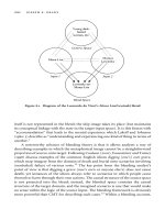
hemisphere. Figure 2.2 (Friedman-Hill, Robertson,
& Treisman, 1995) shows the reconstructed MRI
scan of the patient (R. M.) with Bálint’s syndrome
described in the case report. The lesion involves the
parieto-occipital junction and part of the angular
gyrus of both hemispheres, but spares the temporal
lobe and supramarginal gyrus. A review of other
recent cases of Bálint’s syndrome emphasizes the
consistent involvement of the posterior parietal
lobe and parieto-occipital junction as critical in
producing the syndrome (Coslett & Saffran,
1991; Pierrot-Deseillgny, Gray, & Brunet, 1986;
Verfaellie, Rapcsak, & Heilman, 1990).
Thus Bálint’s syndrome is associated with dis-
eases in which symmetric lesions of the parieto-
occipital junction are typical. For example, Luria
(1959) and Holmes and Horax (1919) have reported
this syndrome after patients received penetrating
wounds from projectiles entering laterally and
traversing the coronal plane through the parieto-
occipital regions. Strokes successively injuring both
hemispheres in the distribution of posterior parietal
branches of the middle cerebral artery are another
common cause (Coslett & Saffran, 1991; Friedman-
Hill et al., 1995; Pierrot-Deseillgny et al., 1986).
Because the parieto-occipital junction lies in the
watershed territory between the middle and the
posterior cerebral arteries, Bálint’s syndrome is a
common sequela of infarction due to global cerebral
hypoperfusion. Another symmetrical pathology is
the “butterfly” glioma—a malignant tumor origi-
Robert Rafal 28
Figure 2.1
Bálint’s drawing of the brain of the patient he described.
(Husain and Stein, 1988).
Figure 2.2
MRI of patient R.M.
nating in one parietal lobe and spreading across the
corpus callosum to the other side.
Radiation necrosis may develop after radiation of
a parietal lobe tumor in the opposite hemisphere in
the tract of the radiation port. Cerebral degenerative
disease, prototypically Alzheimer’s disease, may
begin in the parieto-occipital regions, and there is
now a growing literature reporting cases of classic
Bálint’s syndrome that are due to degenerative dis-
eases (Benson, Davis, & Snyder, 1988; Hof, Bouras,
Constintinidis, & Morrison, 1989, 1990; Mendez,
Turner, Gilmore, Remler, & Tomsak, 1990).
The Symptom Complex of Bálint’s Syndrome
Bálint’s initial description of this syndrome empha-
sized in his patient the constriction of visual atten-
tion, resulting in an inability to perceive more than
one object at a time, and optic ataxia, the inability to
reach accurately toward objects. Bálint used the term
optic ataxia to distinguish it from the tabetic ataxia of
neurosyphilis; tabetic ataxia is an inability to coordi-
nate movements based on proprioceptive input,
while optic ataxia describes an inability to coordinate
movements based on visual input. Many similar
patients have since been reported (Coslett & Saffran,
1991; Girotti et al., 1982; Godwin-Austen, 1965;
Kase, Troncoso, Court, Tapia, & Mohr, 1977;
Luria, 1959; Luria, Pravdina-Vinarskaya, & Yarbuss,
1963; Pierrot-Deseillgny et al., 1986; Tyler, 1968;
Williams, 1970).
In addition to noting the simultanagnosia and
optic ataxia reported by Bálint, Holmes and Horax
emphasized spatial disorientation as the cardinal
feature of the syndrome. Holmes and Horax of-
fered their case “for the record as an excellent
example of a type of special disturbance of vision
. . . which sheds considerable light on those
processes which are concerned in the integration
and association of sensation” (Holmes & Horax,
1919, p. 285).
Constriction of Visual Attention:
Simultanagnosia
In their 1919 report of a 30-year-old World War
I veteran who had a gunshot wound through
the parieto-occipital regions, Holmes & Horax
observed that “the essential feature was his inabil-
ity to direct attention to, and to take cognizance of,
two or more objects” (Holmes & Horax, 1919,
p. 402). They argued that this difficulty “must be
attributed to a special disturbance or limitation of
attention” (p. 402). Because of this constriction
of visual attention (what Bálint referred to as the
psychic field of gaze), the patient could attend to
only one object at a time regardless of the size of
the object. “In one test, for instance, a large square
was drawn on a sheet of paper and he recognized
it immediately, but when it was again shown to him
after a cross had been drawn in its center he saw the
cross, but identified the surrounding figure only
after considerable hesitation; his attention seemed
to be absorbed by the first object on which his eyes
fell” (Holmes & Horax, 1919, p. 390).
Another useful clinical test uses overlapping
figures (figure 2.3). The degree to which local detail
can capture the patient’s attention and exclude all
other objects from his or her attention can be quite
Balint’s Syndrome 29
Figure 2.3
Overlapping figures used to test for simultaneous agnosia.
astonishing. I was testing a patient one day, drawing
geometric shapes on a piece of paper and asking her
to tell me what she saw. She was doing well at
reporting simple shapes until at one point she shook
her head, perplexed, and told me, “I can’t see any
of those shapes now, doctor, the watermark on the
paper is so distracting.”
The visual experience of the patient with Bálint’s
syndrome is a chaotic one of isolated snapshots with
no coherence in space or time. Coslett and Saffran
report a patient whom television programs bewil-
dered “because she could only ‘see’ one person or
object at a time and, therefore, could not determine
who was speaking or being spoken to. She reported
watching a movie in which, after a heated argument,
she noted to her surprise and consternation that the
character she had been watching was suddenly sent
reeling across the room, apparently as a conse-
quence of a punch thrown by a character she had
never seen” (Coslett & Saffran, 1991, p. 1525).
Coslett and Saffran’s patient also illustrated how
patients with Bálint’s syndrome are confounded
in their efforts to read: “Although she read single
words effortlessly, she stopped reading because
the ‘competing words’ confused her” (Coslett &
Saffran, 1991, p. 1525). Luria’s patient reported that
he “discerned objects around him with difficulty,
that they flashed before his eyes and sometimes dis-
appeared from his field of vision. This [was] par-
ticularly pronounced in reading: the words and lines
flashed before his eyes and now one, now another,
extraneous word suddenly intruded itself into the
text.” The same occurred in writing: “[T]he patient
was unable to bring the letters into correlation with
his lines or to follow visually what he was writing
down: letters disappeared from the field of vision,
overlapped with one another and did not coincide
with the limits of the lines” (Luria, 1959, p. 440).
Coslett and Saffran’s patient “was unable to write
as she claimed to be able to see only a single letter;
thus when creating a letter she saw only the tip of
the pencil and the letter under construction and
“lost” the previously constructed letter” (Coslett &
Saffran, 1991, p. 1525).
Figure 2.4 shows the attempts of one of Luria’s
patients to draw familiar objects. When the patient’s
attention was focused on the attempt to draw a part
of the object, the orientation of that part with regard
to the rest of the object was lost, and the rendering
was reduced to piecemeal fragments.
Patients are unable to perform the simplest every-
day tasks involving the comparison of two objects.
They cannot tell which of two lines is longer, nor
which of two coins is bigger. Holmes and Horax’s
patient could not tell, visually, which of two pencils
was bigger, although he had no difficulty doing so
if he touched them. Holmes and Horax made the
important observation that although their patient
could not explicitly compare the lengths of two
lines or the angles of a quadrilateral shape, he had
no difficulty distinguishing shapes whose identity
is implicitly dependent upon such comparisons:
“Though he failed to distinguish any difference in
the length of lines, even if it was as great as 50
percent, he could always recognize whether a
quadrilateral rectangular figure was a square or not.
. . . [H]e did not compare the lengths of its sides but
‘on the first glance I see the whole figure and know
whether it is a square or not’ He could also
appreciate the size of angles; a rhomboid even
when its sides stood at almost right angles was ‘a
square shoved out of shape’” (Holmes & Horax,
1919, p. 394).
Holmes and Horax appreciated the importance of
their observations for the understanding of normal
vision: “It is therefore obvious that though he could
not compare or estimate linear extensions he pre-
served the faculty of appreciating the shape of bidi-
mensional figures. It was on this that his ability
to identify familiar objects depended” (Holmes &
Horax, 1919, p. 394). “[T]his is due to the rule that
the mind when possible takes cognizance of unities”
(Holmes & Horax, 1919, p. 400).
Spatial Disorientation
Holmes and Horax considered spatial disorientation
to be a symptom independent from simultanag-
nosia, and to be the cardinal feature of the syn-
Robert Rafal 30
drome: “The most prominent symptom was his
inability to orient and localize correctly objects
which he saw” (Holmes & Horax, 1919, pp.
390–391). Patients with Bálint’s syndrome cannot
indicate the location of objects, verbally or by point-
ing (optic ataxia, to be discussed later). Holmes
and Horax emphasized that the defect in visual
localization was not restricted to visual objects in
the outside world, but also extended to a defect in
spatial memory: “[H]e described as a visualist does
his house, his family, a hospital ward in which he
had previously been, etc. But, on the other hand, he
had complete loss of memory of topography; he was
unable to describe the route between the house in
a provincial town in which he had lived all his life
and the railways station a short distance away,
explaining ‘I used to be able to see the way but I
can’t see it now ’He was similarly unable to say
how he could find his room in a barracks in which
he had been stationed for some months, or describe
the geography of trenches in which he had served”
(Holmes & Horax, 1919, p. 389).
This gentleman was clearly lost in space: “On one
occasion, for instance, he was led a few yards from
his bed and then told to return to it; after searching
with his eyes for a few moments he identified the
bed, but immediately started off in a wrong direc-
tion” (Holmes & Horax, 1919, p. 395). This patient
showed, then, no recollection of spatial relation-
ships of places he knew well before his injury, and
no ability to learn new routes: “He was never able
to give even an approximately correct description of
the way he had taken, or should take, and though he
passed along it several times a day he never ‘learned
his way’ as a blind man would” (Holmes & Horax,
1919, p. 395).
Holmes and Horax concluded that “The fact that
he did not retain any memory of routes and topo-
graphical relations that were familiar to him before
he received his injury and could no longer recall
Balint’s Syndrome 31
Drawing
Elephant
head
ears
nose
eyes
trunk
feet
feet
body
“I can visualize it well but
my hands don't move properly”
walls
roof
window
door
windows
Copying
Figure 2.4
Drawing by the patient described by Luria (1959).
them, suggests that the cerebral mechanisms con-
cerned with spatial memory, as well as those that
subserve the perception of spatial relations, must
have been involved” (Holmes & Horax, 1919,
p. 404).
Impaired Oculomotor Behavior
Oculomotor behavior is also chaotic in Bálint’s
syndrome, with striking disturbances of fixation,
saccade initiation and accuracy, and smooth-pursuit
eye movements. The patient may be unable to main-
tain fixation, may generate apparently random sac-
cadic eye movements (Luria et al., 1963), and may
seem unable to execute smooth-pursuit eye move-
ments. The disorder of eye movements in Bálint’s
syndrome is restricted to visually guided eye move-
ments. The patient can program accurate eye move-
ments when they are guided by sound or touch:
“When, however, requested to look at his own finger
or to any point of his body which was touched he
did so promptly and accurately” (Holmes & Horax,
1919, p. 387).
Holmes and Horax suggested that the oculomo-
tor disturbances seen in Bálint’s syndrome were
secondary to spatial disorientation: “Some influence
might be attributed to the abnormalities of the
movements of his eyes, but these were an effect
and not the cause” (Holmes & Horax, 1919, p. 401).
“All these symptoms were secondary to and
dependent upon the loss of spatial orientation by
vision” (Holmes & Horax, 1919, p. 405). They
described, similarly, the behavior of a patient with
Bálint’s syndrome when he was tested for smooth-
pursuit eye movements: “When an object at which
he was staring was moved at a slow and uniform
rate he could keep his eyes on it, but if it was jerked
or moved abruptly it quickly disappeared” (Holmes
& Horax, 1919, p. 387).
Optic Ataxia
Figure 2.5 shows misreaching in Bálint’s syndrome.
Even after the patient sees the comb, he doesn’t look
directly at it, and his reaching is inaccurate in depth
as well as being off to the side. He groped for the
comb until his hand bumped into it. Given a pencil
and asked to mark the center of a circle, the patient
with Bálint’s syndrome typically won’t even get the
mark within the circle—and may not be able to even
hit the paper. In part this may be because the patient
cannot take cognizance, simultaneously, of both the
circle and the pencil point; but it is also clear that
the patient doesn’t know where the circle is.
Holmes and Horax considered optic ataxia, like
the oculomotor impairment, to be secondary to the
patient’s “inability to orient and localize correctly
in space objects which he saw. When asked to
take hold of or point to any object, he projected his
hand out vaguely, generally in a wrong direction,
and had obviously no accurate idea of its distance
from him” (Holmes & Horax, 1919, p. 391).
Holmes and Horax again observed that the lack
of access to a representation of space was specific
to vision. Their patient was able to localize sounds
and he did have a representation of peripersonal
space based on kinesthetic input: “The contrast
between the defective spatial guidance he received
from vision and the accurate knowledge of space
that contact gave him, was excellently illustrated
when he attempted to take soup from a small bowl
with a spoon; if he held the bowl in his own hand
he always succeeded in placing the spoon accu-
rately in it, but when it was held by a observer
Robert Rafal 32
Figure 2.5
Optic ataxia in Bálint’s syndrome.
or placed on a table in front of him he could rarely
bring his spoon to it at once, but had to grope for it
till he had located it by touch” (Holmes & Horax,
1919, pp. 391 and 393).
Impaired Depth Perception
Holmes and Horax (1919) also attributed impaired
depth perception to spatial disorientation. They
viewed the loss of depth perception in Bálint’s syn-
drome as a consequence of the loss of topographic
perception, and as a failure to have any appreciation
of distance. In their patient they attributed the loss
of blinking in response to a visual threat to the
patient’s inability to recognize the nearness of the
threatening object. Difficulty in judging distances
also causes another serious problem for patients—
they collide with objects when they walk about.
The impairment of depth perception in Bálint’s
syndrome seems to be due to a failure to appreciate
the relative location of two objects, or of the patient
and the object he or she is looking at. Size cues
seem not to help the patient judge the distance to
an object. However, Holmes and Horax commented
that their patient’s lack of a sense of distance did not
indicate a lack of appreciation of metrics in general
since he could: “indicate by his two hands the exten-
sion of ordinary standards of linear measurement,
as an inch, a foot, or a yard and he could indi-
cate the lengths of familiar objects, as his rifle,
bayonet, etc. (Holmes & Horax, 1919, p. 393).
Nosological Consideration: Bálint’s Syndrome,
Its Neighbors and Relatives
The clinical picture described here is that of Bálint’s
syndrome when it is quite dense and in its pure
form. It reflects the typical presentation of a pa-
tient with bilateral lesions restricted to the parieto-
occipital junction. While strokes and head trauma
may occasionally cause discretely restricted and
symmetrical lesions, it is more commonly the case
that lesions will not respect these territories and
will cause more extensive damage to the occipital,
parietal, and temporal lobes.
Coexisting visual field deficits, hemispatial
neglect, apperceptive or associative agnosia, pro-
sopagnosia, alexia, and other cognitive deficits are
often present in association with Bálint’s syndrome
or some of its constituent elements.
The patient reported by Bálint (1909), for
example, also had left hemispatial neglect, possibly
owing to extension of the lesion into the right tem-
poroparietal junction (figure 2.1): “[T]he attention
of the patient is always directed [by approximately
35 or 40 degrees] to the right-hand side of space
when he is asked to direct his attention to another
object after having fixed his gaze on a first one, he
tends to the right-hand rather than the left-hand
side” (cited by Husain and Stein, 1988, p. 90). In
other cases in which a constriction of visual atten-
tion is also associated with object agnosia, the ten-
dency of the patient to become locked on parts of
objects may contribute to observed agnosic errors
and may result in diagnostic confusion with inte-
grative agnosia (Riddoch & Humphreys, 1987).
It is also the case that a given patient may have
optic ataxia, spatial disorientation, or simultanag-
nosia without other elements of Bálint’s syndrome.
Thus, spatial disorientation may occur without
simultanagnosia (Stark, Coslett, & Saffran, 1996);
optic ataxia may occur without simultanagnosia
or spatial disorientation (Perenin & Vighetto, 1988);
and simultanagnosia may occur without spatial dis-
orientation (Kinsbourne & Warrington, 1962, 1963;
Rizzo & Robin, 1990). It should be borne in mind
that in such cases, the observed symptoms may
result from very different mechanisms than those
that produce them in Bálint’s syndrome. Thus, while
optic ataxia and oculomotor impairment may be
attributable to a loss of spatial representation in
patients with Bálint’s syndrome caused by bilateral
parieto-occipital lesions, optic ataxia from superior
parietal lesions may reflect disruption of the neural
substrates mediating visuomotor transformations
(Milner & Goodale, 1995).
Similarly, simultanagnosia may be caused by
very different kinds of lesions for different reasons.
The term simultanagnosia was originated specifi-
cally to describe a defect in integrating complex
Balint’s Syndrome 33
visual scenes (Wolpert, 1924). As defined by
Wolpert, the term includes, but is more general than,
the constriction of attention seen in Bálint’s syn-
drome. It is seen in conditions other than Bálint’s
syndrome and may result from unilateral lesions.
Hécaen and de Ajuriaguerra describe the difficul-
ties of one of their patients (case 1) on being offered
a light for a cigarette: “[W]hen the flame was
offered to him an inch or two away from the ciga-
rette held between his lips, he was unable to se the
flame because his eyes were fixed on the cigarette”
(Hécaen & de Ajuriaguerra, 1956, p. 374). How-
ever, the mechanism underlying simultanagnosia in
such cases may be different than that which causes
simultanagnosia in Bálint’s syndrome.
Unlike in Bálint’s syndrome, simultanagnosia
caused by unilateral left temporoparietal lesions
appears to be due to a perceptual bottleneck caused
by slowing of visual processing as measured by
rapid, serial, visual presentation (RSVP) tasks
(Kinsbourne & Warrington, 1962, 1963). In con-
trast, patients with Bálint’s syndrome may be able
to recognize a series of individual pictures flashed
briefly in an RSVP test (Coslett & Saffran, 1991).
Implications of Bálint’s Syndrome for
Understanding Visual Cognition
Bálint’s syndrome holds valuable lessons for under-
standing the neural processes involved in control-
ling attention, representing space, and providing
coherence and continuity to conscious visual ex-
perience: (1) attention makes a selection from
object-based representations of space; (2) inde-
pendent neural mechanisms that operate in parallel
orient attention within objects and between objects;
(3) the candidate objects on which attention oper-
ates are generated preattentively by early vision in
the absence of explicit awareness; and (4) attention
is involved in affording explicit (conscious) access
to the spatial representations needed for goal-
directed action and for binding features of objects.
Object- and Space-Based Attention
An appreciation of simultanagnosia in Bálint’s
syndrome has proven influential in helping to
resolve one of the major theoretical controversies
in visual attention research. The issue at stake was
whether visual attention acts by selecting locations
or objects. Work by Michael Posner and others
(Posner, 1980; Posner, Snyder, & Davidson, 1980)
showed that allocating attention to a location in the
visual field enhanced the processing of the visual
signals that appeared at the attended location.
Object-based models of attention, in contrast,
postulate that preattentive processes parse the visual
scene to generate candidate objects (more on this
later) and that attention then acts by selecting one
such object for further processing that can guide
goal-directed action. These models are supported
by experiments in normal individuals that show
better discrimination of two features belonging to
the same object than of features belonging to two
different objects (Duncan, 1984) and that these
object-based effects are independent of the spatial
location of their features (Baylis & Driver, 1995;
Vecera & Farah, 1994).
Physiological recordings have shown that an
object-based attentional set can modulate process-
ing in the extrastriate visual cortex (Chelazzi,
Duncan, Miller, & Desimone, 1998). Recent neu-
roimaging studies have confirmed that attentional
selection of one of two objects results in activation
of brain regions representing other unattended
features of that object (O’Craven, Downing, &
Kanwisher, 2000).
Object-based models predict that brain lesions
could produce an object-based simultanagnosia that
is independent of location. This is precisely the kind
of simultanagnosia that was observed in patients
with Bálint’s syndrome decades before this debate
was joined by psychologists and physiologists.
Moreover, recent experimental work by Humphreys
and colleagues has shown that simultanagnosia can
be manifest in nonspatial domains. In two patients
with parietal lobe lesions and poor spatial localiza-
tion, these authors observed that pictures extin-
Robert Rafal 34
guished words and closed shapes extinguished
open shapes (Humphreys, Romani, Olson, Riddoch,
& Duncan, 1994). Thus the object-based attention
deficit in this syndrome cannot be attributed simply
to the effects of parietal lobe lesions in disrupting
access to spatial representations.
Neural Representations of Objects in Space
The spatial representations upon which attention
operates are determined by objects, or “candidate”
objects, derived from a grouped array of features
by early vision (Vecera & Farah, 1994), and are
not simple Cartesian coordinates of empty space
centered on the observer (Humphreys, 1998).
Humphreys has recently posited that attention
operates on spatial representations determined by
objects, and that there are separate mechanisms,
operating in parallel, for shifting attention within
objects and between objects (Humphreys, 1998).
Shifting attention within an object implies shifting
attention between locations within the object.
Figure 2.6 shows stimuli that Cooper and
Humphreys (2000) used to study shifts of attention
within and between objects in patient G.K. with
Bálint’s syndrome. In conditions 1 and 2, G.K.’s
task was to report whether the upright segments
were the same or different lengths. For the stimuli
in condition 1, in which the comparison was
between two parts of the same object, G.K. was
correct on 84% of the trials, whereas in condition 2
in which the judgment required comparison of two
separate objects, performance was at chance level
(54%).
Visual Processing Outside of Conscious
Awareness
The interaction of spatial and object representations
in determining the allocation of attention requires
that candidate objects be provided by preattentive
processes that proceed in the absence of awareness.
Cumulative observations in patients with hemispa-
tial neglect (see chapter 1) have indeed provided
growing evidence that early vision does separate
figure from ground, group features, and assign
primary axes; it even extracts semantic information
that can assign attentional priorities for subsequent
processing. Here some examples are considered in
which implicit measures of processing in Bálint’s
syndrome have provided strong evidence for exten-
sive processing of visual information outside of
awareness.
Preattentive Representation of Space
Spatial disorientation is a cardinal feature of
Bálint’s syndrome, and one view of the constriction
Balint’s Syndrome 35
Figure 2.6
Figures used by Cooper and Humphreys (2000) to demonstrate grouping in Bálint’s syndrome.
of visual attention posits that it, too, is due to a loss
of a neural representation of space on which atten-
tion may act (Friedman-Hill et al., 1995). However,
as we have seen from the work of Humphreys
et al. (1994), simultanagnosia may also occur for
nonspatial information, such as shifting between
words and pictures. Moreover, recent observa-
tions in patients with both hemispatial neglect
(Danziger, Kingstone, & Rafal, 1998) and Bálint’s
syndrome (Robertson, Treisman, Friedman-Hill,
& Grabowecky, 1997) have shown that parietal
damage does not eliminate representations of spatial
information, but rather prevents explicit access to
this information.
Robertson et al. (1997) showed that although
patient R.M. could not explicitly report the relative
location of two objects, he nevertheless exhibited a
spatial Stroop interference effect. That is, although
he could not report whether the word “up” was in
the upper or lower visual field, he was, nevertheless,
slower to read “up” if it appeared in the lower visual
field than in the upper visual field.
Preattentive Grouping of Features and
Alignment of Principal Axis
As described earlier, observations by Luria (Luria,
1959) and by Humphreys & Riddoch (1993) have
revealed that there is less simultanagnosia when
shapes in the visual field are connected. Other
recent observations by Humphreys and his col-
leagues in patient G.K. have confirmed that group-
ing based on brightness, collinearity, surroundeness,
and familiarity also are generated preattentively, as
is grouping based on alignment of a principal axis.
Figure 2.7 shows G.K.’s performance in reporting
two items; it shows that performance is better
when the items are grouped on the basis of bright-
ness, collinearity, connectedness, surroundness, and
familiarity (Humphreys, 1998).
Preattentive Processing of Meaning of Words
As is the case in hemispatial neglect, neglected
objects do appear to be processed to a high level
of semantic classification in patients with Bálint’s
syndrome. Furthermore, although this information
is not consciously accessible to the patient, it does
influence the perception of objects that are seen. For
example, Coslett & Saffran (1991) simultaneously
presented pairs of words or pictures briefly to their
patient, and asked her to read or name them. When
the two stimuli were not related semantically, the
patient usually saw only one of them, but when they
were related, she was more likely to see them both.
Hence, both stimuli must have been processed to a
semantic level of representation, and the meaning
of the words or objects determined whether one or
both would be perceived.
Words are an example of hierarchical stimuli in
which letters are present at the local level and the
word at the global level. We (Baylis, Driver, Baylis,
& Rafal, 1994) showed patient R.M. letter strings
and asked him to report all the letters he could see.
Since he could only see one letter at a time, he found
this task difficult and, with the brief exposure dura-
tions used in the experiment, he usually only saw a
few of the letters. However, when the letter string
constituted a word, he was able to report more
letters than when it did not. That is, even when the
patient was naming letters and ignoring the word,
the word was processed and helped to bring the con-
stituent letters to his awareness.
Attention, Spatial Representation, and Feature
Integration: Gluing the World Together
I discussed earlier how a single object seen by a
patient is experientially mutable in time. It has no
past or future. Any object that moves disappears. In
addition, objects seen in the present can be per-
plexing to the patient, because other objects that
the patient does not see, and their features, are
processed and impinge upon the experience of the
attended object. Normally, the features of an object,
such as its color and its shape, are correctly con-
joined, because visual attention selects the location
of the object and glues together all the features
sharing that same location (Treisman & Gelade,
1980). For the patient with Bálint’s syndrome,
however, all locations are the same, and all the
Robert Rafal 36
features that impinge on the patient’s awareness are
perceptually conjoined into that object.
Friedman-Hill et al. (1995) showed R.M. pairs of
colored letters and asked him to report the letter he
saw and its color. R.M. saw an exceptional number
of illusory conjunctions (Treisman & Schmidt,
1982), reporting the color of the letter that he did
not see as being the color of the letter that he did
report. Lacking access to a spatial representation
in which colocated features could be coregistered
by his constricted visual attention, visual features
throughout the field were free floating and con-
joined arbitrarily. Since a spatial Stroop effect was
observed in patient R.M. (see earlier discussion),
Robertson and her colleagues (1997) argued that
spatial information did exist and that feature
binding relies on a relatively late stage where
implicit spatial information is made explicitly
accessible. Subsequent observations in patient R.M.
showed, however, that feature binding also occurred
implicitly. Wojciulik & Kanwisher (1998) used a
modification of a Stroop paradigm in which R.M.
was shown two words, one of which was colored,
and asked to report the color and ignore the words.
Although he was not able to report explicitly which
word was colored, there was nevertheless a larger
Stroop interference effect (i.e., he was slower to
name the color) when a word had an incongruent
Balint’s Syndrome 37
Figure 2.7
Figures used by Humphreys (1998) to demonstrate grouping in Bálint’s syndrome.
color. Thus, there was implicit evidence that the
word and its color had been bound, even though
R.M. had no explicit access to the conjunction of
features.
Conclusions and Future Directions
Lost in space, and stuck in a perceptual present con-
taining only one object that he or she cannot find or
grasp, the patient with Bálint’s syndrome is helpless
in a visually chaotic world. Objects appear and dis-
appear and their features become jumbled together.
Contemporary theories of attention and percep-
tion help us to understand the experience of these
patients. At the same time, their experience provides
critical insights into the neural basis of visual atten-
tion and perception, and how they operate together
normally to provide coherent perceptual experience
and efficient goal-directed behavior.
Some of the critical issues remain unresolved and
await future research. It remains unclear whether
simultanagnosia and spatial disorientation are inde-
pendent symptoms in Bálint’s syndrome, or whether
the apparent constriction of visual attention is a
secondary consequence of the lack of access to an
explicit representation of space. The identification
of individual cases in which these two symptoms
are dissociated would resolve this question. It is also
important to learn more about the impairment in
feature binding that causes the generation of illu-
sory conjunctions, and whether this deficit is an
integral component of the syndrome or, rather, is
present only in some patients with a lesion in a spe-
cific part of the parietal-occipital association cortex.
Furthermore, if the component symptoms of the
syndrome are found to be dissociable, we will need
to know more about the neural substrates of each.
References
Bálint, R. (1909). Seelenlahhmung edes “schauens,”
optische ataxie, raumliche storung der aufmerksamkeit.
Montschrife Pscyhiatrie und Neurologie, 25, 51–81.
Baylis, G. C., & Driver, J. (1995). One-sided edge-
assignment in vision: 1. Figure-ground segmentation and
attention to objects. Current Directions in Psychological
Science, 4, 201–206.
Baylis, G. C., Driver, J., Baylis, L. L., & Rafal, R. D.
(1994). Perception of letters and words in Bálint’s
syndrome: Evidence for the unity of words. Neuropsy-
chologia, 32, 1273–1286.
Benson, D. F., Davis, R. J., & Snyder, B. D. (1988).
Posterior cortical atrophy. Archives of Neurology, 45,
789–793.
Chelazzi, L., Duncan, J., Miller, E. K., & Desimone, R.
(1998). Responses of neurons in inferior temporal cortex
during memory-guided visual search. Journal of Neuro-
physiology, 80, 2918–2940.
Cooper, C. G., & Humphreys, G. W. (2000). Coding space
within but not between objects: Evidence from Bálint’s
syndrome. Neuropsychologia 38, 723–733.
Coslett, H. B., & Saffran, E. (1991). Simultanagnosia. To
see but not two see. Brain, 113, 1523–1545.
Danziger, S., Kingstone, A., & Rafal, R. (1998). Reflex-
ive orienting to signals in the neglected visual field.
Psychological Science, 9, 119–123.
Duncan, J. (1984). Selective attention and the organization
of visual information. Journal of Experimental Psychol-
ogy: General, 113, 501–517.
Friedman-Hill, S. R., Robertson, L. C., & Treisman, A.
(1995). Parietal contributions to visual feature binding:
Evidence from a patient with bilateral lesions. Science,
269, 853–855.
Girotti, F., Milanese, C., Casazza, M., Allegranza, A.,
Corridori, F., & Avanzini, G. (1982). Oculomotor distur-
bances in Bálint’s syndrome: Anatomoclinical findings
and electrooculographic analysis in a case. Cortex, 16,
603–614.
Godwin-Austen, R. B. (1965). A case of visual disorien-
tation. Journal of Neurology, Neurosurgery and Psychia-
try, 28, 453–458.
Harvey, M. (1995). Psychic paralysis of gaze, optic ataxia,
spatial disorder of attention. Translated from Bálint
(1909). Cognitive Neuropsychology, 12, 266–282.
Harvey, M., & Milner, A. D. (1995). Bálint’s patient.
Cognitive Neuropsychology, 12, 261–264.
Hécaen, H., & de Ajuriaguerra, J. (1956). Agnosie visuelle
pour les objects inanimes par lesion unilaterle gauche.
Review Neurologique, 94, 222–233.
Robert Rafal 38
Hof, P. R., Bouras, C., Constintinidis, J., & Morrison, J.
H. (1989). Bálint’s syndrome in Alzheimer’s disease:
Specific disruption of the occipito-parietal visual pathway.
Brain Research, 493, 368–375.
Hof, P. R., Bouras, C., Constantinidis, J., & Morrison,
J. H. (1990). Selective disconnection of specific visual
association pathways in cases of Alzheimer’s disease
presenting with Bálint’s syndrome. Journal of Neu-
ropathology and Experimental Neurology, 49, 168–184.
Holmes, G., & Horax, G. (1919). Disturbances of spatial
orientation and visual attention, with loss of stereoscopic
vision. Archives of Neurology and Psychiatry, 1, 385–
407.
Humphreys, G. W. (1998). Neural representation of
objects in space: A dual coding account. Philosophical
Transactions of the Royal Society of London, Ser. B, 353,
1341–1351.
Humphreys, G. W., & Riddoch, M. J. (1993). Interactive
attentional systems in unilateral visual neglect. In
I. H. Robertson & J. C. Marshall (Eds.), Unilateral
neglect: Clinical and experimental studies (pp. 139–168).
Hillsdale, NJ: Lawrence Erlbaum Associates.
Humphreys, G. W., Romani, C., Olson, A., Riddoch,
M. J., & Duncan, J. (1994). Non-spatial extinction fol-
lowing lesions of the parietal lobe in humans. Nature,
372(6504), 357–359.
Husain, M., & Stein, J. (1988). Rezso Bálint and his most
celebrated case. Archives of Neurology 45, 89–93.
Karnath, H O., Ferber, S., Rorden, C., & Driver, J. (2000).
The fate of global information in dorsal simultanagnosia.
NeuroCase 6, 295–306.
Kase, C. S., Troncoso, J. F., Court, J. E., Tapia, F. J., &
Mohr, J. P. (1977). Global spatial disorientation. Journal
of the Neurological Sciences, 34, 267–278.
Kinsbourne, M., & Warrington, E. K. (1962). A disorder
of simultaneous form perception. Brain, 85, 461–486.
Kinsbourne, M., & Warrington, E. K. (1963). The localiz-
ing significance of limited simultaneous visual form
perception. Brain, 86, 461–486.
Luria, A. R. (1959). Disorders of “simultaneous percep-
tion” in a case of bilateral occipito-parietal brain injury.
Brain, 83, 437–449.
Luria, A. R., Pravdina-Vinarskaya, E. N., & Yarbuss, A. L.
(1963). Disorders of ocular movement in a case of
simultanagnosia. Brain, 86, 219–228.
Mendez, M. F., Turner, J., Gilmore, G. C., Remler, B., &
Tomsak, R. L. (1990). Bálint’s syndrome in Alzheimer’s
disease: Visuospatial functions. International Journal of
Neuroscience, 54, 339–346.
Milner, A. D., & Goodale, M. A. (1995). The visual brain
in action. Oxford: Oxford University Press.
O’Craven, K., Downing, P., & Kanwisher, N. (2000).
fMRI evidence for objects as the units of attentional
selection. Nature 401, 584–587.
Perenin, M T., & Vighetto, A. (1988). Optic ataxia: A
specific disruption in visuomotor mechanisms. I. Different
aspects of the deficit in reaching for objects. Brain, 111,
643–674.
Pierrot-Deseillgny, C., Gray, F., & Brunet, P. (1986).
Infarcts of both inferior parietal lobules with impairment
of visually guided eye movements, peripheral visual
inattention and optic ataxia. Brain, 109, 81–97.
Posner, M. I. (1980). Orienting of attention. Quarterly
Journal of Experimental Psychology, 32, 3–25.
Posner, M. I., Snyder, C. R. R., & Davidson, B. (1980).
Attention and the detection of signals. Journal of Experi-
mental Psychology: General, 109, 160–174.
Riddoch, M. J., & Humphreys, G. W. (1987). A case of
integrative visual agnosia. Brain, 110, 1431–1462.
Rizzo, M., & Robin, D. A. (1990). Simultanagnosia: A
defect of sustained attention yields insights on visual
information processing. Neurology, 40, 447–455.
Robertson, L. C., Treisman, A., Friedman-Hill, S. R., &
Grabowecky, M. (1997). The interaction of spatial and
object pathways: Evidence from Bálint’s syndrome.
Journal of Cognitive Neuroscience, 9, 295–317.
Stark, M., Coslett, H. B., & Saffran, E. (1996). Impairment
of an egocentric map of locations: Implications for
perception and action. Cognitive Neuropsychology, 13,
481–523.
Treisman, A., & Gelade, G. (1980). A feature integration
theory of attention. Cognitive Psychology, 12, 97–136.
Treisman, A., & Schmidt, N. (1982). Illusory conjunctions
in the perception of objects. Cognitive Psychology, 14,
107–141.
Tyler, H. R. (1968). Abnormalities of perception with
defective eye movements (Bálint’s syndrome). Cortex, 3,
154–171.
Balint’s Syndrome 39
Vecera, S. P., & Farah, M. J. (1994). Does visual attention
select objects or locations? Journal of Experimental
Psychology: General, 123, 146–160.
Verfaellie, M., Rapcsak, S. Z., & Heilman, K. M. (1990).
Impaired shifting of attention in Bálint’s syndrome. Brain
and Cognition, 12, 195–204.
Williams, M. (1970). Brain damage and the mind.
Baltimore: Penguin Books.
Wojciulik, W., & Kanwisher, N. (1998). Implicit but
not explicit feature binding in a Bálint’s patient. Visual
Cognition, 5, 157–182.
Wolpert, I. (1924). Die simultanagnosie: Storung der
geamtauffassung. Zeitschrift für die gesante Neurologie
und Psychiatrie, 93, 397–415.
Robert Rafal 40
Michael S. Mega
Memory is traditionally divided into implicit and
explicit processes (figure 3.1). Implicit functions
are not under conscious control, while explicit func-
tions are available to our subjective awareness.
Implicit processes include classic conditioning or
associative learning (such as the conditioned eye-
blink response), procedural memory or skill learn-
ing (such as riding a bike or learning the rotary
pursuit task), and the effects of priming that facili-
tate the acquisition of information in the modality
specific to the presentation of the information.
Explicit memory includes both our recall of every-
thing that has happened to us—called “episodic
memory,” and all the information about the mean-
ings of things—called “semantic memory.” The
emphasis here is on patients’ complaints of explicit
memory impairments.
Explicit memory is heuristically divided into the
subprocesses of acquisition, storage, and retrieval
of information. Disorders of learning are assumed
to arise from deficits in either acquisition or storage;
thus poor spontaneous recall could arise from two
distinct problems—either a failure in learning new
information or a deficit in retrieval.
Classically, the amnesic syndrome has been
defined as the presence of a significant isolated
memory disorder in the absence of disturbed atten-
tion, language, visuospatial, or executive function
(executive function is the ability to manipulate pre-
viously acquired knowledge). Although the exact
dysfunctional subprocess producing amnesia is con-
troversial (Bauer, Tobias, & Valenstein, 1993), it is
clinically useful to describe amnesia as a failure to
learn new information, which is distinct from a
retrieval deficit identified by normal recognition.
Recognition, in turn, may depend on two sub-
processes: a feeling of familiarity and an explicit
recollection of some context associated with the
recognized item (Gardiner & Parkin, 1990; Horton,
Pavlick, & Moulin-Julian, 1993; Mandler, 1980).
The recognition tasks that patients with hippo-
campal and perirhinal lesions preferentially fail on
3
Amnesia: A Disorder of Episodic Memory
are those that make greater demands on the later
recollection-based subprocess (Aggleton & Shaw,
1996; Squire & Shimamura, 1986). Successful
recognition performance, after spontaneous recall
has failed, also draws on intact prefrontal resources.
A profound encoding or recognition impairment
appears to require both medial temporal and pre-
frontal disconnection or destruction (Aggleton &
Mishkin, 1983; Gaffan & Parker, 2000).
Case Report
On the first evaluation a 75-year-old right-handed female
was accompanied by her husband to our clinic complain-
ing of a 3–5-year history of declines in memory. The
history was mainly provided by the patient’s husband, who
claimed that 5 years prior to presentation he first noticed
abnormalities in his wife’s ability to operate a new video-
cassette recorder and new 35-mm camera. There was also
a decline in her ability to cook large meals for dinner
parties that began 3 years prior to presentation. Approxi-
mately 2 years prior to presentation, he began noticing
difficulty with his wife’s memory so that she was unable
to shop for food without a detailed list. She would fre-
quently forget conversations that transpired between them
or episodes that might have occurred days prior. Approx-
imately 6 months prior to presentation, the patient’s
husband noted that she forgot what cards were played in
their bridge club when previously she had been an excel-
lent player. The patient agreed with the history of memory
problems, but felt that her cooking was unaffected. Both
the patient and her husband denied any problems with
language function, visuospatial function, or any change
in personality or mood. The patient continued to be quite
active socially, as well as taking part in community activ-
ities. Despite her memory problem, she was still capable
of functioning almost independently with copious list
keeping.
On initial examination the patient had a normal general
medical exam. The patient was well nourished, coopera-
tive, well groomed, and in no apparent distress. The
patient’s attention was intact with six digits forward, five
digits in reverse. The Mini Mental State Exam (MMSE)
(Folstein, Folstein, & McHugh, 1975) score was 28/30; the
patient missed two of the recall questions. Language was
normal for fluency, comprehension, and repetition; and
naming was intact, with fifteen out of fifteen items on a
modified Boston Naming Test correctly identified. Verbal
fluency showed nineteen animals produced in 1 minute,
and reading and writing ability were entirely normal.
Memory testing using a ten-word list (Fillenbaum et al.,
1997) showed a learning curve of four, six, and eight after
three trials; and after a 10-minute interval with interfer-
ence, the patient spontaneously recalled one out of ten
items. Recognition performance, using ten target items
and ten foils, produced five additional items, with three
false positives.
Visuospatial function showed no problems in copying
two-dimensional figures and some mild strategy difficulty
but good copying of a three-dimensional cube. Executive
function showed no problems with calculation ability
for two-digit addition and multiplication; however, word
problems showed some hesitancy and difficulty in
response, although the answers were eventually correct.
Frontal system evaluation showed no perseveration or loss
of set; reciprocal programs, go-no-go, and alternating
programs were all intact. The rest of the neurological
examination, including cranial nerves, motor and sensory
function, coordination, reflexes, and gait was normal.
On the initial evaluation the patient underwent formal
neuropsychological testing, functional imaging with
[
18
F]fluorodeoxyglucose positron emission tomography
(FDG-PET) and structural imaging with 3-D coronal vol-
umetric and double echo magnetic resonance imaging
(MRI).
Neuropsychological evaluation revealed a normal
Wechsler Adult Intelligence Scale-Revised (WAIS-R)
(Wechsler, 1955), a full-scale IQ with normal scores on
all subscales except for arithmetic, which was 1 standard
deviation (S.D.) below age and education norms. Lan-
guage evaluation was intact on the Boston Naming Test
(Kaplan, Goodglass, & Weintraub, 1984), with a score
of 59/60. Controlled oral word fluency (FAS) (Benton &
Hamsher, 1976 revised 1978) and animal naming were
intact, but in the low average range.
Memory testing showed impairments on the California
Verbal Learning Test (CVLT), which demonstrated encod-
ing and storage abnormalities greater than 1.5 S.D. below
age- and education-matched norms. Weschler memory
(Wechsler Memory Scale, WMS) (Wechsler, 1945), logical
memory, and paragraph recall showed significant abnor-
malities in the first and second paragraph delayed-recall
scores. Nonverbal memory was also impaired as noted by
the 30-minute delay on the Rey-Osterrieth Complex
Figure Recall (Osterrieth, 1944; Rey, 1941) as well as the
Benton Visual Retention Test (Benton, 1974). Executive
function showed spotty performance, with normal trails A
and B but Stroop B declines at approximately 1 S.D. below
age- and education-matched norms. The Ruff figural flu-
ency task showed low normal performance (Jones-Gotman
& Milner, 1977). Interpretation of neuropsychological
testing concluded that the patient did not meet the criteria
for dementia according to DSM-IV (APA, 1994); however,
the patient did meet the criteria for amnesic disorder.
FDG-PET showed essentially normal metabolism in
the frontal, parietal, and occipital cortices as well as sub-
cortical structures, but indicated decreased metabolism
in medial temporal lobe regions, the loss in the left being
greater than in the right (figure 3.2). MRI analysis showed
no significant cerebrovascular disease either cortically or
Michael S. Mega 42
Memory
Implicit Explicit
Procedural Priming
Associative
Episodic
Semantic
Figure 3.1
Conceptual organization and terminology of general memory function.