Colonoscopy Principles and Practice - part 3 doc
Bạn đang xem bản rút gọn của tài liệu. Xem và tải ngay bản đầy đủ của tài liệu tại đây (769.22 KB, 67 trang )
124 Section 3: Indications, Contraindications, Screening, and Complications
in 10%; 22.5% of the procedures showed a normal colon
[12]. One study reported an overall yield of 74% in 43
patients with IBD, including one nonneoplastic stenosis,
but no cancer and no polyps (S. Morini, personal com-
munication). Within the context of the EPAGE study, we
assessed, in 6004 patients undergoing colonoscopy, the
diagnostic yield of findings other than IBD in patients
with known ulcerative colitis and known Crohn’s dis-
ease (EPAGE study 2002, unpublished results). In 201
patients with known ulcerative colitis, we found cancer
in 1%, adenomas in 3.5%, nonadenomatous polyps in
2.0%, and diverticulosis in 0.5%. In the 158 patients with
known Crohn’s disease, cancer was found in 0.6%, ade-
nomas in 1.3%, nonadenomatous polyps in 1.3%, and
diverticulosis in 0.6%.
Diagnostic yield of routine ileoscopy
Intubation of the ileum is not routinely performed dur-
ing colonoscopy. Ileal intubation is one way to indicate
completeness of the procedure. A skilled endoscopist
can inspect the terminal ileum in about 90% of cases
in which such examination is needed [1]. In practice,
ileoscopy is not routinely performed. A recent European
multicenter trial in 6004 patients undergoing colono-
scopy found that ileoscopy was performed in 29.6% of
colonoscopies reaching the cecum [138].
40
30
35
25
10
15
20
5
0
Mean + range (%)
–
–
–
–
–
–
Cancer
Adenoma
IBD
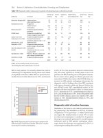
Fig. 11.14 Diagnostic yield of colonoscopy in patients with
abdominal pain or altered bowel habit. Data from 12 studies
(six prospective studies), 3252 patients.
IBD in most patients. We recently studied the yield of
colonoscopy in 1144 patients, among whom was a subset
of 40 patients with known IBD. IBD was present in 65%,
another form of colitis (infectious) in 2.5%, and stenosis
Table 11.8 Diagnostic yield of colonoscopy in patients with abdominal pain or altered bowel habit.
Number of Cancer Adenoma Inflammatory Diverticula Stricture
Reference Comment patients (%) (%) bowel disease (%) (%) (%)
Berkowitz & Kaplan [45] Abdominal pain 55 1.8 11 9.1 16.4
Abnormal bowel habit 79 1.3 10 11.4 21.5 1.3
De Bosset et al. [12] Prospective
Abdominal pain 254 0.8 7 0.8 NS 2
Constipation 73 1.4 6.8 0 NS
Brenna et al. [11] Prospective; gastrointestinal 117 0.8 6 2 NS 3
symptoms
EPAGE study Prospective; unpublished 359 3.5 13 2.9 22.6 NS
results; change in bowel habit
Lasson et al. [131] Prospective; abdominal pain 281 0.7 6.8 8.9 8.5 NS
Liebermann et al. [132] Nonspecific abdominal 1899 7.3 NS NS NS
symptoms; polyp or mass > 1cm
Mulcahy et al. [133] Abdominal pain 389 0.5 2.6 1 NS NS
Neugut et al. [53] Prospective; abdominal pain 311 5 19 NS NS NS
and/or change in bowel habit
Pepin & Ladabaum [134] Constipation 358 2 38 NS NS NS
Rex [9] All patients with one or more 75 0 31 NS NS NS
negative FOBT
Sardinha et al. [135] Patients > 80 years; abdominal 107 2 NS NS NS NS
pain; change in bowel habit
Schmitt et al. [136] Prospective; abdominal pain 794 0.6 7.7 2.6 NS NS
Total 3252* 1.6* 12.6* 3.2
FOBT, fecal occult blood test; NS, not stated.
* Excluding data from Liebermann et al. [132].
Chapter 11: Diagnostic Yield of Colonoscopy by Indication 125
adenomas greater than 1 cm in diameter. Furthermore,
barium enema was falsely positive in 14%. In a large
nonrandomized controlled trial [88] in 21 000 patients
aged over 40 years, barium enema missed 25% of the
lesions found at colonoscopy.
Insufficient procedural competence and experience
on the part of the endoscopist may decrease the value
of colonoscopy [145]. Even in expert hands, there is a
significant miss rate of polyps. Rex and colleagues [146]
performed two colonoscopies on the same day (back-to-
back colonoscopy) in 183 patients randomly assigned
to the same or another endoscopist. The overall miss
rate of adenomas was 24%; it was 27% for adenomas
≤ 5 mm, 13% for adenomas 6–9 mm, and 6% for adeno-
mas ≥ 1 cm. Although considered as the gold standard
in the diagnostic armamentarium of colonic disease,
the performance characteristics of colonoscopy are not
optimal, even in the hands of an expert operator and
under ideal conditions.
The quality and diagnostic reliability of the procedure
are further dependent on several other factors. Much
emphasis has been placed on the duration of colonoscopy
and in particular on the time needed to reach the cecum.
Overall duration may be significant with respect to pro-
cedural efficiency in a context of cost constraints, waiting
lists at endoscopy units, and the need for endoscopists
and endoscopes. However, it is not acceptable that an
overly rapid endoscopic technique should render the
procedure less tolerable or reduce its diagnostic reliab-
ility. Withdrawal time seems to be more critical for diag-
nostic yield, particularly colonic distension, adequate
suctioning and cleaning, and adequate time spent exam-
ining the colon. The quality of withdrawal is critical for
the detection rate of adenomas [147]. In fact, it has
been shown very recently that individual endoscopists’
In one study in 138 consecutive colonoscopies, ileo-
scopy revealed a diagnosis in eight patients (6%). In
half of these patients, the diagnosis was made based
on ileoscopy alone. The yield of ileoscopy was 2.7% in
asymptomatic patients undergoing screening colono-
scopy and 29% in patients complaining of diarrhea [139].
Another prospective study in 295 consecutive patients
[140] reported macroscopic abnormalities of the ileum in
4 of 213 patients (1.8%) in whom ileoscopy was possible,
one-quarter of whom also had abnormal histology of the
ileal mucosa (0.5%). However, this study did not indic-
ate patient symptoms. Ileoscopy is obviously particu-
larly useful in patients with symptoms suggesting IBD
in order to exclude isolated ileal disease or to facilitate
the differential diagnosis between Crohn’s disease and
ulcerative colitis [141]. Furthermore, ileoscopy seems to
be indicated in patients with chronic diarrhea, especially
in HIV-positive patients [142].
Diagnostic reliability of colonoscopy
Colonoscopy is more sensitive than barium enema and
allows biopsies and endoscopic therapy. The sensitivity
of barium enema and colonoscopy for diagnosing colo-
rectal cancer was 84% and 95% in a recent retrospective
study [143]. A controlled and blinded comparison of
both procedures was made in the National Polyp Study
where 580 patients underwent 862 paired examinations
[144]. Barium enema detected a polyp in only 39% of
cases in which a polyp was subsequently found during
colonoscopy, and in only 48% in patients with advanced
(a) (b)
40
30
35
25
10
15
20
5
0
Mean + range (%)
Cancer
Adenoma
IBD
40
30
35
25
10
15
20
5
0
Mean + range (%)
–
–
–
–
–
–
Fig. 11.15 Diagnostic yield of colonoscopy in patients with
abdominal pain or altered bowel habit: (a) EPAGE; (b) all
studies.
126 Section 3: Indications, Contraindications, Screening, and Complications
up after cancer resection, and positive FOBT have a high
diagnostic yield (Fig. 11.16). In contrast, nonbleeding
colonic symptoms (diarrhea, abdominal pain, altered
bowel habit) and surveillance after polypectomy have a
lower yield of cancer (Fig. 11.16). Incidence rates of colo-
rectal cancer increase consistently with age. Patient age
is thus an important predictor of colorectal cancer in
patients referred for colonoscopy.
The yield in the detection of adenomas is less de-
pendent on the indications than the detection of cancer,
due to the high prevalence of polyps found in screening
colonoscopies or in patients with nonspecific symptoms.
The adenoma detection rate is highest in the follow-up
of polyps, follow-up of cancer, in patients with positive
FOBT (Fig. 11.16), and in nonemergency lower gastroin-
testinal bleeding (see Fig. 11.2). IBD is a relatively com-
mon finding in hematochezia and diarrhea.
Although diagnostic yield is important, it must be
kept in mind that colonoscopy may also be beneficial to
patients if it excludes a clinically relevant lesion by con-
ferring reassurance.
References
1 Marshall JH, Barthel JS. The frequency of total colonoscopy
and terminal ileal intubation in the 1990s. Gastrointest
Endosc 1993; 39: 518–20.
2 Rex DK, Bond JH, Winawer S et al. Quality in the technical
performance of colonoscopy and the continuous quality
improvement process for colonoscopy: recommendations
of the U.S. Multi-Society Task Force on Colorectal Cancer.
Am J Gastroenterol 2002; 97: 1296–308.
3 Bat L, Williams CB. Usefulness of pediatric colonoscopes
in adult colonoscopy. Gastrointest Endosc 1989; 35: 329–32.
4 Rex DK, Weddle RA, Lehman GA, Pound DC, O’Connor
KW, Hawes RH. Flexible sigmoidoscopy plus air-contrast
procedure times correlate with the rate at which they
identify multiple or clinically significant polyps [148].
We recently assessed technical aspects of performance
of colonoscopy in 6004 European patients referred for
colonoscopy [138]. The mean overall duration of colono-
scopy was 22.8 min, including a mean withdrawal time
of 10.1 min. In the same study, we found that colon cleans-
ing quality was highly associated with the total duration
of the procedure (P < 0.001), the difficulty of colonoscopy
(P < 0.001), and the overall yield of relevant endoscopic
diagnoses (P = 0.002), particularly of adenomas (P <
0.001) (EPAGE study 2002) [149].
Summary
For the clinician, the yield of relevant diagnoses is one of
the most important outcomes of a diagnostic procedure
such as colonoscopy. While appropriateness of indica-
tions refers to the quality of the indication, the diagnostic
yield refers to endoscopic lesions that are potentially rel-
evant to the patient’s care, in conjunction with clinical
symptoms and signs. Unfortunately, the relationship
between endoscopic findings and clinical presentation is
imperfect, particularly in light of the fact that endoscopic
lesions (e.g. polyps) may be asymptomatic [150]. Patient
age and gender have a major impact on the diagnostic
yield of colonoscopy, increasing age being associated
with a higher rate of lesions.
A main focus of the use of colonoscopy is the diagnosis
and removal of adenomas and the diagnosis of colorectal
cancer. In cancer detection, hematochezia, IDA, follow-
70
60
50
40
30
20
10
0
Mean + range (%)
–
–
–
–
–
–
–
–
–
–
–
–
–
–
–
–
–
–
–
–
–
–
–
–
–
–
– –
Cancer
Polyp
Acute lower
GI bleeding
Non-acute
lower GI
bleeding
Iron
deficiency
anemia
F. up
polyp
F. up
cancer
FOBT pos Diarrhea Non-specific
colonic
symptoms
Fig. 11.16 Diagnostic yield of colonoscopy by indication.
Chapter 11: Diagnostic Yield of Colonoscopy by Indication 127
rectal cancer and age. Implications for screening in older
Americans. Cancer 1995; 75: 775–81.
25 Capocaccia R, De Angelis R, Frova L et al. Estimation and
projections of colorectal cancer trends in Italy. Int J
Epidemiol 1997; 26: 924–32.
26 Chak A, Post AB, Cooper GS. Clinical variables associated
with colorectal cancer on colonoscopy: a prediction model.
Am J Gastroenterol 1996; 91: 2483–8.
27 American Society for Gastrointestinal Endoscopy. The
rôle of endoscopy in patients with lower GI bleeding.
Gastrointest Endosc 1998; 48: 685–8.
28 Gonvers JJ, De Bosset V, Froehlich F, Dubois RW, Burnand
B, Vader JP. Appropriateness of colonoscopy: hema-
tochezia. Endoscopy 1999; 31: 631–6.
29 Jensen DM, Machicado GA. Diagnosis and treatment of
severe hematochezia. The role of urgent colonoscopy after
purge. Gastroenterology 1998; 95: 1569–74.
30 Richter JM, Christensen MR, Kaplan LM, Nishioka
NS. Effectiveness of current technology in the diagnosis
and management of lower gastrointestinal hemorrhage.
Gastrointest Endosc 1995; 41: 93–8.
31 Vernava AM, Moore BA, Longo WE, Johnson FE. Lower
gastrointestinal bleeding. Dis Colon Rectum 1997; 40:
846–58.
32 Makela JT, Kiviniemi H, Laitinen S, Kairaluoma MI.
Diagnosis and treatment of acute lower gastrointestinal
bleeding. Scand J Gastroenterol 1993; 28: 1062–6.
33 Rhee JC, Lee KT. The causes and management of lower GI
bleeding: a study based on clinical observations at Hanyang
University Hospital. Gastroenterol Jpn 1991; 26: 101–6.
34 Caos A, Benner KG, Manier J et al. Colonoscopy after
Golytely preparation in acute rectal bleeding. J Clin
Gastroenterol 1986; 8: 46–9.
35 Church JM. Analysis of the colonoscopic findings in
patients with rectal bleeding according to the pattern of
their presenting symptoms. Dis Colon Rectum 1991; 34:
391–5.
36 Forde KA. Colonoscopy in acute rectal bleeding. Gas-
trointest Endosc 1981; 27: 219–20.
37 Guillem JD, Forde KA, Treat MR, Neugut AI, Bodian CA.
The impact of colonoscopy on the early detection of
colonic neoplasms in patients with rectal bleeding. Ann
Surg 1987; 206: 606–11.
38 Jensen DM, Machicado GA. Colonoscopy for diagnosis
and treatment of severe lower gastrointestinal bleeding.
Gastroenterol Clin North Am 1997; 7: 477–98.
39 Rossini FP, Ferrari A, Spandre M et al. Emergency colono-
scopy. World J Surg 1989; 13: 190–2.
40 Dent OF, Goulston KJ, Zubrzycki J, Chapuis PH. Bowel
symptoms in an apparently well population. Dis Colon
Rectum 1986; 29: 243–7.
41 Crosland A, Jones R. Rectal bleeding: prevalence and con-
sultation behaviour. BMJ 1995; 311: 486–8.
42 Silman AJ, Mitchell P, Nicholls RJ et al. Self-reported dark
red bleeding as a marker comparable with occult blood
testing in screening for large bowel neoplasms. Br J Surg
1983; 70: 721–4.
43 Acosta JA, Fournier TK, Knutson CO, Ragland JJ. Colo-
noscopic evaluation of rectal bleeding in young adults. Am
Surg 1994; 60: 903–6.
44 Bat L, Pines A, Shemesh E et al. Colonoscopy in patients
aged 80 years or older and its contribution to the evalu-
ation of rectal bleeding. Postgrad Med J 1992; 68: 355–8.
barium enema versus colonoscopy for suspected lower
gastrointestinal bleeding. Gastroenterology 1990; 98: 855–
61.
5 Waye JD, Bashkoff E. Total colonoscopy: is it always pos-
sible? Gastrointest Endosc 1991; 37: 152–4.
6 Kim WH, Cho YJ, Park JY, Min PK, Kang JK, Park IS.
Factors affecting insertion time and patient discomfort
during colonoscopy. Gastrointest Endosc 2000; 52: 600–5.
7 Reichelderfer M. Colonoscopy preparation: is it better
from above or below? Gastrointest Endosc 1986; 32: 301–2.
8 Lieberman DA, De Garmo PL, Fleischer DE, Eisen GM,
Helfand M. Patterns of endoscopy use in the United States.
Gastroenterology 2000; 118: 619–24.
9 Rex DK. Colonoscopy: a review of its yield for cancers
and adenomas by indication. Am J Gastroenterol 1995; 90:
353–65.
10 Gonvers JJ, Froehlich F, Burnand B, Vader JP, Wietlisbach
V and the European EPAGE Study Group. A European
view of appropriateness and diagnostic yield of colono-
scopy: a multicenter study. Gastroenterology 2002; 122:
A574.
11 Brenna E, Skreden K, Waldum HL et al. The benefit of
colonoscopy. Scand J Gastroenterol 1990; 25: 81–8.
12 De Bosset V, Froehlich F, Rey JP et al. Do explicit appropri-
ateness criteria enhance the diagnostic yield of colo-
noscopy? Endoscopy 2002; 34: 360–8.
13 Winawer SJ, Zauber AG, O’Brien MJ et al. The National
Polyp Study. Cancer 1992; 70: 1236–45.
14 Morini S, Hassan C, Meucci G, Toldi A, Zullo A, Minoli G.
Diagnostic yield of open access colonoscopy according to
appropriateness. Gastrointest Endosc 2001; 54: 175–9.
15 Froehlich F, Pache I, Burnand B et al. Performance of
panel-based criteria to evaluate the appropriateness of
colonoscopy: a prospective study. Gastrointest Endosc 1998;
48: 128–36.
16 Davenport PM, Morgan AG, Darnborough A, de Dombal
FT. Can preliminary screening of dyspeptic patients allow
more effective use of investigational techniques? BMJ
1985; 290: 217–20.
17 Holdstock G, Harman M, Machin D, Patel C, Lloyd
RS. Prospective testing of a scoring system designed to
improve case selection for upper gastrointestinal endo-
scopy. Gastroenterology 1986; 90: 1164–9.
18 Hungin AP, Thomas PR, Bramble MG et al. What happens
to patients following open access gastroscopy? An out-
come study from general practice. Br J Gen Pract 1994; 44:
519–21.
19 Bytzer P, Hansen JM, Schaffalitzky de Muckadell OB.
Empirical H
2
blocker therapy or prompt endoscopy in
management of dyspepsia. Lancet 1994; 343: 811–16.
20 Jones RH, Lydeard SE, Hobbs FD et al. Dyspepsia in
England and Scotland. Gut 1990; 31: 401–5.
21 Lambert R. Digestive endoscopy: relevance of negative
findings. Ital J Gastroenterol Hepatol 1999; 31: 761–72.
22 Delaney BC, Wilson S, Roalfe A et al. Cost effectiveness of
initial endoscopy for dyspepsia in patients over age 50
years: a randomized controlled trial in primary care. Lancet
2000; 356: 1965–9.
23 Parkin DM, Whelan SL, Ferlay J, Raymond L, Young
J. Cancer Incidence in Five Continents. IARC Scientific Pub-
lications, 1997: 143.
24 Cooper GS, Yuan Z, Landefeld CS, Johanson JF, Rimm AA.
A national population-based study of incidence of colo-
128 Section 3: Indications, Contraindications, Screening, and Complications
64 Cook IJ, Pavli P, Riley JW et al. Gastrointestinal investiga-
tion of iron deficiency anaemia. BMJ 1986; 292: 1380–2.
65 Wilcox CM, Alexander LN, Clark WS. Prospective evalu-
ation of the gastrointestinal tract in patients with iron
deficiency and no systemic or gastrointestinal symptoms
or signs. Am J Med 1997; 103: 405–9.
66 Gordon SR, Smith RE, Power GC. The role of endoscopy in
the evaluation of iron deficiency anemia in patients over
the age of 50. Am J Gastroenterol 1994; 89: 1963–7.
67 Hardwick RH, Armstrong CP. Synchronous upper and
lower gastrointestinal endoscopy is an effective method of
investigating iron-deficiency anaemia. Br J Surg 1997; 84:
1725–8.
68 Bini EJ, Micale PL, Weinshel EH. Evaluation of the gas-
trointestinal tract in premenopausal women with iron
deficiency anemia. Am J Med 1998; 105: 281–6.
69 Till SH, Grundman MJ. Prevalence of concomitant disease
in patients with iron deficiency anaemia. BMJ 1997; 314:
206–8.
70 Lee JG, Sahagun G, Oehlke MA, Lieberman DA. Serious
gastrointestinal pathology found in patients with serum fer-
ritin values < 50 ng/ml. Am J Gastroenterol 1998; 93: 772–6.
71 McIntyre AS, Long RC. Prospective survey of investiga-
tions in outpatients referred with iron deficiency anaemia.
Gut 1993; 34: 1102–7.
72 Zuckerman G, Benitez J. A prospective study of bi-direc-
tional endoscopy (colonoscopy and upper endoscopy) in
the evaluation of patients with occult gastrointestinal
bleeding. Am J Gastroenterol 1992; 87: 62–6.
73 De Bosset V, Gonvers JJ, Burnand B, Dubois RW,
Vader JP, Froehlich F. Appropriateness of colonoscopy:
iron-deficiency anemia. Endoscopy 1999; 31: 627–30.
74 Alemayehu G, Jarnerot G. Same-day upper and lower
endoscopy in patients with occult bleeding, melena,
hematochezia, and/or microcytic anemia. A retrospective
study of 224 patients. Scand J Gastroenterol 1993; 28: 667–72.
75 Joosten E, Ghesquiere B, Linthoudt H et al. Upper and
lower gastrointestinal evaluation of elderly inpatients who
are iron deficient. Am J Med 1999; 107: 24–9.
76 Nahon S, Lahmek P, Lesgourgues B et al. Predictive factors
of GI lesions in 241 women with iron deficiency anemia.
Am J Gastroenterol 2002; 97: 590–3.
77 Nakama H, Zhang B, Fattah ASMA, Zhang X. Colorectal
cancer in iron deficiency anemia with a positive result on
immunochemical fecal occult blood. Int J Colorectal Dis
2000; 15: 271–4.
78 Bond JH for the Practice Parameters Committee of
the American College of Gastroenterology. Polyp guide-
line: diagnosis, treatment, and surveillance for patients
with colorectal polyps. Am J Gastroenterol 2000; 95: 3053–
63.
79 Blumberg D, Opelka FG, Hicks TC, Timmcke AE, Beck DE.
Significance of a normal surveillance colonoscopy in
patients with a history of adenomatous polyps. Dis Colon
Rectum 2000; 43: 1084–92.
80 Citarda F, Tomaselli G, Capocaccia R, Barcherini S, Crespi
M. Efficacy in standard clinical practice of colonoscopic
polypectomy in reducing colorectal cancer incidence. Gut
2001; 48: 812–15.
81 Jorgensen OD, Kronborg O, Fenger C. The Funen adenoma
follow-up study. Incidence and death from colorectal car-
cinoma in an Adenoma Surveillance Program. Scand J
Gastroenterol 1993; 28: 869–74.
45 Berkowitz I, Kaplan M. Indications for colonoscopy. An
analysis based on indications and diagnostic yield. S Afr
Med J 1993; 83: 245–8.
46 Brand EJ, Sullivan BH, Sivak MV, Rankin GB. Colonoscopy
in the diagnosis of unexplained rectal bleeding. Ann Surg
1980; 192: 111–13.
47 Eckardt VF, Schmitt T, Kanzler G, Eckardt AJ, Bernhard G.
Does scant hematochezia necessitate the performance of
total colonoscopy? Endoscopy 2002; 34: 599–603.
48 Fine KD, Nelson AC, Ellington RT, Mossburg A. Com-
parison of the color of fecal blood with the anatomical
location of gastrointestinal bleeding lesions: potential mis-
diagnosis using only flexible sigmoidoscopy for bright
red blood per rectum. Am J Gastroenterol 1999; 94: 3201–10.
49 Goulston KJ, Cook I, Dent OF
et al. How important is rectal
bleeding in the diagnosis of bowel cancer and polyps?
Lancet 1986; 2: 261–4.
50 Graham DJ, Pritchard TJ, Bloom AD. Colonoscopy for
intermittent rectal bleeding: impact on patient manage-
ment. J Surg Res 1993; 54: 136–9.
51 Irvine EJ, O’Connor J, Frost RA et al. Prospective compar-
ison of double contrast barium enema plus flexible sigmoi-
doscopy v colonoscopy in rectal bleeding: barium enema v
colonoscopy in rectal bleeding. Gut 1988; 29: 1188–93.
52 Mulcahy HE, Patel RS, Postic G et al. Yield of colonoscopy
in patients with nonacute rectal bleeding: a multicenter
database study of 1766 patients. Am J Gastroenterol 2002; 97:
328–33.
53 Neugut AI, Garbowski GC, Waye JD et al. Diagnostic yield
of colorectal neoplasia with colonoscopy for abdominal
pain, change in bowel habits, and rectal bleeding. Am J
Gastroenterol 1993; 88: 1179–83.
54 Segal WN, Greenberg PD, Rockey DC, Cello JP, McQuaid
KR. The outpatient evaluation of hematochezia. Am J
Gastroenterol 1998; 93: 179–82.
55 Swarbrick ET, Fevre DI, Hunt RH, Thomas BM, Williams
CB. Colonoscopy for unexplained rectal bleeding. BMJ
1978; 2: 1685–7.
56 Teague RH, Thornton JR, Manning AP, Salmon PR, Read
AE. Colonoscopy for investigation of unexplained rectal
bleeding. Lancet 1978; 1: 1350–2.
57 Shinya H, Cwern M, Wolf G. Colonoscopic diagnosis and
management of rectal bleeding. Surg Clin North Am 1982;
62: 897–903.
58 Niederau C, Niederau CM, Lange S et al. Screening for
hemochromatosis and iron deficiency in employees and
primary care patients in Western Germany. Ann Intern
Med 1998; 128: 337–45.
59 Rockey DC. Occult gastrointestinal bleeding. N Engl J Med
1999; 341: 38–46.
60 American Gastroenterological Association. Technical
review on the evaluation and management of occult and
obscure gastrointestinal bleeding. Gastroenterology 2000;
118: 201–20.
61 Rockey DC, Cello JP. Evaluation of the gastrointestinal
tract in patients with iron-deficiency anemia. N Engl J Med
1993; 329: 1691–5.
62 Kepczyk T, Kadakia SC. Prospective evaluation of gas-
trointestinal tract in patients with iron-deficiency anemia.
Dig Dis Sci 1995; 40: 1283–9.
63 Bampton PA, Holloway RH. A prospective study of the
gastroenterological causes of iron deficiency anaemia in a
general hospital. Aust N Z J Med 1996; 26: 793–9.
Chapter 11: Diagnostic Yield of Colonoscopy by Indication 129
100 Patchett SE, Mulcahy SE, O’Donoghue DP. Colonoscopic
surveillance after curative resection for colorectal cancer.
Br J Surg 1993; 80: 1330–2.
101 Pietra N, Sarli L, Costi R, Ouchemi C, Grattarola M,
Peracchia A. Role of follow-up in management of local
recurrences of colorectal cancer: a prospective, random-
ized study. Dis Colon Rectum 1998; 41: 1127–33.
102 Schoemaker D, Black R, Giles L, Toouli J. Yearly colono-
scopy, liver CT, and chest radiography do not influence
5-year survival of colorectal cancer patients. Gastroenterology
1998; 114: 7–14.
103 Unger SW, Wanebo HJ. Colonoscopy: an essential monit-
oring technique after resection of colorectal cancer. Am J
Surg 1983; 145: 71–6.
104 Weber CA, Deveney KE, Pellegrini CA, Way LW. Routine
colonoscopy in the management of colorectal carcinoma.
Am J Surg 1986; 152: 87–92.
105 Ahlquist DA, Wieand HS, Moertel CG et al. Accuracy of
fecal occult blood screening for colorectal neoplasia: a
prospective study using hemoccult and HemoQuant tests.
JAMA 1993; 269: 1262–7.
106 Frommer DJ, Kapparis A, Brown MK. Improved screening
for colorectal cancer by immunological detection of occult
blood. BMJ 1988; 296: 1092–4.
107 Frühmorgen P, Demling L. Early detection of colorectal
carcinoma with a modified guaiac test. A screening exam-
ination in 6000 humans. Acta Gastroenterol Belg 1978; 41:
682–7.
108 Grazzini G, Castiglione G, Isu A et al. Colorectal can-
cer screening by fecal occult blood testing: results of a
population-based experience. Tumori 2000; 86: 384–8.
109 Hardcastle JD, Chamberlain JO, Robinson MHE et al. Ran-
domised controlled trial of faecal-occult-blood screening
for colorectal cancer. Lancet 1996; 348: 1472–7.
110 Hardcastle JD, Thomas WM, Chamberlain J et al. Ran-
domised, controlled trial of faecal occult blood screening
for colorectal cancer. Results for first 107,349 subjects.
Lancet 1989; i: 1160–4.
111 Kewenter J, Engaras B, Haglind E, Jensen J. Value of retest-
ing subjects with a positive Hemoccult in screening for
colorectal cancer. Br J Surg 1990; 77: 1349–51.
112 Kronborg O, Fenger C, Sondergaard O, Pedersen KM,
Olsen J. Initial mass screening for colorectal cancer with
fecal occult blood test. Scand J Gastroenterol 1987; 22:
677–86.
113 Lieberman DA, Weiss DG for the Veterans Affairs
Cooperative Study Group 380. One-time screening for
colorectal cancer with combined fecal occult-blood testing
and examination of the distal colon. N Engl J Med 2001; 345:
555–60.
114 Mandel JS, Church TR, Bond JH et al. The effect of fecal
occult-blood screening on the incidence of colorectal can-
cer. N Engl J Med 2000; 343: 1603–7.
115 Rasmussen M, Kronborg O. Upper gastrointestinal cancer
in a population-based screening program with fecal occult
blood test for colorectal cancer. Scand J Gastroenterol 2002;
37: 95–8.
116 Winawer SJ, Flehinger BJ, Schottenfeld D, Miller DG.
Screening for colorectal cancer with fecal occult blood test-
ing and sigmoidoscopy. J Natl Cancer Inst 1993; 85: 1311–18.
117 Elliot MS, Levenstein JH, Wright JP. Faecal occult blood
testing in the detection of colorectal cancer. Br J Surg 1984;
71: 785–6.
82 Lund JN, Scholefield JH, Grainge MJ et al. Risks, costs, and
compliance limit colorectal adenoma surveillance: lessons
from a randomised trial. Gut 2001; 49: 91–6.
83 Matek W, Guggenmoos-Holzmann I, Demling L. Follow-
up of patients with colorectal adenomas. Endoscopy 1985;
17: 175–81.
84 McFarland RJ, Becciolini C, Lallemand RC. The value of
colonoscopic surveillance following a diagnosis of colorec-
tal cancer or adenomatous polyp. Eur J Surg Oncol 1991; 17:
514–18.
85 Nava H, Carlsson G, Petrelli NJ, Herrera L, Mittelman A.
Follow-up colonoscopy in patients with colorectal adeno-
matous polyps. Dis Colon Rectum 1987; 30: 465–8.
86 Von Stolk RU, Beck GJ, Baron JA, Haile R, Summers R for
the Polyp Prevention Study Group. Adenoma character-
istics at first colonoscopy as predictors of adenoma recur-
rence and characteristics at follow-up. Gastroenterology
1998; 115: 13–18.
87 Waye JD, Braunfeld S. Surveillance intervals after colono-
scopic polypectomy. Endoscopy 1982; 14: 79–81.
88 Winawer SJ, Zauber AG, O’Brien MJ et al. Randomized
comparison of surveillance intervals after colonoscopic
removal of newly diagnostic adenomatous polyps. N Engl
J Med 1993; 328: 901–6.
89 Woolfson IK, Eckholdt GJ, Wetzel CR et al. Usefulness of
performing colonoscopy one year after endoscopic poly-
pectomy. Dis Colon Rectum 1990; 35: 389–93.
90 Jorgensen OD, Kronborg O, Fenger C. A randomized sur-
veillance study of patients with pedunculated and small
sessile tubular and tubulovillous adenomas. The Funen
adenoma follow-up study. Scand J Gastroenterol 1995; 30:
686–92.
91 Carlsson G, Petrelli NJ, Nava HR, Herrera L, Mittelman A.
The value of colonoscopic surveillance after curative resec-
tion for colorectal cancer or synchronous adenomatous
polyps. Arch Surg 1987; 122: 1261–3.
92 Kronborg O, Hage E, Deichgraeber E. The remaining colon
after radical surgery for colorectal cancer: the first three
years of a prospective study. Dis Colon Rectum 1983; 26:
172–6.
93 Brady PG, Straker RJ, Goldschmid S. Surveillance colo-
noscopy after resection for colon carcinoma. South Med J
1990; 83: 765–8.
94 Jahn H, Joergensen OD, Kronborg O, Fenger C. Can
Hemoccult-II replace colonoscopy in surveillance after rad-
ical surgery for colorectal cancer and after polypectomy?
Dis Colon Rectum 1992; 35: 253–6.
95 Juhl G, Larson GM, Mullins R, Bond S, Polk HC Jr. Six-year
results of annual colonoscopy after resection of colorectal
cancer. World J Surg 1990; 14: 255–61.
96 Nava HR, Pagana TJ. Postoperative surveillance of colo-
rectal carcinoma. Cancer 1982; 19: 1043–7.
97 Hall C, Griffin J, Dykes PW, Williams JA, Keighley MRB.
Haemoccult does not reduce the need for colonoscopy in
surveillance after curative resection for colorectal cancer.
Gut 1993; 34: 227–9.
98 Kjeldsen BJ, Kronborg O, Fenger C, Jorgensen OD. A
prospective randomized study of follow-up after rad-
ical surgery for colorectal cancer. Br J Surg 1997; 84:
666–9.
99 Larson GM, Bond SJ, Shallcross C, Mullins R, Polk HC.
Colonoscopy after curative resection of colorectal cancer.
Arch Surg 1986; 121: 535–40.
130 Section 3: Indications, Contraindications, Screening, and Complications
135 Sardinha TC, Nogueras JJ, Ehrenpreis ED et al. Colono-
scopy in octogenarians: a review of 428 cases. Int J Colo-
rectal Dis 1999; 14: 172–6.
136 Schmitt T, Bernhard G, Eckardt VF. Colonoscopy for the
evaluation of abdominal pain in an open access endoscopy
unit: how often is significant pathology found? Gastrointest
Endosc 2001; 53: A180.
137 Tedesco FJ, Pickens CA, Griffin JW, Sivak MV, Sullivan BH.
Role of colonoscopy in patients with unexplained melena:
analysis of 53 patients. Gastrointest Endosc 1981; 27: 221–3.
138 Wietlisbach V, Burnand B, Vader JP, Froehlich F, Gonvers
JJ and the European EPAGE Study Group. Variations in
technical performance and quality of use of colonoscopy
throughout Europe: the EPAGE multicenter study. Gas-
trointest Endosc 2002: 55: A82.
139 Zwas FR, Bonheim NA, Berken CA, Gray S. Diagnostic yield
of routine ileoscopy. Am J Gastroenterol 1995; 90: 1441–3.
140 Kundrotas LW, Clement DJ, Kubik CM, Robinson AB,
Wolfe PA. A prospective evaluation of successful terminal
ileum intubation during routine colonoscopy. Gastrointest
Endosc 1994; 40: 544–6.
141 Geboes K, Ectors N, D’Haens G, Rutgeerts P. Is ileoscopy
with biopsy worthwhile in patients presenting with symp-
toms of inflammatory bowel disease? Am J Gastroenterol
1998; 93: 201–6.
142 Gonvers JJ, Bochud M, Burnand B, Froehlich F, Dubois
RW, Vader JP. Appropriateness of colonoscopy: diarrhea.
Endoscopy 1999; 31: 641–6.
143 Rex DK, Rahmani EY, Haseman JH, Lemmel GT, Kaster S,
Buckley JS. Relative senitivity of colonoscopy and barium
enema for detection of colorectal cancer in clinical practice.
Gastroenterology 1997; 112: 17–23.
144 Winawer S, Stewart ET, Zauber AG et al. A comparison
of colonoscopy and double-contrast barium enema for
surveillance after polypectomy. N Engl J Med 2000; 342:
1766–72.
145 Winawer SJ, Fletcher RH, Miller L et al. Colorectal cancer
screening: clinical guidelines and rationale. Gastroentero-
logy 1997; 112: 1060–3.
146 Rex DK, Cutler CS, Lemmel GT et al. Colonoscopic miss
rates of adenomas determined by back-to-back colono-
scopies. Gastroenterology 1997; 112: 24–8.
147 Rex DK. Colonoscopic withdrawal technique is associated
with adenoma miss rates. Gastrointest Endosc 2001; 51: 33–6.
148 Sanchez W, Petersen BT, Herrick L. Evaluation of diag-
nostic yield in relation to procedure time of screening or sur-
veillance colonoscopy. Gastrointest Endosc 2002: 55: A213.
149 Froehlich F, Gonvers JJ, Vader JP, Wietlisbach V, Burnand
B. and the EPAGE study group. Impact of colon cleansing
on quality and diagnostic yield of colonoscopy: EPAGE
European multicenter study. Gastrointest Endosc 2003; 57:
AB104.
150 Froehlich F, Burnand B, Vader JP, Gonvers JJ. Endoscopies:
too many and not enough! Endoscopy 1997; 29: 652–4.
118 Hobbs FDR, Cherry RC, Fielding JWL et al. Acceptability
of opportunistic screening for occult gastrointestinal blood
loss. BMJ 1992; 304: 483–6.
119 Dupont HL. Guidelines on acute infectious diarrhea in
adults. Am J Gastroenterol 1997; 92: 1962–75.
120 Kalra L, Hamlyn AN. Comparative evaluation of invest-
igations for colorectal carcinoma in symptomatic patients.
Postgrad Med J 1988; 64: 666–8.
121 Fine KD, Seidel RH, Do K. The prevalence, anatomic distri-
bution, and diagnosis of colonic causes of chronic diarrhea.
Gastrointest Endosc 2000; 51: 318–26.
122 Patel Y, Pettigrew NM, Grahame GR, Bernstein CN. The
diagnostic yield of lower endoscopy plus biopsy in non-
bloody diarrhea. Gastrointest Endosc 1997; 46: 338–43.
123 Shah RJ, Fenoglio-Preiser Bleau BL, Giannella RA. Use-
fulness of colonoscopy with biopsy in the evaluation of
patients with chronic diarrhea. Am J Gastroenterol 2001; 96:
1091–5.
124 Marshall JB, Singh R, Diaz-Arias AA. Chronic, unex-
plained diarrhea: are biopsies necessary if colonoscopy is
normal? Am J Gastroenterol 1995; 90: 372–6.
125 Prior A, Lessells AM, Whorwell PJ. Is biopsy necessary if
colonoscopy is normal? Dig Dis Sci 1987; 32: 673–6.
126 Yusoff IF, Ormonde DG, Hoffmann NE. Routine colonic
mucosal biopsy and ileoscopy increases diagnostic yield
in patients undergoing colonoscopy for diarrhea. J Gastro-
enterol Hepatol 2002; 17: 276–80.
127 Bernstein CN, Ridell RH. Colonoscopy plus biopsy in the
inflammatory bowel diseases. Gastrointest Endosc Clin
North Am 2000; 10: 755–71.
128 Bini EJ, Cohen J. Diagnostic yield and cost-effectiveness of
endoscopy in chronic human immuno-deficiency virus
related diarrhoea. Gastrointest Endosc 1998; 48: 354–61.
129 Bond JH. Is referral for colonoscopy underutilized by
primary care physicians? Gastrointest Endosc 2000; 52: 693–
6.
130 De Bosset V, Gonvers JJ, Vader JP, Dubois RW, Burnand B,
Froehlich F. Appropriateness of colonoscopy: lower abdom-
inal pain or constipation. Endoscopy 1999; 31: 637–40.
131 Lasson A, Kilander A, Stotzer PO. Diagnostic yield of
colonoscopy based on symptoms. Gastroenterology 2002;
55: A266.
132 Liebermann DA, De Garmo PL, Fleischer DE, Eisen GM,
Chan BKS, Helfand M. Colonic neoplasia in patients with
nonspecific GI symptoms. Gastrointest Endosc 2000; 51:
647–51.
133 Mulcahy HE, Patel RS, Mohkashi MS et al. Low yield of
significant pathology when colonoscopy is performed for
isolated abdominal pain: a multicenter database analysis
of 15,550 patients. Gastrointest Endosc 2000: 51: A135.
134 Pepin C, Ladabaum U. The yield of lower endoscopy in
patients with constipation: survey of a university hospital,
a public county hospital, and a Veterans Administration
medical center. Gastrointest Endosc 2002; 56: 325–32.
131
itself should be the preferred screening test [8], others
have argued that it should be one of several screening
options [4,7,9].
This chapter reviews the rationale for considering
colonoscopy as a primary screening test in average-risk
populations and discusses implementation issues includ-
ing compliance, resources, and cost.
Rationale for screening
Screening with colonoscopy should be considered in the
context of other screening tests. For each test we should
ask the following questions.
1 What is the likelihood that the test will detect the
target lesion (advanced adenoma or early cancer)?
2 Are there programmatic issues, such as need for
repeat testing, which impact effectiveness?
3 What are the potential harms?
Fecal occult blood test
Three randomized controlled trials have compared
population screening using the fecal occult blood test
(FOBT) with no screening [11–13]. Although there were
differences in study methods, the findings are consistent
across all the studies. Cancers are detected at earlier
stages in screened compared with unscreened subjects,
and this translates into significant mortality reduction of
15–33% over time [11–13]. Rehydration of FOBT slides
increases sensitivity but reduces specificity, so that
many more patients will receive colonoscopy for false-
positive results over time. In the Minnesota study [11],
38% of subjects in the FOBT arm received colonoscopy
during the first 13 years of the study. One analysis has
suggested that some of the benefit of the FOBT could
be explained by random assignment to screening colo-
noscopy [14].
In the Veterans Affairs (VA) Cooperative Study
[15], average risk subjects (n = 2885) had both one-time
rehydrated FOBT and screening colonoscopy. FOBT was
positive in 50% of patients with cancer, consistent with
other studies [16,17]. However, among patients with
advanced neoplasia without invasive cancer (defined as
adenoma with high-grade dysplasia or villous histology,
Introduction
Colorectal cancer (CRC) is the second leading cause of
cancer death in North America and western Europe [1].
As populations live longer due to advances in medicine
and public health, rates of CRC are likely to increase.
The biology of CRC offers an opportunity for both early
detection and prevention. Most cancers evolve from pre-
malignant adenomas over a period of many years;
spread of malignancy from the colon to sites outside the
colon likewise occurs over years. Screening of asymp-
tomatic populations has demonstrated that cancers can
be detected at early, more curable stages compared with
unscreened controls. Furthermore, studies have demon-
strated that detection and removal of premalignant
adenomas can prevent incident cancers [2,3]. Therefore,
if screening tests could identify patients with high-risk
adenomas, many cancers could be prevented, mortality
reduced, and the burden of caring for patients with can-
cer diminished. If the target of screening is the advanced
adenoma, we should ask: how effectively do screening
tests identify patients with advanced adenomas?
There is consensus that colonoscopy should be the
preferred screening test for individuals known to have
higher than average risk [4]. Higher-risk categories
include individuals with familial hereditary syndromes
(familial polyposis, hereditary nonpolyposis CRC),
chronic colitis due to ulcerative colitis or Crohn’s dis-
ease, and a family history of CRC in a first-degree relat-
ive. Patients with a personal history of adenoma or
cancer should receive colonoscopic surveillance, and are
not considered part of a screening cohort.
Recent studies [5,6] have raised questions about
whether colonoscopy should also be a preferred
screening test in average-risk individuals. The perform-
ance characteristics of several screening modalities in
average-risk populations have been scrutinized by the
United States Preventive Services Task Force (USPSTF)
and by expert multidisciplinary panels [4,7–10]. All the
expert panels strongly recommend that population
screening should begin for average-risk individuals at
age 50 years. They have noted that colonoscopy is more
effective than other screening tests for polyp detection.
Although some experts have argued that colonoscopy
Chapter 12
Screening Colonoscopy:
Rationale and Performance
David Lieberman
Colonoscopy Principles and Practice
Edited by Jerome D. Waye, Douglas K. Rex, Christopher B. Williams
Copyright © 2003 Blackwell Publishing Ltd
132 Section 3: Indications, Contraindications, Screening, and Complications
reduce CRC mortality, particularly from tumors in the
distal colon. An important limitation is that a large por-
tion of the colon is not examined at sigmoidoscopy. If
most patients with advanced neoplasia in the proximal
colon had index adenomas in the distal colon, which
would lead to complete colonoscopy, then sigmoido-
scopy would be a sensitive screening test.
Two screening colonoscopy studies reported the find-
ings of complete colonoscopy, and estimated the poten-
tial findings of screening sigmoidoscopy in average-risk
subjects [5,6]. Advanced neoplasia was more likely to be
found in the distal colon (55% in the Indiana study; 53%
in the VA study). Both studies found that more than 50%
of patients with advanced proximal neoplasia (beyond
the reach of the sigmoidoscope) would not have been
identified with sigmoidoscopy, even assuming that any
index adenoma would lead to colonoscopy. In addition,
both studies found that as average-risk subjects age, they
are more likely to harbor advanced proximal neoplasia
and that these are less likely to be identified with sigmoi-
doscopy alone.
Sigmoidoscopy is able to detect advanced adenomas
and early cancers in the area examined. The key limita-
tion of sigmoidoscopy is that a large portion of the colon
is not examined; some patients with advanced proximal
neoplasia would go undetected. There is also concern
that with increasing age, sigmoidoscopy may be less
effective.
Combined flexible sigmoidoscopy and FOBT
The American Cancer Society has long recommended
screening with both FOBT and flexible sigmoidoscopy
beginning at age 50 years [9], among other options.
Intuitively, this combined approach should have a
greater impact on CRC mortality than either test alone.
In one study [23], patients were offered sigmoidoscopy
with or without FOBT. Although the patients were not
randomly assigned to groups, the groups were com-
parable. Follow-up was irregular and compliance with
follow-up testing poor. During the 9-year follow-up, 144
cases of CRC were found but only 28 were actually
detected through screening. The major finding was that
patients screened with both FOBT and sigmoidoscopy
had better long-term survival after detection of cancer
compared with controls, suggesting a benefit from evalu-
ation of positive screening tests. The overall mortality
rate of the two groups was similar.
In the VA Cooperative Study [15], combined screening
with one-time FOBT and sigmoidoscopy would have
identified 76% of patients with advanced neoplasia, only
slightly better than sigmoidoscopy alone (70%). With
increasing age, there was a trend for decreasing efficacy
of the combined screening approach. Modeling [24–26]
has suggested that the combined approach could be
or tubular adenoma ≥ 1 cm), FOBT was positive in only
21.6%. Moreover, it is likely that if rehydration had not
been used, the positive rate would have been lower.
These results suggest that one-time FOBT has serious lim-
itations for detection of high-risk adenomas. If FOBT is
to be used for screening, a program of repeat screening
must be developed. Compliance with repeat screening is
poor. There is some concern that patients may be falsely
reassured after a negative test, and not return for repeat
testing [7]. If the FOBT is positive, there is consensus that
patients should undergo complete colonoscopy. This
represents a second step during which compliance can
break down.
These studies support the hypothesis that population
screening of average-risk subjects could reduce CRC
mortality. FOBT is a poor test for detection of advanced
adenomas. Although there is some evidence that screen-
ing with FOBT can lead to reduction in cancer incidence
(due to polyp detection and removal) [3], this reduction
is modest. The need for frequent repeat testing and
appropriate follow-up of positive tests with colonoscopy
represent important program limitations.
Flexible sigmoidoscopy
There is evidence from two case–control studies [18,19]
that exposure to sigmoidoscopy is associated with a
reduction in colon cancer mortality, in that portion of
the colon examined. In these studies, patients with death
due to CRC were ascertained and an age-matched
control group without CRC was used for comparison.
Selby and colleagues [18] compared 261 patients with
fatal rectosigmoid cancers (within reach of the sigmoi-
doscope) to 868 age- and sex-matched controls; 8.8% of
cases had sigmoidoscopy compared with 24.2% of con-
trols, suggesting that endoscopic sigmoid screening
could reduce the risk of fatal cancers within the range
of the sigmoidoscope (odds ratio 0.41). Moreover, the
benefit remained strong even when the most recent
examination was 9–10 years earlier. Newcomb and col-
leagues [19] found similar results. Both studies did
not find that sigmoidoscopy reduced the likelihood of
fatal cancers of the right colon, perhaps because such
tumors would not be readily detected with sigmoi-
doscopy. Muller and Sonnenberg [20] reported another
case–control study in a VA population to determine the
impact of either sigmoidoscopy or colonoscopy on CRC
risk. Compared with controls, patients with CRC were
less likely to have had prior endoscopic examinations
of the colon (odds ratio 0.51 for colon cancer; 0.55 for
rectal cancer). Two ongoing randomized trials using
flexible sigmoidoscopy will report findings in the next
few years [21,22].
These case–control data provide compelling evid-
ence that screening sigmoidoscopy could substantially
Chapter 12: Screening Colonoscopy: Rationale and Performance 133
The case for screening with colonoscopy
Rationale
Colonoscopy can examine the entire colon in more than
90–95% of procedures, if performed by a fully trained
endoscopist. Polypectomy can be performed at the same
time. Given these obvious advantages, we should ask:
why not perform screening colonoscopy?
Arguments against screening with colonoscopy
General criteria for screening tests applied to the popula-
tion are summarized in Table 12.1. Colonoscopy is an
invasive and expensive test. The risk of perforation, seri-
ous bleeding, and cardiopulmonary events is low when
performed by experienced endoscopists (0.3–0.5%), but
if applied to the general population could account for
considerable morbidity [29]. If only 5–6% of the adult
population will develop CRC during life, most patients
will not benefit from colonoscopy. Ideal screening would
target colonoscopy at the patients most likely to have
advanced neoplasia or cancer, and would not employ
an expensive invasive test to populations with a relat-
ively low pretest probability of disease. However, the
ideal simple test has been elusive. Lacking the perfectly
sensitive and adequately specific noninvasive screening
test, screening with colonoscopy is now recommended
as a screening option by all expert panels in the USA,
though not in Canada, Europe, or Australia.
Arguments for screening with colonoscopy
Relative to other screening tests, there is substantial
evidence that colonoscopy is very accurate for detection
of significant neoplasia. In two tandem colonoscopy
studies, in which patients had two colonoscopies per-
formed during the same session, the miss rate for polyps
greater than 1 cm was less than 10% [30,31]. Since these
studies were performed by experts, it is possible that in
clinical practice more lesions are missed by less expert
endoscopists. Specificity for detection of neoplasia ap-
proaches 100%, because biopsies are usually obtained
that confirm the histologic presence of neoplasia.
The ability to prevent incident cancers or reduce
mortality with primary screening colonoscopy has never
been tested in a clinical trial. However, there are several
lines of indirect evidence which endorse the potential
effectiveness of colonoscopy. First, the FOBT trials all
recommended colonoscopy as the follow-up test after
a positive FOBT. It was colonoscopy which identified
the early cancers that led to a survival advantage in
screened populations. Lang and Ransohoff [14] per-
formed a posthoc analysis of the Minnesota FOBT study,
in which 38% of subjects in the screened group received
more effective and less costly than other screening
approaches if tests are performed programmatically and
on a regular basis, as is recommended (annual FOBT and
sigmoidoscopy every 5 years). However, the models
require assumptions about compliance with initial test-
ing and follow-up colonoscopy after positive tests,
which may not be realistic in clinical practice.
Radiographic colon imaging with barium, computed
tomography and magnetic resonance imaging
No large studies have evaluated colon imaging with
barium in an average-risk population. The USPSTF rates
barium as “unknown” with regard to effectiveness
in reducing incidence and mortality from CRC, and
only “fair” with regard to ability to detect cancer and
advanced neoplasia. The National Polyp Study found
that the sensitivity of barium studies for detection of
polyps larger than 1 cm was 48% [27].
The data on computed tomography or magnetic
resonance imaging of the colon are preliminary and
the technology is still evolving. The range of sensitiv-
ity for large polyps is 40–96%, suggesting wide vari-
ation in either skill or technique. Currently, no review
panel has recommended screening with these modal-
ities, although they have captured the attention of the
public.
Possible future tests
There are other screening modalities that show promise.
When specific gene mutations were identified in patients
with familial polyposis (adenomatous polyposis coli
gene on chromosome 5) and hereditary nonpolyposis
CRC (mismatch repair gene mutations), there was great
hope that molecular genetics would provide a simple
blood test to risk-stratify otherwise average-risk sub-
jects. Such screening was touted to the public in the New
York Times in the 1990s. The reality of genetic testing to
date has been sobering, but there has been recent pro-
gress. Several groups have identified genetic mutations
in stool samples. If tumors slough cells with genetic
mutations into the bowel lumen and if these mutations
can be identified, it may be possible to select individuals
for colonoscopy based on the stool profile. This “needle
in a haystack” approach is complicated by the fact that
there is no single mutation which identifies all high-risk
patients. New tests that search for several of the most
common genetic alterations associated with CRC are
under study [28]. With the development of the Human
Genome Project has come the science of proteomics:
understanding of the relationship of a gene mutation to
specific protein product. If altered protein products are
circulating in the blood, it may be possible to screen
patients with blood tests.
134 Section 3: Indications, Contraindications, Screening, and Complications
regarding accuracy, compliance, and harms. The con-
clusion of the most recent analyses is that colon cancer
incidence could be reduced by 58–86% and that CRC
mortality could be reduced by 64–90% [32].
Patient acceptance
Patient acceptance of colonoscopy as a screening test
is unknown. Colonoscopy is well accepted when recom-
mended for evaluation of other positive screening tests
and other gastrointestinal symptoms. In the VA Cooper-
ative Study, nearly two-thirds of eligible subjects who
were offered colonoscopy completed the examination.
The VA population may not be generalizable, but this
study does demonstrate that good compliance can be
obtained when procedures are fully explained. Accept-
ance of sigmoidoscopy is estimated to be 25–50% [33].
Although acceptance of one-time FOBT may exceed 75%,
compliance with repeat FOBT is poor. A colonoscopy
screening program may require only one or two exam-
inations in a lifetime, a factor that may enhance program
performance compared with other programs requiring
frequent repeat testing and colonosocopic follow-up of
positive screening tests.
Benefits/harms
The largest study to report complications of colonoscopy
is VA Cooperative Study 380 [29]. Serious complications,
definitely attributed to colonoscopy, occurred in 0.3%
of patients receiving screening colonoscopy. The most
common serious complications were serious bleeding
colonoscopy over 13 years of study. They attributed
much of the mortality reduction to high rates of colo-
noscopy, with only a portion of benefit derived from
performance of FOBT. In the follow-up of the Minnesota
study, the subsequent incidence of CRC was reduced in
patients who had been screened, a benefit attributed by
the authors to colonoscopy with polypectomy [3]. The
second line of evidence is extrapolated from the case–
control studies of sigmoidoscopy. These studies found a
significant reduction in fatal colon cancers in that por-
tion of the colon examined. There was no reduction in
mortality from proximal colon cancers [18]. It is logical to
assume that if more colon is examined, the benefit could
be extended to as much of the colon as can be examined.
The third line of evidence comes from the National
Polyp Study [2], in which patients underwent complete
colonoscopy with polypectomy and were followed over
the next 5 years. When compared with reference popula-
tions, the incident rates of CRC were reduced by 76–90%
in the study subjects. Although the comparison groups
differed from the study subjects, the marked reduction
in expected incidence is compelling. Finally, a case–
control study in the VA population found that patients
diagnosed with CRC were less likely to have had prior
colonoscopy compared with patients without CRC [20].
The risk reduction of 53% for colon cancer and 39% for
rectal cancer was significant. These studies provide com-
pelling indirect evidence that screening colonoscopy
could be effective, i.e. reduce colon cancer mortality and
incidence.
Several investigators have modeled colon cancer
screening and evaluated a broad range of assumptions
Table 12.1 Criteria for population-based screening test.
General criteria Colorectal cancer
Relatively common disease 150 000 new cases/year
5–6% risk in adults
Serious consequences 55 000 deaths/year
Second most common cause of cancer death
Recognized as problem Increasing public and provider recognition
Asymptomatic phase during which screening could detect disease Yes
Screening test is noninvasive, low risk Yes for FOBT
Imaging requires preparation, but is low risk
Colonoscopy invasive, with higher risk
Screening test is inexpensive Costs are similar for all screening programs
Tests are sensitive enough to detect disease at curable phase Yes for all tests
Colonoscopy more sensitive than other tests
Tests are specific (high false-positive rates increase cost if patients have Nearly 100% for endoscopic tests
expensive evaluations) FOBT: rehydration increases false-positive rate
Poor specificity for imaging studies
Available health services for diagnostic follow-up of positive tests Uncertain if there are enough fully trained endoscopists
Therapy during asymptomatic phase will favorably alter natural history Yes
Effective therapy if cancer found Yes, if early stage
FOBT, fecal occult blood test.
Chapter 12: Screening Colonoscopy: Rationale and Performance 135
They assumed that some patients would have comorbid
conditions which would preclude screening, some
would have examinations to evaluate symptoms, and a
large number would be noncompliant. In a “best-case”
scenario, 60% of the population would be compliant
with screening. Therefore, rather than a stampede to
screening colonoscopy, the demand may more closely
resemble a traffic jam. If traffic patterns are understood,
most traffic jams have engineering solutions. To offer
colonoscopy services with existing resources, Rex and
Lieberman made several recommendations.
1 Improve the efficiency of delivering colonoscopy. Most
endoscopy units are not efficient with regard to room
scheduling and turnover. Endoscopists could develop
open-access systems for screening of otherwise healthy
individuals, and use physician extenders to obtain con-
sent and perform initial history and physical examina-
tions. Support personnel could handle much of the
postprocedure follow-up with patients who do not have
complex pathology.
2 Shift current colonoscopy resources. In the USA, 20–25%
of colonoscopy procedures are performed for surveil-
lance of prior adenomas (D. Lieberman, unpublished
data from the Clinical Outcomes Research Initiative
[CORI] database). Based on the VA Cooperative Study
[5], more than 70% of patients found to have adenomas
at screening will have only small (< 1 cm) tubular
adenomas. The Indiana colonoscopy study found that
65% of patients with neoplasia had only small tubular
adenomas [6]. Data from the National Polyp Study [35]
suggest that these patients may have a low risk of serious
pathology at surveillance examinations. Extending the
interval for surveillance of patients with low-risk lesions
could shift considerable resources towards screening.
Rex [36] estimated that screening will have a greater
yield than surveillance (64 colonoscopies to detect one
cancer for screening average-risk male vs. 317 colono-
scopies to detect one cancer in postpolypectomy surveil-
lance). If specialists in gastroenterology spend more time
performing colonoscopy and less with flexible sigmoi-
doscopy, this will allow some resource shifting. This
trend is currently observed in the CORI database, which
shows a significant decline in sigmoidoscopy as a frac-
tion of endoscopic practice by gastrointestinal specialists
(D. Lieberman, unpublished data).
In summary, existing resources can be provided more
efficiently and selectively to increase the capacity for
screening colonoscopy (or colonoscopy to evaluate other
positive screening tests).
Cost
Several recent analyses of colon screening costs have
reached similar conclusions: screening with any of the
recommended tests is cost-effective relative to other
and myocardial infarction or serious arrhythmia. Most
of the serious complications occurred in association with
polypectomy. The serious complication rate of a diag-
nostic colonoscopy was 0.1%. Less serious complica-
tions were common, including vasovagal events (5.4%),
transient oxygen desaturation (4.4%), abdominal pain
requiring termination of the procedure (0.9%), and minor
gastrointestinal bleeding that did not require hospital-
ization or intervention (0.2%). Since these procedures
were performed by experts, it is not known if complica-
tions would be more common in community practice.
Studies are currently underway to measure 30-day com-
plication rates in diverse clinical practice settings.
Resources
The algorithm of every CRC screening program even-
tually leads to colonoscopy to evaluate positive tests.
Public and provider awareness of the benefits of colon
screening has increased over the past few years. A
Gallup poll in 1998 indicated that nearly 90% of the pub-
lic was aware of the potential benefits of colon screening.
In March 2000, a prominent television personality had a
screening colonoscopy performed on her program, with
the goal of diminishing public fear of the test. An aggress-
ive public education campaign followed the program.
Despite this increased public awareness, compliance
with screening has been poor: only 30–40% of the age-
eligible population have had the recommended screen-
ing. However, there are indications that this may improve
over the next few years. In 1998, the Department of Health
and Human Services added colon screening with FOBT
or sigmoidoscopy as a Medicare benefit for average-
risk individuals, and colonoscopy for individuals with
a positive family history. In July 2001, the federal gov-
ernment extended the benefit to include colonoscopy
screening for all. Healthcare systems such as the Depart-
ment of Veterans Affairs have initiated annual reminders
to primary providers to encourage FOBT. Health main-
tenance organizations like Kaiser have enrolled all age-
eligible patients into flexible sigmoidoscopy screening
programs. The National Cancer Institute and the Centers
for Disease Control are dedicating resources to study
strategies which will improve compliance.
By 2000, most gastrointestinal practices in the USA
were confronted with increased demand for colonoscopy
services. During this same time period of the late 1990s,
there was a decline in the number of gastrointestinal
fellowship positions in the USA. The shifting demand
for colonoscopy and the decline in newly trained en-
doscopists has raised concerns about whether there are
sufficient resources to provide colonoscopy screening to
the general population.
Rex and Lieberman [34] examined some of the
assumptions that underlie the demand for services.
136 Section 3: Indications, Contraindications, Screening, and Complications
by colon cancer screening, then the benefit may not offset
harm. For example, let us assume that a hypothetical
individual would have died from CRC at age 80 years. If
his colon cancer is prevented by screening but he has
a myocardial infarction and dies at age 80, is there a
benefit? Although society is spared the cost of caring for
a patient with cancer, would the resources spent for
screening have been better spent on some other form of
healthcare? These are difficult questions to answer in
clinical trials. The modeling analyses are helpful because
they account for all causes of death, and consistently
show that there is a benefit from screening. A clinical
trial to resolve this issue would require 10–20 years,
large numbers of patients, and an enormous budget.
As in other areas of medicine, we may lack precise
information for medical decision-making. As new data
become available from the VA Study follow-up and the
CONCeRN trial [43] in women, they can be incorporated
into the models and reduce areas of uncertainty.
The appropriate timing for screening colonoscopy
is uncertain and has implications for cost, resource util-
ization, and benefit. Imperiale and colleagues [44] found
that detection of serious pathology is uncommon in
asymptomatic persons aged 40–49 years who had
screening colonoscopy. Ness and colleagues [39] found
that screening colonoscopy at age 50–54 years would be
cost-effective compared with no screening. The VA
Cooperative Study data showed that the prevalence
of any advanced pathology in men aged 50–59 years
was 5.7%, and few had cancer. Only 2% had advanced
proximal neoplasia and most of these patients would
have been detected with sigmoidoscopy [5]. In contrast,
4.9% of patients aged 60–69 years and 5.9% of patients
aged 70–74 years had advanced proximal neoplasia. Less
than half of these patients would have been detected
with sigmoidoscopy. Therefore, a strategy of screening
sigmoidoscopy during the sixth decade followed by
complete colonoscopy at age 60 years might be a cost-
effective screening strategy in men.
Expert groups have recommended that colonoscopy
screening be performed at 10-year intervals, based
largely on the expected natural history of progression of
colonic neoplasia. There has not been a study evaluating
a 10-year interval. Rex and colleagues [45] performed
follow-up colonoscopy at 5.5 years in 154 average-risk
persons who had a negative baseline colonoscopy; only
one patient had an adenoma greater than 1 cm. These
data suggest that a 6-year interval is quite safe. Would a
negative screening colonoscopy at age 60 years identify a
low-risk person who does not need further screening?
These data are crucial to decision-making about when
to stop screening. The VA Cooperative Study will follow
its population for 10 years, and will provide some pro-
gnostic information in men who have had a baseline
screening colonoscopy. For now, there is some uncer-
tainty about the appropriate screening interval.
medical interventions, and could even be cost-saving
if large numbers of cancers can be prevented [24–26,
37–41]. The analyses show that various screening tests
are quite similar in programmatic costs over life, roughly
$20 000 per life-year saved. The analysis of these studies
by the USPSTF stated that the current evidence is insuf-
ficient to determine the most effective or cost-effective
strategy for screening [32].
Important assumptions in these analyses include the
rate of cancer prevention and the cost of cancer care.
In the USA, the cost of care for patients with CRC prob-
ably exceeds $50 000 [42]. This cost includes diagnostic
studies, cancer surgery, chemotherapy or radiation
therapy, postcancer surveillance, and end-of-life care if
detection is late. As the cost of cancer care increases,
averting this cost by detection and removal of advanced
adenomas will probably result in cost-saving. In each
model, colonoscopy results in the greatest potential for
cancer prevention because of the highly accurate detec-
tion and removal of adenomas.
If cost differences between the screening tests are
small, why are many insurers reluctant to include
colonoscopy screening as a benefit to their clients? From
the standpoint of the insurer, screening is a large invest-
ment with potential downstream benefit. If cancers are
averted, then the cost of cancer care can be reduced,
although this benefit may not be realized for many years.
If individuals change insurance coverage frequently, the
insurer may not wish to make a large “up-front” invest-
ment for a downstream benefit that may occur after the
individual is no longer covered by the insurer. Among
the screening test options, colonoscopy would represent
the largest up-front investment. If we approach the
screening from a societal point of view (a lifetime, single-
payer system), an effective cancer prevention program
would be a worthwhile investment.
Screening colonoscopy:
areas of uncertainty
Colonoscopy screening has not been studied in a clin-
ical trial. Therefore, the balance of benefits and harms
remains uncertain. Although there is little doubt that
colonoscopy is beneficial in the evaluation of other
positive screening tests (FOBT, sigmoidoscopy, imag-
ing), it is uncertain if whole-population colonoscopy
screening would necessarily confer the degree of bene-
fit that would justify the risk and resource utilization.
For colonoscopy to be effective, the examinations will
need to be accurate and complete, and performed with
minimal risk. The overall success rate and risk of colono-
scopy in community practice is unknown and requires
study. Future advances in colonoscopy technology may
improve success rates and reduce risk.
The “holy grail” of screening is mortality reduction.
Some may argue that if all-cause mortality is not reduced
Chapter 12: Screening Colonoscopy: Rationale and Performance 137
4 Winawer SJ, Fletcher RH, Miller L et al. Colorectal cancer
screening: clinical guideline and rationale. Gastroenterology
1997; 112: 594–642.
5 Lieberman DA, Weiss DG, Bond JH et al. Use of
colonoscopy to screen asymptomatic adults for colorectal
cancer. N Engl J Med 2000; 343: 162–8.
6 Imperiale TF, Wagner DR, Lin CY, Larkin GN, Rogge JD,
Ransohoff DF. Risk of advanced proximal neoplasms in
asymptomatic adults according to the distal colorectal
findings. N Engl J Med 2000; 343: 169–74.
7 Pignone M, Rich M, Teutsch SM, Berg AO, Lohr KN.
Screening for colorectal cancer in adults at average risk: a
summary of the evidence for the US Preventive Services
Task Force. Ann Intern Med 2002; 137: 132–41.
8 Rex DK, Johnson DA, Lieberman DA, Burt RW, Sonnenberg
A. Colorectal cancer prevention 2000: screening recommen-
dations of the American College of Gastroenterology. Am J
Gastroenterol 2000; 95: 868–77.
9 Smith RA, von Eschenbach C, Wender R et al. American
Cancer Society guidelines for early detection of cancer. CA
Cancer J Clin 2001; 51: 38–75.
10 American Society for Gastrointestinal Endoscopy. Guide-
lines for colorectal cancer screening and surveillance.
Gastrointest Endosc 2000; 51: 777–82.
11 Mandel JS, Bond JH, Church TR et al. Reducing mortality
from colorectal cancer by screening for fecal occult blood. N
Engl J Med 1993; 328: 1365–71.
12 Kronborg O, Fenger C, Olsen J, Jorgensen OD, Sondergaard
O. Randomised study of screening for colorectal cancer
with faecal occult blood test. Lancet 1996; 148: 1467–71.
13 Hardcastle JD, Chamberlain J, Robinson MHE et al.
Randomised, controlled trial of faecal occult blood screen-
ing for colorectal cancer. Lancet 1996; 148: 1472–7.
14 Lang CA, Ransohoff DF. Fecal occult blood screening for
colorectal cancer. Is mortality reduced by chance selection
for screening colonoscopy? JAMA 1994; 271: 1011–3.
15 Lieberman DA, Weiss DG, for the Veterans Affairs
Cooperative Study Group 380. One-time screening for col-
orectal cancer with combined fecal occult-blood test and
examination of the distal colon. N Engl J Med 2001; 345:
555–60.
16 Allison JE, Feldman R, Rekawa IS. Hemoccult screening in
detecting colorectal neoplasm: sensitivity, specificity, and
predictive value. Ann Intern Med 1990; 112: 328–33.
17 Ahlquist DA, Wiend HS, Moertel CG et al. Accuracy of fecal
occult blood screening for colorectal neoplasia. A prospect-
ive study using Hemoccult and HemoQuant tests. JAMA
1993; 269: 1262–7.
18 Selby JV, Friedman GD, Quesenberry CP Jr, Weiss NS. A
case-control study of screening sigmoidoscopy and mortal-
ity from colorectal cancer. N Engl J Med 1992; 326: 653–7.
19 Newcomb PA, Norfleet RG, Storer BE, Surawicz TS, Marcus
PM. Screening sigmoidoscopy and colorectal cancer mortal-
ity. J Natl Cancer Inst 1992; 84: 1572–5.
20 Muller AD, Sonnenberg A. Prevention of colorectal cancer
by flexible endoscopy and polypectomy: a case-control
study of 32,702 veterans. Ann Intern Med 1995; 123: 904–10.
21 Atkin WS, Hart A, Edwards R et al. Uptake, yield of neopla-
sia, and adverse effects of flexible sigmoidoscopy screening.
Gut 1998; 42: 560–5.
22 Kramer BS, Gohagan J, Prorok PC, Smart C. A National
Cancer Institute sponsored screening trial for prostatic,
lung, colorectal and ovarian cancers. Cancer 1993; 71: 589–
93.
Will screening colonoscopy likely be replaced by new
methods of screening? This is an important question
because of resource utilization. If society determines
that screening colonoscopy should be offered to every-
one, significant resources will need to be dedicated to
provide endoscopy services and train endoscopists. If
colonoscopy is subsequently replaced, then there will
be issues of excess capacity and wasted resources. The
ideal screening test of the future will target colonoscopy
precisely at those patients most likely to develop can-
cer. If a genetic or biologic marker could successfully
risk-stratify patients, colonoscopy may only need to be
offered to the 10–20% of the population who develop
high-risk lesions. For patients with sporadic CRC, this
ideal test remains in the distant future. In the best-case
scenario, once a marker was identified, years of testing
would likely precede widespread acceptance. Imaging
studies are not likely to provide precise targeting be-
cause they will identify patients with advanced and non-
advanced lesions. Unless clinicians are willing to ignore
small polyps found on imaging studies, these tests are
not likely to reduce the need for colonoscopy services.
Therefore, for the next generation, colonoscopy will be
the most accurate test for assessing risk and enhancing
prevention.
Summary
CRC screening with colonoscopy in average-risk popu-
lations could have a significant impact on CRC incid-
ence and mortality [32]. Advantages over other forms
of screening include the ability to examine the entire
colon and remove pathology during the examination.
Uncertainties exist about the application of the proced-
ure in practice. Would completion rates and complica-
tion rates be similar to those reported from clinical trials?
Further study is needed in community practice. Would
one or two examinations during a lifetime be sufficient
if they are negative? Are there sufficient resources to
provide colonoscopy to large populations? Despite these
questions, there is little doubt that colonoscopy screen-
ing would have a large impact on CRC incidence and
mortality. Until selective screening can be targeted at
those individuals most likely to develop CRC, colono-
scopy screening may offer the most effective means for
reducing mortality.
References
1 American Cancer Society. Cancer Facts and Figures. Atlanta:
American Cancer Society, 2002.
2 Winawer SJ, Zauber AG, Ho MN et al. Prevention of colorec-
tal cancer by colonoscopic polypectomy. N Engl J Med 1993;
329: 1977–81.
3 Mandel JS, Church TR, Bond JH et al. The effect of fecal
occult-blood screening on the incidence of colorectal cancer.
N Engl J Med 2000; 343: 1603–7.
138 Section 3: Indications, Contraindications, Screening, and Complications
35 Winawer SJ, Zauber AG, O’Brien MJ et al. Randomized com-
parison of surveillance intervals after colonoscopic removal
of newly diagnosed adenomatous polyps. N Engl J Med
1993; 328: 901–6.
36 Rex DK. Colonoscopy: a review of its yield for cancers and
adenomas by indication. Am J Gastroenterol 1995; 90: 353–
65.
37 Lieberman DA. Cost-effectiveness model for colon cancer
screening. Gastroenterology 1995; 109: 1781–90.
38 Sonnenberg A, Delco F, Inadomi JM. Cost-effectiveness of
colonoscopy in screening for colorectal cancer. Ann Intern
Med 2000; 133: 573–84.
39 Ness RM, Holmes AM, Klein R, Dittus R. Cost-utility of one-
time colonoscopy screening for colorectal cancer at various
ages. Am J Gastroneterol 2000; 95: 1800–11.
40 Loeve F, Brown ML, Boer R, van Ballegooijen M, van
Oortmarssen GJ, Habbema JD. Endoscopic colorectal cancer
screening: a cost-saving analysis. J Natl Cancer Inst 2000; 92:
557–63.
41 Khandker RK, Dulski JD, Kilpatrick JB, Ellis RP, Mitchell JB,
Baine WB. A decision model and cost-effectiveness analysis
of colorectal cancer screening and surveillance guidelines
for average-risk adults. Int J Technol Assess Health Care 2000;
16: 799–810.
42 Taplin SH, Barlow W, Urban N et al. State, age, comorbidity,
and direct costs of colon, prostate and breast cancer care. J
Natl Cancer Inst 1995; 87: 417–26.
43 Schoenfeld P, Cash B, Dobhan R et al. Colorectal neoplasia
screening with colonoscopy in average-risk women at
regional Naval medical centers: the CONCERN trial.
Gastrointest Endosc 2002: 55: A99.
44 Imperiale TF, Wagner DR, Lin CY, Larkin GN, Rogge JD,
Ransohoff DF. Results of screening colonoscopy among
persons 40–49 years of age. N Engl J Med 2002; 346: 1781–
5.
45 Rex DK, Cummings OW, Helper DJ et al. 5-year incidence
of adenomas after negative colonoscopy in asymptomatic
average-risk persons. Gastroenterology 1996; 111: 1178–81.
23 Winawer SJ, Flehinger BJ, Schottenfeld D, Miller DG.
Screening for colorectal cancer with fecal occult blood test-
ing and sigmoidoscopy. J Natl Cancer Inst 1993; 85: 1311–18.
24 Wagner JL, Tunis S, Brown M et al. Cost effectiveness of col-
orectal cancer screening in average-risk adults. In: Young
GP, Rozen P, Levin B, eds. Prevention and Early Detection of
Colorectal Cancer. London: WB Saunders, 1996: 321–56.
25 Frazier AL, Colditz GA, Fuchs CS, Kuntz KM. Cost-
effectiveness of screening colorectal cancer in the general
population. JAMA 2000; 284: 1954–61.
26 Vijan S, Hwang EW, Hofer TP, Hayward RA. Which colon
cancer screening test? A comparion of costs, effectiveness
and compliance. Am J Med 2001; 111: 593–601.
27 Winawer SJ, Stewart ET, Zauber AG et al. A comparison of
colonoscopy and double-contrast barium enema for surveil-
lance after polypectomy. N Engl J Med 2000; 342: 1766–72.
28 Ahlquist DA, Skoletsky JE, Boynton KA et al. Colorectal
cancer screening by detection of altered human DNA in
stool: feasibility of a multitarget assay panel. Gastroenterology
2000; 119: 1219–27.
29 Nelson DB, McQuaid KR, Bond JH et al. Procedural success
and complications of large-scale screening colonoscopy.
Gastrointest Endosc 2002; 55: 307–14.
30 Rex DK, Cutler CS, Lemmel GT et al
. Colonoscopic miss
rates of adenomas determined by back-to-back colono-
scopies. Gastroenterology 1997; 112: 24–8.
31 Hixson LS, Fennerty MB, Sampliner RE, McGee D, Garewal
H. Prospective study of the frequency and size distribution
of polyps missed by colonoscopy. J Natl Cancer Inst 1990; 82:
1769–72.
32 Pignone M, Saha S, Hoerger T, Mandelblatt J. Cost-effective-
ness analyses of colorectal cancer screening: a systematic
review for the U.S. Preventive Services Task Force. Ann
Intern Med 2002; 137: 96–104.
33 Ransohoff DF, Sandler RS. Screening for colorectal cancer. N
Engl J Med 2002; 346: 40–4.
34 Rex DK, Lieberman DA. Feasibility of colonoscopy screen-
ing: discussion of issues and recommendations regarding
implementation. Gastrointest Endosc 2001; 54: 662–7.
139
QALY is encouraged, because it potentially allows
one to compare the cost-effectiveness of a large variety
of health measures, e.g. appendectomy, vaccination, or
tooth filling, and design policies that distribute scarce
healthcare resources according to an objectively meas-
ured need.
Effectiveness of screening is measured in terms of
life-years saved through prevention of colorectal cancer
and improved survival by detecting cancer at earlier
cancer stages. The cost-effectiveness of colonoscopy or
other alternatives for screening is calculated as the aver-
age costs per life-years saved (average cost-effectiveness
ratio, ACER). Rather than calculate an absolute value,
cost-effectiveness is frequently calculated in comparison
with other strategies. The marginal or incremental cost-
effectiveness ratio (ICER) corresponds to the additional
costs needed to spend in order to save one additional
life-year in comparison with another strategy. In math-
ematical terms:
[13.1]
where the indices 1 and 2 refer to the first and second
medical strategy, respectively, compared with each
other. Frequently, the cost-effectiveness of prevention is
compared with a strategy of no cancer prevention or no
screening.
It has been argued that in making a decision, a
policy-maker could rely directly on the various cost-
effectiveness ratios published in the medical literature
[2]. The outcome of CEA is heavily dependent on the
types of variables considered by the analysis and the
assumptions built into the decision model. Despite mul-
tiple efforts at standardization, no two diseases are alike
and submit to similar types of cost analyses. If one truly
tries to include all factors that contribute to the manage-
ment of a disease and tally even its most remote implica-
tions, the actual medical question becomes diluted by a
large variety of nonmedical issues, such as cab fare to the
hospital or patient placement in a nursing home. QALY
is an effectiveness parameter that does not apply to all
diseases and that does not provide a reliable measure for
all medical achievements, e.g. in treating dental cavit-
ies, managing irritable bowel syndrome, or just ruling
ICER
costs costs
QALY QALY
12
12
=
−
−
Introduction
Colonoscopy has a wide range of clinical applications,
from its use as the primary diagnostic tool for all colonic
diseases to a treatment modality in an ever-increasing
variety of clinical indications. In each instance, the use of
colonoscopy is governed by the interplay between its
medical effectiveness and costs, as well as the availabil-
ity of other competing medical options. A large portion
of the clinical use of colonoscopy is still focused on the
diagnosis and prevention of colorectal cancer and this
chapter deals with the cost-effectiveness of colonoscopy
in the prevention of colorectal cancer.
The primary goal of any medical intervention relates
to medical success rather than inexpensive management.
Cost is only of secondary relevance compared with the
primary concerns about the most efficacious prevention,
diagnosis, or therapy. Costs become relevant if assessed
in conjunction with medical effectiveness. In compar-
ing two competing management options, four potential
scenarios can arise. If the first option is cheaper and bet-
ter than the second option, the decision in its favor is
easy. The decision against the first option is similarly
easy if the first option is more expensive and worse than
its alternative option. It is the mixture of medically better
but more expensive or medically worse but less expens-
ive outcomes that are difficult to decide unequivocally.
Ideally, comparison of cost-effectiveness ratios would
provide a means for comparison of such options [1,2].
General principles of cost-effectiveness
analyses
In cost-effectiveness analyses (CEA), one calculates the
ratio of costs per effectiveness of the medical inter-
vention [1,2]. The effectiveness is measured in terms of
quality adjusted life-years (QALY) gained through the
intervention. Health-related quality of life (HRQL) is
measured on a scale between 0 (death) and 1 (perfect
health) and is used as a multiplier for life-years to adjust
for the different values of lifetime spent in various
disease states. A low cost-effectiveness ratio indicates
a highly cost-effective medical intervention with low
investment costs per yield. The general use of CEA and
Chapter 13
Cost-effectiveness of Colonoscopy
Screening
Amnon Sonnenberg
Colonoscopy Principles and Practice
Edited by Jerome D. Waye, Douglas K. Rex, Christopher B. Williams
Copyright © 2003 Blackwell Publishing Ltd
140 Section 3: Indications, Contraindications, Screening, and Complications
ally small when compared with their potential benefit.
For instance, a single colonoscopy costing $1000 can save
a life worth, say, $1 000 000. However, the relatively
inexpensive test must be applied multiple times to a
large population of subjects in order to gain a benefit in
one or few patients. In other words, colonoscopies may
need to be performed 1000 times before one single can-
cer case is detected in a timely fashion to save a single
life. On one hand, this balance may shift in disfavor of
screening if the screening procedure itself results in
life-threatening and costly complications. On the other
hand, if screening is associated not only with life-saving
measures in one patient but also with extended life in
another case and prevention of cancer altogether in yet
another set of patients, the balance may shift yet again,
this time in favor of screening.
To avoid the contentious issue of assigning a monet-
ary value to human life, health economists have largely
abandoned cost–benefit analyses and resorted to CEA.
Instead of translating medical benefits into costs, the end
result is usually measured in terms of QALY, life-years
saved, or some other outcome parameter indicating the
effectiveness of screening. The investment in screening
and its potential risks and adverse effects are still meas-
ured as monetary cost. Cost lends itself to be used as
the common denominator for the multitude of hetero-
geneous entities touched upon by the analysis. It is a
widely used means of measurement and people are
intimately familiar with its meaning and ubiquitous
applicability. These items include, among others, cost of
the screening procedure and its potential complications,
costs of cancer care, and absenteeism from work.
The tree in Fig. 13.1 represents only a crude presenta-
tion of the issues involved in screening. In Fig. 13.2, the
initial tree is expanded to depict in more detail other
potential events and outcomes associated with screen-
ing. Besides screening, the option of nonscreening is
added as a lower branch to the tree. The results of screen-
ing are broken down into true and false. Lastly the more
detailed outcomes differentiate between patients who
do and do not fully benefit from prevention. It is obvious
that even this tree is far from complete and that it could
be expanded much further, e.g. by considering the
influence of repeat screening procedures, the concom-
itant or subsequent use of different screening tests, or
patient noncompliance with the screening procedure.
Cost-effectiveness of decennial
colonoscopy
The decision tree shown in Fig. 13.2 becomes an un-
helpful instrument when tackling decision problems
that involve many screening options, test outcomes, and
therapies. The tree is difficult to appreciate and the
reader is overwhelmed by the amount of detail. It is also
out the presence of a serious disease with a negative
test. Although costs were initially introduced only as an
accounting trick to make the heterogeneous variety of
social and medical items commensurable, their intro-
duction into the analysis has brought economists into
the fray who harbor a completely different set of inter-
ests from physicians.
Economists are primarily concerned with scarcity of
resources and the maximization of resource utilization.
Although in many CEA the focus has shifted from a
medical to an economic perspective, physicians may still
misread them to provide guidance on the best med-
ical management. From the perspective of a physician,
medical arguments should prevail over economic argu-
ments. Costs are only relevant to a physician as a gen-
eral way of accounting for the large variety of otherwise
incommensurable quantities that may bear on a medical
decision. CEA are helpful and most reliable when striv-
ing to compare different medical management strat-
egies, such as fecal occult blood testing vs. colonoscopy
or repeat vs. single colonoscopy. If done properly,
comparative CEA subject all management options to a
similar set of constraints, assumptions, and costs. CEA
are less suited to providing absolute measures of cost-
effectiveness or general guidance about the actual imple-
mentation of a particular medical strategy.
Small costs in a great many as opposed
to great effects in a small few
At least in principle, the decision for or against screen-
ing is governed by a rather simple balance: the end result
must justify the initial investment in screening. The
risks and costs of screening affect the entire population,
whereas the preventive measures benefit only a small
fraction of the population (Fig. 13.1). This interplay
between the entire population and a subfraction prone to
develop cancer adds the perspective of probability to the
analysis. The costs of the screening procedure are usu-
Benefit of
prevention
Cost of
screening
No benefit
Few positives
Many negatives
Fig. 13.1 Decision tree demonstrating the general principles of
screening. The costs of screening are accrued in the entire
population, whereas its benefits affect only a minority of
patients.
Chapter 13: Cost-effectiveness of Colonoscopy Screening 141
cer is equal to the screening interval. The population in
each state is also subject to the annual age-specific death
rate of the US population.
The transition probabilities built into the model are
taken from the literature, including a 40% mortality from
colorectal cancer, a 75% efficacy of colonoscopy in pre-
venting colorectal cancer, and a 1% annual incidence rate
of colorectal adenomas. In a set of sensitivity analyses
less well suited for handling the impact of decisions and
events that occur repetitively and change over time, such
as surveillance colonoscopy after polypectomy, time-
dependent decline in compliance, or age-related rise in
cancer incidence. The transition from polyp to cancer
and the time-dependent evolution of cancer are better
modeled by a Markov process [3]. In a Markov process,
medical events are modeled as transitions of patients
among a set of predefined health states, the occurrence
of each transition being governed by a probability value
(Fig. 13.3). The circles in Fig. 13.3 symbolize the vari-
ous health states, while the arrows symbolize transition
probabilities among them. The time frame of the ana-
lysis is divided into equal increments of 1 year, during
which patients may cycle from one state to another.
The initial population comprises 100 000 subjects aged
50 years who at the start are offered screening colono-
scopy. Depending on the initial compliance rate, subjects
undergo a colonoscopy or enter the pool of noncom-
pliant persons. After a normal colonoscopy (without
adenomatous polyp), subjects enter a new state labeled
“status post colonoscopy.” In subjects compliant with a
repeat screening, a colonoscopy is scheduled every 10
years. In the case of an adenomatous polyp, surveillance
colonoscopy is repeated every 3 years until adenomat-
ous polyps are no longer found. Subjects in any Markov
state can develop colorectal cancer, the probability
being given by the age-specific incidence rate. The likeli-
hood of developing cancer is reduced in subjects after a
normal colonoscopy or after polypectomy, depending
on the rate of preventive efficacy assigned to the pro-
cedure. The length of time for which colonoscopy plus
polypectomy provide protection against colorectal can-
Screening
Cure
Extended life
True positive
True negative
Positive
Negative
False positive
False negative
Death
Cure
Extended life
Death
$ screen
$ tests
$ intervention
$ Therapy
Start
s/p
polypec-
tomy
Non-
compliant
CRC
Death
3 yrs
polyp
Colono-
scopy
10 yrs no polyp
s/p
colono-
scopy
Fig. 13.2 Expanded decision tree of
screening. The arrows symbolize
transitions that are associated with
costs.
Fig. 13.3 Markov state diagram of screening for colorectal
cancer (CRC) by repeat colonoscopy. The arrows symbolize
transitions between the various states. Noncompliant, subjects
noncompliant with repeat colonoscopy. s/p, status post.
142 Section 3: Indications, Contraindications, Screening, and Complications
From the age of 50–64 years and after the age of 75 years,
subjects are exposed to the age-specific incidence rate of
colorectal cancer without any potential protection from
colonoscopy and polypectomy. The smaller fraction
of cancers prevented by a single screening colonoscopy
is associated with fewer life-years saved. The ACER
of a single colonoscopy is $55 400. Compared with no
screening, a single colonoscopy represents an extremely
cost-effective screening strategy of less than $3000 per
life-year saved.
Flexible sigmoidoscopy
The cost-effectiveness of screening by flexible sigmoi-
doscopy is modeled similarly to screening by decennial
colonoscopy (Fig. 13.4) [3]. Instead of colonoscopy,
the simulation is started with 100 000 subjects being
offered screening through flexible sigmoidoscopy. The
transitions out of this initial state depend on whether a
polyp is found during sigmoidoscopy. After a normal
(negative) flexible sigmoidoscopy without adenomatous
polyps, subjects stay in the pool waiting for the next
screening sigmoidoscopy in 5 years. The remainder of
the model is similar to that of colonoscopy. Besides the
states shown in Fig. 13.4, the actual model was simulated
with an additional status to account for noncompliant
patients regarding repeat flexible sigmoidoscopies or
follow-up colonoscopies after a positive flexible sigmoi-
doscopy. About 45% of all polyps are within the reach of
flexible sigmoidoscopy [11–13]. According to the model,
screening by flexible sigmoidoscopy prevents 34% of all
colorectal cancers. Although the investments in screen-
ing with flexible sigmoidoscopy reduce the number of
these values are varied over a wide range. The costs
for medical, surgical, and diagnostic services represent
the average payments allowed for each service by the
U.S. Health Care Finance Administration. The costs
also include the possibility of hospitalization for bleed-
ing or perforation after colonoscopy with or without
polypectomy and cost estimates for the medical care of
subjects with colorectal cancer. The costs accrue every
time subjects pass through transitions that are associated
with healthcare utilization. Effectiveness is measured in
terms of life-years that accumulate in subjects who stay
alive after each cycle. The number of life-years saved
through screening corresponds to the difference in life-
years lost from cancer-related deaths between two
Markov models with and without screening. All future
costs arising from screening or care of colorectal cancer
and all future life-years saved through screening are dis-
counted at an annual rate of 3% [4].
The ACER of colonoscopy every 10 years is $28 000
per life-year saved. The ICER of colonoscopy compared
with no screening amounts to $11 000 per life-year
saved. In comparison with other medical interventions,
such ACER and ICER values are considered quite cost-
effective and colonoscopy appears a strategy worth-
while pursuing [5]. More frequent colonoscopies, e.g.
every 5 instead of 10 years, increase the ACER and ICER
of cancer prevention by making the screening procedure
more costly. Changes in the surveillance interval after
polypectomy exert only a small influence without affect-
ing the relative differences among competing screening
programs. Any decrease in the efficacy of colonoscopy
plus polypectomy in preventing colorectal cancer also
increases the ACER and ICER of colonoscopy.
Cost-effectiveness of alternative
screening procedures
Single colonoscopy
Although repeat colonoscopies every 5–10 years repres-
ent the most effective screening strategy for colorectal
cancer, it has not been widely used because of its asso-
ciated high costs and relatively low patient compliance.
To escape these shortcomings, some authors have sug-
gested a one-time only screening colonoscopy after age
50 [6–9]. In a previous study it was shown that the best
period to schedule a single colonoscopy lies between
65 and 70 years of age [9]. At this age a balance is
achieved between a declining life expectancy (leading to
a reduced impact of any life-saving measures) and a ris-
ing incidence of colorectal cancer. Screening by a single
colonoscopy at age 65 is modeled similarly to screening
by multiple colonoscopies, as shown by Fig. 13.3 [10].
However, no repeat colonoscopy is scheduled after the
initial colonoscopy or after a successful polypectomy.
Start
s/p
FS
Non-
compliant
CRC
Death
5 yrs
3 yrs
No polyp
FS
10 yrs polyp
Colono-
scopy
Fig. 13.4 Markov state diagram of screening for colorectal
cancer (CRC) by flexible sigmoidoscopy (FS). s/p, status post.
Chapter 13: Cost-effectiveness of Colonoscopy Screening 143
rise in costs (from baseline $3.50) to $7 or $14 raises
the ICER of FOBT in comparison with no screening to
$12 600 or $18 400, respectively. An increase in both test
sensitivity and specificity reduces the ICER. Within the
broad ranges tested in the sensitivity analysis, their over-
all influence on the ICER does not exceed $2000. Because
screening based on flexible sigmoidoscopy or FOBT both
depend on colonoscopy as their final arbiter, shortening
of the interval between repeat colonoscopies also ren-
ders these two screening strategies more expensive and
less cost-effective. Similarly, any decrease in the efficacy
of colonoscopy plus polypectomy in preventing colorec-
tal cancer increases the ACER and ICER of other screen-
ing methods as well.
Under base case conditions, patient compliance with
the screening program is assumed to be perfect. Since the
initial compliance determines how many persons enter
the screening program, it influences the overall number
of cancers prevented and the total costs in a linear fash-
ion. However, the initial compliance rate does not affect
the cost-effectiveness ratio of any individual program.
A decrease in compliance associated with test repeti-
tion results in higher costs per life-year saved. FOBT
is particularly sensitive to changes in compliance with
repeat testing because it is done more frequently than
colonoscopy. Only a slight decrease in compliance with
repeat FOBT increases its ICER (compared with no
screening) far above the ICER of colonoscopy. A low
compliance with colonoscopy following a positive FOBT
also renders the initial FOBT less efficacious and in-
creases its associated costs per saved life-year. Because it
depends on several types of patient compliance, screen-
ing by FOBT is generally far more sensitive to changes in
compliance than a colonoscopy screening program.
CEA comparison of competing
screening strategies
Table 13.1 illustrates a comparison of various analyses
that use variations of a similar Markov process to estim-
ate the cost-effectiveness of competing strategies to
prevent colorectal cancer in the general population. All
analyses assume perfect compliance and do not include
costs or quality adjustment of the life years saved. As
highlighted in the previous sections, all values shown
in Table 13.1 can easily be shifted upward or downward
by assuming a set of more or less favorable costs and
prevention outcomes. Based on the limited evidence
provided by the analyses, the ACERs of all six medical
interventions fall between $28 000 and $82 000. When
compared with no prevention, single colonoscopy, FOBT,
and decennial colonoscopy are associated with the small-
est ICER and appear the most cost-effective strategies.
Compared with no screening, a single colonoscopy
represents a very cost-effective screening strategy of less
colonoscopies used for screening, an ACER of $74 000
and an ICER of $36 500 compared with no screening
make this strategy far more expensive than any strategy
using colonoscopy. Screening by flexible sigmoidoscopy
is most sensitive to the costs of the procedure itself. The
only way to salvage sigmoidoscopy as a screening pro-
cedure would be to offer it at very low cost. Currently,
flexible sigmoidoscopy costs $400, compared with $695
for a simple colonoscopy and $1004 colonoscopy plus
polypectomy. If the cost of flexible sigmoidoscopy drops
below a threshold of $170, its ICER (compared with no
screening or with screening using fecal occult blood test)
makes it a cost-effective alternative to colonoscopy as
a secondary screening procedure. Moreover, if polyps
can be removed during flexible sigmoidoscopy without
need for a follow-up colonoscopy, this strategy also
becomes a cost-effective screening alternative.
Fecal occult blood test
In prospective trials the fecal occult blood test (FOBT)
was shown to reduce colorectal cancer-related mortality,
on average by 18% [14]. Since it also appears to be a
cheap test, it has been suggested that it would represent
a cost-effective alternative for screening colorectal can-
cer [6,15–18]. In our own analysis, its cost-effectiveness
was assessed using a Markov process very similar to the
one shown in Fig. 13.4 for flexible sigmoidoscopy [3]. In
the case of FOBT, the lower two states of Fig. 13.4 repres-
ent “FOBT” and “status post FOBT.” The simulation is
started with 100 000 subjects being offered screening
through FOBT. The transitions out of the initial FOBT
state depend on whether the test is negative or positive.
After a negative FOBT, subjects stay in the pool waiting
for the test repetition in a year’s time. In case of a positive
FOBT, subjects undergo a colonoscopy. After a negative
colonoscopy, FOBT is abandoned for a period of 10
years. In addition to many similar transition probabilit-
ies from the previous models, the present Markov pro-
cess assumes a 40% sensitivity and 97.5% specificity of
FOBT for colorectal cancer.
Compared with no intervention, screening by FOBT
prevents 16% of all colorectal cancers. In detecting
earlier cancer stages, FOBT leads to an additional 2%
reduction in mortality beyond cancer prevention alone.
Compared with no screening, only $9700 (ICER) are
spent to save one additional life-year. FOBT represents
a relatively cost-effective option when compared with
no screening.
The outcome of the simulation is mostly influenced by
the costs of the FOBT itself and the test characteristics.
The baseline cost of $3.50 for the FOBT may be overly
optimistic because it does not include any cost for phy-
sician visit and test management. The ICER of FOBT is
linearly dependent on the costs of delivering the test. A
144 Section 3: Indications, Contraindications, Screening, and Complications
(CT) or magnetic resonance imaging (MRI) to create two-
and three-dimensional scans of the colon. Advanced
imaging software creates axial and reformatted two-
dimensional images of the colon, as well as simulated
endoluminal images. When computer-generated endo-
luminal images are displayed at a fast rate of 15–30/s,
virtual colonoscopy provides the illusion of traveling
through the colon. For both techniques the bowel needs
to be cleansed in the same way as for a barium enema
or colonoscopy. The colon is then inflated with a single
contrast of gas or a water-based enema. Both techniques
have been reported to yield a sensitivity over 75% and
a specificity over 90% in detecting colorectal cancer
and polyps of 10 mm or more in size [19–21]. Using CT
or MRI colonography for screening of colorectal cancer
would reduce the number of colonoscopies. This cost
saving is gained at the expense of exposing all subjects
with suspected polyps or cancers to two procedures, i.e.
colonography plus colonoscopy. In principle, screen-
ing CT or MRI colonography is associated with a similar
situation as all other screening procedures whose find-
ings need to be followed by a subsequent colonoscopy.
After a positive FOBT, for instance, a colonoscopy is
needed to assess the colon for the presence of neoplasm.
In the USA, polyps found during flexible sigmoidoscopy
result inevitably in a colonoscopy administered for poly-
pectomy and to evaluate the remainder of the colon.
than $3000 per life-year saved. Repeat decennial colono-
scopies could save two to three times more lives than
a screening program based on a single colonoscopy. If
third-party payers are able to provide the financial re-
sources and subjects are willing to participate, a screen-
ing program comprising repeat colonoscopies represents
the better yet more expensive alternative. Under tighter
economic conditions with only limited funds available
for cancer screening, a single colonoscopy between the
age of 65 and 70 years offers a relatively cheap and highly
cost-effective means of screening for colorectal cancer.
The ICER of colonoscopy compared with no screening
amounts to $11 000 per life-year saved, which is only
slightly higher than the ICER of FOBT. Colonoscopy is
also associated with a relatively modest ICER when com-
pared as an add-on to FOBT alone, i.e. $11 400 per life-
year saved. In screening using flexible sigmoidoscopy,
the costs saved on colonoscopies are partly offset by the
additional expenses for two procedures in all patients
with distal polyps and the higher expenses for cancer
care among patients with missed proximal cancers.
Costs of inconclusive tests and
colonoscopy as the final arbiter
Computer-assisted colonography is a new technique
that uses data generated from computed tomography
Table 13.1 Outcomes of competing programs to prevent colorectal cancer.
No Single Decennial
prevention FOBT Sigmoidoscopy colonoscopy colonoscopy
Effectiveness
Expected number of CRC without screening 5904 5904 5904 5904 5904
Number of prevented CRC 0 926 2027 1352 4428
Life-years saved 0 1896 3636 2604 7952
Reduction in mortality (%) 0 18 34 23 75
Resource utilization
Number of FOBT 0 2 464 606 0 0 0
Number of sigmoidoscopies 0 0 623 597 0 0
Number of colonoscopies 0 69 794 27 319 86 184 365 456
Costs
Costs of FOBT ($) 0 5 497 809 0 0 0
Costs of sigmoidoscopy ($) 0 0 163 313 218 0 0
Costs of colonoscopy ($) 0 33 640 016 16 281 508 41 091 209 189 667 598
Cost of care for CRC ($) 136 452 922 115 715 753 89 619 575 103 124 901 34 113 230
Total costs ($) 136 452 922 154 853 577 269 214 301 144 216 110 223 780 829
Cost-effectiveness
ACER costs/saved life-years ($) ∞ 81 678 74 032 55 386 28 143
ICER compared with no prevention ($) 9705 36 509 2981 10 983
Numbers in the table relate to a cohort of 100 000 persons aged 50 and followed on average for 28.5 years until the time of death.
Future life-years saved and future costs were discounted using an annual rate of 3%.
ACER, average cost-effectiveness ratio; CRC, colorectal cancer; FOBT, fecal occult blood test; ICER, incremental
cost-effectiveness ratio.
Chapter 13: Cost-effectiveness of Colonoscopy Screening 145
tion of subjects with normal findings. For instance,
expecting 30% of all subjects at age 50 years to harbor
polyps and a colonoscopy to cost $1000, P = 70% and the
alternative procedure < $700.
The usefulness of this type of threshold analysis
relates to the fact that it can be similarly applied to many
different screening techniques that require a colonoscopy
as a follow-up test for their positive results. As indic-
ated above, such screening techniques include FOBT,
flexible sigmoidoscopy, and newer stool tests for tumor-
specific DNA sequences. It needs to be kept in mind
that threshold analysis represents a rather crude, back-
of-the-envelope type of calculation that ignores, for
instance, the different costs associated with simple
colonoscopy vs. polypectomy and the impact of false-
positive or false-negative outcomes of the alternative test
procedure. Of course, the analysis could be refined by
using more detailed assumptions or one could use a
more detailed Markov modeling, as done previously to
assess the cost-effectiveness of flexible sigmoidoscopy
or FOBT [3]. Using a Markov process, we also did not
find (MRI or CT) colonography to be a cost-effective
method that could presently compete with colonoscopy
[24]. However, it is conceivable that further refine-
ments and simplifications of the technique will lower
its costs and render it a cost-effective alternative in the
future.
Surveillance and prevention in
ulcerative colitis
Patients with long-standing extensive ulcerative colitis
harbor an increased risk of developing colorectal cancer
[25–28]. After 40 years about 30% of all patients with
pancolitis have developed colorectal cancer [25–28],
compared with a 6% cumulative risk over lifetime for
such cancer in the general population [29]. Because of the
increased risk of cancer, surveillance colonoscopy in
long-standing pancolitis has been widely recommended
[30–32]. The rationale of surveillance colonoscopy is to
detect cancer at an early stage when treatment is more
likely to be curative. Little evidence exists, however, that
surveillance is truly efficacious and cost-effective in
preventing deaths from colorectal cancer. Considering
the obstacles to a clinical resolution, one can again use
the techniques of medical decision analysis to assess the
feasibility and usefulness of surveillance. However, the
values of ACER or ICER would provide little guidance
to the clinician, since no other comparative measures of
prevention are available to put such parameters in per-
spective. Instead of expressing the outcome of a simula-
tion in terms of ACER or ICER, a threshold analysis
is used similarly to the example given above [33]. The
analysis tries to answer the following question: How
high does the cumulative probability of colorectal cancer
Newer tests designed to screen stool specimens for a vari-
ety of cancer-related genes face a similar need for colono-
scopy as final arbiter in the case of a positive finding [22].
In Fig. 13.5, this medical scenario is modeled as a
simple decision tree. A threshold analysis is used to
determine the probability of a normal finding, i.e. no
colorectal polyps or cancers that would render screen-
ing with colonography the less expensive approach [23].
The decision between colonoscopy or another screening
alternative is symbolized by the small filled square on
the left-hand side. The upper branch representing the
screening alternative to colonoscopy has two possible
outcomes, both governed by chance. The alternative test
can reveal a normal finding or a neoplasm. The prob-
abilities associated with these two outcomes are P and
1–P respectively. In the case of a normal finding, no fur-
ther testing is necessary. In the case of a positive test out-
come, a subsequent colonoscopy becomes necessary. The
lower decision branch representing screening colono-
scopy results in the same two outcomes and probabilities
as in the upper branch but with different implications.
No further diagnostic work-up is needed in case of neo-
plasm or any other positive finding. In summary, the
decision tree weighs the higher costs of a colonoscopy
against the savings obtained through a cheaper alternat-
ive procedure with the occasional need to perform two
procedures in patients with positive findings. For the
upper branch to cost less than the lower branch:
Alternative + (1 – P) × Colonoscopy < Colonoscopy
[13.2]
Simple algebraic manipulations yield:
Alternative < P × Colonoscopy [13.3]
In essence, any alternative test procedure needs to cost
less than colonoscopy multiplied by the expected frac-
Negative: $0
Alternative
procedure
Colonoscopy
Positive:
$colonoscopy
Negative: $0
Positive: $0
P
P
1-P
1-P
Fig. 13.5 Decision tree for calculating the threshold
probability when a test becomes a viable alternative to
colonoscopy. Both procedures (colonoscopy or its alternative)
are associated with the same two potential outcomes, i.e.
negative vs. positive finding. A positive finding of the
alternative procedure needs to be followed by an additional
colonoscopy.
146 Section 3: Indications, Contraindications, Screening, and Complications
against surveillance. It has only two possible outcomes
governed by the probability of developing cancer. In the
case of cancer, the outcome is identical to that of a missed
cancer as a consequence of FN surveillance tests. With-
out cancer, the outcome is identical to the outcome
associated with TN surveillance tests, i.e. life unaffected
by cancer or proctocolectomy.
The decision tree is applied to the example of a 45-
year-old patient with ulcerative colitis first diagnosed
at age 35. Since the patient’s life expectancy is about 34
years, a biannual surveillance program would require 17
colonoscopies at a total cost for surveillance of 17 ×
$1000 = $17 000. Using the human capital approach, the
value of life is equated with the average annual earnings
multiplied by the life expectancy, i.e. life = 34 × $25 000 =
$850 000. The HRQL after proctocolectomy is assumed
to be 95% compared with 100% of an unoperated healthy
individual. A recent study reported a cancer mortality
rate of 15% (mort
1
) in a population with surveillance as
opposed to 45% (mort
2
) in a population without surveil-
lance [34]. The sensitivity of colonoscopy in detecting
premalignant lesions and preventing cancer-related
death is estimated as TP = 80%, while the specificity is
estimated as TN = 60% based on data taken from Con-
nell and colleagues [35].
need to be for biannual surveillance to be more beneficial
than nonsurveillance?
This question is translated into the decision tree
shown in Fig. 13.6. Its structure is explained proceed-
ing from left to right and from top to bottom. The filled
square on the left-hand side symbolizes the initial
decision for or against surveillance. The chances for or
against the development of colorectal cancer are denoted
as P and 1 – P respectively. In the case of cancer, surveil-
lance colonoscopy plus histology can yield true-positive
(TP) or false-negative (FN) test results. Cancers pre-
vented or detected as a consequence of surveillance are
associated with proctocolectomy. The mortality (mort
1
=
15%) reflects the impact of cancers that cannot be pre-
vented through surveillance and proctocolectomy. The
life gained becomes reduced by the impaired HRQL
after proctocolectomy. The final outcomes of TP and FN
surveillance tests are quite similar, except for the higher
mortality rate (mort
2
= 45%) associated with cancers
missed during surveillance. Colonoscopy in patients
without dysplasia can yield true-negative (TN) or false-
positive (FP) tests. Life and its quality remain unaffected
by TN surveillance procedures. FP tests lead to an un-
necessary proctocolectomy and a reduction in HRQL.
The main lower branch of the tree represents the decision
CRC, Timely operation
life x HRQL x (1-mort
1
)
$686,375
a
Missed CRC, late operation
life x HRQL x (1-mort
2
)
$444,125
b
No CRC
life
$850,000
c
No CRC, unneeded operation
life x HRQL
$807,500
d
CRC, late operation
life x HRQL x (1-mort
2
)
$444,125
e
No CRC
life
$850,000
f
FP
TN
TP
FN
1-P
P
60%
20%
80%
1-P
No CRC
No CRC
CRC
CRC
No
surveillance
Surveillance
P
40%
Fig. 13.6 Decision tree for threshold
analysis of surveillance in patients
with ulcerative colitis. P, cumulative
probability of developing colorectal
cancer (CRC); TP, true-positive
(sensitivity); FN, false-negative; TN,
true-negative (specificity); FP, false-
positive results of surveillance
colonoscopy; HRQL, health-related
quality of life (95%); mort
1
(15%) and
mort
2
(45%), cancer-related mortality
in patients with and without
surveillance respectively.
Chapter 13: Cost-effectiveness of Colonoscopy Screening 147
TP = 70%. The resulting threshold value P = 45% lies
outside the cumulative lifetime risk of the patient to ever
develop colorectal cancer. The second set of assumptions
is by no means extreme and seems to fall well within a
reasonable range that might be expected by a widely dis-
tributed surveillance program.
As these examples show, one can conceive sim-
ilarly reasonable sets of assumptions that result either
in excessively high or low thresholds. Based on one’s
preferences, one can use the decision analysis to do
both, defend or refute the usefulness of surveillance
colonoscopy. To narrow down the possible range of each
assumption built into the decision analysis, a better set
of data would be needed that can only be obtained
through clinical studies. More refined decision models
can be envisaged that account for the time-dependent
development of dysplastic lesions and their multistep
transition into cancer [37]. Instead of comparing the
two main branches of Fig. 13.6, one can compare the
outcome of two separate Markov chains that simulate
the age- and time-dependent occurrence of colorectal
cancer. Such more complicated models also consider the
transition from dysplasia to cancer, the expenditures
arising from medical and surgical therapy, as well as the
indirect costs of surveillance. However, a more detailed
model provides a similar answer as the present thresh-
old analysis, i.e. the argument in favor or against sur-
veillance depends on the assumptions built into the
model, for which definitive data are lacking [37]. The
decision analysis shows which factors are most relevant
for the success of a surveillance program, but fails to pro-
vide a clear-cut answer as to whether such a program
would be truly beneficial.
Limitations of CEA
The decision for or against screening and prevention of
colorectal cancer depends on many partly interrelated
factors. These factors include:
• family history of adenomatous polyps and cancer;
• patient demographics;
• presence of other comorbid conditions;
• incidence and prevalence of colorectal polyps;
• progression of various polyp types and other pre-
malignant conditions to cancer;
• sensitivity and specificity of competing diagnostic
techniques;
• invasiveness and risks of various diagnostic modalities;
• surgical success at different disease stages;
• adverse effects, disability, and mortality from surgery;
• effectiveness of other treatments;
• availability of medical interventions;
• medical and nonmedical costs;
• natural history, including mortality, of colorectal
cancer.
For surveillance to be the preferred management strat-
egy, the upper main branch of the decision tree should
result in a higher yield than the lower main branch:
– Surveillance + P ·TP·a + P ·FN·b + (1 – P)·
TN · c + (1 – P)·FP·d ≥ P · e + (1–P)·f [13.4]
where a–f are used as short forms to indicate the various
outcomes. The cost for surveillance enters the equation
with a minus sign, as opposed to the plus sign associated
with the benefit of life-years saved. Equation 13.4 can be
solved for the probability value of P:
[13.5]
Although the formula may look daunting, the P-value
is readily calculated on a spreadsheet. A probability of
P = 16% is obtained using the values introduced in the
preceding paragraph. This P-value suggests that if the
probability for developing cancer exceeds 16%, surveil-
lance would represent a decision preferred over no
surveillance. In the present example of a 45-year-old
patient with a 10-year history of ulcerative colitis, the
cumulative probability of developing cancer over the
patient’s remaining lifetime of 34 years equals:
P = 10 years × 0.5% + 10 years × 1.0% + 14 years
× 1.5% = 36% [13.6]
Considering the high risk of developing colitis-related
cancer, biannual surveillance appears to be the better
medical decision to make. Since the 36% probability
of developing cancer exceeds the threshold P-value of
16%, surveillance becomes the preferred management
strategy. The strength of the argument in favor of sur-
veillance is directly proportional to the threshold value.
A low threshold value would argue strongly in favor
of surveillance. Vice versa, a high threshold value that
exceeds the lifetime probability of developing cancer
would speak against the use of surveillance colonoscopy.
Since the value of life appears as variable in the final
outcomes of all six branches in the decision tree shown
in Fig. 13.6, the actual costs calculated by the human
capital approach exert little influence on the outcome of
the analysis. The cost of surveillance pales in comparison
with the benefit of life-years saved. Therefore, vari-
ations in the cost of surveillance also exert relatively little
influence on the threshold probability. However, the
outcome of the analysis very much depends on the other
assumptions built into the model. It has been suggested,
for instance, that HRQL remains largely unaffected by
proctocolectomy [36]. Increasing HRQL from baseline
95% to 100% halves the threshold value from baseline
16% to 8%. In the baseline analysis shown in Fig. 13.6,
the following set of values were chosen: mort
1
= 15%,
HRQL = 95%, TP = 80%. Slight variations lead to a
second set of values, such as mort
1
= 25%, HRQL = 85%,
P
cdf
cdf a be
≥
−+⋅+⋅−
⋅+ ⋅− − ⋅+ ⋅−
surveillance (TN FP )
(TN FP ) (TP FN )
148 Section 3: Indications, Contraindications, Screening, and Complications
and present their outcomes as definitive answers to lin-
gering medical problems. They restrict the ranges tested
in the sensitivity analyses or avoid pointing out vari-
ables that shift the model out of balance. The investig-
ators make their results appear more conclusive than
they really are and advertise them as mathematically
derived rigorous evidence for or against a particular
medical strategy. Various medical specialists and their
professional organizations pursue a political and eco-
nomic agenda. Gastroenterologists, for instance, are
interested in studies that confirm the relevance associ-
ated with endoscopic procedures, whereas radiologists
want to emphasize the benefit of their imaging tech-
niques, and generalists want to preserve the use of FOBT
and flexible sigmoidoscopy as screening methods acces-
sible to the nonspecialist. Rather than look at details of
the analysis, CEA are often accepted based on their out-
come alone and whether they succeed in confirming a
set of preconceived notions. However, medical decision
analyses are generally less suitable for implementing
a specific policy, but more suitable for highlighting
which variables are important in influencing the med-
ical decision or its outcome. It needs to be kept in mind
that models only serve as guidance for assessing the
potential outcome of a medical strategy. Economic and
decision models do not obviate the primacy of clinical
data gathered through controlled clinical trials.
Summary
In comparing two competing screening strategies, the
following scenarios may arise. If one strategy is cheaper
and more effective than the alternative one, the decision
in favor of the cheaper and more effective strategy is
made easy. The mixtures of more effective but more
expensive or less effective but less expensive outcomes
are sometimes difficult to decide without a formal cost-
effectiveness analysis. However, physicians should not
misread a cost-effectiveness analysis as guidance toward
the best medical strategy. Compared with no screening,
a single colonoscopy represents a very cost-effective
screening strategy of less than $3000 per life-year saved.
Repeat decennial colonoscopies save two to three times
more lives than a screening program based on a single
colonoscopy. The ICER of decennial colonoscopy com-
pared with no screening amounts to $11 000 per life-year
saved. Colonoscopy is also associated with a relatively
modest ICER when used in addition to FOBT, i.e.
$11 400 per life-year saved. In screening using flexible
sigmoidoscopy, the costs saved on colonoscopies are
partly offset by the additional expenses for two proced-
ures in all patients with distal polyps and the higher
expenses for cancer care among patients with missed
proximal cancers. Economic and decision models are
generally unreliable in predicting the exact outcomes
Many of these parameters do not remain constant but
vary as the patient ages and the disease progresses. For
instance, the sensitivity and specificity of all screening
methods improve as the disease progresses from a small
mucosal lesion, to polyp, to small and eventually large
cancer [38,39].
For the vast majority of associations, sufficiently reli-
able data do not exist. Different factors contribute differ-
ently to the disease and its prevention. Medical decision
analysis helps to weigh the contribution of these factors
and to choose between competing management options.
Because the available evidence is often crude or incom-
plete, economic and medical decision analyses have to
include many assumptions in their models. Although
the individual assumption may have a small margin of
error, the sheer multitude of assumptions built into a
model can render its overall outcome susceptible to large
variations. Even if the influence of individual factors is
known and clinically well established, their interaction
and joint influence often remain untested and unknown.
For instance, it is known that colonoscopy is a good tech-
nique for diagnosing colon cancers and removing
polyps, but less conclusive evidence exists that these
single achievements actually prevent cancers or cancer-
related deaths [40–42]. Even if screening colonoscopy
prevented deaths from colorectal cancer, it would still
remain to be proven that such a strategy actually saved
lives and extended life expectancy [43,44]. It may well
be that patients who are saved from death through colo-
rectal cancer soon succumb to other diseases. Screening
itself could be associated with untoward medical or
social effects that, in the final balance, completely negate
its seemingly obvious benefits [45].
In the review process of decision analyses submitted
for publication, reviewers almost invariably suggest
additions that make already complex models even more
complicated and difficult to appreciate. The inclusion of
many less relevant side issues distract from the few
important associations. Unfortunately, simple models
are often misjudged as being primitive or inconclusive
rather than transparent, insightful, or elegant. There is a
general failure among medical reviewers to understand
that instead of painting a detailed picture of reality, ideal
models are supposed to contain a simplified and con-
densed representation of a medicine that focuses on the
few essential parameters. It does not help the clinician
to have the complexity of his or her medical reality be
replaced by the black box of an overly complicated
model whose conclusions have to be taken at face value,
because the model has become too large and too detailed
to fit the confinements of a journal article. Even experts
may find it difficult to disentangle the intricacies of indi-
vidual models and compare their outcomes [46].
To publish their decision analyses investigators are
forced to oversell the relevance of their modeling efforts