Báo cáo y học: "Mannose-binding lectin deficiency is associated with early onset of polyarticular juvenile rheumatoid arthritis: a cohort study" pdf
Bạn đang xem bản rút gọn của tài liệu. Xem và tải ngay bản đầy đủ của tài liệu tại đây (258.08 KB, 11 trang )
Available online />
Research article
Vol 10 No 2
Open Access
Mannose-binding lectin deficiency is associated with early onset
of polyarticular juvenile rheumatoid arthritis: a cohort study
Koert M Dolman1,2, Nannette Brouwer2, Florine NJ Frakking1, Berit Flatø3, Paul P Tak4,
Taco W Kuijpers1,2, Øystein Førre3 and Anna Smerdel-Ramoya3
1Department
of Pediatric Hematology, Immunology and Infectious diseases, Emma Children's Hospital, Academic Medical Center, University of
Amsterdam, Meibergdreef, Amsterdam, 1105 AZ, The Netherlands
2Department of Blood Cell Research, Sanquin Research at CLB, and Landsteiner Laboratory, University of Amsterdam, Plesmanlaan, Amsterdam,
1066 CX, The Netherlands
3Department of Rheumatology, Rikshospitalet University Hospital, Sognsvannsveien, Oslo, NO-0027, Norway
4Division of Clinical Immunology and Rheumatology, Academic Medical Center, University of Amsterdam, Meibergdreef, Amsterdam, 1105 AZ, The
Netherlands
Corresponding author: Florine NJ Frakking,
Received: 18 Dec 2007 Revisions requested: 6 Feb 2008 Revisions received: 29 Feb 2008 Accepted: 11 Mar 2008 Published: 11 Mar 2008
Arthritis Research & Therapy 2008, 10:R32 (doi:10.1186/ar2386)
This article is online at: />© 2008 Dolman et al.; licensee BioMed Central Ltd.
This is an open access article distributed under the terms of the Creative Commons Attribution License ( />which permits unrestricted use, distribution, and reproduction in any medium, provided the original work is properly cited.
Abstract
Background Mannose-binding lectin (MBL) is an innate
immune protein. The aim of our study was to determine whether
genetically determined MBL deficiency is associated with
susceptibility to juvenile rheumatoid arthritis (JRA) and whether
MBL2 genotypes are associated with JRA severity.
Methods In a retrospective cohort study of 218 patients with
polyarthritis (n = 67) and oligoarthritis (n = 151), clinical and
laboratory disease variables were obtained by clinical
examination and chart reviews. Healthy Caucasian adults (n =
194) served as control individuals. MBL2 gene mutations were
determined by Taqman analysis to identify genotypes with high,
medium and low expression of MBL. Functional MBL plasma
concentrations were measured using enzyme-linked
immunosorbent assay. Associations between clinical and
laboratory variables and MBL2 genotypes were determined by
Kruskal-Wallis and χ2 tests.
Results MBL2 genotype frequencies were similar in
polyarthritis and oligoarthritis patients as compared with control
Introduction
Juvenile rheumatoid arthritis (JRA), also known as juvenile idiopathic arthritis (JIA), is a rheumatic disease of childhood, and
includes a heterogeneous group of patients with differing
characteristics, clinical manifestations, serological parameters
individuals. MBL plasma concentrations were associated with
the high, medium and low MBL genotype expression groups (P
< 0.01). In polyarthritis patients, the presence of low-expressing
(deficient) MBL2 genotypes was associated with early age at
onset of disease (P = 0.03). In oligoarthritis patients, patients
with low-expressing MBL2 genotypes were more often in
remission (81%) than patients in the medium (54%) and high
(56%) genotype groups (P = 0.02). The remaining clinical and
laboratory variables, such as arthritis severity index, presence of
radiographic erosions and antinuclear antibody positivity, were
not associated with MBL2 genotypes.
Conclusion Genetically determined MBL deficiency does not
increase susceptibility to JRA, but MBL deficiency is associated
with a younger age at onset of juvenile polyarthritis. On the other
hand, MBL-deficient children with juvenile oligoarthritis are more
often in remission. Therefore, MBL appears to play a dual role in
JRA.
and genetic background. Although the aetiology of JRA
remains unknown, it appears to be a combined action of environmental, hormonal and genetic factors [1-3]. It is generally
believed that infections play an important role in the pathogenesis of JRA [4].
ANA = antinuclear antibody; CHAQ = Childhood Health Assessment Questionnaire; CRP = C-reactive protein; IQR = interquartile range; MBL =
mannose-binding lectin; JIA = juvenile idiopathic arthritis; JRA = juvenile rheumatoid arthritis; PGA = physician's global assessment; RA = rheumatoid
arthritis; RF = rheumatoid factor; SNP = single nucleotide polymorphism.
Page 1 of 11
(page number not for citation purposes)
Arthritis Research & Therapy
Vol 10 No 2
Dolman et al.
Mannose-binding lectin (MBL) is a serum protein, produced in
the liver, that plays an important role in innate immunity and
functions as an opsonin, recognizing sugar structures on a
wide variety of micro-organisms [5]. Serum MBL can directly
opsonize micro-organisms and enhance the uptake by phagocytic cells via activation of the lectin pathway of the complement system [6,7]. Genetically determined functional MBL
serum levels vary within the population. Six single nucleotide
polymorphisms (SNPs) in the MBL2 gene on chromosome 10
are known to influence MBL plasma levels. Reduced or deficient MBL plasma levels are seen in individuals with heterozygous or homozygous SNPs in codons 54 (B mutation), 52
(D mutation), or 57 (C mutation) of exon 1 of the MBL2 gene
[5,8,9]. The variant alleles occur with a combined phenotype
frequency of about 25% to 30% in the Caucasian population
[10,11]. The wild-type is called A, whereas the common designation for the variant alleles is O. In addition, MBL plasma
concentrations fluctuate in the presence or absence of three
SNPs (position -550: H and L alleles; position -221: X and Y
alleles; and position +4: P and Q alleles) in the promoter
region of the MBL2 gene [12,13]. However, only the X/Y variant has a pronounced influence; the X allele is associated with
decreased plasma MBL levels and the Y variant with high
plasma MBL levels. Subsequently, intermediately decreased
MBL serum levels are seen in individuals with the genotypes
XA/XA and YA/O, whereas very low or undetectable serum
MBL levels are seen in individuals with genotypes XA/O and
O/O. Individuals with YA/YA and YA/XA haplotypes have high
or normal MBL levels. Therefore, patients can be classified into
high (YA/YA and YA/XA), medium (XA/XA and YA/O) and low
(XA/O and O/O) MBL genotype expression groups [10,14].
MBL deficiency has been associated with increased susceptibility to and severity of infections, especially in children
[15,16]. In addition, it has been suggested that MBL modulates inflammation and autoimmune disease; for example, variant MBL alleles are risk factors for systemic lupus
erythematosus [17,18]. It has also been suggested that MBL
deficiency is associated with joint erosions and early disease
onset of adult rheumatoid arthritis (RA) [19-23], although
other investigators were unable to confirm such an association
[24,25]. Moreover, it is believed that MBL plays an important
role in innate immunity. Although unproven, it has been hypothesized that infection may trigger JRA in genetically susceptible
patients [26]; this viewpoint suggests that MBL deficiency can
predispose to JRA. In a recently reported study [27], there was
no significant difference in genotypic frequencies of MBL2
codon 54 SNPs between 93 patients with JIA and 48 healthy
control individuals. Codon 57 SNPs were not found. The other
MBL2 SNPs were not investigated in this study. In addition, no
association of MBL2 haplotypes was found between the subgroups of patients with JIA and control individuals.
The aim of the present study was to determine whether genetically determined MBL deficiency is associated with suscepti-
Page 2 of 11
(page number not for citation purposes)
bility to JRA and whether MBL2 genotypes are associated
with severity of JRA, as assessed based on patient characteristics and disease variables.
Materials and methods
Patients and samples
Eligible patients participated in a larger cohort study of Caucasian Norwegian children with JRA and visited the Department
of Rheumatology of Rikshospitalet University Hospital (Oslo,
Norway) for the first time between January 1980 and September 1985 [28,29]. JRA was defined as meeting the American
College of Rheumatology criteria for JRA [30]. The 236
patients from whom blood was drawn were stratified according to JRA subgroup, because disease variables vary within
these groups. Patients with systemic arthritis (n = 2) and juvenile spondylarthropathy (juvenile ankylosing spondylitis [n = 3],
seronegative enthesopathy [n = 4], juvenile psoriatic arthritis
[n = 11], or inflammatory bowel disease associated arthritis [n
= 1]) were excluded because these subgroups consisted of
too few individuals to permit reliable statistic analysis. Of the
218 remaining patients, 151 had oligoarthritis and 67 had polyarthritis. The patients were examined and interviewed after a
median disease duration of 14.8 years (interquartile range
[IQR] 13.5 to 16.2 years) and their medical records were
reviewed for variables associated with the onset and course of
disease.
Plasma samples were immediately frozen at -80°C. Genomic
DNA was isolated from heparinized/EDTA blood according to
standard procedures. The study is compliant with the Helsinki
Declaration. It was approved by the Regional Ethics Committee for Medical Research and written informed consent was
given by the parents. Routine laboratory investigations
included C-reactive protein (CRP) level and erythrocyte sedimentation rate, and detection of IgM-rheumatoid factor (RF)
and antinuclear antibodies (ANAs). In addition, MBL plasma
concentrations and genotypes were determined in 194
healthy adult volunteers, who served as control individuals
[10].
Clinical data
Demographic and clinical outcome variables were recorded
from the charts at the follow-up visit. Onset of disease was
defined as the date that arthritis was documented by a physician for the first time. The clinical examination included a physician's global assessment (PGA) of overall disease activity
(ranging from 0 to 5) as well as assessment of numbers of
actively involved (swollen or tender and mobility-restricted)
and affected (swollen or mobility-restricted) joints, disease
remission status (current remission, active disease after previous remission, or continuously active disease) and presence
of uveitis. Furthermore, the number of cumulative affected
joints and the arthritis severity index score were recorded. The
Childhood Health Assessment Questionnaire (CHAQ) was
used to measure physical disability at follow up [31]. It
Available online />
measures physical functioning in the following areas: dressing
and grooming, arising, eating, walking, hygiene, reaching, gripping and activities. The mean CHAQ score ranges from 0 to 3,
where 0 represents no disability and values above 1.5 represent severe disability.
Radiographic examinations
Radiographs of the sacroiliac joints, hips, ankles and tarsi
were obtained at follow up of all patients, and examined by two
radiologists, who were blinded to patient information and had
no access to earlier radiographic, clinical, or laboratory data.
Radiographs of other affected joints were obtained when clinically indicated. The radiographic changes were classified as
joint erosions (grades III to V) or no joint erosions (grades 0 to
II).
MBL assays
MBL measurements were performed at Sanquin Research
and the Landsteiner Laboratory (Academic Medical Center,
Amsterdam, The Netherlands). MBL plasma levels were measured using an enzyme-linked immunosorbent assay, as previously described [14,32]. Briefly, mannose was coated to the
solid phase, and after incubation with plasma, biotinylated
mouse-anti-human MBL IgG (10 μg/ml; Tacx and coworkers
[32], Amsterdam) was used as detection antibody [32].
Genotyping of the promoter polymorphisms and exon 1 SNPs
was performed by allelic discrimination using a Taqman assay,
using specific primers and minor groove binding probes for
each SNP [14,33]. Genotyping was performed independently
of the clinical data collection and MBL plasma level measurements. Patients were classified into three MBL2 genotype
groups with high, medium and low expression of MBL. The
influence of the X/Y allele was also determined by studying six
'extended' genotype groups: YA/YA, YA/XA, XA/XA, YA/O,
XA/O and O/O.
Statistical analysis
Data are presented as median and IQR because clinical and
laboratory variables were not normally distributed. Consequently, the nonparametric Kruskal-Wallis and Mann-Whitney
U tests were used for comparison of these variables. Frequencies between groups were compared by the χ2 or Fisher's
exact test, where appropriate. Multivariate binominal logistic
regression was used to study the association between MBL2
genotype and remission status (active/remission) after adjustment for disease duration. The odds ratio and 95% confidence
interval were calculated. P < 0.05 was considered statistically
significant. Patients were stratified according to remission status (active/remission) to explore further the association
between CRP levels and MBL2 genotype in oligoarthritis
patients. For statistical analysis SPSS 12.0.1 software was
used (SPSS Inc., Chicago, IL, USA).
Results
Demographics
The patient group consisted of 59 boys (27%) and 159 girls
(73%), with a median age at diagnosis of 8.0 years (range 0.8
to 15.4 years; Table 1). The median (IQR) follow-up time was
14.8 (13.6 to 16.2) years. Table 1 shows that most patient
characteristics differ between polyarthritis and oligoarthritis
patients (P < 0.05). Therefore, the association between MBL2
genotype and disease was analyzed in the two JRA subsets
separately (see below).
MBL genotype and functional MBL levels in relationship
to disease
The median (range) MBL plasma concentration was 1.23
(0.01 to 7.59) μg/ml in the 218 JRA patients. Frequencies of
the B, C and D exon 1 mutations in these JRA patients did not
differ significantly from those in control individuals (P = 0.89,
P = 1.00 and P = 0.37, respectively; Table 2). No deviation
from Hardy-Weinberg equilibrium was observed in JRA
patients or healthy control individuals (data not shown). Of the
218 JRA patients, 113 (52%) were in the high genotype
expression group, 71 (33%) were in the medium genotype
group and 34 (16%) were in the low genotype expression
group (Table 2). The frequency of MBL deficiency was similar
in JRA patients and control individuals (odds ratio 1.1, 95%
confidence interval 0.9 to 1.4; P = 0.37). The distribution of
the extended MBL2 haplotypes in the 218 JRA patients was
as follows: 62 (28%) YA/YA haplotype, 51 (23%) YA/XA haplotype, 15 (7%) XA/XA haplotype, 56 (26%) YA/O haplotype,
25 (12%) XA/O haplotype and 9 (4%) O/O haplotype. These
frequencies did not differ significantly from those in control
individuals (P = 0.89) or between the two JRA subgroups (P
= 0.69). MBL plasma concentrations were highest in the YA/
YA genotype group and almost absent in XA/O and O/O
groups (Figure 1). In JRA patients with high, medium and low
expressing haplotypes, the median (IQR) MBL plasma level
was 1.86 (1.23 to 3.26) μg/ml, 0.77 (0.38 to 1.41) μg/ml and
0.07 (0.04 to 0.15) μg/ml, respectively (P < 0.01; Table 2).
The MBL plasma concentrations of the six extended genotype
groups did not differ between polyarthritis and oligoarthritis
patients (P > 0.46).
MBL association with disease parameters
Polyarthritis group
In the 67 patients with polyarthritis, patients in the low MBL2
genotype group were younger (4.4 years, IQR 3.6 to 7.0
years) at onset of disease than the patients in the medium
(10.1 years, IQR 8.4 to 13.0 years) and high (9.5, IQR 5.6 to
13.0 years) genotype groups (P = 0.05; Table 3). This association was even stronger after exclusion of the 11 IgM-RF positive patients (P = 0.02; data not shown). The same
association was found in the ANA-negative (P < 0.01) but not
in the ANA-positive patients (P = 0.47; data not shown). In the
high genotype expression group, four patients (11%) were
IgM-RF positive, as compared with seven patients (30%) in
Page 3 of 11
(page number not for citation purposes)
Arthritis Research & Therapy
Vol 10 No 2
Dolman et al.
Table 1
Demographic, clinical, and laboratory characteristics of JRA patients, according to disease onset subtype
Characteristic
All JRA patients (n = 218)
JRA subgroups
Polyarthritis (n = 67)
P
Oligoarthritis (n = 151)
Demographic variables
Males (n [%])
59 (27%)
19 (28%)
40 (27%)
0.87
Age (years) at onset
8.0 (3.7 to 11.6)
9.4 (5.5 to 12.9)
7.3 (3.1 to 11.5)
<0.01
Disease duration (years) at follow up
14.8 (13.6 to 16.2)
14.6 (13.4 to 16.3)
15.0 (13.8 to 16.2)
0.68
Number of cumulative affected joints
5 (2 to 15)
20 (11 to 34)
4 (2 to 6)
<0.01
Arthritis severity index
2 (0 to 11)
12 (2 to 37)
2 (0 to 5)
<0.01
Physician global assessment
1 (1 to 2)
2 (1 to 3)
1 (1 to 2)
<0.01
Childhood Health Assessment Questionnaire score
0 (0 to 0.4)
0.1 (0 to 0.6)
0 (0 to 0.3)
<0.01
Patients with uveitis (n [%])
42 (19%)
10 (15%)
34 (23%)
0.27
Current remission
122 (56%)
32 (48%)
90 (60%)
<0.01
Active but previous remission
55 (25%)
14 (21%)
41 (27%)
Continuously active
41 (19%)
21 (31%)
20 (13%)
51 (23%)
30 (45%)
21 (14%)
<0.01
Erythrocyte sedimentation rate (mm/hour)
6 (4 to 13)
7 (4 to 22)
6 (4 to 11)
0.19
C-reactive protein (mg/l)
5 (3 to 6)
5 (3 to 14)
5 (1 to 5)
<0.01
Antinuclear antibody positivity
79 (36%)
17 (26%)
62 (41%)
0.03
IgM-rheumatoid factor positivity
11 (5%)
11 (16%)
0 (0%)
<0.01
Clinical variables
Remission status at follow-up (n [%])
Radiographic erosions grade III to IV (n [%])
Laboratory variables
Continuous variables are presented as median (interquartile range [IQR]). JRA, juvenile rheumatoid arthritis.
the medium genotype group and none in the low genotype
group (P = 0.06). We did not find any association of MBL
genotype groups with other clinical features, such as number
of cumulative affected joints, arthritis severity index, PGA,
CHAQ scores, or number of patients with uveitis, remission, or
severe radiographic erosions, or with laboratory tests such as
ANAs, erythrocyte sedimentation rate, and IgM-RF (Table 3).
CRP levels were similar in the high, medium and low MBL2
genotype group (Table 3), even after stratification for remission status (P > 0.10; Figure 2). No differences in clinical or
laboratory variables were found between patients with the A/
A, the A/O and the O/O MBL2 genotypes either (data not
shown).
Oligoarthritis group
In the 151 oligoarthritis patients, age at onset was similar in
the high, medium and low genotype expression groups (P =
Page 4 of 11
(page number not for citation purposes)
0.66; Table 4). Patients with oligoarthritis carrying the low
MBL expression genotype were more often in remission (81%)
than patients in the medium (54%) and high (56%) genotype
groups (P = 0.02; Table 4). Multivariate analysis revealed that,
after adjustment for disease duration, patients in the low genotype groups had an odds ratio of 2.5 (95% confidence interval 1.1 to 5.7) of being in remission at follow up, as compared
with patients in the high genotype group (P = 0.04; data not
shown). The median CRP level was 5 mg/l at follow up in the
three genotype groups, but the CRP value distribution differed
statistically significantly (P < 0.01; Table 4) between these
three groups. Figure 2 shows CRP levels and MBL2 genotypes in patients with a current remission and patients with
active disease with or without a previous remission. When the
patients were stratified according to remission status (remission versus active), median CRP levels remained statistically
significantly different in patients with active disease as com-
Available online />
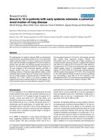
Figure 1
juvenile polyarthritis to oligoarthritis
MBL level accordingand(extended) MBL2 haplotypes in patients with
juvenile polyarthritis and oligoarthritis. Median mannose-binding lectin
(MBL) plasma levels, represented by horizontal lines, differ between
extended haplotype groups (P < 0.01), but not between patients with
oligoarthritis (n = 151) and polyarthritis (n = 67) who had similar haplotypes (P > 0.46).
pared with those with a current remission. In these patients,
the median (IQR) CRP level was 4 (1 to 5) mg/l in the high
genotype group versus 5 (4 to 10) mg/l in the medium and 5
(5 to 9) mg/l in the low genotype groups (P < 0.01).
The remaining clinical and laboratory variables did not differ
between the patients in the high, medium and low MBL2 genotype groups (Table 4). The differences found in CRP level
and remission status were also present in patients with the A/
A, the A/O and the O/O MBL2 genotype. Other clinical and
laboratory variables did not differ between these patients (data
not shown).
Discussion
In this study we demonstrated that the frequency of MBL deficiency was not increased in 218 Norwegian Caucasian children with JRA as compared with 194 Dutch Caucasian control
individuals. Our observations are in agreement with the only
previous study of MBL conducted in JIA patients [27]. In that
study no association between MBL2 codon 54 mutations and
JIA was found. We have now shown that JRA is also not associated with any of the other five known MBL2 SNPs.
The frequency of these mutations also did not differ from the
frequencies identified in previously published Danish Caucasian control populations [10,11]. Over the past few years studies have been published that consistently reported similar
frequencies in Caucasian populations of different countries
[10,11,34]. Therefore, we assume that the frequencies of
MBL2 gene polymorphism in the Caucasian Norwegian population do not differ from those in other Caucasian populations. Therefore, our present observations suggest that
genetically determined MBL deficiency is not associated with
increased susceptibility to JRA. Based on the number of
included patients and control individuals, this study has 80%
power when an odds ratio of 1.71 or greater for MBL deficiency is found.
Interestingly, children in the low MBL2 genotype group developed polyarthritis at a younger age than did children in the
medium or high genotype groups. Previously, Garred and
coworkers [23] showed that MBL2 exon 1 variant allele carrier
status was associated with early age at onset of RA, which is
the adult counterpart of polyarthritis [26]. Garred and coworkers hypothesized that MBL may delay the onset of RA but that
it does not prevent the disease. The mechanism by which MBL
deficiency might promote inflammation in immune-mediated
inflammatory diseases such as RA and JRA is as yet unknown.
MBL deficiency might lead to a diminished innate immunity,
and subsequent increased risk for infections, as was previously remonstrated [15,16]. These infections may trigger JRA,
as has been hypothesized previously [26]. Another possibility
is that MBL is involved in the recognition of an infectious agent
in the pathophysiology of JRA. Low or absent MBL plasma
concentration leads to decreased complement activation and
ineffective clearance of the pathogen or pathogen-derived
antigens. The prolonged presence of infectious agents in the
host may enhance synovial inflammation because of the proinflammatory effects of bacterial DNA and bacterial cell wall
fragments [35,36]. Anti-MBL autoantibodies may also play a
role, because elevated levels of anti-MBL autoantibodies were
found in the sera of RA patients [37]. It is unclear at present
whether MBL deficiency is indeed involved in the pathogenesis of RA or JRA, because the data reported are variable.
Furthermore, MBL deficiency does not appear to play a role
once polyarthritis has developed, because no associations
were found between MBL2 genotype and the laboratory variables or the remaining disease severity related clinical variables, such as PGA, CHAQ score, number of actively involved
or affected joints, and number of patients with uveitis or remission. Consistent with the previous report by Barton and coworkers [25] on RA and MBL polymorphisms, we did not find an
association between erosive joint destruction and MBL polymorphisms in patients with JRA.
In the oligoarthritis group, patients in the low genotype group
were in remission more often (81%) than were the children in
the medium or high genotype group (54% to 56%). In this
regard, lack of the protein MBL in serum appears to be associated with a milder disease course or decreased inflammation. The possible explanation for these findings might be that
MBL has an immunomodulating effect. MBL is present in synovial fluid and can bind potential causative agents in JRA
Page 5 of 11
(page number not for citation purposes)
Arthritis Research & Therapy
Vol 10 No 2
Dolman et al.
Table 2
MBL concentrations and MBL2 genotypes
Control individuals
All JRA patients
JRA subgroups
Polyarthritis
Oligoarthritis
Exon 1 mutations
Sum A/A
120 (62)
128 (59)
43(64)
85 (56)
Sum A/O
65 (33)
81 (37)
22 (33)
59 (39)
A/B
40 (21)
43 (20)
14 (21)
29 (19)
A/C
5 (3)
7 (3)
0 (0)
7 (5)
A/D
20 (10)
31 (14)
8 (12)
23 (15)
Sum O/O
9 (5)
9 (4)
2 (2)
7 (5)
B/B
4 (2)
4 (2)
1 (1)
3 (2)
B/C
1 (0)
0 (0)
0 (0)
0 (0)
B/D
2 (1)
3 (1)
1 (1)
2 (1)
C/D
0 (0)
1 (0)
0 (0)
1 (1)
D/D
2 (1)
1 (0)
0 (0)
1 (1)
194 (100)
218 (100)
67 (100)
151 (100)
110 (57)
113 (52)
36 (54)
77 (51)
YA/YA
60 (31)
62 (28)
21 (31)
41 (27)
YA/XA
50 (26)
51 (23)
15 (22)
36 (24)
52 (27)
71 (33)
23 (34)
48 (32)
XA/XA
10 (5)
15 (7)
7 (10)
8 (5)
YA/O
42 (22)
56 (26)
16 (24)
40 (27)
32 (16)
34 (16)
8 (12)
26 (17)
XA/O
23 (12)
25 (12)
6 (9)
19 (13)
O/O
9 (4)
9 (4)
2 (3)
7 (5)
194 (100)
218 (100)
67 (100)
151 (100)
High
1.65 (1.20 to 2.69)
1.86 (1.23 to 3.26)
1.87 (1.14 to 3.15)
1.85 (1.32 to 3.67)
Medium
0.52 (0.40 to 0.92)
0.77 (0.38 to 1.41)
0.89 (0.32 to 1.79)
0.73 (0.38 to 1.43)
Low
0.04 (0.02 to 0.13)
0.07 (0.04 to 0.15)
0.10 (0.05 to 0.15)
0.07 (0.04 to 0.17)
Total
Genotype groups
High
Medium
Low
Total
MBL concentration
Norwegian Caucasian children with juvenile polyarthritis (n = 67) and oligoarthritis (n = 151) are compared with 194 healthy Dutch Caucasian
adult control individuals. Values are expressed as number (%) or, for continuous variables, as median (interquartile range). Median mannosebinding lectin (MBL) concentrations and frequencies of exon 1 mutations and MBL2 genotype groups did not differ between all juvenile
rheumatoid arthritis (JRA) patients and healthy control individuals or within the polyarthritis and oligoarthritis groups (P values > 0.05). A is the
designation for wild-type; O is the common designation for the variant alleles B (codon 54), C (codon 57) and D (codon 52).
Page 6 of 11
(page number not for citation purposes)
Available online />
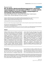
Figure 2
CRP and MBL2 genotype: remission versus active disease. Shown are serum C-reactive protein (CRP) concentrations (mg/l) and mannose-binding
versus active disease
lectin (MBL) genotype in patients with a current remission versus active disease (either active disease with a previous remission or continuously
active disease). *Only CRP values of oligoarthritis patients with active disease (as compared with patients with a current remission) differed statistically significantly (P < 0.01).
including micro-organisms, cellular debris, and agalactosyl
IgG (IgG-G0) [38,39]. Binding of MBL to agalactosyl IgG
immune complexes may result in local complement activation
and subsequent increased inflammation and thus active disease, whereas this is absent in the presence of very low levels
of MBL [40]. Recently, Troelsen and colleagues [41] found
that high serum levels of MBL and agalactosyl IgG were risk
factors for ischaemic heart disease in RA patients. Besides,
RA patients had higher MBL levels than did their relatives, suggesting that high MBL may trigger RA [39]. Harmful effects of
high MBL levels have been shown in other disease entities as
well. For instance, MBL deposits in the glomeruli can cause
histological damage of kidneys, and activation of the lectin
pathway by MBL can induce vascular tissue damage in myocardial ischemia-reperfusion injury and diabetes [42-44]. On
the other hand, MBL deficiency might be associated with
defective clearance of immune complexes and apoptotic cells,
as seen in individuals with C1q deficiency. Because MBL and
C1q are molecules with similar characteristics this might
explain why during active disease CRP levels were increased
in children in the low compared with the medium and high genotype groups. Remission rates were not associated with
MBL2 genotype in patients with polyarthritis, possibly
because more joints were affected.
Conclusion
MBL appears to play a dual role in JRA. Genetically determined MBL deficiency does not increase susceptibility to JRA,
but MBL does appear to have an immunomodulating effect.
On the one hand children with low levels of MBL develop pol-
yarthritis at younger age. In the case of MBL deficiency, potential explanations for this younger age at onset are increased
susceptibility to infections, as a potential trigger of polyarthritis, or ineffective clearance of infectious agents in the pathophysiology of JRA. On the other hand, the low MBL2
expressing genotypes appear to be beneficial once oligoarthritis has developed, because they are associated with increased
frequency of remission. An explanation may be that the local
MBL itself may lead to complement-mediated inflammation in
the synovium, sustaining active disease. If we are to discover
the possible contribution of MBL to JRA disease severity, then
we must study molecular mechanisms such as the interaction
of MBL with immune complexes, the presence of anti-MBL
autoantibodies and the role of activation of the complement
system.
Competing interests
The authors declare that they have no competing interests.
Authors' contributions
The study was designed by KD, TK, PT and AS. They were all
involved in the management of the study and in supporting
other contributors. BF, OF and AS collected the clinical data.
NB conducted the laboratory investigations. FF analyzed the
data statistically and interpreted the results. She completed
the first draft, written by KD. Finally, each author contributed to
the writing of the final manuscript. They all read and approved
this version of the manuscript and take full responsibility for it.
Page 7 of 11
(page number not for citation purposes)
Arthritis Research & Therapy
Vol 10 No 2
Dolman et al.
Table 3
Association of demographic, clinical, and laboratory characteristics and MBL2 genotype expression groups: juvenile polyarthritis
Characteristic
Pa
MBL genotype expression
groups
Pb
High (n = 36)
Medium (n = 23)
Low (n = 8)
Males
10 (28%)
4 (17%)
5 (63%)
0.05
0.04
Age (years) at onset
9.5 (5.6 to 13.0)
10.1 (8.4 to 13.0)
4.4 (3.6 to 7.0)
0.03
<0.01
Disease duration (years) at follow up
14.6 (13.5 to 16.3)
14.5 (13.2 to 16.4)
15.8 (13.4 to 16.6)
NS
NS
NS
NS
Demographic variables
Clinical variables
Cumulative affected joints
18 (10 to 32)
22 (10 to 36)
23 (12 to 39)
NS
NS
Actively involved joints
1 (0 to 4)
2 (0 to 8)
0 (0 to 2)
NS
NS
Affected joints
6 (2 to 18)
8 (0 to 20)
8 (1 to 27)
NS
NS
Arthritis severity index
10 (2 to 31)
17 (0 to 46)
19 (2 to 54)
NS
NS
Physician global assessment
2 (1 to 3)
2 (1 to 4)
1 (1 to 2)
NS
NS
Childhood Health Assessment Questionnaire score
0.1 (0.0 to 0.6)
0.3 (0.0 to 1.2)
0 (0.0 to 0.3)
NS
NS
Patients with uveitis
5 (14%)
4 (17%)
1 (13%)
NS
NS
NS
NS
NS
NS
NS
NS
Remission status at follow up
Current remission
18 (50%)
10 (44%)
4 (50%)
Active, but previous remission
8 (22%)
4 (17%)
2 (25%)
Continuously active
10 (28%)
9 (39%)
2 (25%)
16 (44%)
10 (44%)
4 (50%)
Radiographic erosions grade III to IV
Laboratory variables
Erythrocyte sedimentation rate (mm/hour)
8 (4 to 20)
8 (5 to 25)
3 (0 to 23)
NS
NS
C-reactive protein (mg/l)
5 (3 to 9)
7 (3 to 18)
5 (4 to 17)
NS
NS
Antinuclear antibody positivity
10 (28%)
6 (27%)
1 (13%)
NS
NS
IgM-rheumatoid factor positivity
4 (11%)
7 (30%)
0 (0%)
NS
NS
Included in this analysis are 67 patients with juvenile polyarthritis. Values are expressed as number (%) or, for continuous variables, as median
(interquartile range). aComparison of the high, medium and low genotype expression groups by means of the two-sided Fisher's exact test and
Kruskal-Wallis test. bComparison of the high and medium genotype group versus the low genotype expression group by means of the two-sided
Fisher's exact test and Mann-Whitney U-test. NS, not significant.
Page 8 of 11
(page number not for citation purposes)
Available online />
Acknowledgements
Table 4
Association of demographic, clinical, and laboratory characteristics and MBL2 genotype expression groups: oligoarthritis
Characteristic
Pa
MBL genotype expression
groups
Pb
High (n = 77)
Medium (n = 48)
Low (n = 26)
Males
22 (29%)
10 (21%)
8 (31%)
NS
NS
Age (years) at onset
6.6 (3.4 to 10.6)
8.7 (2.6 to 12.1)
6.6 (2.9 to 12.6)
NS
NS
Disease duration (years) at follow up
14.5 (13.6 to 15.9)
15.1 (13.2 to 16.1)
15.3 (14.1 to 16.4)
NS
NS
NS
NS
Demographic variables
Clinical variables
Cumulative affected joints
3 (2 to 6)
4 (2 to 6)
2 (2 to 4)
NS
NS
Actively involved joints
0 (0 to 1)
0 (0 to 1)
0 (0 to 0)
NS
NS
Affected joints
1 (0 to 3)
1 (0 to 2)
1 (0 to 3)
NS
NS
Arthritis severity index
2 (0 to 6)
2 (0 to 5)
1.5 (0 to 5)
NS
NS
Physician global assessment
1 (1 to 2)
1 (1 to 2)
1 (1 to 2)
NS
NS
Childhood Health Assessment Questionnaire score
0.0 (0.0 to 0.3)
0.0 (0.0 to 0.1)
0.0 (0.0 to 0.4)
NS
NS
Patients with uveitis
12 (16%)
16 (33%)
6 (23%)
NS
NS
0.02
0.01
NS
NS
NS
NS
Remission status at follow up
Current remission
43 (56%)
26 (54%)
21 (81%)
Active, but previous remission
27 (35%)
13 (27%)
1 (4%)
Continuously active
7 (9%)
9 (19%)
4 (15%)
11 (14%)
8 (17%)
2 (8%)
Radiographic erosions grade III to IV
Laboratory variables
Erythrocyte sedimentation rate (mm/hour)
6 (4 to 11)
8 (5 to 13)
5 (4 to 11)
NS
NS
C-reactive protein (mg/l)
5 (1 to 5)
5 (3 to 6)
5 (5 to 9)
<0.01
0.01
Antinuclear antibody positivity
33 (43%)
18 (38%)
11 (42%)
NS
NS
IgM-rheumatoid factor positivity
0
0
0
-
-
Included in this analysis are 151 patients with juvenile oligoarthritis. Values are expressed as number (%) or, for continuous variables, as median
(interquartile range). aComparison of the high, medium and low genotype expression groups by means of the two-sided Fisher's exact test and
Kruskal-Wallis test. bComparison of the high and medium genotype group versus the low genotype expression group by means of the two-sided
Fisher's exact test and Mann-Whitney U-test. NS, not significant.
Page 9 of 11
(page number not for citation purposes)
Arthritis Research & Therapy
Vol 10 No 2
Dolman et al.
We thank Professor Ben Dijkmans for his intermediary support and
Michel van Houdt for excellent technical assistance.
21.
References
1.
2.
3.
4.
5.
6.
7.
8.
9.
10.
11.
12.
13.
14.
15.
16.
17.
18.
19.
20.
Burgos-Vargas R, Vazquez-Mellado J: The early clinical recognition of juvenile-onset ankylosing spondylitis and its differentiation from juvenile rheumatoid arthritis. Arthritis Rheum 1995,
38:835-844.
Ploski R, Vinje O, Ronningen KS, Spurkland A, Sorskaar D, Vartdal
F, Forre O: HLA class II alleles and heterogeneity of juvenile
rheumatoid arthritis. DRB1*0101 may define a novel subset of
the disease. Arthritis Rheum 1993, 36:465-472.
Schaller JG: Pauciarticular arthritis of childhood (pauciarticular
juvenile rheumatoid arthritis). Ann Pediatr (Paris) 1983,
30:557-563.
Wilder RL, Crofford LJ: Do infectious agents cause rheumatoid
arthritis? Clin Orthop Relat Res 1991, 265:36-41.
Turner MW: Mannose-binding lectin: the pluripotent molecule
of the innate immune system.
Immunol Today 1996,
17:532-540.
Neth O, Jack DL, Dodds AW, Holzel H, Klein NJ, Turner MW: Mannose-binding lectin binds to a range of clinically relevant
microorganisms and promotes complement deposition. Infect
Immun 2000, 68:688-693.
Saifuddin M, Hart ML, Gewurz H, Zhang Y, Spear GT: Interaction
of mannose-binding lectin with primary isolates of human
immunodeficiency virus type 1. J Gen Virol 2000, 81:949-955.
Lipscombe RJ, Sumiya M, Hill AV, Lau YL, Levinsky RJ, Summerfield JA, Turner MW: High frequencies in African and non-African populations of independent mutations in the mannose
binding protein gene. Hum Mol Genet 1992, 1:709-715.
Madsen HO, Garred P, Kurtzhals JA, Lamm LU, Ryder LP, Thiel S,
Svejgaard A: A new frequent allele is the missing link in the
structural polymorphism of the human mannan-binding
protein. Immunogenetics 1994, 40:37-44.
Brouwer N, Dolman KM, van Zwieten R, Nieuwenhuys E, Hart M,
Aarden LA, Roos D, Kuijpers TW: Mannan-binding lectin (MBL)mediated opsonization is enhanced by the alternative pathway
amplification loop. Mol Immunol 2006, 43:2051-2060.
Kronborg G, Weis N, Madsen HO, Pedersen SS, Wejse C,
Nielsen H, Skinhoj P, Garred P: Variant mannose-binding lectin
alleles are not associated with susceptibility to or outcome of
invasive pneumococcal infection in randomly included
patients. J Infect Dis 2002, 185:1517-1520.
Madsen HO, Garred P, Thiel S, Kurtzhals JA, Lamm LU, Ryder LP,
Svejgaard A: Interplay between promoter and structural gene
variants control basal serum level of mannan-binding protein.
J Immunol 1995, 155:3013-3020.
Madsen HO, Satz ML, Hogh B, Svejgaard A, Garred P: Different
molecular events result in low protein levels of mannan-binding lectin in populations from southeast Africa and South
America. J Immunol 1998, 161:3169-3175.
Frakking FN, van de Wetering MD, Brouwer N, Dolman KM,
Geissler J, Lemkes B, Caron HN, Kuijpers TW: The role of mannose-binding lectin (MBL) in paediatric oncology patients with
febrile neutropenia. Eur J Cancer 2006, 42:909-916.
Koch A, Melbye M, Sorensen P, Homoe P, Madsen HO, Molbak K,
Hansen CH, Andersen LH, Hahn GW, Garred P: Acute respiratory tract infections and mannose-binding lectin insufficiency
during early childhood. JAMA 2001, 285:1316-1321.
Summerfield JA, Sumiya M, Levin M, Turner MW: Association of
mutations in mannose binding protein gene with childhood
infection in consecutive hospital series.
BMJ 1997,
314:1229-1232.
Garred P, Larsen F, Madsen HO, Koch C: Mannose-binding lectin deficiency – revisited. Mol Immunol 2003, 40:73-84.
Lee YH, Witte T, Momot T, Schmidt RE, Kaufman KM, Harley JB,
Sestak AL: The mannose-binding lectin gene polymorphisms
and systemic lupus erythematosus: two case-control studies
and a meta-analysis. Arthritis Rheum 2005, 52:3966-3974.
Graudal NA, Homann C, Madsen HO, Svejgaard A, Jurik AG,
Graudal HK, Garred P: Mannan binding lectin in rheumatoid
arthritis. A longitudinal study. J Rheumatol 1998, 25:629-635.
Graudal NA, Madsen HO, Tarp U, Svejgaard A, Jurik G, Graudal
HK, Garred P: The association of variant mannose-binding lec-
Page 10 of 11
(page number not for citation purposes)
22.
23.
24.
25.
26.
27.
28.
29.
30.
31.
32.
33.
34.
35.
36.
37.
tin genotypes with radiographic outcome in rheumatoid
arthritis. Arthritis Rheum 2000, 43:515-521.
Jacobsen S, Madsen HO, Klarlund M, Jensen T, Skjodt H, Jensen
KE, Svejgaard A, Garred P: The influence of mannose binding
lectin polymorphisms on disease outcome in early polyarthritis. TIRA Group. J Rheumatol 2001, 28:935-942.
Saevarsdottir S, Vikingsdottir T, Vikingsson A, Manfredsdottir V,
Geirsson AJ, Valdimarsson H: Low mannose binding lectin predicts poor prognosis in patients with early rheumatoid arthritis. A prospective study. J Rheumatol 2001, 28:728-734.
Garred P, Madsen HO, Marquart H, Hansen TM, Sørensen SF,
Petersen J, Volck B, Svejgaard A, Graudal NA, Rudd PM, Dwek
RA, Sim RB, Andersen V: Two edged role of mannose binding
lectin in rheumatoid arthritis: a cross sectional study. J
Rheumatol 2000, 27:26-34.
Stanworth SJ, Donn RP, Hassall A, Dawes P, Ollier W, Snowden
N: Absence of an association between mannose-binding lectin
polymorphism and rheumatoid arthritis. Br J Rheumatol 1998,
37:186-188.
Barton A, Platt H, Salway F, Symmons D, Lunt M, Worthington J,
Silman A: Polymorphisms in the mannose binding lectin (MBL)
gene are not associated with radiographic erosions in rheumatoid or inflammatory polyarthritis. J Rheumatol 2004,
31:442-447.
Ravelli A, Martini A: Juvenile idiopathic arthritis. Lancet 2007,
369:767-778.
Kang M, Wang HW, Cheng PX, Yin ZD, Li XO, Shi H, Hu XF: Lack
of association between mannose-binding lectin gene polymorphisms and juvenile idiopathic arthritis in a Han population from the Hubei province of China. Arthritis Res Ther 2006,
8:R85.
Flato B, Smerdel A, Johnston V, Lien G, Dale K, Vinje O, Egeland
T, Sorskaar D, Forre O: The influence of patient characteristics,
disease variables, and HLA alleles on the development of radiographically evident sacroiliitis in juvenile idiopathic arthritis.
Arthritis Rheum 2002, 46:986-994.
Flato B, Lien G, Smerdel A, Vinje O, Dale K, Johnston V, Sorskaar
D, Moum T, Ploski R, Forre O: Prognostic factors in juvenile
rheumatoid arthritis: a case-control study revealing early predictors and outcome after 14.9 years. J Rheumatol 2003,
30:386-393.
Brewer EJ Jr, Bass J, Baum J, Cassidy JT, Fink C, Jacobs J, Hanson
V, Levinson JE, Schaller J, Stillman JS: Current proposed revision
of JRA Criteria. JRA Criteria Subcommittee of the Diagnostic
and Therapeutic Criteria Committee of the American Rheumatism Section of The Arthritis Foundation. Arthritis Rheum 1977,
20:195-199.
Singh G, Athreya BH, Fries JF, Goldsmith DP: Measurement of
health status in children with juvenile rheumatoid arthritis.
Arthritis Rheum 1994, 37:1761-1769.
Tacx AN, Groeneveld AB, Hart MH, Aarden LA, Hack CE: Mannan
binding lectin in febrile adults: no correlation with microbial
infection and complement activation. J Clin Pathol 2003,
56:956-959.
Bernig T, Breunis W, Brouwer N, Hutchinson A, Welch R, Roos D,
Kuijpers T, Chanock S: An analysis of genetic variation across
the MBL2 locus in Dutch Caucasians indicates that 3' haplotypes could modify circulating levels of mannose-binding
lectin. Hum Genet 2005:1-12.
Garcia-Laorden MI, Pena MJ, Caminero JA, Garcia-Saavedra A,
Campos-Herrero MI, Caballero A, Rodriguez-Gallego C: Influence
of mannose-binding lectin on HIV infection and tuberculosis in
a Western-European population.
Mol Immunol 2006,
43:2143-2150.
Schrijver IA, Melief MJ, Tak PP, Hazenberg MP, Laman JD: Antigen-presenting cells containing bacterial peptidoglycan in
synovial tissues of rheumatoid arthritis patients coexpress
costimulatory molecules and cytokines. Arthritis Rheum 2000,
43:2160-2168.
van der Heijden I, Wilbrink B, Tchetverikov I, Schrijver IA, Schouls
LM, Hazenberg MP, Breedveld FC, Tak PP: Presence of bacterial
DNA and bacterial peptidoglycans in joints of patients with
rheumatoid arthritis and other arthritides. Arthritis Rheum
2000, 43:593-598.
Gupta B, Raghav SK, Agrawal C, Chaturvedi VP, Das RH, Das HR:
Anti-MBL autoantibodies in patients with rheumatoid arthritis:
Available online />
38.
39.
40.
41.
42.
43.
44.
prevalence and clinical significance. J Autoimmun 2006,
27:125-133.
Saevarsdottir S, Vikingsdottir T, Valdimarsson H: The potential
role of mannan-binding lectin in the clearance of self-components including immune complexes. Scand J Immunol 2004,
60:23-29.
Saevarsdottir S, Steinsson K, Grondal G, Valdimarsson H:
Patients with rheumatoid arthritis have higher levels of mannan-binding lectin than their first-degree relatives and unrelated controls. J Rheumatol 2007, 34:1692-1695.
Malhotra R, Wormald MR, Rudd PM, Fischer PB, Dwek RA, Sim
RB: Glycosylation changes of IgG associated with rheumatoid
arthritis can activate complement via the mannose-binding
protein. Nat Med 1995, 1:237-243.
Troelsen LN, Garred P, Madsen HO, Jacobsen S: Genetically
determined high serum levels of mannose-binding lectin and
agalactosyl IgG are associated with ischemic heart disease in
rheumatoid arthritis. Arthritis Rheum 2007, 56:21-29.
Roos A, Bouwman LH, Munoz J, Zuiverloon T, Faber-Krol MC, Fallaux-van den Houten FC, Klar-Mohamad N, Hack CE, Tilanus MG,
Daha MR: Functional characterization of the lectin pathway of
complement in human serum. Mol Immunol 2003, 39:655-668.
Hansen TK, Tarnow L, Thiel S, Steffensen R, Stehouwer CD,
Schalkwijk CG, Parving HH, Flyvbjerg A: Association between
mannose-binding lectin and vascular complications in type 1
diabetes. Diabetes 2004, 53:1570-1576.
Jordan JE, Montalto MC, Stahl GL: Inhibition of mannose-binding lectin reduces postischemic myocardial reperfusion injury.
Circulation 2001, 104:1413-1418.
Page 11 of 11
(page number not for citation purposes)