Báo cáo khoa học: " Counting colonies of clonogenic assays by using densitometric software" pot
Bạn đang xem bản rút gọn của tài liệu. Xem và tải ngay bản đầy đủ của tài liệu tại đây (418.85 KB, 3 trang )
BioMed Central
Page 1 of 3
(page number not for citation purposes)
Radiation Oncology
Open Access
Methodology
Counting colonies of clonogenic assays by using densitometric
software
Maximilian Niyazi*
1
, Ismat Niyazi
2
and Claus Belka
1
Address:
1
CCC Tübingen, Department of Radiation Oncology, Hoppe-Seyler-Str. 3, 72076 Tübingen, Germany and
2
Bureau for Technique and
Documentation, Paracelsusstr. 21, 70599 Stuttgart, Germany
Email: Maximilian Niyazi* - ; Ismat Niyazi - ; Claus Belka -
* Corresponding author
Abstract
Clonogenic assays are a useful tool to test whether a given cancer therapy can reduce the
clonogenic survival of tumour cells. A colony is defined as a cluster of at least 50 cells which can
often only be determined microscopically. The process of counting colonies is very extensive work
and so we developed software that is able to count the colonies automatically from scanned flasks.
This software is made freely available by us with a detailed description how to use and install the
necessary features.
Background
Everyone who has already counted colonies from a clono-
genic assay knows that this is hard work which takes a lot
of time. Many groups have thought about an improve-
ment of the counting system [1-8] and the cited publica-
tions certainly won't cover all attempts, but no method
has achieved a widespread use at all.
One may ask what the reasons are. Two major reasons are
certainly the following: all presented methods had prob-
lems when faced with clustered colonies. Some systems
have managed to avoid this problem by constructing
ingenious scanning systems, but these systems are only
commercially available.
For this purpose we developed a new software that is able
to evaluate the clonogenic assays by using densitometric
methods and other special parameters. This revealed
highly fitting results which were compared to manual
countings with the microscope.
Procedure and results
The newly designed software requires normally scanned
six-well-plates or flasks (200 dpi is optimal for excellent
results) which should be scanned as grey-scaled photos
and saved as jpeg-file. Even better is a reflection light scan-
ner but this is no condition for good results. It is anyhow
recommended to avoid (as much as possible) disturbing
shades which are caused by scanners, although the soft-
ware can usually recognize and neglect these shades.
Flasks can be scanned and evaluated without editing the
scanned pictures. In our institute it is a routine activity to
archive the clonogenic assays by scanning them so that
this is no additional work.
When launching the program (a more detailed descrip-
tion is given in the manual of the program, see additional
file 1) you are asked to select a data file. After selecting a
file the program will start (see fig. 1). The area to be
counted can be selected and copied to the working area.
By counting the first time you usually won't get the result
you expected. So you have to adjust the available parame-
Published: 09 January 2007
Radiation Oncology 2007, 2:4 doi:10.1186/1748-717X-2-4
Received: 13 December 2006
Accepted: 09 January 2007
This article is available from: />© 2007 Niyazi et al; licensee BioMed Central Ltd.
This is an Open Access article distributed under the terms of the Creative Commons Attribution License ( />),
which permits unrestricted use, distribution, and reproduction in any medium, provided the original work is properly cited.
Radiation Oncology 2007, 2:4 />Page 2 of 3
(page number not for citation purposes)
ters. This presumes that you have counted one well of the
plate (usually the one that contains the minimum
amount of colonies). In our experiments we had several
dilutions of cells with the same treatment; so for each
treatment we counted one well with around 20 cells and
could adjust the parameters for the program.
The software provides three independent parameters: the
gray level, the maximum size of one colony (defined by
the area) and another parameter that considers the distri-
bution of the gray colour within the colony. This is done
by writing every cluster of the scanned area into several
matrices which contain information on localisation and
gray level.
One needs some experience to find the right parameters,
but with the help of some tricks (see the manual) you can
manage it in a short time.
But it is not only the match of the counted numbers which
gives evidence on the right parameters. The program dis-
plays which colonies were counted and which not. This
allows a manual correction with respect to the golden
standard (the microscope).
We compared this attempt with the microscopical evalua-
tion and found a relative mistake < 5%.
Discussion
We developed a new system for automated counting of
colonies on cell culture flasks that uses an algorithm that
has not been used before.
This attempt requires three parameters which are deter-
mined by comparison to the microscopical evaluation of
single wells which leads to high economy of time.
The results showed excellent matching (relative mistake <
5%) as we compared that to manual counting (dependent
on the person, but normally 5 – 10%) or to the golden
standard (microscope) which has after all an interindivid-
ual difference of around 2%. Another argument is the fact
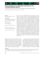
Screenshot of the programFigure 1
Screenshot of the program. Left panel: the scanned flask, the circle marks the region that will be counted; right panel the
result: green areas denote a colony, red points mark the clusters which are too small. Parameters and counted points are dis-
played below.
Publish with BioMed Central and every
scientist can read your work free of charge
"BioMed Central will be the most significant development for
disseminating the results of biomedical research in our lifetime."
Sir Paul Nurse, Cancer Research UK
Your research papers will be:
available free of charge to the entire biomedical community
peer reviewed and published immediately upon acceptance
cited in PubMed and archived on PubMed Central
yours — you keep the copyright
Submit your manuscript here:
/>BioMedcentral
Radiation Oncology 2007, 2:4 />Page 3 of 3
(page number not for citation purposes)
that this relative mistake is made for all wells and as (for
a clonogenic assay) the plating efficiency is considered,
this does not play a key role according to the overall result.
The program is written in Java and can be downloaded. It
is freely available and we are interested in feedback and
whether you can use the program for your practical work.
Detailed advice for installation and operation is given in
the manual.
One may ask why one has to make new adjustments for
every treatment. For similar treatments this is in fact not
necessary. But some cell lines change their shape follow-
ing very different treatments. Sometimes cells grow under
treatment and so one has to set another cut off for the
minimum size compared to the control.
But this did not influence the inherent advantage of auto-
mated counting according to economy of time.
Additional material
Acknowledgements
Supported by a grant from the Federal Ministry of Education and Research
(Fö. 01KS9602) and the Interdisciplinary Center of Clinical Research Tübin-
gen (IZKF).
References
1. Barber PR, Vojnovic B, Kelly J, Mayes CR, Boulton P, Woodcock M,
Joiner MC: Automated counting of mammalian cell colonies.
Phys Med Biol 2001, 46:63-76.
2. Biston MC, Corde S, Camus E, Marti-Battle R, Esteve F, Balosso J: An
objective method to measure cell survival by computer-
assisted image processing of numeric images of Petri dishes.
Phys Med Biol 2003, 48:1551-63.
3. Dahle J, Kakar M, Steen HB, Kaalhus O: Automated counting of
mammalian cell colonies by means of a flat bed scanner and
image processing. Cytometry A 2004, 60:182-8.
4. Herman CJ, Pelgrim OE, Kirkels WJ, Verheijen R, Debruyne FM, Ken-
emans P, Vooijs GP: In-use evaluation of the Omnicon auto-
mated tumor colony counter. Cytometry 1983, 3:439-42.
5. Mukherjee DP, Pal A, Sarma SE, Majumder DD: Bacterial colony
counting using distance transform. Int J Biomed Comput 1995,
38:131-40.
6. Parry RL, Chin TW, Donahoe PK: Computer-aided cell colony
counting. Biotechniques 1991, 10:772-4.
7. Salmon SE, Young L, Lebowitz J, Thomson S, Einsphar J, Tong T, Moon
TE: Evaluation of an automated image analysis system for
counting human tumor colonies. Int J Cell Cloning 1984, 2:142-60.
8. Wilson IG: Use of the IUL Countermat Automatic Colony
Counter for Spiral Plated Total Viable Counts. Appl Environ
Microbiol 1995, 61:3158-3160.
Additional File 1
Clono-Counter. Contains a manual for the program, the program itself
and an example.
Click here for file
[ />717X-2-4-S1.zip]