Báo cáo y học: "Activation of WNT and BMP signaling in adult human articular cartilage following mechanical injury" potx
Bạn đang xem bản rút gọn của tài liệu. Xem và tải ngay bản đầy đủ của tài liệu tại đây (4.75 MB, 13 trang )
Open Access
Available online />Page 1 of 13
(page number not for citation purposes)
Vol 8 No 5
Research article
Activation of WNT and BMP signaling in adult human articular
cartilage following mechanical injury
Francesco Dell'Accio
1
, Cosimo De Bari
1
, Noha MF El Tawil
1
, Francesca Barone
1
,
Thimios A Mitsiadis
2
, John O'Dowd
3
and Costantino Pitzalis
1
1
Department of Rheumatology, King's College London, London, UK
2
Department of Craniofacial Development, King's College London, London, UK
3
Guy's and St Thomas's Hospitals, London, UK
Corresponding author: Francesco Dell'Accio,
Received: 17 Feb 2006 Revisions requested: 4 Apr 2006 Revisions received: 2 May 2006 Accepted: 7 Aug 2006 Published: 7 Aug 2006
Arthritis Research & Therapy 2006, 8:R139 (doi:10.1186/ar2029)
This article is online at: />© 2006 Dell'Accio et al.; licensee BioMed Central Ltd.
This is an open access article distributed under the terms of the Creative Commons Attribution License ( />),
which permits unrestricted use, distribution, and reproduction in any medium, provided the original work is properly cited.
Abstract
Acute full thickness joint surface defects can undergo repair,
which involves tissue patterning and endochondral bone
formation. Molecular signals regulating this process may
contribute to the repair outcome, chronic evolution and,
eventually, the onset of osteoarthritis. We tested the hypothesis
that mechanical injury modulates morphogenetic pathways in
adult human articular cartilage explants. Adjacent articular
cartilage explants were obtained from preserved areas of the
femoral condyles of patients undergoing arthroplasty for
osteoarthritis, or from a normal joint of a patient undergoing
lower limb amputation. Paired explants were individually
maintained in explant culture. From each pair, one explant was
mechanically injured and the other left uninjured as a control.
Cultures were terminated at different time points for
histochemistry, immunohistochemistry and gene expression
analysis by reverse transcription real time PCR. Bone
morphogenetic protein 2 (BMP-2) mRNA was upregulated in
the injured explants. We detected phosphorylation of SMAD-1
and SMAD-5, consistent with activation of the bone
morphogenetic protein (BMP) pathway. FRZB-1 mRNA was
downregulated in the injured explants, suggesting de-repression
of WNT signaling. Accordingly, expression of the canonical
WNT target genes Axin-2 and c-JUN was upregulated in the
injured explants. Activation of the canonical WNT signaling
pathway by LiCl treatment induced upregulation of COL2A1
and Aggrecan mRNA, suggesting an anabolic effect.
Phosphorylation of SMAD-1/-5 and downregulation of FRZB
were confirmed in vivo in a mouse model of joint surface injury.
Taken together, these data show modulation of the BMP and
WNT pathways following mechanical injury in vitro and in vivo,
which may play a role in the reparative response of the joint
surface. These pathways may, therefore, represent potential
targets in protocols of biological joint surface defect repair.
Introduction
Chronic symptomatic full thickness defects of the joint surface
are commonly regarded to have a poor repair capacity. There-
fore, surgical treatment is provided for symptomatic relief and
in an attempt to avoid possible evolution towards osteoarthritis
(OA) [1]. The natural history of acute full thickness joint sur-
face defects (JSDs), however, is not yet well known. Scattered
clinical and animal studies have suggested that acute full
thickness JSDs exhibit potential for repair, which is dependent
on age, the size of the lesion, and biomechanical factors.
In two independent, long term, prospective studies, acute trau-
matic chondral lesions in young athletes had a good to excel-
lent clinical outcome in 78% of the cases in the absence of
specific surgical treatments [2,3]. In addition, Koshino and col-
leagues [4] reported significant regeneration of chronic JSDs
associated with genu varu at 2 years after correction of knee
malalignment by valgus osteotomy. Age dependent spontane-
ous repair has been reported in patients with osteochondritis
dissecans [5]. Likewise, age dependent spontaneous repair of
relatively small experimental full thickness JSDs has been
reported in rabbits [6,7] and dogs [8]. In rabbits, this repair
process entails invasion of the fibrin clot, filling the defect by
BMP = bone morphogenetic protein; glycogen synthase kinase 3 = GSK-3; DAPI = 49,6-diamidino-2-phenylindole; FBS = fetal bovine serum; JSD
= joint surface defect; MMP = metalloproteinase; OA = osteoarthritis; Q-PCR = quantitative real time PCR; RT-PCR = reverse transcription PCR;
TBST = tris buffered saline; TCF/LEF = T-cell factor/lymphoid enhancer factor.
Arthritis Research & Therapy Vol 8 No 5 Dell'Accio et al.
Page 2 of 13
(page number not for citation purposes)
mesenchymal progenitors, chondrogenesis, and endochon-
dral bone formation. Bone formation is polarized towards the
joint surface, and preserves a layer of articular cartilage [6].
Although the repair tissue is not always durable and advance-
ment of the bone front at the expense of stable articular carti-
lage sometimes occurs, this repair process, under specific
conditions, can restore joint surface homeostasis.
The patterning and morphogenesis that joint surface repair
entails implies a stepwise cellular and molecular program.
Thus, failure of the signaling mechanisms governing this proc-
ess may be a factor contributing to a poor repair outcome.
Such signals may represent therapeutic targets to support
spontaneous repair or complement existing biological joint
resurfacing techniques.
The current surgical approaches for localized full thickness
lesions of the joint surface are autologous chondrocyte
implantation, microfracture, and mosaicplasty. However, clini-
cal outcomes suffer from some degree of variability [9-11]. In
addition, there is still no satisfactory biological regeneration
protocol for non-localized lesions. An alternative or comple-
mentary approach for joint tissue repair would be the control-
led delivery of molecular signals to mesenchymal progenitors
reported within the joint environment [12-18] with support of
the subsequent steps of repair, including proliferation, pattern-
ing, and differentiation in vivo.
In this study, we have tested the hypothesis that the adult
human articular cartilage is a source of morphogenetic signals
upon injury. To this end, we have used an in vitro model of
Figure 1
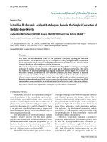
Ex vivo model of mechanical injury to adult human articular cartilage explantsEx vivo model of mechanical injury to adult human articular cartilage explants. (a) Adjacent explants from human adult articular cartilage were dis-
sected and placed in culture in separate bacteriological Petri dishes. After 6 days, 1 explant was injured. At different time points the cultures were
terminated for gene expression analysis, histochemistry and immunohistochemistry. (b) Safranin O staining of: a, freshly dissected normal articular
cartilage; b, an adjacent explant after 7 days in culture; c, a further adjacent explant after 6 days in culture before injury plus 1 additional day after
injury; and d, a typical freshly dissected explant from a preserved area from a patient who had undergone joint arthroplasty for osteoarthritis. (c,d)
Time course of metalloproteinase (MMP)-3 and MMP-13 mRNA differential expression in injured versus uninjured explants. Values are normalized
for the housekeeping gene
β
actin and expressed as fold change of gene expression in the injured explants from paired uninjured controls. Dia-
monds indicate samples from preserved areas from joints affected by osteoarthritis; open squares indicate sample pairs from healthy cartilage. *p <
0.05; **p < 0.01. D, day(s); h, hours.
Available online />Page 3 of 13
(page number not for citation purposes)
mechanical injury to the adult human articular cartilage to
screen signaling pathways potentially involved in the repair
response. In particular, we have focused on the bone morpho-
genetic protein (BMP) and the canonical WNT pathways,
which are known to play a crucial role in joint morphogenesis
and homeostasis as well as in repair processes [19-21].
BMPs are secreted molecules belonging to the transforming
growth factor β superfamily of morphogens. Upon binding
their ligands, BMP receptors phosphorylate the carboxy-termi-
nal domain of SMAD-1, SMAD-5 and SMAD-8. Phosphor-
ylated SMADS translocate to the nucleus where they
participate in the transcriptional regulation of target genes
[20].
WNTs constitute a large family of morphogens. WNT ligands
transduce their signal through different intracellular pathways.
In the β catenin-dependent (canonical) pathway, in the
absence of WNT ligands, glycogen synthase kinase 3 (GSK-
3) constitutively phosphorylates β catenin, which then is
degraded through the proteasome pathway. When WNT lig-
ands bind to their receptors (called FRZD), GSK-3 is inhibited
and β catenin is, therefore, stabilized and accumulates in the
cytoplasm and translocates into the nucleus, where it binds to
members of the T-cell factor/lymphoid enhancer factor (TCF/
LEF) family of transcription factors, thereby activating tran-
scription of target genes [22].
Materials and methods
Ex vivo cartilage injury model and tissue culture
Well-preserved (modified Mankin score 5 or less) cartilage
samples were obtained from patients who underwent total
knee replacement for unicompartmental OA (e.g., lateral con-
dyle in genu varu). The average age was 67.5 ± 8.9 years old
and the study included 3 males and 5 females. In one case
(male, 49 years old), we obtained cartilage explants from a
patient who had undergone limb amputation due to a road traf-
fic accident and was free from OA. In this case, therefore, the
cartilage was considered normal. Paired adjacent explants of
approximately 6 × 6 mm were maintained in culture in 4 ml of
Dulbecco's modeified Eagle's medium/HAMF12 1:1 (Invitro-
gen, Paisley, UK) in the presence or in the absence of 10%
FBS (Invitrogen) and antibiotics/antimycotics (Invitrogen) in
individual 33 mm bacteriological Petri dishes (BD Falcon™,
BD Biosciences, Le Pont De Claix, France). We used bacteri-
ological Petri dishes to avoid spreading of cells from the
explants. After 6 days, the medium was replaced and one of
each pair of adjacent samples was cut using a scalpel at 1 mm
intervals. The other explant of each pair was left uninjured (Fig-
ure 1a). At different time points, the explants were used for
RNA extraction and one aliquot was processed for histology
and immunohistochemistry.
For experiments investigating activation of the WNT/β catenin
canonical pathway by means of LiCl treatment, the explants
were maintained for 6 days in complete culture medium con-
taining 10 mM NaCl. At the end of this period, the explants
were either switched to medium containing 10 mM LiCl or, for
control explants, the medium was replaced with fresh medium
containing 10 mM NaCl. The experiments were then termi-
nated after one day. All procedures received approval from the
local ethics committee.
RNA extraction, reverse transcription PCR and
quantitative real time RT-PCR
Cartilage samples were snap-frozen in liquid nitrogen, pow-
dered with a mortar and pestle in liquid nitrogen, and subse-
quently homogenized in Trizol reagent (Life Technologies,
Invitrogen, Paisley, UK) using a polytron homogenizer. Total
RNA was extracted using Trizol reagent. Reverse transcription
PCR (RT-PCR) was performed as described elsewhere [23].
Quantitative real time RT-PCR (Q-PCR) was performed using
hot start DNA polymerase (Quiagen Ltd, Crawley, UK) in the
presence of 0.1X SYBR Green (Molecular Probes, Invitrogen,
Paisley, UK) utilizing the DNA Engine Opticon
®
2 System (MJ
Research, Alpha technologies Ltd, Northern Ireland). Reac-
tions were performed in duplicate and repeated in the rare
cases when the Ct of the duplicates differed for more than 1
cycle. A serial dilution of a cDNA from early passage human
articular chondrocytes was used for a standard curve. Gene
expression was calculated using a standard curve and normal-
ized for the expression of the housekeeping gene β actin. To
simplify the representation of time course analyses, the gene
expression data normalized for β actin are shown as fold
increase from uninjured paired control.
Primers and expected amplicon size are:
β
-actin
(GeneBank:BC014861
), forward 5'-CACGGCTGCTTC-
CAGCTC-3', reverse 5'-CACAGGACTCCATGCCCAG-3',
134 base pairs (bp); MMP-3 (GeneBank:NM_002422
), for-
ward 5'-CAACCGTGAGGAAAATCGATGCAG-3', reverse
5'-CGGCAAGATACAGATTCACGCTCAA-3', 440 bp;
MMP13 (GeneBank:NM_002427
), forward 5'-ACGGAC-
CCATACAGTTTGAATACAGC-3', reverse 5'-CCATTTGT-
GGTGTGGGAAGTATCATC-3, 360 bp; BMP-2
(GeneBank:NM_001200
), forward 5'-CGT-
CAAGCCAAACACAAACAGCG-3', reverse 5'- CAC-
CCACAACCCTCCACAACCAT-3', 341 bp; FRZB
(GeneBank:U24163
), forward 5'GGGCTATGAAGATGAG-
GAACGT-3', reverse 5'-ACCGAGTCGATCCTTCCACTT-3',
79 bp;
β
catenin (GeneBank:X87838), forward 5'-
CCAGCCGACACCAAGAAGCA-3', reverse 5'-GCG-
GGACAAAGGGCAAGATT-3', 151 bp; WNT1
(GeneBank:NM-005430
), forward 5'-CTGCCTCTCTTCTTC-
CCCTT-3', reverse 5'-TCACAGCTGTTCAATGGCTC-3',
251 bp; WNT5A (GeneBank:L20861
), forward 5'-CCACCT-
TCCTCTTCACACTG-3', reverse 5'-CGAACAAGTAAT-
GCCCTCTC-3', 770 bp; WNT5B (GeneBank:AB060966
),
forward 5'- CCGCCTCTGCAACAAGACCT-3', reverse 5'-
AACTTGCAGTGGCAGCGCTC-3', 111 bp; WNT14
Arthritis Research & Therapy Vol 8 No 5 Dell'Accio et al.
Page 4 of 13
(page number not for citation purposes)
(GeneBenk:NM_003395), forward 5'- TGAGAAGAACT-
GCGAGAGCA -3', reverse 5'- CTGTGTGCAATGCCTG-
TACC -3', 285 bp; WNT16 (GeneBank:NM_016087
),
forward 5'- AAAGAAATGTTTCCCTGCCC -3', reverse 5'-
GACATTTTCCATGGGTTTGC -3', 106 bp; FRZD-1
(GeneBank:NM_003505
), forward 5'- TTCAGCAGCACAT-
TCTGAGG-3', reverse 5'- CCTGCACACATTTTCCCTTT-3',
154 bp; FRZD-7 (GeneBank:NM_003507
), forward 5'-
CTGGAGTTCTTTGAAATGTGCT-3', reverse 5'- AAGGT-
TAGCTCCCATGATTCTC-3', 133 bp; LEF-1
(GeneBank:NM_016269
), forward 5'- CAGAGAAAGGAG-
CAGGAGCCAA -3', reverse 5'- TGATGTCAGTGTTCCTTT-
GGCG -3', 481 bp; TCF-1 (GeneBank:NM000545
), forward
5'- CTCATCACCGACACCACCAACC-3', reverse 5'-
TCCCACGAAGCAGCGACAGT -3', 608 bp; COL2A1
(GeneBank:NM_033150
), forward 5'- CCCTGAGTGGAA-
GAGTGGAG -3', reverse 5'- GAGGCGTGAGGTCTTCT-
GTG -3', 511 bp; Aggrecan (GeneBank:NM-001135
),
forward 5'- GTTGTCATCAGCACCAGCATC -3', reverse 5'-
ACCACACAGTCCTCTCCAGC -3', 509 bp; c-JUN
(GeneBank:NM_002228
), forward 5'-CCCCAAGATCCT-
GAAACAGA-3', reverse 5'- CCGTTGCTGGACTGGATTAT-
3'.
Histology, histochemistry and immunohistochemistry
Tissues were fixed overnight in 4% buffered paraformaldehyde
at 4°C, dehydrated and embedded in paraffin. Sections (5 µm
thick) were used for hematoxylin-eosin and safranin O staining
according to standard protocols. The degree of OA was eval-
uated using a modified Mankin score [24] in which the sub-
score related to the tide mark was not included. For immuno-
histochemistry, paraffin sections were deparaffinized and
hydrated in xylene and an ethanol series, post-fixed with 4%
paraformaldehyde, and washed twice in phosphate-buffered
saline. For antigen retrieval in the detection of FRZB and β cat-
enin, the sections were first equilibrated in 0.02% HCl for 7
minutes, digested in 3 mg/ml pepsin (Sigma-Aldrich Company
Ltd., Gillingham, UK) in 0.02% HCl for 45 minutes at 37°C,
washed in water and allowed to air dry for 20 minutes. Sec-
tions were washed twice in 0.2% Tween-20 in tris buffered
saline (TBST), blocked in 0.5% bovine serum albumin in TBST
for 1 hour at room temperature, blotted, and incubated over-
night with the primary antibody (goat anti-mouse/human FRZB
(R&D Systems, Abingdon, UK), or mouse anti-human β catenin
(BD Transduction Laboratories, BD, Cowley, Oxford, UK) at a
final concentration of 1 µg/ml in 0.5% bovine serum albumin
in TBST. Sections were then washed twice in TBST, and incu-
bated for 1 hour with the secondary antibody. For FRZB immu-
nostaining, the secondary antibody was a biotin-conjugated
rabbit anti-goat antibody (DAKO UK Ltd., Ely Cambridgeshire,
UK) diluted 1:300. For β catenin immunostaining, we used
either a cy™2 conjugated goat anti-mouse antibody (Jackson
ImmunoResearch Laboratories, Inc. West Grove, PA, USA)
diluted 1:200 for indirect immunofluorescence, or the Strept-
ABComplex/AP kit (DAKO) for signal amplification and Vec-
tor
®
Red substrate kit (Vector Laboratories UK, Peterborough,
UK) as a chromogenic substrate of alkaline phosphatase, in
the presence of 0.2 mM levamisole to inhibit endogenous alka-
line phosphatase. For the detection of phosphorylated SMAD-
1 and SMAD-5, we used the same protocol with the following
modifications. For antigen retrieval, instead of pepsin diges-
tion, we boiled the sections for 10 minutes in sodium citrate
buffer, pH 6; we quenched endogenous peroxidase by incu-
bating for 10 minutes with 9% H
2
O
2
; we used the PS-1 antise-
rum [25] (a kind gift of P ten Dijke and C-H Heldin, Ludwig
Institute for Cancer Research, Uppsala, Sweden) as primary
antibody; as secondary antibody we used biotin-conjugated
sheep anti-rabbit antibody (Serotec UK, Oxford, UK) diluted
1:200; we used the StreptABComplex/AP kit (DAKO) as an
amplification system, and Liquid DAB Substrate Chromogen
System (DAKO) as peroxidase substrate. Sections were
mounted in mowiol (Calbiochem, Merck Biosciences Ltd, Not-
tingham, UK) containing 49,6-diamidino-2-phenylindole
(DAPI; ICN, Stretton Scientific Ltd., Stretton, UK) for nuclear
counterstaining. In positive cells the DAB precipitate
quenched the DAPI fluorescence. Image processing was per-
formed using Adobe Photoshop version 6 (Adobe). Negative
controls were sections in which isotype and species-matched
non-specific immunoglobulins or normal rabbit serum (for
phospho-SMAD-1/-5) were used instead of the primary anti-
body.
Statistical analysis
Normally distributed data sets from paired samples were com-
pared using the paired t test. When the values did not have a
normal distribution, they were either transformed into their log-
arithms before analysis or, if this still did not result in a normal
distribution, they were analyzed using the Wilcoxon matched
pair test.
Joint surface injury in mice
Seven week old C57BL/6 male mice were utilized for these
experiments. The mice were anesthetized and subjected to
medial para-patellar arthrotomy. The patellar groove was
exposed by lateral patellar dislocation. A longitudinal full thick-
ness injury was made in the patellar groove using a custom
made device in which the length of a 26G needle was limited
by a glass bead (injured knee). The patellar dislocation was
then reduced and the joint capsule and the skin sutured in sep-
arate layers. The mice were then allowed to walk freely in
standard cages and maintained on free diet. Control mice
were subjected to the arthrotomy and to the patellar disloca-
tion, but no cartilage injury was made (sham operated con-
trols). The animals were killed at different time-points and the
knees dissected for histological and histochemical analysis.
The same procedure has been performed in 9 month old mice
of the same strain and sex and produced analogous results.
Available online />Page 5 of 13
(page number not for citation purposes)
Results
An in vitro model of mechanical injury to adult human
articular cartilage
To screen for signaling molecules regulated by mechanical
damage in adult human articular cartilage we have adapted an
in vitro model of mechanical cartilage injury (Figure 1a). Under
our experimental conditions, uninjured explants preserved
metachromatic staining with safranin O (Figure 1b) and toluid-
ine blue (not shown) for at least 6 days. To validate this in vitro
assay, we tested if we could detect in this injury model upreg-
ulation of metalloproteinase (MMP)-3 and MMP-13, as has
been reported following mechanical cartilage injury in vitro and
in vivo [26-28]. Under our experimental conditions, expression
of MMP-3 and MMP-13 mRNA was significantly upregulated
in the injured explants of each pair at the day 1 (p < 0.05) and
day 6 (p < 0.01 for MMP-3; p < 0.05 for MMP-13) time points
(Figure 1c,d).
Morphogenetic pathways modulated by mechanical
injury
We then performed a differential gene expression analysis by
Q-PCR, comparing the injured versus the paired uninjured
explants by focusing on molecular pathways known to play a
role in embryonic skeletogenesis and in the repair of other tis-
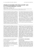
sues. We detected statistically highly significant upregulation
of BMP-2 mRNA (Figure 2a) and down-regulation of the
secreted WNT inhibitor FRZB mRNA (Figure 2b) 1 day after
injury (p < 0.01).
Mechanical injury is associated with modulation of the
BMP pathway
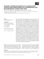
To determine the temporal window of BMP-2 mRNA regula-
tion, we performed a time course gene expression analysis at
5 hours, 1 day, and 6 days after injury. Statistically significant
(p < 0.05) upregulation of BMP-2 was detected already 5
hours after wounding and tended to subside within 6 days
(Figure 3a). Similar results were obtained in the absence of
serum, where a statistically significant (p < 0.05) upregulation
of BMP-2 mRNA was present 5 hour after injury (Figure 3b),
indicating that, under our experimental conditions, the regula-
tion of BMP-2 expression in response to mechanical injury is
not serum dependent.
To test whether the adult cartilage tissue is itself a target of
BMP signaling, we performed immunohistochemistry using an
antibody that recognizes the phosphorylated form of the MAD
homology domain 2 of SMAD-1 and SMAD-5 [25]. In the
explant pair obtained from normal articular cartilage, we
detected phospho-SMAD-1/-5-positive chondrocytes in all
cartilage layers in the uninjured as well as the injured explants
(83% in the uninjured explant versus 100% in the injured) (Fig-
ure 3f–h). However, in adjacent uncultured freshly dissected
articular cartilage, the proportion of phospho-SMAD-1/-5-pos-
itive cells was 41%, with nearly all positive cells localized in the
intermediate layer (Figure 3c–e,h). These results suggest that
the dissection of the cartilage explants from the joints may be
associated with a molecular response to wounding, which the
resting period in culture reverted only partially. Consistent with
this hypothesis, BMP-2, MMP-3, and MMP-13 mRNA levels
were lowest in the freshly dissected cartilage, intermediate in
the uninjured cultured explant, and highest in the injured
explant, while FRZB mRNA levels had an opposite trend. Sim-
ilar results for the proportions of phospho-SMAD-1/-5-positive
cells were found in injured and uninjured cartilage explants
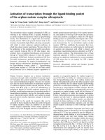
from OA cartilage. Finally, SMAD-1/5 phosphorylation was
confirmed in vivo in a mouse model of mechanical joint surface
injury (Figure 4). Full characterization of this model represents
an ongoing effort in our laboratory.
Activation of the WNT pathway following cartilage
mechanical injury
In a time course analysis, FRZB mRNA was already down-reg-
ulated in some but not all explant pairs 5 hours after injury (Fig-
ure 5a). Similar results were obtained with serum free culture
conditions (Figure 5b), thereby demonstrating that, under our
experimental conditions, FRZB mRNA regulation in response
to mechanical injury was not dependent on the presence of
FBS in the culture medium. Statistical analysis confirmed a
highly significant difference (p < 0.01) at the day 1 time point
in the presence of FBS and a significant difference (p < 0.05)
at the 5 hour and day 1 time points in the absence of serum.
At the protein level, FRZB was present in both injured and
uninjured explants as evaluated by immunohistochemistry (Fig-
ure 5c–f). The proportion of FRZB positive cells was signifi-
cantly lower (p < 0.05) in the injured explant in three
independent explant pairs, confirming at the protein level the
down-regulation of FRZB expression in the injured explants
(Figure 5g). Downregulation of FRZB was confirmed at protein
Figure 2
Differential expression of bone morphogenetic protein (BMP)-2 and FRZB mRNA following mechanical injuryDifferential expression of bone morphogenetic protein (BMP)-2 and
FRZB mRNA following mechanical injury. (a) BMP-2 mRNA was signif-
icantly upregulated and (b) FRZB mRNA significantly down-regulated
in most injured samples compared to uninjured adjacent controls. Val-
ues were calculated using a standard curve and normalized for the
housekeeping
β
actin gene. Diamonds indicate samples from pre-
served areas from joints affected by osteoarthritis; open squares indi-
cate the sample pair from healthy cartilage.
Arthritis Research & Therapy Vol 8 No 5 Dell'Accio et al.
Page 6 of 13
(page number not for citation purposes)
Figure 3
Activation of the bone morphogenetic protein (BMP) signaling pathwayActivation of the bone morphogenetic protein (BMP) signaling pathway. (a,b) Time course of the differential expression of BMP-2 mRNA in injured
versus uninjured explants in (a) the presence or (b) the absence of fetal bovine serum (FBS) in the culture medium. Values are normalized for the
housekeeping
β
actin gene and expressed as fold change of gene expression in the injured explants from paired uninjured controls. Diamonds indi-
cate samples from preserved areas from joints affected by osteoarthritis; open squares indicate the sample pair from healthy cartilage. (c-g) Immu-
nostaining for phosphorylated SMAD-1/-5 in: (c) freshly dissected normal cartilage; (g) the adjacent injured explant at day 1 after injury; (f) and the
adjacent uninjured control at the same time-point. (d) Larger magnification of the area shown in the square in (c). In the freshly dissected sample,
phosphorylated SMAD-1/-5-positive cells were detected predominantly in the intermediate layer indicated by the bracket in (c). (e) Image obtained
by false coloring in red the image in (d) and superimposing it on the fluorescent image in the blue channel documenting the nuclear DAPI counter-
stain. The DAB precipitate in the phosphorylated SMAD-1/-5-positive cells quenched the DAPI fluorescence and, therefore, in this panel, phosphor-
ylated SMAD-1/-5-positive cells appear red and the nuclei of negative cells appear blue. The top insets in (f,g) are large magnifications of the
corresponding squared areas. (h) A graphic summary of the proportion of phospho-SMAD-1/-5-positive cells and the expression of BMP-2, FRZB,
metalloproteinase (MMP)-3 and MMP-13 mRNAs in this experiment with normal adult human articular cartilage. Values are expressed as: percent of
positive cells for phospho-SMAD-1/-5; relative gene expression normalized for the housekeeping
β
actin gene; percent of the day 6 time point for
BMP-2, MMP-3 and MMP-13 mRNA; and percent of the freshly dissected cartilage for FRZB. *p < 0.05; **p < 0.01. D, day(s); H, hours; SF, serum
free medium.
Available online />Page 7 of 13
(page number not for citation purposes)
level in vivo in a mouse model of joint surface injury (Figure 4).
The down-regulation of the secreted inhibitor FRZB suggests
de-repression of WNT signaling. Thus, we next investigated
whether the expression of components of the WNT pathway
that are present in cartilage during mouse embryonic develop-
ment [29,30] is maintained in adult human articular cartilage.
We detected mRNA encoding WNT ligands (WNT-1, WNT-
5a, WNT-5b, WNT-9a/14, and WNT16), receptors (FRZD-1
and FRZD-7), intracellular mediators such as β-catenin, and
downstream transcription factors such as TCF and LEF-1
(data not shown). The presence of β-catenin was also con-
firmed at the protein level (Figure 5h–m).
We then investigated whether mechanical injury resulted in a
net activation of the canonical WNT pathway by performing
gene expression analysis of the WNT target genes Axin-2 [31]
and c-JUN [32,33]. Consistent with our hypothesis and with
the activation of the WNT/β-catenin signaling pathway, Axin-2
mRNA was upregulated 1 day after mechanical injury (Figure
6a), with a statistically highly significant difference (p < 0.01).
Figure 4
A figure showing modulation of the BMP and WNT pathway after mechanical injury in vivo in miceA figure showing modulation of the BMP and WNT pathway after mechanical injury in vivo in mice. Modulation of BMP and WNT pathway after
mechanical injury in vivo in mice. 7 week old C57BL/6 male mice were challenged in a model of joint surface injury in vivo. In this model the knee joint
surface is exposed by medial para-patellar arthrotomy and lateral patellar dislocation. A full thickness injury is made in the patellar groove using a
custom made device in which the length of a 26G needle is limited by a glass bead (injured knee), or left uninjured (sham operated control). In either
case the patellar dislocation is then reduced and the joint capsule and the skin sutured in separate layers and the mice allowed to walk freely. The
animals were killed at different time-points for histological and histochemical analysis. A-B immunohistochemistry for FRZB in sham operated (A) and
injured (B) articular cartilage 1 day after the operation. C-D immunohistochemistry for phosphorylated SMAD-1 in sham operated (A) and injured (B)
articular cartilage 6 days after the operation. The asterisk indicates the site of injury (occupied by debris). The dashed line indicates the margin of the
injury site.
Arthritis Research & Therapy Vol 8 No 5 Dell'Accio et al.
Page 8 of 13
(page number not for citation purposes)
c-JUN [32,33] was also significantly (p < 0.05) upregulated in
the injured explants, although to a lesser extent then Axin-2
(Figure 6b). To confirm that Axin-2 and c-JUN mRNA are
WNT targets in adult articular cartilage and under our experi-
mental conditions, we monitored the expression of these
genes after treatment with 10 mM LiCl, an inhibitor of GSK-3
Figure 5
Components of the canonical WNT pathway in adult human articular cartilageComponents of the canonical WNT pathway in adult human articular cartilage. (a,b) Time course of the differential expression of FRZB mRNA in
injured versus uninjured explants in (a) the presence or (b) the absence of fetal bovine serum (FBS) in the culture medium. Values were calculated
using a standard curve, normalized for the housekeeping
β
actin gene and expressed as fold change of gene expression in the injured explants from
paired uninjured controls. Diamonds indicate samples from preserved areas from joints affected by osteoarthritis; open squares indicate sample
pairs from healthy cartilage. (c-f) Immunohistochemical staining for FRZB protein (red) in (c) uninjured and (d) injured explants at the day 1 time
point. Haematoxylin was used as a nuclear counterstain. (e,f) Larger magnifications of the boxed areas in (c) and (d), respectively. (g) Percentage of
FRZB-positive cells in injured explants and in the paired uninjured controls from 3 independent donors as evaluated by immunohistochemistry. (h)
Haematoxylin-eosin and (i) safranin O stainings of an explant with a relatively high degree of osteoarthritis (modified Mankin score 5). (j-m) Immunos-
taining for β catenin in parallel, non-consecutive sections of (h) and (i). (j-l) Indirect immunofluorescence stainings for β catenin from a parallel sec-
tion in the area of (h) boxed with the dashed line (top). (k) β catenin (green). (l) DAPI counterstain of the same section (blue). (j) The superimposition
of (k) and (l). In this tissue, which is commonly called pannus, there were cells with a nuclear localization of β catenin. (m) Immunohistochemistry
showing the cytoplasmic localization of β catenin in chondrocytes of the basal layer (area in (h) boxed with a solid line). *p < 0.05; **p < 0.01. D,
day(s); H, hours; SF, serum free Medium.
Available online />Page 9 of 13
(page number not for citation purposes)
and, therefore, an activator of the β catenin-dependent WNT
signaling pathway [34]. The expression of Axin-2 and c-JUN
was consistently and significantly (p < 0.05) upregulated in
the LiCl-treated explants compared with the paired control
explants treated with NaCl (Figure 6c,d). To test the effects of
the activation of the canonical WNT pathway in adult human
articular cartilage, we determined the expression of the carti-
lage markers COL2A1 and Aggrecan in LiCl treated and con-
trol cultures. Under our experimental conditions, LiCl
treatment significantly (p < 0.05) upregulated COL2A1 and
Aggrecan mRNA, suggesting an anabolic effect (Figure 6e,f).
Discussion
The articular cartilage of adult individuals is commonly
regarded as a passive target of different pathogenic elements,
such as mechanical wear and inflammation, leading to carti-
lage matrix breakdown and loss of chondrocytes. However,
acute, small, full thickness JSDs appear to have repair capacity
in animals and humans, especially in young individuals [2,3,5-
8]. Repair of full thickness JSDs involves coordination of pat-
terning and tissue maturation that recapitulates some aspects
of embryonic skeletal development [6], thereby requiring mor-
phogenetic signaling. Here we have tested the hypothesis that
the injured articular cartilage may be a source of morphoge-
netic signals activated by damage. To this end we have used
an ex vivo model to investigate the modulation of gene expres-
sion induced by mechanical injury to adult human articular car-
tilage explants. We have detected upregulation of BMP-2
mRNA after injury. Several factors can determine activation of
BMP signaling independently of the expression of one ligand,
including secretion and solubility of the ligand(s), its/their bind-
ing to matrix molecules, the presence of secreted or intracellu-
lar inhibitors and receptor regulation [35]. Our data showing
phosphorylation of SMAD-1/-5 suggest activation of BMP sig-
naling.
BMPs elicit a well-documented anabolic response on cartilage
explants [20], and genetic evidence has been provided that
the BMP pathway is needed for joint homeostasis in adulthood
[36]. Indeed, targeted deletion of the gene encoding BMP
receptor 1A in the articular cartilage in mice results in joint sur-
face degeneration resembling OA [36]. In addition, BMPs
have been shown to regulate recruitment of chondroprogeni-
tors [37], synthesis of cartilage matrix, and endochondral bone
formation [20] during embryonic skeletogenesis. Finally, the
expression of BMP-2 mRNA is associated with the capacity of
in vitro expanded adult human articular chondrocytes to form
stable cartilage in vivo, resistant to vascular invasion and
endochondral bone formation [23]. Therefore, the recruitment
of progenitor cells, the regulation of endochondral bone forma-
tion and cartilage extracellular matrix synthesis, as well as the
preservation of the phenotypic stability of articular chondro-
cytes are all potential roles of BMP signaling in JSD repair.
However, it must be underscored that BMP signaling also
plays a part in the pathogenesis of joint diseases such as oste-
Figure 6
Activation of the WNT/β catenin canonical pathway following mechani-cal injuryActivation of the WNT/β catenin canonical pathway following mechani-
cal injury. (a) Axin-2 and (b) c-JUN mRNAs, two known transcriptional
targets of the WNT/β catenin canonical pathway, were upregulated 1
day after injury compared to uninjured controls. (c-f) Paired cartilage
explants were cultured in the presence of either 10 mM LiCl or 10 mM
NaCl for 1 day and then terminated for gene expression analysis by
quantitative real time PCR. Culture in the presence of LiCl induced the
upregulation of axin-2 (c) and c-JUN (d) mRNAs, thereby confirming
that these two genes are targets of the WNT/β catenin canonical path-
way in this experimental system. LiCl treatment also upregulated aggre-
can and COL2A1 mRNA (e,f). **p < 0.01. D, day(s); h, hours.
Arthritis Research & Therapy Vol 8 No 5 Dell'Accio et al.
Page 10 of 13
(page number not for citation purposes)
ophyte formation in OA [38] and enthesopathy [39]. Finally,
upregulation of BMP-2 has already been reported following
exposure of cartilage explants to interleukin 1 and tumor
necrosis factor alpha [40]. It is possible, therefore, that upreg-
ulation of BMP-2 may represent a response of the articular car-
tilage to different types of injuries.
In addition to the upregulation of BMP-2 mRNA, we have doc-
umented a consistent injury-associated down-regulation of the
secreted WNT inhibitor FRZB, suggesting de-repression of
the WNT signaling pathway. Consistently, we have detected,
in the injured explants, upregulation of mRNA encoding the
WNT/β catenin transcriptional targets Axin-2 and c-JUN. The
WNT signaling pathway can be regulated at multiple levels
[22] and, therefore, our experimental setup does not allow
determining whether the decreased expression of FRZB
mRNA is responsible for the detected upregulation of the
WNT/β catenin target genes. Nevertheless, the functional
importance of the regulation of FRZB expression in the context
of joint homeostasis is underscored by the observation that a
single nucleotide polymorphism causing loss of function of the
FRZB gene product is associated with hip OA in humans [41].
The function of WNT signaling in the context of joint surface
defect repair is still poorly understood. Studies on embryonic
tissues indicate that the activation of the canonical β catenin
pathway plays an important role in joint specification [30,42]
and in the regulation of chondrocyte differentiation inhibiting
chondrogenesis in immature mesenchymal cells and enhanc-
ing terminal differentiation in mature chondrocytes [29,32].
However, while the data in embryonic tissues suggest a gen-
eral inhibitory effect of canonical WNT signaling on chondro-
genesis, in experimental models utilizing adult cells, the
activation of the β catenin-dependent canonical WNT path-
way, under specific experimental conditions, rather appears to
promote chondrogenesis and cartilage differentiation [43-45].
This is in line with our findings that adult human articular carti-
lage explants cultured in the presence of LiCl upregulate
COL2A1 and aggrecan mRNA. Since in other organ systems
WNTs are involved in supporting repair processes by main-
taining a stem cell pool and specifying cell fates [19,46,47], it
is tempting to speculate that the canonical WNT pathway
would play a similar function in the repair of osteochondral
defects. Finally, there is also evidence that WNTs, at least
through the non-canonical pathway, may be implicated in joint
inflammation and may be detrimental for cartilage integrity
[48]. The most likely interpretation of these apparently con-
trasting data is that a tight regulation of the WNT and the BMP
pathways is necessary for proper joint homeostasis and repair
and that, in postnatal life, the same mechanisms that are set
into action to support repair may also play a pathogenic role
when de-regulated or when restoration of homeostasis fails. In
this regard, it is interesting that gain or loss of function of β cat-
enin in the developing skeleton both result in severe chondro-
dysplasia, although through different mechanisms [49].
We have encountered a high variability in the molecular
responses to injury in different pairs of cartilage explants. This
variability can be explained by the heterogeneity of tissues
from patient to patient, and by our inability to obtain adequately
'homogeneous' preparation of the explants. Analogous varia-
bility has been reported in the molecular response of cartilage
explants to inflammatory cytokines [50]. Indeed, the variability
in the molecular response to injury could be a factor contribut-
ing to the variability in the clinical outcome of untreated acute
articular cartilage injuries.
In some experiments, the differences in gene expression were
of small magnitude. However, we have observed a reproduci-
ble upregulation of WNT reporter genes, including Axin-2, fol-
lowing injury or LiCl treatment, which indicate that the
modulation of the wnt signaling was sufficient to induce a tran-
scriptional response. Axin-2 upregulation of approximately the
same magnitude was reported to be associated with
increased bone mass in osteoporotic lrp5
-/-
mice following oral
administration of LiCl [51]. Remarkably, the plasma levels of
LiCl achieved in that study were only 0.4 to 0.5 mM, which are
insufficient to trigger detectable wnt responses in the classic
assays such as β catenin nuclear localization or activation of
the TOP-FLASH reporter. It is reasonable that this magnitude
of wnt activation in adult animals is probably more physiologi-
cal than that achieved in overexpression experiments [51].
Indeed, in postnatal life, morphogenetic events take place at a
much lower rate than in embryonic development and, there-
fore, slight changes in the balance of the morphogenetic path-
ways can result in significant biological effects.
The in vitro culture conditions may influence the molecular
response to injury, potentially introducing artifacts. However,
the reproducibility of FRZB and BMP-2 mRNA regulation in
response to damage regardless of the presence of serum in
the culture medium suggests that this response is largely not
dependent on culture conditions. In addition, it is possible that
the response to injury in vivo will be more vibrant than that in
vitro because the resting period does not appear to be suffi-
cient to completely reverse the response due to the initial dis-
section of the explants. In this respect, Vincent and colleagues
[52] reported rapid phosphorylation of ERK following dissec-
tion of porcine articular cartilage explants, which was com-
pletely reverted after 48 hours of "resting" in culture. In our
study, the modulation of BMP-2 and FRZB mRNA appear to
last longer than 48 hours. This is also supported by the analy-
sis of the sample in Figure 3h, in which the expression levels
of all molecules tested and the number of phospho-SMAD-1/-
5-positive cells in the rested explant were intermediate
between the freshly dissected explant and the explant re-
injured after the resting period. Most importantly, we have
shown phosphorylation of SMAD-1/-5 and downregulation of
FRZB expression in vivo in a mouse model of joint surface
injury (Figure 4) not only confirming our data in vivo, but also
suggesting that such mechanisms are evolutionarily con-
Available online />Page 11 of 13
(page number not for citation purposes)
served. Functional studies are being performed to evaluate the
role of these molecular mechanisms in the context of cartilage
damage and repair. Full characterization of this model is an
ongoing effort in our laboratory.
Injuries to the articular cartilage result in activation of the bone
marrow and subchondral bone remodeling [53], suggesting
the presence of molecular signals that are released and target
the neighboring tissues. We have demonstrated that mechan-
ical injury in vitro can elicit the activation of two of the most
important signaling pathways involved in embryonic skele-
togenesis and joint morphogenesis, suggesting that the artic-
ular cartilage is capable of triggering a signaling machinery
that may play a role in joint surface repair.
Although several risk factors for OA have been identified,
including the nature and entity of the injury, age, genetic pre-
disposition, and joint congruity, it is still not clear why some
individuals can efficiently repair JSDs while some others will
develop chronic symptomatic lesions requiring surgical inter-
vention and possibly evolving into OA [1]. Failure of repair sig-
naling may contribute to evolution towards OA. Our data
suggest that morphogenetic pathways are transiently acti-
vated early following acute injury, as has been reported in
other organ systems [19]. Insufficient or untimely activation of
this machinery may result in repair failure. It is important, there-
fore, to study these events in a temporally dynamic fashion,
and it is possible that the early post-traumatic signals may be
critical for the final repair outcome. Understanding the molec-
ular mechanisms of repair may help us define a more focused
indication for biological JSD repair. On the other hand, the
modulation of these signaling pathways (e.g., by controlled
release of bioactive molecules from scaffolding biomaterials)
may complement the available tissue engineering approaches
to enhance specific aspects of repair. Finally, the persistence
in adulthood of locally residing stem cells within several joint
tissues, including bone marrow [54], synovial membrane [16],
periosteum [13], and articular cartilage [12,14,15,18], opens
the possibility to recruit and guide these cells locally using
appropriate molecular signals to enhance repair. This would
circumvent a number of problems associated with ex vivo cell
manipulation, including phenotypic instability, high costs, non-
optimal consistency, and complex regulation of the cellular
products [55].
Conclusion
Our data show modulation of the WNT and BMP signaling
pathways in adult human and mouse articular cartilage follow-
ing mechanical injury in vitro and in vivo. These molecular
events may contribute to trigger or support a repair response
and failure to promptly activate these reparative signals may
contribute to poor repair and poor clinical outcome. Hence,
activation of the WNT and BMP pathways in response to injury
may represent a prognostic marker and at the same time a
therapeutic target to enhance the early response of the joint
surface to acute injury.
Competing interests
The authors declare that they have no competing interests.
Authors' contributions
FD designed the study, performed the experiments and
drafted the manuscript. CD was involved in the study design,
in data interpretation, and drafting the manuscript. NE contrib-
uted to immunohistochemical stainings for FRZB. FB contrib-
uted to the optimization of the phospho-SMAD-1 staining. TM
critically revised the manuscript for important intellectual con-
tent. JO critically revised the manuscript for important intellec-
tual content. CP was involved in the study design,
interpretation of the results and has critically reviewed the
manuscript.
Acknowledgements
We wish to thank the Arthritis Research Campaign (ARC) for funding
this work (grant no. D0603) and Dr Dell'Accio's Fellowship; the ortho-
pedic surgeons at Guy's Hospital for providing cartilage samples; and
Frank P Luyten for critically reviewing the manuscript.
References
1. Buckwalter JA, Saltzman C, Brown T, Schurman DJ: The impact
of osteoarthritis: implications for research. Clin Orthop
2004:S6-S15.
2. Messner K, Maletius W: The long-term prognosis for severe
damage to weight-bearing cartilage in the knee: a 14-year clin-
ical and radiographic follow-up in 28 young athletes. Acta
Orthop Scand 1996, 67:165-168.
3. Shelbourne KD, Jari S, Gray T: Outcome of untreated traumatic
articular cartilage defects of the knee: a natural history study.
J Bone Joint Surg Am 2003, 85-A(Suppl 2):8-16.
4. Koshino T, Wada S, Ara Y, Saito T: Regeneration of degenerated
articular cartilage after high tibial valgus osteotomy for medial
compartmental osteoarthritis of the knee. Knee 2003,
10:229-236.
5. Linden B: Osteochondritis dissecans of the femoral condyles:
a long-term follow-up study. J Bone Joint Surg Am 1977,
59:769-776.
6. Shapiro F, Koide S, Glimcher MJ: Cell origin and differentiation
in the repair of full-thickness defects of articular cartilage. J
Bone Joint Surg Am 1993, 75:532-553.
7. Wei XF, Gao JF, Messner K: Maturation-dependent repair of
untreated osteochondral defects in the rabbit knee joint. J
Biomed Mater Res 1997, 34:63-72.
8. Breinan HA, Hsu HP, Spector M: Chondral defects in animal
models: effects of selected repair procedures in canines. Clin
Orthop Relat Res 2001:S219-S230.
9. Bentley G, Biant LC, Carrington RW, Akmal M, Goldberg A, Wil-
liams AM, Skinner JA, Pringle J: A prospective, randomised com-
parison of autologous chondrocyte implantation versus
mosaicplasty for osteochondral defects in the knee. J Bone
Joint Surg Br 2003, 85:223-230.
10. Knutsen G, Engebretsen L, Ludvigsen TC, Drogset JO, Grontvedt
T, Solheim E, Strand T, Roberts S, Isaksen V, Johansen O: Autol-
ogous chondrocyte implantation compared with microfracture
in the knee. A randomized trial. J Bone Joint Surg Am 2004, 86-
A:455-464.
11. Roberts S, McCall IW, Darby AJ, Menage J, Evans H, Harrison PE,
Richardson JB: Autologous chondrocyte implantation for carti-
lage repair: monitoring its success by magnetic resonance
imaging and histology. Arthritis Res Ther 2003, 5:R60-R73.
12. Barbero A, Ploegert S, Heberer M, Martin I: Plasticity of clonal
populations of dedifferentiated adult human articular
chondrocytes. Arthritis Rheum 2003, 48:1315-1325.
Arthritis Research & Therapy Vol 8 No 5 Dell'Accio et al.
Page 12 of 13
(page number not for citation purposes)
13. De Bari C, Dell'accio F, Vanlauwe J, Eyckmans J, Khan IM, Archer
CW, Jones EA, McGonagle D, Mitsiadis TA, Pitzalis C, et al.: Mes-
enchymal multipotency of adult human periosteal cells dem-
onstrated by single-cell lineage analysis. Arthritis Rheum 2006,
54:1209-1221.
14. Alsalameh S, Amin R, Gemba T, Lotz M: Identification of mesen-
chymal progenitor cells in normal and osteoarthritic human
articular cartilage. Arthritis Rheum 2004, 50:1522-1532.
15. Dell'accio F, Bari CD, Luyten FP: Microenvironment and pheno-
typic stability specify tissue formation by human articular car-
tilage-derived cells in vivo. Exp Cell Res 2003, 287:16-27.
16. De Bari C, Dell'accio F, Tylzanowski P, Luyten FP: Multipotent
mesenchymal stem cells from adult human synovial mem-
brane. Arthritis Rheum 2001, 44:1928-1942.
17. De Bari C, Dell'accio F, Vandenabeele F, Vermeesch JR, Raymack-
ers JM, Luyten FP: Skeletal muscle repair by adult human mes-
enchymal stem cells from synovial membrane. J Cell Biol
2003, 160:909-918.
18. Dowthwaite GP, Bishop JC, Redman SN, Khan IM, Rooney P,
Evans DJR, Haughton L, Bayram Z, Boyer S, Thomson B, et al.:
The surface of articular cartilage contains a progenitor cell
population. J Cell Sci 2004, 117:889-897.
19. Beachy PA, Karhadkar SS, Berman DM: Tissue repair and stem
cell renewal in carcinogenesis. Nature 2004, 432:324-331.
20. Luyten FP, Lories R, De Bari C, Dell'accio F: Bone morphoge-
netic proteins and the synovial joints. In Progress in Inflamma-
tion Research Edited by: Vukicevic S, Sampath TK. Basel/
Switzerland: Birkhauser Verlag; 2002:223-248.
21. Goldring MB, Tsuchimochi K, Ijiri K: The control of chondrogen-
esis. J Cell Biochem 2006, 97:33-44.
22. Nelson WJ, Nusse R: Convergence of Wnt, beta-catenin, and
cadherin pathways. Science 2004, 303:1483-1487.
23. Dell'accio F, De Bari C, Luyten FP: Molecular markers predictive
of the capacity of expanded human articular chondrocytes to
form stable cartilage in vivo. Arthritis Rheum 2001,
44:1608-1619.
24. Mankin HJ, Dorfman H, Lippiello L, Zarins A: Biochemical and
metabolic abnormalities in articular cartilage from osteo-
arthritic human hips. II. Correlation of morphology with bio-
chemical and metabolic data. J Bone Joint Surg Am 1971,
53:523-537.
25. Persson U, Izumi H, Souchelnytskyi S, Itoh S, Grimsby S, Engstrom
U, Heldin CH, Funa K, ten Dijke P: The L45 loop in type I recep-
tors for TGF-beta family members is a critical determinant in
specifying Smad isoform activation. FEBS Lett 1998,
434:83-87.
26. Hembry RM, Dyce J, Driesang I, Hunziker EB, Fosang AJ, Tyler JA,
Murphy G: Immunolocalization of matrix metalloproteinases in
partial-thickness defects in pig articular cartilage. A prelimi-
nary report. J Bone Joint Surg Am 2001, 83-A:826-838.
27. Lee JH, Fitzgerald JB, Dimicco MA, Grodzinsky AJ: Mechanical
injury of cartilage explants causes specific time-dependent
changes in chondrocyte gene expression. Arthritis Rheum
2005, 52:2386-2395.
28. Flannelly J, Chambers MG, Dudhia J, Hembry RM, Murphy G,
Mason RM, Bayliss MT: Metalloproteinase and tissue inhibitor
of metalloproteinase expression in the murine STR/ort model
of osteoarthritis. Osteoarthritis Cartilage 2002, 10:722-733.
29. Hartmann C, Tabin CJ: Dual roles of Wnt signaling during chon-
drogenesis in the chicken limb. Development 2000,
127:3141-3159.
30. Guo X, Day TF, Jiang X, Garrett-Beal L, Topol L, Yang Y: Wnt/
beta-catenin signaling is sufficient and necessary for synovial
joint formation. Genes Dev 2004, 18:2404-2417.
31. Yan D, Wiesmann M, Rohan M, Chan V, Jefferson AB, Guo L,
Sakamoto D, Caothien RH, Fuller JH, Reinhard C, et al.: Elevated
expression of axin2 and hnkd mRNA provides evidence that
Wnt/beta -catenin signaling is activated in human colon
tumors. Proc Natl Acad Sci USA 2001, 98:14973-14978.
32. Ryu JH, Kim SJ, Kim SH, Oh CD, Hwang SG, Chun CH, Oh SH,
Seong JK, Huh TL, Chun JS: Regulation of the chondrocyte phe-
notype by beta-catenin. Development 2002, 129:5541-5550.
33. Mann B, Gelos M, Siedow A, Hanski ML, Gratchev A, Ilyas M, Bod-
mer WF, Moyer MP, Riecken EO, Buhr HJ, et al.: Target genes of
beta-catenin-T cell-factor/lymphoid-enhancer-factor signaling
in human colorectal carcinomas. Proc Natl Acad Sci USA
1999, 96:1603-1608.
34. Sato N, Meijer L, Skaltsounis L, Greengard P, Brivanlou AH: Main-
tenance of pluripotency in human and mouse embryonic stem
cells through activation of Wnt signaling by a pharmacological
GSK-3-specific inhibitor. Nat Med 2004, 10:55-63.
35. Canalis E, Economides AN, Gazzerro E: Bone morphogenetic
proteins, their antagonists, and the skeleton. Endocr Rev
2003, 24:218-235.
36. Rountree RB, Schoor M, Chen H, Marks ME, Harley V, Mishina Y,
Kingsley DM: BMP receptor signaling is required for postnatal
maintenance of articular cartilage. PLoS Biol 2004, 2:e355.
37. Tsumaki N, Tanaka K, Arikawa-Hirasawa E, Nakase T, Kimura T,
Thomas JT, Ochi T, Luyten FP, Yamada Y: Role of CDMP-1 in
skeletal morphogenesis: promotion of mesenchymal cell
recruitment and chondrocyte differentiation. J Cell Biol 1999,
144:161-173.
38. Scharstuhl A, Vitters EL, van der Kraan PM, van Den Berg WB:
Reduction of osteophyte formation and synovial thickening by
adenoviral overexpression of transforming growth factor
beta/bone morphogenetic protein inhibitors during experi-
mental osteoarthritis. Arthritis Rheum 2003, 48:3442-3451.
39. Lories RJ, Derese I, Luyten FP: Modulation of bone morphoge-
netic protein signaling inhibits the onset and progression of
ankylosing enthesitis. J Clin Invest 2005, 115:1571-1579.
40. Fukui N, Zhu Y, Maloney WJ, Clohisy J, Sandell LJ: Stimulation of
BMP-2 expression by pro-inflammatory cytokines IL-1 and
TNF-alpha in normal and osteoarthritic chondrocytes. J Bone
Joint Surg Am 2003, 85-A(Suppl 3):59-66.
41. Loughlin J, Dowling B, Chapman K, Marcelline L, Mustafa Z,
Southam L, Ferreira A, Ciesielski C, Carson DA, Corr M: Func-
tional variants within the secreted frizzled-related protein 3
gene are associated with hip osteoarthritis in females. Proc
Natl Acad Sci USA 2004, 101:9757-9762.
42. Hartmann C, Tabin CJ: Wnt-14 plays a pivotal role in inducing
synovial joint formation in the developing appendicular skele-
ton. Cell 2001, 104:341-351.
43. Yates KE: Demineralized bone alters expression of Wnt net-
work components during chondroinduction of post-natal
fibroblasts. Osteoarthritis Cartilage 2004, 12:497-505.
44. Zhou S, Eid K, Glowacki J: Cooperation between TGF-beta and
Wnt pathways during chondrocyte and adipocyte differentia-
tion of human marrow stromal cells. J Bone Miner Res 2004,
19:463-470.
45. Yano F, Kugimiya F, Ohba S, Ikeda T, Chikuda H, Ogasawara T,
Ogata N, Takato T, Nakamura K, Kawaguchi H, et al.: The canon-
ical Wnt signaling pathway promotes chondrocyte differentia-
tion in a Sox9-dependent manner. Biochem Biophys Res
Commun 2005, 333:1300-1308.
46. Hill TP, Spater D, Taketo MM, Birchmeier W, Hartmann C: Canon-
ical Wnt/beta-catenin signaling prevents osteoblasts from dif-
ferentiating into chondrocytes. Dev Cell 2005, 8:727-738.
47. Day TF, Guo X, Garrett-Beal L, Yang Y: Wnt/beta-catenin signal-
ing in mesenchymal progenitors controls osteoblast and
chondrocyte differentiation during vertebrate skeletogenesis.
Dev Cell 2005, 8:739-750.
48. Sen M, Lauterbach K, El Gabalawy H, Firestein GS, Corr M, Car-
son DA: Expression and function of wingless and frizzled
homologs in rheumatoid arthritis. Proc Natl Acad Sci USA
2000, 97:2791-2796.
49. Akiyama H, Lyons JP, Mori-Akiyama Y, Yang X, Zhang R, Zhang Z,
Deng JM, Taketo MM, Nakamura T, Behringer RR, et al.: Interac-
tions between Sox9 and beta-catenin control chondrocyte dif-
ferentiation. Genes Dev 2004, 18:1072-1087.
50. Kobayashi M, Squires GR, Mousa A, Tanzer M, Zukor DJ, Antoniou
J, Feige U, Poole AR: Role of interleukin-1 and tumor necrosis
factor alpha in matrix degradation of human osteoarthritic car-
tilage. Arthritis Rheum 2005, 52:128-135.
51. Clement-Lacroix P, Ai M, Morvan F, Roman-Roman S, Vayssiere B,
Belleville C, Estrera K, Warman ML, Baron R, Rawadi G: Lrp5-
independent activation of Wnt signaling by lithium chloride
increases bone formation and bone mass in mice. Proc Natl
Acad Sci USA 2005, 102:17406-17411.
52. Vincent T, Hermansson M, Bolton M, Wait R, Saklatvala J: Basic
FGF mediates an immediate response of articular cartilage to
mechanical injury. Proc Natl Acad Sci USA 2002,
99:8259-8264.
53. Vasara AI, Hyttinen MM, Lammi MJ, Lammi PE, Langsjo TK, Lindahl
A, Peterson L, Kellomaki M, Konttinen YT, Helminen HJ, et al.:
Available online />Page 13 of 13
(page number not for citation purposes)
Subchondral bone reaction associated with chondral defect
and attempted cartilage repair in goats. Calcif Tissue Int 2004,
74:107-114.
54. Luyten FP, Dell'accio F, De Bari C: Skeletal tissue engineering:
opportunities and challenges. Best Pract Res Clin Rheumatol
2001, 15:759-769.
55. De Bari C, Pitzalis C, Dell'accio F: Reparative Medicine: from tis-
sue engineering to joint surface regeneration. Regenerative
Med 2006, 1:59-69.