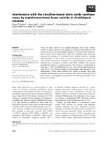
Báo cáo khoa học: "Experience with adjuvant chemotherapy for pseudomyxoma peritonei secondary to mucinous adenocarcinoma of the appendix with oxaliplatin/fluorouracil/leucovorin (FOLFOX4)" docx
Bạn đang xem bản rút gọn của tài liệu. Xem và tải ngay bản đầy đủ của tài liệu tại đây (583.88 KB, 7 trang )
BioMed Central
Page 1 of 7
(page number not for citation purposes)
World Journal of Surgical Oncology
Open Access
Case report
Experience with adjuvant chemotherapy for pseudomyxoma
peritonei secondary to mucinous adenocarcinoma of the appendix
with oxaliplatin/fluorouracil/leucovorin (FOLFOX4)
Chin-Fan Chen
1,4
, Che-Jen Huang*
1,3
, Wan-Yi Kang
2
and Jan-Sing Hsieh
1,3
Address:
1
Department of Surgery, Kaohsiung Medical University Hospital, Kaohsiung 807, Taiwan,
2
Department of Pathology, Kaohsiung Medical
University Hospital, Kaohsiung 807, Taiwan,
3
Faculty of Medicine, College of Medicine, Kaohsiung Medical University, Kaohsiung 807, Taiwan
and
4
Department of Surgery, Pingtung Hospital, Department of Health, Executive Yuan, Ping-Tung 900, Taiwan
Email: Chin-Fan Chen - ; Che-Jen Huang* - ; Wan-Yi Kang - ; Jan-
Sing Hsieh -
* Corresponding author
Abstract
Background: Pseudomyxoma peritonei (PMP) is a rare condition characterized by mucinous
tumors, disseminated intra-peritoneal implants, and mucinous ascites. So far its diagnosis remains
challenging to most clinicians.
Case presentation: A 55-year-old male patient had suffered from acute onset of abdominal pain
and abdominal distension for one day prior to his admission. Physical examination revealed
tenderness over the right lower quadrant of the abdomen without diffuse muscle guarding. A large
amount of ascites was identified by abdominal computed tomography (CT) scan. Paracentesis
showed the appearance of sticky mucinous ascites. He underwent laparotomy under the
impression of pseudomyxoma peritonei. There was a lot of mucinous ascites, one appendiceal
tumor and multiple peritoneal implants disseminated from the subphrenic space to the recto-
vesicle pouch. Pseudomyxoma Peritonei caused by mucinous adenocarcinoma of appendiceal
origin, was confirmed by histopathology. We performed an excision of the appendiceal tumor
combined with copious irrigation and debridement. After the operation, he received 10 cycles of
systemic chemotherapy with FOLFOX4 regimen, without specific morbidity. Follow-up of
abdominal CT and colonoscopy at post-operative 17 months showed excellent response without
evidence of local recurrence or distal metastasis. He made an uneventful recovery (up to the
present) for 21 months after the operation.
Conclusion: This case report emphasizes the possible new role of systemic chemotherapy in the
treatment of patients with this rare clinical syndrome.
Background
Pseudomyxoma peritonei (PMP) is a rare condition char-
acterized by mucinous tumors, disseminated intra-perito-
neal implants, and mucinous ascites. It may represent a
pathologic diagnostic term to both benign and malignant
mucinous neoplasms that produce abundant extracellular
mucin. Therefore, poorly predictable clinical course and
variable prognosis could be expected. A definitive diagno-
sis of PMP requires the presence of mucinous neoplastic
epithelium and mucinous ascites, and may also include
Published: 11 November 2008
World Journal of Surgical Oncology 2008, 6:118 doi:10.1186/1477-7819-6-118
Received: 3 July 2008
Accepted: 11 November 2008
This article is available from: />© 2008 Chen et al; licensee BioMed Central Ltd.
This is an Open Access article distributed under the terms of the Creative Commons Attribution License ( />),
which permits unrestricted use, distribution, and reproduction in any medium, provided the original work is properly cited.
World Journal of Surgical Oncology 2008, 6:118 />Page 2 of 7
(page number not for citation purposes)
the diffuse mucinous implants [1]. In spite of more
detailed understanding of PMP based on the clinical case
series, there is still some debate about its clinical behavior,
pathogenesis, and treatment strategy. We report our clini-
cal experience concerning systemic chemotherapy
(FOLFOX4 regimen) for one case of pseudomyxoma peri-
tonei secondary to appendiceal mucinous adenocarci-
noma and review the literature.
Case presentation
A 55-year-old male patient had suffered from acute onset
of abdominal pain and abdominal distension for one day
prior to his admission. He had previously been healthy
without any specific underlying disease. Unfortunately,
nausea and vomiting were noted twelve hours after his
onset of abdominal pain. There was no fever, chills, or
diarrhea. The characteristics of his abdominal pain
included steady dull pain over the periumbilical and
lower abdomen. On general physical examination, we
found the patient presenting abdominal distension,
hypoactive bowel sound, and diffuse tenderness over the
whole abdomen, and localized muscle guarding over the
right lower abdomen. Laboratory data showed predomi-
nant neutrophil (94%) without leukocytosis (white blood
cell count 9160/μl). A large amount of ascites was identi-
fied by abdominal sonography and paracentesis was
done. It showed the appearance of sticky mucinous ascites
with the result of monocyte predominant in the ascites
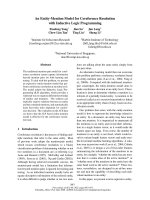
study. Abdominal computed tomography (CT) showed
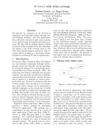
mucin septations (Fig. 1), thickening of the omentum and
scalloping of hepatic and splenic margins (Fig. 2), which
were compatible with the characteristics of the image of
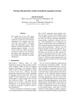
pseudomyxoma peritonei (PMP). He underwent laparot-
omy and much yellowish-greenish jelly-like material in
the peritoneal cavity was noted (Fig. 3). Besides, one
appendiceal tumor and multiple peritoneal implants dis-
seminated from subphrenic space to the recto-vesicle
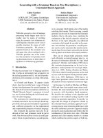
pouch. Intra-operative frozen section of appendiceal
tumor and one of the peritoneal implants confirmed
mucinous adenocarcinoma of the appendix (Fig. 4 &5).
The diagnosis of PMP was confirmed by the final histopa-
thology. Instead of the aggressive peritonectomy proce-
dures, we performed excision of the appendiceal tumor;
local debridement and copious irrigation. After the oper-
ation, he received 10 cycles of systemic chemotherapy
with FOLFOX4 regimen (oxaliplatin 85 mg/m
2
as a two-
hour infusion on day 1, leucovorin 200 mg/m
2
as a two-
hour infusion concurrently with oxaliplatin on day 1, fol-
lowed by a bolus of 5-FU 400 mg/m
2
and continuous
infusion of 5-FU 600 mg/m
2
over 22 hours), without spe-
cific morbidity [2]. Abdominal CT and colonoscopy at
post-operative 17 months showed complete response
without evidence of local recurrence or distal metastasis
(Fig. 6 &7). Follow-up serum carcinoembryonic antigen
(CEA) level also showed no progressive activity of his dis-
ease. He remains well currently, and receives follow-up
regularly in our outpatient clinic.
CT scan of abdomen showing pseudomyxoma peritonei with mucin septations (arrows)Figure 1
CT scan of abdomen showing pseudomyxoma peri-
tonei with mucin septations (arrows).
CT scan of abdomen showing pseudomyxoma peritonei with scalloping of hepatic margin (arrows)Figure 2
CT scan of abdomen showing pseudomyxoma peri-
tonei with scalloping of hepatic margin (arrows).
World Journal of Surgical Oncology 2008, 6:118 />Page 3 of 7
(page number not for citation purposes)
Discussion
Pseudomyxoma peritonei (PMP) is a rare clinical syn-
drome with an estimated incidence of approximately one
per million per year and preferentially affects women (2–
3 times more common than men) [3,4]. Since Werth [5]
first described PMP produced by an ovarian neoplasm
Multiple peritoneal implants (arrows) over visceral perito-neumFigure 3
Multiple peritoneal implants (arrows) over visceral
peritoneum. Mucinous ascites with yellowish-greenish
materials (arrow head) in peritoneal cavity.
The pathological findings of the resected appendixFigure 4
The pathological findings of the resected appendix.
Mucinous adenocarcinoma (arrows) exhibiting abundant
acellular mucin pooling (arrow head), with scarce well-differ-
entiated mucin producing epithelium embedded in a fibrous
matrix or as lining epithelium.
The pathological findings of the resected appendix extra-cel-lular mucinous materials (arrow) showing light blue in color distributed in fibrous stromaFigure 5
The pathological findings of the resected appendix
extra-cellular mucinous materials (arrow) showing
light blue in color distributed in fibrous stroma.
CT scan of abdomen – postoperative 17 months, compared with Fig 1Figure 6
CT scan of abdomen – postoperative 17 months,
compared with Fig 1. No local recurrence or metastatic
lesion is identified.
World Journal of Surgical Oncology 2008, 6:118 />Page 4 of 7
(page number not for citation purposes)
and Frankel [6] first reported on the association of PMP
with an appendiceal mucocele, there have been many
reports focusing on the pathogenesis, diagnosis, treat-
ment and prognosis of PMP.
The primary origin of the mucinous peritoneal implants
in PMP has remained controversial for a long time. Some
reports indicated that ovarian tumor is more likely to be
the primary neoplasm of PMP [3,7], however, others favor
the appendiceal tumor as the answer [4,8,9]. Because
immunohistochemical stains and molecular genetic stud-
ies both show the evidence of these tumors being second-
ary to appendiceal neoplasms [10-12], most people agree
that the primary tumor of PMP is predominately a muci-
CT scan of abdomen – postoperative 17 months, compared with Fig 2Figure 7
CT scan of abdomen – postoperative 17 months, compared with Fig 2. No local recurrence or metastatic lesion is
identified.
World Journal of Surgical Oncology 2008, 6:118 />Page 5 of 7
(page number not for citation purposes)
nous epithelial neoplasm of the appendix. Other possible
primary sites being reported include: colorectum, gall-
bladder, pancreas, urachus, urinary bladder, breast and
lung, but these are uncommon [13,14]. PMP is character-
ized by an abundant amount of mucinous ascites pro-
duced by adenomucinous tumor cells in implants on
peritoneal surfaces. These implants are the final stage of a
distribution process following the rupture of the muci-
nous neoplasm. The key associated finding is the presence
of epithelium outside the appendix in association with
the mucin and the peritoneal implants [1].
Ronnett et al [15] first described a widely accepted and
useful definition of PMP. They classified PMP into three
pathological subtypes with different pathological charac-
teristics and different prognosis: disseminated peritoneal
adenomucinosis (DPAM), peritoneal mucinous carcino-
matosis (PMCA), and an intermediate subtype (PMCA-I).
Histopathologically, DPAM is characterized by an abun-
dance of mucus with focally adenomucinous epithelium
without atypia or mitotic activity. In contrast to this,
PMCA is characterized by peritoneal tumor composed of
more abundant mucinous tumor cells with the architec-
ture and cytological features of carcinoma. Finally, the
intermediate subtype PMCA-I is characterized by an abun-
dance of DPAM lesions, but with focal areas with PMCA
lesions [15,16].
Besides the histopathological difference, their clinical
behaviors are also quite different. DPAM remains poten-
tially non-invasive and stays localized to the abdomen
without metastatic behavior. In contrast to this, PMCA
behaves with invasive and metastatic potential, as is char-
acteristic of mucinous adenocarcinoma. Patients with
PCMA are associated with the possibilities of liver, lung
and lymph node metastatic disease [17].
As symptoms remain non-specific, PMP presents a great
diagnostic challenge to clinicians. A precise diagnosis is
difficult due to the lack of specific symptoms in the early
stages of the disease. The most important symptom is a
gradually increasing abdominal girth. Patients may
present a typical "jelly belly" appearance [18]. Sometimes
patients present symptoms mimicking appendicitis with
intra-operative identification of a perforated appendiceal
mucocele. In other cases, they present an inguinal herni-
ated sac or an ovarian mass [18]. For 30% of female
patients, the first symptom is an ovarian mass [16].
Routine laboratory studies are seldom helpful in making
this diagnosis. Ultrasound is useful for initial establish-
ment of the diagnosis. Echo-guided paracentesis may
reveal mucinous ascites. Abdominal computed tomogra-
phy (CT) scan may demonstrate the characteristics of
mucinous ascites by analyzing the density properties
(Hounsfield Units [H.U.]), as it is significantly higher (5–
20 H.U.) than normal ascites (0 H.U.) [16]. CT may also
show the characteristic of "scalloping effect" on the sur-
face of the visceral organs resulting from compression by
the viscous mucinous secretions [1]. In the majority of
cases, however, PMP is often an unexpected finding of
laparoscopy or exploratory laparotomy [19].
When mucinous tumors on the peritoneal surface or
mucinous ascites are visualized on CT or during abdomi-
nal surgery, treatment of PMP should be performed since
untreated PMP patients will eventually suffer from intesti-
nal obstruction and mortality [20]. In spite of the contro-
versial standard treatment strategies for PMP, the current
mainstay of the treatment remains surgical resection of
the lesions. Alternative non-surgical treatment, such as:
peritoneal washing with 5% dextrose and systemic chem-
otherapy, have been reported; however, their roles in PMP
are still uncertain because of the limited case number and
short follow-up time [21,22].
Repeated cytoreductive surgical de-bulking procedures as
treatment for PMP have been described in earlier litera-
tures. Since the 1990s, a new combined treatment
approach was introduced by Sugarbaker et al [23]. They
defined it as peritonectomy procedures in combination
with intra-operative hyperthermic intra-peritoneal chem-
otherapy (HIPEC). Now this new combination treatment
is increasingly performed as treatment for PMP patients,
with promising results [24,25]. The available evidence
suggests that cytoreductive surgery with perioperative
intraperitoneal chemotherapy should replace serial de-
bulking as the standard of care for patients with peritoneal
spread of appendiceal epithelial neoplasms [20]. PMCA
behaves like peritoneal carcinomatosis from the original
colorectal adenocarcinoma; however, its poor prognosis
in comparison with DPAM after the similar management
by cytoreductive surgery and HIPEC was still demon-
strated in the two studies conducted by Sugarbaker et al
[26]. and Smeenk et al [27]. Moreover, Verwaal et al, also
showed a similar result in their randomized study, that
patients with peritoneal carcinomatosis of colorectal can-
cer (CRC) origin combined with cancer implant involve-
ment of six or more regions of the abdominal cavity, got
little survival benefit after the cytoreductive surgical pro-
cedures and intra-operative HIPEC [28].
Herein, further clinical trials with investigations of differ-
ent treatment strategies for patients with PMCA, are still
needed. This contributes to the application of systemic
chemotherapy for the patient in our case report since the
new therapeutic agent Oxaliplatin and its combination
with 5-fluorouracil/leucovorin (FOLFOX4 regimen) has
been used widely as first-line treatment in patients with
advanced CRC, and the promising results in these patients
World Journal of Surgical Oncology 2008, 6:118 />Page 6 of 7
(page number not for citation purposes)
have been demonstrated in several randomized studies
[29,30].
The effect of systemic chemotherapy in PMP seems ques-
tionable. Jones et al [22] reported their experience in the
treatment of pseudomyxoma peritonei of ovarian origin
with cisplatinum, doxorubicin, and cyclophosphamide,
with excellent responses. On the other hand, Smeenk et al
[31] reported the poor response of six patients (3 patients
with DPAM, another 3 patients with PMCA-I, and all 6
patients with lesions diffusely spread throughout the
abdomen) after 5-FU based systemic chemotherapy, and
subsequent poor prognosis was noted in the study.
Regarding the benefit of new therapeutic agents (includ-
ing Capecitabine, Oxaliplatin, Irinotecan and Bevacizu-
mab) and modern schedules for patients with metastatic
CRC, clinical experience with the use of these agents for
PMP are still absent, and it is questionable whether they
will do any better in this situation, especially for patients
with PMCA. Due to the limited experience and indetermi-
nate effects of systemic chemotherapy in PMP, some stud-
ies still suggest that systemic therapy should be reserved
for a palliative setting in patients with recurrent or pro-
gressive disease [20,32].
Because of the high grade mucinous adenocarcinoma of
the appendix with disseminated peritoneal lesions both
confirmed by histopathology, instead of the treatment
strategies (including aggressive peritonectomy proce-
dures), we used oxaliplatin/5-FU/leucovorin combina-
tion systemic chemotherapy (FOLFOX4 regimen) in our
patient after less invasive surgery, as the treatment for the
metastatic colorectal cancer. After 10 cycles of FOLFOX4
chemotherapy, excellent response was achieved. Colonos-
copy and abdominal CT scan 17 months after the opera-
tion both showed no evidence of development of local
recurrent or metastatic lesions. The patient had an une-
ventful recovery up to now without any disease-related
morbidity. We believe that our experience is one of the
few reports about effective treatment of PMP with sys-
temic chemotherapy. More clinical experience and further
studies are still needed for determination of the benefit of
systemic chemotherapy for these patients.
Conclusion
We report our clinical experience regarding the use of sys-
temic chemotherapy (FOLFOX4 regimen) for one case of
pseudomyxoma peritonei secondary to mucinous adeno-
carcinoma of the appendix. This case report emphasizes
the possible new role of systemic chemotherapy in the
treatment of patients with this rare clinical syndrome.
Consent
Written informed consent was taken from the patient for
publication of this case report.
Competing interests
The authors declare that they have no competing interests.
Authors' contributions
CFC performed the initial surgery, conceptualized the case
report, gathered the data, reviewed the literature and
drafted the manuscript. CJH performed the initial surgery,
took responsibility for the patient's postoperative care and
revised the manuscript. WYK assessed the histological
specimens and prepared the histological slides. JSH
reviewed the clinical data and helped to draft and revise
the manuscript. All authors read and approved the final
manuscript.
References
1. Li C, Kanthan R, Kanthan SC: Pseudomyxoma peritonei – a
revisit: report of 2 cases and literature review. World J Surg
Oncol 2006, 4:60.
2. de Gramont A, Figer A, Seymour M, Homerin M, Hmissi A, Cassidy J,
Boni C, Cortes-Funes H, Cervantes A, Freyer G, Papamichael D, Le
Bail N, Louvet C, Hendler D, de Braud F, Wilson C, Morvan F, Bonetti
A: Leucovorin and fluorouracil with or without oxaliplatin as
first-line treatment in advanced colorectal cancer. J Clin Oncol
2000, 18:2938-2947.
3. Galani E, Marx GMF, Steer CB, Culora G, Harper PG: Pseu-
domyxoma peritonei: the 'controversial' disease. Int J Gynecol
Cancer 2003, 13:413-418.
4. Mukherjee A, Parvaiz A, Cecil TD, Moran BJ: Pseudomyxoma peri-
tonei usually originates from the appendix: a review of the
evidence. Eur J Gynaecol Oncol 2004, 25:411-414.
5. Werth R: Klinische und anatomische untersuchungen zur
lehre von den bauchgeschwuelsten und der laparotomie.
Arch Gynaecol Obstet 1884, 24:100-118.
6. Frankel E: Uber das sogenannte pseudomyxoma peritonei.
Med Wochenschr 1901, 48:965-970.
7. Ronnett BM, Kurman RJ, Zahn CM, Shmookler BM, Jablonski KA,
Kass ME, Sugarbaker PH: Pseudomyxoma peritonei in women:
a clinicopathologic analysis of 30 cases with emphasis on site
of origin, prognosis, and relationship to ovarian mucinous
tumors of low malignant potential. Hum Pathol 1995,
26:509-524.
8. van Ruth S, Acherman YIZ, Vijver MJ van de, Hart AAM, Verwaal VJ,
Zoetmulder FAN: Pseudomyxoma peritonei: a review of 62
cases. Eur J Surg Oncol 2003, 29:682-688.
9. Young RH, Gilks CB, Scully RE: Mucinous tumors of the appendix
associated with mucinous tumors of the ovary and pseu-
domyxoma peritonei. A clinicopathological analysis of 22
cases supporting an origin in the appendix. Am J Surg Pathol
1991, 15:415-429.
10. Prayson RA, Hart WR, Petras RE: Pseudomyxoma peritonei. A
clinicopathologic study of 19 cases with emphasis on site of
origin and nature of associated ovarian tumors. Am J Surg
Pathol 1994, 18:591-603.
11. Szych C, Staebler A, Connolly DC, Wu R, Cho KR, Ronnett BM:
Molecular genetic evidence supporting the clonality and
appendiceal origin of pseudomyxoma peritonei in women.
Am J Pathol 1999, 154:1849-1855.
12. Ronnett BM, Shmookler BM, Diener-West M, Sugarbaker PH, Kur-
man RJ: Immunohistochemical evidence supporting the
appendiceal origin of pseudomyxoma peritonei in women.
Int J Gynecol Pathol 1997, 16(1):1-9.
13. de Bree E, Witkamp A, Vijver M Van De, Zoetmulde F: Unusual ori-
gins of Pseudomyxoma peritonei. J Surg Oncol 2000, 75:270-274.
14. Sherer DM, Abulafia O, Fliakim R: Pseudomyxoma peritonei. A
review of current literature. Gynecol Obstet Invest 2001, 51:73-80.
15. Ronnett BM, Zahn CM, Kurman RJ, Kass ME, Sugarbaker PH,
Shmookler BM: Disseminated peritoneal adenomucinosis and
peritoneal mucinous carcinomatosis. A clinicopathologic
analysis of 109 cases with emphasis on distinguishing patho-
logic features, site of origin, prognosis, and relationship to
pseudomyxoma peritonei. Am J Surg Pathol 1995, 19:1390-1408.
Publish with BioMed Central and every
scientist can read your work free of charge
"BioMed Central will be the most significant development for
disseminating the results of biomedical research in our lifetime."
Sir Paul Nurse, Cancer Research UK
Your research papers will be:
available free of charge to the entire biomedical community
peer reviewed and published immediately upon acceptance
cited in PubMed and archived on PubMed Central
yours — you keep the copyright
Submit your manuscript here:
/>BioMedcentral
World Journal of Surgical Oncology 2008, 6:118 />Page 7 of 7
(page number not for citation purposes)
16. Smeenk RM, Verwaal VJ, Zoetmulder FAN: Tumor Review-pseu-
domyxoma peritonei. Cancer Treatment Reviews 2007,
33:138-145.
17. Bradley RF, Stewart JH, Russell GB, Levine EA, Geisinger KR: Pseu-
domyxoma peritonei of appendiceal origin: a clinicopatho-
logic analysis of 101 patients uniformly treated at a single
institution, with literature review. Am J Surg Pathol 2006,
30:551-559.
18. Esquivel J, Sugarbaker PH: Clinical presentation of the pseu-
domyxoma peritonei syndrome. Br J Surg 2000, 87:1414-1418.
19. Sugarbaker PH: New standard of care for appendiceal epithe-
lial neoplasms and pseudomyxoma peritonei syndrome? Lan-
cet Oncol 2006, 7:69-76.
20. Sugarbaker PH: Pseudomyxoma peritonei. Cancer Treat Res 1996,
81:105-119.
21. Green N, Gancedo H, Smith R, Bernett G: Pseudomyxoma peri-
tonei – nonoperative management and biochemical findings.
A case report. Cancer 1975, 36:1834-1837.
22. Jones CM III, Homesley HD: Successful treatment of pseu-
domyxoma peritonei of ovarian origin with cisplatinum, dox-
orubicin, and cyclophosphamide. Gynecol Oncol 1985,
22:257-259.
23. Sugarbaker PH, Ronnett BM, Archer A, Averbach AM, Bland R, Chang
D, Dalton RR, Ettinghausen SE, Jacquet P, Jelinek J, Koslowe P, Kur-
man RJ, Shmookler B, Stephens AD, Steves MA, Stuart OA, White S,
Zahn CM, Zoetmulder FA: Pseudomyxoma peritonei syn-
drome. Adv Surg 1996, 30:233-280.
24. Sugarbaker PH: Cytoreductive surgery and peri-operative
intraperitoneal chemotherapy as a curative approach to
pseudomyxoma peritonei syndrome. Eur J Surg Oncol 2001,
27:239-243.
25. Loungnarath R, Causeret S, Bossard N, Faheez M, Sayag-Beaujard AC,
Brigand C, Gilly F, Glehen O: Cytoreductive surgery with intra-
peritoneal chemohyperthermia for the treatment of pseu-
domyxoma peritonei: a prospective study. Dis Colon Rectum
2005, 48:1372-1379.
26. Sugarbaker PH, Chang D: Results of treatment of 385 patients
with peritoneal surface spread of appendiceal malignancy.
Ann Surg Oncol 1999, 6:727-731.
27. Smeenk RM, Verwaal VJ, Antonini N, Zoetmulder FAN: Survival
analysis of pseudomyxoma peritonei patients treated by
cytoreductive surgery and hyperthermic intraperitoneal
chemotherapy. Annals of Surgery 2007, 25(1):104-109.
28. Verwaal VJ, van Ruth S, de Bree E, van Sloothen GW, van Tinteren H,
Boot H, Zoetmulder FA: Randomized trial of cytoreduction and
hyperthermic intraperitoneal chemotherapy versus sys-
temic chemotherapy and palliative surgery in patients with
peritoneal carcinomatosis of colorectal cancer. J Clin Oncol
2003, 21:3737-3743.
29. Goldberg RM, Sargent DJ, Morton RF, Fuchs CS, Ramanathan RK,
Williamson SK, Findlay BP, Pitot HC, Alberts S: A randomized con-
trolled trial of fluorouracil plus leucovorin, irinotecan, and
oxaliplatin combinations in patients with previously
untreated metastatic colorectal cancer. J Clin Oncol 2004,
22:23-30.
30. Nordlinger B, Sorbye H, Glimelius B, Poston GJ, Schlag PM, Rougier
P, Bechstein WO, Primrose JN, Walpole ET, Finch-Jones M, Jaeck D,
Mirza D, Parks RW, Collette L, Praet M, Bethe U, van Cutsem E,
Scheithauer W, Gruenberger T: Perioperative chemotherapy
with FOLFOX4 and surgery versus surgery alone for resect-
able liver metastases from colorectal cancer (EORTC Inter-
group trial 40983): a randomised controlled trial. Lancet 2008,
371:1007-1016.
31. Smeenk RM, Verwaal VJ, Antonini N, Zoetmulder FA: Progression
of pseudomyxoma peritonei after combined modality treat-
ment: Management and outcome. Annals of Surgical Oncology
2006, 14(2):493-499.
32. Smith JW, Kemeny N, Caldwell C, Banner P, Sigurdson E, Huvos A:
Pseudomyxoma peritonei of appendiceal origin. The Memo-
rial Sloan-Kettering Cancer Center experience. Cancer 1992,
70:396-401.