Báo cáo y học: "egulatory T cells in rheumatoid arthritis" ppsx
Bạn đang xem bản rút gọn của tài liệu. Xem và tải ngay bản đầy đủ của tài liệu tại đây (129.77 KB, 7 trang )
93
GITR = tumor necrosis factor receptor family-related protein; IFN = interferon; IL = interleukin; JIA = juvenile idiopathic arthritis; MHC = major histo-
compatibility complex; PBMC = peripheral blood mononuclear cells; RA = rheumatoid arthritis; TGF-β = transforming growth factor-β; Treg = regu-
latory T cell; TNF = tumor necrosis factor.
Available online />Abstract
Apart from the deletion of autoreactive T cells in the thymus,
various methods exist in the peripheral immune system to control
specific human immune responses to self-antigens. One of these
mechanisms involves regulatory T cells, of which CD4
+
CD25
+
T cells are a major subset. Recent evidence suggests that
CD4
+
CD25
+
T cells have a role in controlling the development of
autoimmune diseases in animals and in humans. The precise
delineation of the function of CD4
+
CD25
+
T cells in autoimmune
inflammation is therefore of great importance for the understanding
of the pathogenesis of autoimmune diseases. Moreover, the ability
to control such regulatory mechanisms might provide novel
therapeutic opportunities in autoimmune disorders such as
rheumatoid arthritis. Here we review existing knowledge of
CD4
+
CD25
+
T cells and discuss their role in the pathogenesis of
rheumatic diseases.
Introduction
The development of autoimmune diseases requires the
breakdown of immunologic self-tolerance that usually controls
self and non-self discrimination [1]. The primary mechanism
that leads to tolerance to self-antigens is the thymic deletion
of self-reactive T cells (‘negative selection’). However,
because some self-reactive T cells escape this process
physiologically and autoreactive CD4
+
T cells are present in
the peripheral circulation of healthy individuals, where they
retain their capacity to initiate autoimmune inflammation [2],
negative selection in the thymus is not sufficient to prevent
the activation of self-reactive T cells in the periphery [3].
Thus, regulatory mechanisms in the peripheral immune
system are required to protect against both the generation of
self-directed immune responses and the consequence of this,
namely the initiation of autoimmune diseases. It is likely that
one such mechanism of peripheral tolerance involves the
active suppression of T cell responses by CD4
+
T cells with
regulatory capacity, of which a major subset are the
CD4
+
CD25
+
regulatory T cells.
Phenotype and function of mouse regulatory
T cells
Regulatory T cells were first discovered in experimental
animal models and were subsequently identified in humans. In
1971, a unique subpopulation of T cells was described that
was capable of downregulating or suppressing the functions
of other cells [4]. These regulatory (‘suppressor’) T cells had
the capacity to transfer antigen-specific tolerance to naive
animals. However, the concept of active suppression by
T cells lost acceptance because of several technical problems.
For example, it was not possible to identify specific cell-
surface markers associated with suppressor T cells. Further,
when T cell receptor genes were analyzed, suppressor T cells
did not seem to have functional gene rearrangements [5].
Most remarkably, soluble suppressor factors, which were
believed to be the molecular mechanism of action of
suppressor T cells, were thought to be encoded by the
murine I–J locus of the major histocompatibility complex
(MHC) region. But when molecular studies with hybrid DNA
technology failed to identify the I–J region within the MHC
[6], the concept of T cell suppression was discarded.
Nevertheless, various experimental observations remained
difficult to interpret without postulating an active form of
downregulation during an immune response [7]. For many
years it was not clear whether distinct specialized T cells
exerted this regulatory function or whether this phenomenon
was a function of ‘non-specialized’ T cells. In the mid-1990s a
phenotypic description of regulatory T cells eventually
became available. Sakaguchi and colleagues [8] showed that
injection of CD4
+
T cells from Balb/c mice that had been
depleted of the fraction of cells coexpressing CD25 (the IL-2
receptor α-chain) into athymic Balb/c mice resulted in the
development of various organ-specific autoimmune diseases
such as thyroiditis, gastritis, colitis and insulin-dependent
autoimmune diabetes. Furthermore, co-transfer of CD4
+
CD25
+
Review
Regulatory T cells in rheumatoid arthritis
Jan Leipe
1
, Alla Skapenko
1
, Peter E Lipsky
2
and Hendrik Schulze-Koops
1,2
1
Nikolaus Fiebiger Center for Molecular Medicine, University of Erlangen-Nuremberg, Erlangen, Germany
2
National Institute of Arthritis and Musculoskeletal and Skin Diseases, National Institutes of Health, Bethesda, MD, USA
Corresponding author: Hendrik Schulze-Koops,
Published: 9 March 2005 Arthritis Research & Therapy 2005, 7:93-99 (DOI 10.1186/ar1718)
This article is online at />© 2005 BioMed Central Ltd
94
Arthritis Research & Therapy June 2005 Vol 7 No 3 Leipe et al.
with the pathogenic CD4
+
CD25
–
T cells prevented the
development of experimentally induced autoimmune diseases
[9,10]. These data implied that murine CD4
+
CD25
+
T cells
are actively able to regulate the responsiveness of auto-
reactive T cells that have escaped central tolerance, which
distinguishes them from other mechanisms of peripheral
tolerance including T cell depletion [11], T cell anergy [12]
and immunologic ignorance [13].
CD4
+
CD25
+
T cells are characterized by a low proliferative
capacity after triggering with polyclonal or allogeneic
stimulation, and by their ability to suppress CD4
+
and CD8
+
immune responses by means of cell-contact dependent
mechanisms [14]. CD4
+
CD25
+
T cells have therefore been
named regulatory T cells (Tregs). They are typified by the
expression of an array of surface molecules, of which several
have been implicated in contributing to the suppressive
function of Tregs. Although not unique to Tregs, the array of
these surface molecules makes it possible to identify Tregs
phenotypically. For example, CTLA4 and CD25, which are
upregulated on naive and memory T cells after activation, are
constitutively expressed on the surface of Tregs. In mice, an
important role of CTLA4 in the function of Tregs can be
inferred from the ability of CTLA4-specific antibodies to
abrogate the CD25
+
T cell-mediated protection of auto-
immune gastritis [15] and the CD45RB
low
T cell-mediated
inhibition of colitis in the appropriate animal model [16].
However, it is as yet uncertain whether these findings can be
explained by the concept that CTLA4 transduces ‘negative’
signals to activated effector T cells.
Glucocorticoid-induced tumor necrosis factor receptor family-
related protein (GITR) is another membrane-associated
receptor that was identified during the characterization of the
phenotype and function of CD25
+
Tregs [17]. GITR is the
specific antigen of an antibody that was generated after
immunization with CD25
+
T cells. Although antibodies
against GITR abrogate CD25
+
CD4
+
T cell-mediated
suppression in vitro and in vivo [18], the mechanism behind
these activities still remains to be determined. However, it
should be emphasized that similarly to CD25 and CTLA-4,
GITR is not Treg-specific and is upregulated on effector/
memory cells after antigen-driven activation. Recently, LAG-3,
an MHC class II-binding CD4 homologue was shown to be
selectively upregulated on Tregs, and antibodies against
LAG-3 inhibited suppression by Tregs, both in vitro and in
vivo [19]. LAG-3 expression remains high on Tregs and
decreases shortly after activation in memory T cells,
indicating that LAG-3 might mark cells with regulatory activity
and is not simply an activation marker. However, it is at
present not clear whether LAG-3 selectively marks only
certain Treg subsets analyzed in that study.
The transcription factor Foxp3 has been shown to be
selectively expressed by Tregs. Foxp3 was first identified as
the gene responsible for the defect in scurfy mice, which die
early in life from CD4 T cell-mediated lymphoreticular
disease, and was subsequently shown to be important in
murine Treg development and function [20]. Patients with the
IPEX syndrome (for ‘immune dysregulation, polyendo-
crinopathy, enteropathy, and X-linked inheritance’), a clinical
syndrome presenting with autoimmune diseases similar to
that developing in mice after depletion of CD25
+
CD4
+
regulatory cells, have mutations in Foxp3 [21,22]. This
observation provided a first correlation between Tregs and
T cell-mediated autoimmune diseases in humans and mice
caused by a genetic defect in a defined transcription factor
that is essential for the development of the function of Tregs.
However, despite these indications there is still a concern
that CD4
+
CD25
+
Tregs from mice that are kept in germ-free
facilities with low levels of endogenous T cell activation are
not identical with human CD4
+
CD25
+
T cells [23]. In
particular, it is at present unclear whether human
CD4
+
CD25
+
Tregs are able to suppress immune responses
in vivo, as their counterparts do in the mouse.
Phenotype and function of human
CD4
+
CD25
+
Tregs
In humans, a population of CD4
+
CD25
+
Tregs has been
identified in the peripheral circulation [24-28] and in the
thymus [29,30]. In general, the characteristics of human and
mouse CD4
+
CD25
+
T cells are very similar. As in mice, 5 to
15% of human peripheral blood CD4
+
T cells constitutively
express CD25. It has been proposed that the suppressive
effects of human CD4
+
CD25
+
T cells may reside in the
CD25
high
CD4
+
T cell fraction [28]; however, this finding is
not uniformly accepted [31]. After isolation and in vitro
allogeneic [25,26], polyclonal [27,29] or antigen-specific
[32] stimulation, human CD4
+
CD25
+
T cells do not
proliferate – that is, they are anergic [33] – and when cultured
with CD4
+
CD25
–
cells, CD4
+
CD25
+
T cells suppress the
CD4
+
CD25
–
T cell response in a cell-contact-dependent
manner [25] (Fig. 1).
Although CD4
+
CD25
+
cells are unresponsive to mitogenic
stimulation, they do proliferate in the presence of exogenous
IL-2 [34]. CD4
+
CD25
+
T cells have a differentiated pheno-
type (CD45RA
–
RO
+
in humans), indicating that they have
been stimulated in their internal environment. Evidence
suggests that, once these cells are activated, their suppressor
function is antigen-nonspecific because CD4
+
CD25
+
Tregs
suppress not only T cells stimulated with the same antigens
but also T cells activated by other antigens [35]. Thus, Tregs
might be able to act as bystander suppressors through
contact-dependent mechanisms.
Controversial data exist as to whether and which cytokines
are produced by CD4
+
CD25
+
Tregs. Whereas some
investigators describe that these cells do not produce
immunomodulatory cytokines [28], others demonstrate that
they are able to produce IL-10 [27,29,36], transforming
95
growth factor-β (TGF-β) [26,36] and IL-4 [29]. As shown in
Fig. 1, however, Tregs exert their inhibitory function indepen-
dently of the production of potentially immunoregulatory
cytokines. Nevertheless, it is widely accepted that Tregs do
not produce IL-2.
CD4
+
CD25
+
Tregs in rheumatoid arthritis
The development of assays to evaluate the function of human
CD4
+
CD25
+
Tregs in vitro has provided the opportunity to
analyze the role of Tregs in human autoimmune diseases
such as rheumatoid arthritis (RA). A series of recent articles
has focused on the role of Tregs in rheumatoid inflammation
and has indicated that CD4
+
CD25
+
T cells might function as
potential regulators of immune responses in RA.
Phenotype of peripheral blood CD4
+
CD25
+
T cells in RA
Controversy exists with regard to the frequency of
CD4
+
CD25
+
T cells in the peripheral circulation of patients
with RA in comparison with healthy individuals [31,37,38].
The divergent results might be in part related to different
definitions of CD4
+
CD25
+
T cells, because some investigators
focused on the CD25
bright
T cells [39], whereas others
analyzed the total population of CD25
+
T cells [31]. In
patients with a different but related inflammatory joint
disease, juvenile idiopathic arthritis (JIA), the frequency of
CD25
bright
CD4
+
cells in the peripheral blood was lower than
in healthy controls [40]. Patients with a self-limiting form of
JIA had an increased frequency of CD25
bright
CD4
+
T cells
with higher levels of FoxP3 mRNA in the peripheral blood
than in patients with the subtype of the disease with a less
favorable prognosis, suggesting a functional role for
CD25
bright
CD4
+
T cells in JIA. Although it is difficult to
transfer the findings from one inflammatory joint disease to
another, JIA and RA are related in their mechanisms of
disease pathogenesis and their clinical presentation,
suggesting that the findings in JIA might at least in part
represent the situation in RA adequately.
A significant correlation was found between the frequency of
CD4
+
CD25
+
T cells in the peripheral blood of patients with
RA, the erythrocyte sedimentation rate [31] and the level of
C-reactive protein [38], which suggests that in active disease
the frequency of CD4
+
CD25
+
T cells increases. In contrast,
no associations were detected between the frequency of
CD4
+
CD25
+
T cells in the peripheral blood and the use of
methotrexate, corticosteroids or tumor necrosis factor (TNF)-
neutralizing agents [31,37]. However, in a subsequent study
a significant increase in the number of CD4
+
CD25
high
T cells
was observed after anti-TNF treatment in patients with RA
[38] who responded to therapy, but not in those patients who
failed to respond to therapy.
Phenotype of synovial CD4
+
CD25
+
T cells in
RA
In contrast to the situation in the peripheral blood, there is
clear evidence that the frequencies of CD4
+
CD25
+
T cells in
the synovial fluid of patients with RA are elevated compared
with those in the peripheral blood (Fig. 2) [31,39].
CD25
bright
CD4
+
T cells are enriched in the synovial fluid not
only in patients with RA but also in patients with spondyl-
arthropathies or with JIA [37,40].
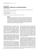
Available online />Figure 1
Phenotype and function of CD25
+
CD4
+
regulatory T cells from human peripheral blood. (a) CD25
+
CD4
+
T cells are anergic. Purified CD25
+
and
CD25
–
CD4
+
T cells from the peripheral blood of a healthy individual were stimulated with a monoclonal antibody against CD3, and proliferation
was assessed by incorporation of
3
H-labeled thymidine into newly synthesized DNA after 96 hours of culture. (b) CD25
+
CD4
+
T cells inhibit the
proliferation of autologous peripheral blood mononuclear cells (PBMC). Human PBMC were stimulated with monoclonal antibodies against CD3 in
the absence or presence of autologous purified CD25
+
or CD25
–
CD4
+
T cells. Proliferation was assessed as described in (a). (c) The regulatory
capacity of CD25
+
CD4
+
T cells is inhibited by exogenous IL-2. Human PBMC were stimulated as in (b) in the presence of a non-mitogenic
concentration of human IL-2. Proliferation was assessed as in (a). (d) Suppression by CD25
+
CD4
+
T cells is contact-dependent and independent
of regulatory cytokines. Human PBMC were stimulated with a monoclonal antibody against CD3 in the presence of autologous CD25
+
CD4
+
T
cells and neutralizing monoclonal antibodies against IL-10 (αIL-10) or IL-4 (αIL-4), or separated from CD25
+
CD4
+
T cells by an insert (‘transwell’).
Proliferation was assessed as described in (a).
Proliferation (cpm x 1
0)
–3
80
70
60
0
40
20
10
30
50
CD25
–
CD25
+
(a) (b) (c) (d)
Proliferation (cpm x 10 )
–3
9
8
7
6
0
4
2
1
3
5
Proliferation (cpm x 10 )
–3
PBMC PBMC
CD25
+
PBMC
CD25
–
PBMC PBMC
CD25
+
PBMC
CD25
–
10
0
40
20
30
50
60
αIL-10
α
IL-4
control
transwell
I
nhibition (
%
)
0
40
70
20
60
10
30
50
80
96
Several alternative mechanisms might contribute to the
enrichment of CD4
+
CD25
+
T cells in the synovial fluid of
patients with rheumatic diseases. A preferential migration of
these cells into the inflamed joint might be inferred from the
observation that CD4
+
CD25
+
T cells specifically express the
chemokine receptors CXCR4, CCR4 and CCR8 [41]. The
CCR4 ligands CCL17 and CCL22 are highly expressed in
synovial tissue [42], and it has been suggested that dendritic
cells are able to ‘chemoattract’ cells by the secretion of
CCL17 and CCL22 [41]. However, it should be pointed out
that although CCR4
+
T cells can be detected in the peripheral
blood of healthy individuals and in the synovial fluid of patients
with RA, the vast majority of T cells in the rheumatoid synovial
fluid do not express CCR4 [43], making the CCR4–CCL17-
mediated recruitment of Tregs into the rheumatoid joint rather
unlikely. The ligand for CXCR4, stromal-derived factor-1 (SDF-
1), is expressed on synovial endothelial cells [44], and
persistent expression of the chemokine receptor CXCR4 on
synovial CD4 T cells mediates their active retention within the
rheumatoid synovium [45]. Because human CD4
+
CD25
+
Tregs traffic to and are retained in the bone marrow through
interactions involving CXCR4 [46], it is also conceivable that
CD4
+
CD25
+
T cells are selectively recruited to and retained
in the rheumatoid joint through interactions involving CXCR4.
In line with the hypothesis that CD4
+
CD25
+
T cells are
effectively recruited to sites of chronic inflammation,
CD25
+
CD4
+
T cells are found in inflammatory infiltrates of
C57BL/6 mice infected with Leishmania major [47] and of
Balb/c mice infected with Candida albicans [48]. The data
therefore suggest that the accumulation of CD4
+
CD25
+
T cells during an inflammatory immune response might be a
physiologic control mechanism of potentially dangerous
effector functions to prevent tissue damage.
A second mechanism leading to the accumulation of
CD4
+
CD25
+
T cells in the rheumatic joint might relate to the
fact that inflammatory cytokines such as IL-2 and
costimulatory molecules cause CD4
+
CD25
+
T cells to revert
to an anergic phenotype [34] (Fig. 1c). Because the synovial
fluid contains high levels of inflammatory cytokines and of
antigen-presenting cells that are able to engage costimulatory
molecules on synovial T cells, CD25
+
CD4
+
T cells might
expand locally in the rheumatoid joint. However, in the
rheumatoid synovium it was found that T cells display low
proliferative responses [49], and in patients with JIA the
T cells in the synovial fluid are not actively dividing [50].
A third alternative method for the enrichment of CD4
+
CD25
+
T cells in the rheumatoid joint is related to the observation
that synovial T cells are actively inhibited from undergoing
apoptosis, thereby expanding their lifespan compared with
their peripheral counterparts. An integrin–ligand interaction is
involved in the fibroblast-mediated survival of synovial T cells
[51]. Fibroblast-secreted IFN-β is also able to inhibit
apoptosis, and in particular that of CD4
+
CD25
+
T cells [24].
A final explanation for the increased frequencies of CD25
+
T
cells in the synovium derives from the characteristic of CD25
to be upregulated on activated T cells. Thus, the sole deter-
mination of CD25 does not make it possible to discriminate
Tregs from activated effector cells. Because synovial T cells
express an array of activation markers and effector functions,
it is likely that most CD25-expressing T cells from the synovial
fluid constitute an effector population actively engaged in
driving synovial inflammation.
Recent evidence suggests that the CD4
+
CD25
+
Tregs from
the synovial fluid are different from those in the peripheral
circulation. CD25
bright
CD4
+
T cells from the synovial fluid in
RA contain higher frequencies of cells expressing CTLA-4
and GITR than those from the peripheral blood of healthy
donors and of patients with RA [31,37]. Tregs from synovial
fluid also display an activated phenotype with a higher
expression of CD69 and MHC class II than CD4
+
CD25
+
cells in the peripheral blood of matched individuals.
Intermittent flares in disease activity are typical of RA.
Whether the frequency of regulatory CD25
bright
CD4
+
T cells
fluctuate over time or are correlated with disease activity is
therefore of considerable interest. Although the frequency of
synovial CD25
bright
CD4
+
T cells varies between patients, the
numbers of these cells do not vary significantly over time in a
single joint [39]. Similar stable frequencies of synovial
CD25
bright
CD4
+
T cells over time were also observed in
patients with JIA, psoriatic arthritis and spondylarthropathies
[37]. Moreover, the frequencies of synovial CD25
bright
CD4
+
T cells in patients with RA was not correlated with clinical
parameters such as disease duration, the presence of
rheumatoid factor, the level of C-reactive protein and the
presence of erosions [31,37]. In addition, no association was
Arthritis Research & Therapy June 2005 Vol 7 No 3 Leipe et al.
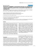
Figure 2
CD25
+
CD4
+
T cells are enriched in the synovial fluid in rheumatoid
arthritis. Mononuclear cells were isolated from the peripheral blood
(PB) or the synovial fluid (SF) of a patient with rheumatoid arthritis,
stained with monoclonal antibodies against CD4 and CD25 and
analyzed by flow cytometry. The numbers denote the frequency of cells
in the gate as defined by the expression of CD4 and CD25.
10
3
10
1
10
2
10
0
10
-1
10
3
10
1
10
2
10
0
10
-1
34.3
10
3
10
1
10
2
10
0
10
-1
10
3
10
1
10
2
10
0
10
-1
4.9
CD25
CD4
PB
SF
97
found between the use of methotrexate, corticosteroids or
anti-TNF therapy and the frequency of CD4
+
CD25
+
T cells in
the synovial fluid [31]. These data suggest that the presence
of CD4
+
CD25
+
T cells in the rheumatoid synovium is a
function of the disease and is characteristic of a particular
patient but unrelated to treatment, clinical course and disease
activity. These results might therefore question the
importance of CD4
+
CD25
+
Tregs in the regulation of synovial
inflammation.
Together, the data suggest that CD4
+
CD25
+
T cells in
chronically inflamed rheumatoid joints might enrich and
persist as a result of preferential recruitment, rescue from cell
death and activation by their specific antigen. Consequently,
the determination of frequencies of CD25
+
T cells in the
synovial fluid without complementary functional studies does
not make it possible to draw meaningful conclusions about
the role of CD4
+
CD25
+
Tregs in rheumatoid inflammation.
Function of synovial CD4
+
CD25
+
T cells in RA
When examined in conventional in vitro assays, synovial
CD4
+
CD25
bright
T cells are able to suppress the proliferation
of autologous CD4
+
CD25
–
(responder) T cells of synovial
and peripheral origin [31,37,39]. Synovial CD4
+
CD25
+
T cells display an even increased suppressive capacity
compared with blood CD4
+
CD25
+
T cells in RA [31] and in
JIA [40]. It is of interest that CD4
+
CD25
intermediate
T cells
enhance rather than suppress the proliferation of synovial
responder CD4
+
CD25
–
T cells, which might suggest that
CD25
intermediate
T cells represent effector T cells.
The major question that these results immediately bring up is
why inflammation occurs in the rheumatoid joints despite
elevated frequencies of apparently functional CD4
+
CD25
+
T cells with an even enhanced suppressive capacity in assays
in vitro.
One possible explanation for this seeming paradox might be
an active inhibition of the function of Tregs in the rheumatoid
joint. For example, several constituents of the inflamed
synovial environment, such as IL-2 and IL-7, have been shown
to abrogate the function of Tregs [34,52], suggesting that
Tregs are inhibited at sites of inflammation from performing
their regulatory function by pro-inflammatory cytokines.
Similarly, although shown only for peripheral blood, it has
been suggested that CD4
+
CD25
+
T cells display functional
differences before and after treatment with anti-TNF [38].
CD4
+
CD25
high
cells isolated from the peripheral blood of
patients with active RA suppress the proliferative response of
responder CD4 T cells but not the secretion of inflammatory
cytokines such as IFN-γ and TNF. In contrast, CD4
+
CD25
high
cells isolated from the patients’ blood after anti-TNF therapy
suppress (like CD4
+
CD25
high
cells in healthy individuals) not
only the proliferation but also the secretion of these cytokines
from responder CD4 T cells derived from anti-TNF-treated
patients. Thus, these findings indicate a functional deficit of
CD4
+
CD25
high
T cells from patients with active RA with
regard to their ability to suppress pro-inflammatory cytokine
production that reverts after treatment with TNF-neutralizing
agents. Additional evidence for an inhibitory function of TNF
on Tregs in RA derives from experiments in which the
depletion of CD4
+
CD25
high
T cells from peripheral blood
mononuclear cells (PBMC) from patients with active RA did
not alter the frequency of cells producing TNF or IL-10 in a 2-
day cell culture, whereas an increase in TNF-secreting cells
and a reduction in IL-10-secreting cells occurred in the
culture of PBMC derived from anti-TNF-treated patients with
RA that were depleted of Tregs [38]. Together, these data
might underline the potential role of cytokines in maintaining
chronic inflammation in vivo.
An alternative explanation for persistent synovial inflammation
despite enriched numbers of CD4
+
CD25
+
T cells with
enhanced suppressive capacity in vitro is provided by the
finding that synovial responder T cells express a decreased
susceptibility to the regulatory effect of CD4
+
CD25
+
Tregs in
comparison with peripheral blood responder T cells, thereby
‘compensating’ for the enhanced regulatory capacity of the
synovial Tregs [31]. IL-6, which is known to be found in large
amounts in the rheumatoid synovium [53], has been shown to
enhance the resistance of T effector cells to the suppressive
effects of Tregs [54]. Finally, although suppression by Tregs
is probably not antigen-specific but might involve neighboring
T cells in a ‘bystander’ fashion [35], Tregs require activation
through their T-cell antigen receptor to deliver their regulatory
function. Thus, if the specific antigen for the synovial Tregs is
not presented either in the secondary lymphoid organs or in
the inflamed synovia, or, alternatively, if Tregs in RA express
an altered threshold for antigen-specific activation, synovial
Tregs, although present, will not become activated and will
therefore fail to inhibit ongoing inflammation.
Together, these arguments indicate that rheumatoid inflam-
mation occurs in the presence of Tregs that express an
impaired regulatory function in vivo, despite their enhanced
regulatory capacity in vitro. Although it is tempting to
speculate that synovial inflammation is the consequence of an
inadequate ability of synovial Tregs to downmodulate local
inflammation, several observations indicate clearly that
synovial Tregs are functional and actively dampen the
inflammatory immune response in vivo. For example, in JIA the
frequencies of CD4
+
CD25
+
synovial T cells are inversely
correlated with the clinical outcome, and the expression of
FoxP3 mRNA, a ‘marker’ for Treg function, is elevated in mild
cases in comparison with severe forms of the disease [50]. In
collagen-induced arthritis, depletion of CD4
+
CD25
+
T cells
accelerates the onset of severe disease, and transfer of
syngeneic CD4
+
CD25
+
T cells into Treg-depleted mice
reverses the increased severity [55]. Thus, the local
expansion in the CD4
+
CD25
+
Treg cell population in the
rheumatoid synovium might reflect a mechanism for resolving
the inflammatory immune response. Although not sufficient to
Available online />98
prevent inflammatory activity in the joint, the CD4
+
CD25
+
Tregs in the inflamed rheumatoid synovium might
nevertheless be important for a downmodulation of the
inflammation, thereby delaying further tissue damage and
impeding erosive inflammation. These findings might be of
relevance in validating and fostering the development of
clinical applications of in vitro-generated Tregs in auto-
immune diseases in the near future by means of personalized
cellular therapy.
It should be noted that other subsets of CD4 T cells have
been identified that are capable of suppressing specific
immune responses. The most prominent of these are termed
Treg 1 (Tr1) and T helper type 3 (Th3) cells. Th3 cells
produce predominantly TGF-β. They are generated in vivo by
immunization through an oral or other mucosal route [35], and
have been detected in patients with multiple sclerosis after
oral administration of myelin basic protein [56]. Groux and
colleagues first isolated mouse and human Tr1 cells that have
immune-regulatory activities both in vitro and in vivo [57,58].
These regulatory CD4
+
T cells secrete IL-10 and have been
generated in vitro by repeated antigenic stimulations of
human and murine CD4
+
cells in the presence of IL-10
[26,59,60] or by activation through immature antigen-
presenting cells that lack potent costimulatory activity [61].
However, comprehensive analyses of Tr1 and Th3 cells in
humans are not available, so the precise role of these subsets
in human autoimmune disease has not been defined.
Conclusions
In conclusion, human CD4
+
CD25
+
Tregs that are capable of
suppressing CD4 T cell proliferation in vitro are enriched in
the synovial fluid of patients with RA. Synovial Tregs express
an increased regulatory capacity in comparison with Tregs
derived from the peripheral blood, in assays in vitro. In the
synovium, Tregs might be inhibited by different mechanisms
such as inflammatory cytokines including TNF, or stimulation
by antigen-presenting cells, which in concert might allow
synovial inflammation to evolve and persist despite the
enhanced frequencies of synovial Tregs. However, because
evidence suggests that synovial Tregs, although not sufficient
to ameliorate disease activity completely, are involved in
regulating synovial inflammation in vivo, future treatment
strategies of autoimmune diseases can be envisaged in
which Tregs generated and/or expanded in vitro will be
employed in an attempt to control local and systemic
autoimmune inflammation.
Competing interests
The author(s) declare that they have no competing interests.
Acknowledgments
This work was supported in part by the Deutsche Forschungsgemein-
schaft (Grants Schu 786/2-3 and 2-4) and by the Interdisciplinary
Center for Clinical Research (IZKF) at the University hospital of the Uni-
versity of Erlangen-Nuremberg (Projects B27 and B3).
References
1. Bach JF, Chatenoud L: Tolerance to islet autoantigens in type 1
diabetes. Annu Rev Immunol 2001, 19:131-161.
2. Ota K, Matsui M, Milford EL, Mackin GA, Weiner HL, Hafler DA:
T-cell recognition of an immunodominant myelin basic protein
epitope in multiple sclerosis. Nature 1990, 346:183-187.
3. Sakaguchi S: Regulatory T cells: key controllers of immuno-
logic self-tolerance. Cell 2000, 101:455-458.
4. Gershon RK, Kondo K: Infectious immunological tolerance.
Immunology 1971, 21:903-914.
5. Hedrick SM, Germain RN, Bevan MJ, Dorf M, Engel I, Fink P, Gas-
coigne N, Heber-Katz E, Kapp J, Kaufmann Y, et al.: Rearrange-
ment and transcription of a T-cell receptor beta-chain gene in
different T-cell subsets. Proc Natl Acad Sci USA 1985, 82:531-
535.
6. Kronenberg M, Steinmetz M, Kobori J, Kraig E, Kapp JA, Pierce
CW, Sorensen CM, Suzuki G, Tada T, Hood L: RNA transcripts
for I-J polypeptides are apparently not encoded between the
I-A and I-E subregions of the murine major histocompatibility
complex. Proc Natl Acad Sci USA 1983, 80:5704-5708.
7. Janeway CA Jr: Do suppressor T cells exist? A reply. Scand J
Immunol 1988, 27:621-623.
8. Sakaguchi S, Sakaguchi N, Asano M, Itoh M, Toda M: Immuno-
logic self-tolerance maintained by activated T cells express-
ing IL-2 receptor alpha-chains (CD25). Breakdown of a single
mechanism of self-tolerance causes various autoimmune dis-
eases. J Immunol 1995, 155:1151-1164.
9. McHugh RS, Shevach EM: Cutting edge: depletion of
CD4+CD25+ regulatory T cells is necessary, but not suffi-
cient, for induction of organ-specific autoimmune disease. J
Immunol 2002, 168:5979-5983.
10. Mottet C, Uhlig HH, Powrie F: Cutting edge: cure of colitis by
CD4+CD25+ regulatory T cells. J Immunol 2003, 170:3939-
3943.
11. Miller JF, Basten A: Mechanisms of tolerance to self. Curr Opin
Immunol 1996, 8:815-821.
12. Schwartz RH: Models of T cell anergy: is there a common mol-
ecular mechanism? J Exp Med 1996, 184:1-8.
13. Miller JF, Heath WR: Self-ignorance in the peripheral T-cell
pool. Immunol Rev 1993, 133:131-150.
14. Thornton AM, Shevach EM: Suppressor effector function of
CD4+CD25+ immunoregulatory T cells is antigen nonspecific.
J Immunol 2000, 164:183-190.
15. Takahashi T, Tagami T, Yamazaki S, Uede T, Shimizu J, Sakaguchi
N, Mak TW, Sakaguchi S: Immunologic self-tolerance main-
tained by CD25
+
CD4
+
regulatory T cells constitutively
expressing cytotoxic T lymphocyte-associated antigen 4. J
Exp Med 2000, 192:303-310.
16. Read S, Malmstrom V, Powrie F: Cytotoxic T lymphocyte-asso-
ciated antigen 4 plays an essential role in the function of
CD25
+
CD4
+
regulatory cells that control intestinal inflamma-
tion. J Exp Med 2000, 192:295-302.
17. McHugh RS, Whitters MJ, Piccirillo CA, Young DA, Shevach EM,
Collins M, Byrne MC: CD4
+
CD25
+
immunoregulatory T cells:
gene expression analysis reveals a functional role for the glu-
cocorticoid-induced TNF receptor. Immunity 2002, 16:311-323.
18. Shimizu J, Yamazaki S, Takahashi T, Ishida Y, Sakaguchi S: Stim-
ulation of CD25
+
CD4
+
regulatory T cells through GITR breaks
immunological self-tolerance. Nat Immunol 2002, 3:135-142.
19. Huang CT, Workman CJ, Flies D, Pan X, Marson AL, Zhou G,
Hipkiss EL, Ravi S, Kowalski J, Levitsky HI, et al.: Role of LAG-3
in regulatory T cells. Immunity 2004, 21:503-513.
20. Hori S, Nomura T, Sakaguchi S: Control of regulatory T cell
development by the transcription factor Foxp3. Science 2003,
299:1057-1061.
21. Bennett CL, Christie J, Ramsdell F, Brunkow ME, Ferguson PJ,
Whitesell L, Kelly TE, Saulsbury FT, Chance PF, Ochs HD: The
immune dysregulation, polyendocrinopathy, enteropathy, X-
linked syndrome (IPEX) is caused by mutations of FOXP3. Nat
Genet 2001, 27:20-21.
22. Gambineri E, Torgerson TR, Ochs HD: Immune dysregulation,
polyendocrinopathy, enteropathy, and X-linked inheritance
(IPEX), a syndrome of systemic autoimmunity caused by
mutations of FOXP3, a critical regulator of T-cell homeostasis.
Curr Opin Rheumatol 2003, 15:430-435.
23. Baecher-Allan C, Hafler DA: Suppressor T cells in human dis-
eases. J Exp Med 2004, 200:273-276.
Arthritis Research & Therapy June 2005 Vol 7 No 3 Leipe et al.
99
24. Taams LS, Smith J, Rustin MH, Salmon M, Poulter LW, Akbar AN:
Human anergic/suppressive CD4
+
CD25
+
T cells: a highly dif-
ferentiated and apoptosis-prone population. Eur J Immunol
2001, 31:1122-1131.
25. Jonuleit H, Schmitt E, Stassen M, Tuettenberg A, Knop J, Enk AH:
Identification and functional characterization of human
CD4
+
CD25
+
T cells with regulatory properties isolated from
peripheral blood. J Exp Med 2001, 193:1285-1294.
26. Levings MK, Sangregorio R, Roncarolo MG: Human CD25
+
CD4
+
T regulatory cells suppress naive and memory T cell prolifera-
tion and can be expanded in vitro without loss of function. J
Exp Med 2001, 193:1295-1302.
27. Dieckmann D, Plottner H, Berchtold S, Berger T, Schuler G: Ex
vivo isolation and characterization of CD4
+
CD25
+
T cells with
regulatory properties from human blood. J Exp Med 2001,
193:1303-1310.
28. Baecher-Allan C, Brown JA, Freeman GJ, Hafler DA:
CD4+CD25high regulatory cells in human peripheral blood. J
Immunol 2001, 167:1245-1253.
29. Stephens LA, Mottet C, Mason D, Powrie F: Human CD4
+
CD25
+
thymocytes and peripheral T cells have immune suppressive
activity in vitro. Eur J Immunol 2001, 31:1247-1254.
30. Annunziato F, Cosmi L, Liotta F, Lazzeri E, Manetti R, Vanini V,
Romagnani P, Maggi E, Romagnani S: Phenotype, localization,
and mechanism of suppression of CD4
+
CD25
+
human thymo-
cytes. J Exp Med 2002, 196:379-387.
31. van Amelsfort JM, Jacobs KM, Bijlsma JW, Lafeber FP, Taams LS:
CD4
+
CD25
+
regulatory T cells in rheumatoid arthritis: differ-
ences in the presence, phenotype, and function between
peripheral blood and synovial fluid. Arthritis Rheum 2004, 50:
2775-2785.
32. Taams LS, Vukmanovic-Stejic M, Smith J, Dunne PJ, Fletcher JM,
Plunkett FJ, Ebeling SB, Lombardi G, Rustin MH, Bijlsma JW, et
al.: Antigen-specific T cell suppression by human CD4+CD25+
regulatory T cells. Eur J Immunol 2002, 32:1621-1630.
33. Woo EY, Yeh H, Chu CS, Schlienger K, Carroll RG, Riley JL,
Kaiser LR, June CH: Cutting edge: regulatory T cells from lung
cancer patients directly inhibit autologous T cell proliferation.
J Immunol 2002, 168:4272-4276.
34. Thornton AM, Shevach EM: CD4+CD25+ immunoregulatory T
cells suppress polyclonal T cell activation in vitro by inhibiting
interleukin 2 production. J Exp Med 1998, 188:287-296.
35. Weiner HL: Oral tolerance: immune mechanisms and the gen-
eration of Th3-type TGF-beta-secreting regulatory cells.
Microbes Infect 2001, 3:947-954.
36. Nakamura K, Kitani A, Strober W: Cell contact-dependent
immunosuppression by CD4
+
CD25
+
regulatory T cells is
mediated by cell surface-bound transforming growth factor
beta. J Exp Med 2001, 194:629-644.
37. Cao D, van Vollenhoven R, Klareskog L, Trollmo C, Malmstrom V:
CD25
bright
CD4
+
regulatory T cells are enriched in inflamed
joints of patients with chronic rheumatic disease. Arthritis Res
Ther 2004, 6:R335-346.
38. Ehrenstein MR, Evans JG, Singh A, Moore S, Warnes G, Isenberg
DA, Mauri C: Compromised function of regulatory T cells in
rheumatoid arthritis and reversal by anti-TNF
αα
therapy. J Exp
Med 2004, 200:277-285.
39. Cao D, Malmstrom V, Baecher-Allan C, Hafler D, Klareskog L,
Trollmo C: Isolation and functional characterization of regula-
tory CD25
bright
CD4
+
T cells from the target organ of patients
with rheumatoid arthritis. Eur J Immunol 2003, 33:215-223.
40. de Kleer IM, Wedderburn LR, Taams LS, Patel A, Varsani H, Klein
M, de Jager W, Pugayung G, Giannoni F, Rijkers G, et al.:
CD4+CD25bright regulatory T cells actively regulate inflam-
mation in the joints of patients with the remitting form of juve-
nile idiopathic arthritis. J Immunol 2004, 172:6435-6443.
41. Iellem A, Mariani M, Lang R, Recalde H, Panina-Bordignon P, Sini-
gaglia F, D’Ambrosio D: Unique chemotactic response profile
and specific expression of chemokine receptors CCR4 and
CCR8 by CD4
+
CD25
+
regulatory T cells. J Exp Med 2001, 194:
847-853.
42. Radstake TR, Van Der Voort R, Ten Brummelhuis M, De Waal
Malefijt M, Schreurs W, Looman M, Sloetjes A, Figdor CG, Van
Den Berg WB, Barrera P, et al.: Increased expression of CCL18,
CCL19, and CCL17 by dendritic cells from patients with
rheumatoid arthritis and regulation by Fc gamma receptors.
Ann Rheum Dis 2004.
43. Suzuki N, Nakajima A, Yoshino S, Matsushima K, Yagita H,
Okumura K: Selective accumulation of CCR5+ T lymphocytes
into inflamed joints of rheumatoid arthritis. Int Immunol 1999,
11:553-559.
44. Buckley CD, Amft N, Bradfield PF, Pilling D, Ross E, Arenzana-
Seisdedos F, Amara A, Curnow SJ, Lord JM, Scheel-Toellner D, et
al.: Persistent induction of the chemokine receptor CXCR4 by
TGF-beta 1 on synovial T cells contributes to their accumula-
tion within the rheumatoid synovium. J Immunol 2000, 165:
3423-3429.
45. Nanki T, Hayashida K, El-Gabalawy HS, Suson S, Shi K, Girschick
HJ, Yavuz S, Lipsky PE: Stromal cell-derived factor-1-CXC
chemokine receptor 4 interactions play a central role in CD4+
T cell accumulation in rheumatoid arthritis synovium. J
Immunol 2000, 165:6590-6598.
46. Zou L, Barnett B, Safah H, Larussa VF, Evdemon-Hogan M,
Mottram P, Wei S, David O, Curiel TJ, Zou W: Bone marrow is a
reservoir for CD4+CD25+ regulatory T cells that traffic
through CXCL12/CXCR4 signals. Cancer Res 2004, 64:8451-
8455.
47. Belkaid Y, Piccirillo CA, Mendez S, Shevach EM, Sacks DL:
CD4+CD25+ regulatory T cells control Leishmania major per-
sistence and immunity. Nature 2002, 420:502-507.
48. Montagnoli C, Bacci A, Bozza S, Gaziano R, Mosci P, Sharpe AH,
Romani L: B7/CD28-dependent CD4+CD25+ regulatory T
cells are essential components of the memory-protective
immunity to Candida albicans. J Immunol 2002, 169:6298-
6308.
49. Petersen J, Andersen V, Ingemann-Hansen T, Halkjaer-Kristensen
J, Wiik A, Thyssen H: Synovial fluid and blood monocyte influ-
ence on lymphocyte proliferation in rheumatoid arthritis and
traumatic synovitis. Scand J Rheumatol 1983, 12:299-304.
50. Black AP, Bhayani H, Ryder CA, Gardner-Medwin JM, Southwood
TR: T-cell activation without proliferation in juvenile idiopathic
arthritis. Arthritis Res 2002, 4:177-183.
51. Salmon M, Scheel-Toellner D, Huissoon AP, Pilling D, Sham-
sadeen N, Hyde H, D’Angeac AD, Bacon PA, Emery P, Akbar AN:
Inhibition of T cell apoptosis in the rheumatoid synovium. J
Clin Invest 1997, 99:439-446.
52. van Amelsfort JM, Noordegraaf M, Bijlsma JWJ, Taams LS,
Lafeber FPJG: Influence of the inflammatory milieu on the
suppressive function of CD4+CD25+ T cells in rheumatoid
arthritis. Arthritis Rheum 2004, 50:S526.
53. Hirano T, Matsuda T, Turner M, Miyasaka N, Buchan G, Tang B,
Sato K, Shimizu M, Maini R, Feldmann M, et al.: Excessive pro-
duction of interleukin 6/B cell stimulatory factor- 2 in rheuma-
toid arthritis. Eur J Immunol 1988, 18:1797-1801.
54. Pasare C, Medzhitov R: Toll pathway-dependent blockade of
CD4+CD25+ T cell-mediated suppression by dendritic cells.
Science 2003, 299:1033-1036.
55. Morgan ME, Sutmuller RP, Witteveen HJ, van Duivenvoorde LM,
Zanelli E, Melief CJ, Snijders A, Offringa R, de Vries RR, Toes RE:
CD25+ cell depletion hastens the onset of severe disease in
collagen-induced arthritis. Arthritis Rheum 2003, 48:1452-
1460.
56. Fukaura H, Kent SC, Pietrusewicz MJ, Khoury SJ, Weiner HL,
Hafler DA: Induction of circulating myelin basic protein and
proteolipid protein-specific transforming growth factor-beta1-
secreting Th3 T cells by oral administration of myelin in multi-
ple sclerosis patients. J Clin Invest 1996, 98:70-77.
57. Groux H, Bigler M, de Vries JE, Roncarolo MG: Interleukin-10
induces a long-term antigen-specific anergic state in human
CD4+ T cells. J Exp Med 1996, 184:19-29.
58. Groux H, O’Garra A, Bigler M, Rouleau M, Antonenko S, de Vries
JE, Roncarolo MG: A CD4
+
T-cell subset inhibits antigen-spe-
cific T-cell responses and prevents colitis. Nature 1997, 389:
737-742.
59. Asseman C, Powrie F: Interleukin 10 is a growth factor for a
population of regulatory T cells. Gut 1998, 42:157-158.
60. Cottrez F, Hurst SD, Coffman RL, Groux H: T regulatory cells 1
inhibit a Th2-specific response in vivo. J Immunol 2000, 165:
4848-4853.
61. Jonuleit H, Schmitt E, Schuler G, Knop J, Enk AH: Induction of
interleukin 10-producing, nonproliferating CD4
+
T cells with
regulatory properties by repetitive stimulation with allogeneic
immature human dendritic cells. J Exp Med 2000, 192:1213-
1222.
Available online />