Báo cáo khoa học: "Prognostic value of HMGB1 overexpression in resectable gastric adenocarcinomas" docx
Bạn đang xem bản rút gọn của tài liệu. Xem và tải ngay bản đầy đủ của tài liệu tại đây (807.58 KB, 7 trang )
WORLD JOURNAL OF
SURGICAL ONCOLOGY
Bao et al. World Journal of Surgical Oncology 2010, 8:52
/>Open Access
RESEARCH
© 2010 Bao et al; licensee BioMed Central Ltd. This is an Open Access article distributed under the terms of the Creative Commons At-
tribution License ( which permits unrestricted use, distribution, and reproduction in any
medium, provided the original work is properly cited.
Research
Prognostic value of HMGB1 overexpression in
resectable gastric adenocarcinomas
Guoqiang Bao, Qing Qiao, Huadong Zhao and Xianli He*
Abstract
Introduction: HMGB1(High mobility group box 1), originally described as a nuclear protein, is now regarded as a
multifunctional protein with a paradoxical dual effect in tumors. In the present study, HMGB1 overexpression and its
correlation with the clinicopathologic characteristics and recurrence-free survival were evaluated in gastric
adenocarcinomas.
Methods: 76 gastric adenocarcinomas surgically removed entered the study. The immunohistochemical staining was
used to assess HMGB1 expression through tissue microarray procedure. The clinicopathologic characteristics of all
patients were recorded, and the regular follow-up was made for all patients.
Results: Almost all the gastric adenocarcinomas showed HMGB1 positive staining mainly in the nucleus, and the
overexpression of HMGB1 was found in cancerous tissues with higher strong reactivity rate, compared with non-
cancerous tissues (total expression score ≥ 9, 42.0% vs. 9.0%, P < 0.001). Survival analysis revealed that tumor stage
negatively correlated with cancer-free survival (P = 0.022). Furthermore, HMGB1 overexpression positively associated
with cancer-free survival of resectable gastric adenocarcinomas (P = 0.023).
Conclusions: The overexpression of HMGB1 protein indicates that HMGB1 may play a role in the tumorigenesis of
gastric adenocarcinomas. And the overexpression of HMGB1 may be a marker of good prognosis of gastric
adenocarcinoma given curative resection combined with adjuvant chemotherapy.
Introduction
Gastric cancer (GC) is the second most common cause of
cancer-related death in the world. Many Asian countries,
including China, have very high rates of GC. For patients
in advanced stages, the five-year survival rate is only
about 20 percent. There are many factors that limit the
prognosis of the disease. High mobility group box l
(HMGB1), a nuclear DNA-binding protein, originally
described as a nuclear protein that binds to and modifies
DNA, stabilizes the structure and function of chromatin
and regulates gene transcription. It has been realized that
HMGB1 can act either as a DNA binding protein or
extracellularly as a cytokine-like danger signal, which is
either actively secreted or passively released by necrotic
cells[1]. Now HMGB1 is regarded as a central mediator of
inflammation by acting as a cytokine, which has been
reported as a "late" proinflammatory mediator in sepsis
[2,3].
HMGB1 plays a role in many clinical conditions such as
autoimmunity, acute ischemia-reperfusion injury, cardio-
vascular disease and cancer [4]. Recent evidences suggest
that HMGB1 plays critical roles in the development and
progression of numerous tumors [5]. HMGB1 modulates
the transcriptional activity in the nucleus, but it is also
present in the cytoplasm and outside the cell in certain
conditions, associated with the proliferation and metasta-
sis of many tumors, including breast cancer, colon carci-
noma, and melanoma[6]. More recently, HMGB1 has
been recognized as a proangiogenic factor [7].
In the case of tumors, HMGB1 recognition has a para-
doxical dual effect: the reparative inflammatory response
promotes tumor neoangiogenesis, cell survival, expan-
sion, and metastases; on the other hand, it triggers pro-
tective anti-neoplastic T-cell responses[8,9]. Tumor cell
death triggered by chemotherapy or radiotherapy initi-
ates an immunoadjuvant pathway that contributes to the
success of cytotoxic treatments. The interaction of
* Correspondence:
1
Department of general surgery, Tangdu Hospital, The Fourth Military Medical
University, Xi'an 710038, China
Full list of author information is available at the end of the article
Bao et al. World Journal of Surgical Oncology 2010, 8:52
/>Page 2 of 7
HMGB1 released from dying tumor cells with Toll-like
receptor 4 (TLR4) on dendritic cells (DCs) was required
for the cross-presentation of tumor antigens and the pro-
motion of tumor specific cytotoxic T-cell responses
[10,11]. HMGB1 plays roles in various disease conditions
mainly through RAGE (the receptor for advanced glyca-
tion end products). HMGB1-RAGE interactions have
been found to be important in a number of cancers,
which involves the MAPK/ERK pathway[12].
HMGB1 has emerged as a candidate for therapeutic
intervention in various disease conditions [13]. However,
further basic and clinical studies are warranted to con-
firm the roles played by HMGB1 in clinical cancer medi-
cine. In the present study, the expression of HMGB1
protein was evaluated with tissue microarray(TMA) and
immunohistochemical(IHC) staining procedures to study
the prognostic significance of HMGB1 and its correlation
with the clinical and histopathologic features of resect-
able gastric adenocarcinomas.
Patients and methods
Patients
TMAs were prepared for IHC test from a total of 78 con-
secutive cases of gastric adenocarcinomas operated in
our department from December 2007 to October 2008.
All the patients was given the radical resection and
D1+or D2 lymphadenectomy followed by adjuvant che-
motherapy with the regimen ECF (Epirubicin, cisplatin
and 5-FU). To all patients, no preoperative therapy was
given. The pathologic staging were made according to
American Joint Committee on Cancer (AJCC) TNM
staging system. The follow-up end point was defined as
the recurrence or metastasis of the cancer. The use of the
tissue samples in TMA analyses and clinical data was
approved by Medical Ethics Committee of The Fourth
Military Medical University and the patients. Patients'
clinical and histopathologic data were summarized in
Table 1.
Tissue Microarrays
For each case, we selected the tumor foci for the TMA
construction during routine diagnosis by marking them
on the more representative hematoxylin-eosin (H & E)-
stained slide with a waterproof pencil.
The advanced tissue arrayer (ATA-100, Chemicon
International, Tamecula, CA, USA) was used to create
holes in a recipient paraffin block and to acquire cylindri-
cal core tissue biopsies with a diameter of 1 mm from the
specific areas of the "donor" block. The tissue core biop-
sies were transferred to the recipient paraffin block at
defined array positions. The TMAs contained tissue sam-
ples from 78 formalin-fixed paraffin-embedded cancer
specimens with known diagnosis, and correlated non-
cancerous tissues from the same patients.
The block was incubated in an oven at 45°C for 20 min
to allow complete embedding of the grafted tissue cylin-
ders in the paraffin of the recipient block, and then stored
at 4°C until microtome sectioning.
Immunohistochemical staining
Rabbit-derived anti-human HMGB1 antibodies were
used for IHC detection of HMGB1 protein in TMAs.
TMA sections were processed for IHC demonstration of
HMGB1 protein by the Biotin-Avidin-Peroxidase detec-
tion system (Sigma). The anti-HMGB1 antibodies were
used at 1:200 dilutions. Endogenous peroxidase was
inhibited by incubation with freshly prepared 3% hydro-
gen peroxide with 0.1% sodium azide. Nonspecific stain-
ing was blocked with 0.5% casein and 5% normal goat
serum. TMAs were incubated with biotinylated goat anti-
rabbit antibodies and ExtrAvidin-conjugated horseradish
peroxidase. Staining was developed with diaminobenzi-
dine substrate and sections were counterstained with
hematoxylin. Normal mouse serum or PBS replaced anti-
HMGB1 antibodies in negative controls.
Table 1: Clinical and histopathologic data of the patients.
Variables Number of cases(%)
Number of patients 78(100%)
Age(y)
≤ 60 44(56.4%)
> 60 34(43.6%)
Gender
Male 55(70.5%)
Female 23(28.5%)
Tumor localisation
Proximal 33(42.3%)
Distal 45(57.7%)
Histologic grade
Undifferentiated(G4) 13(16.7%)
Poorly differentiated(G3) 27(34.6%)
Moderately differentiated(G2) 29(37.2%)
Well differentiated(G1) 9(11.5%)
Tumor stage
Stage I + II 35(44.9%)
Stage III + IV 43(55.1%)
Primary tumor
T1-2 12(15.4%)
T3-4 66(84.6%)
Regional lymph nodes
N0 34(43.6%)
N1-3 44(56.4%)
Bao et al. World Journal of Surgical Oncology 2010, 8:52
/>Page 3 of 7
The quantification evaluation of HMGB1 protein expression
HMGB1 expression was semiquantitatively estimated as
the total HMGB1 immunostaining score, which was cal-
culated as the sum of a proportion score and an intensity
score. The propotion score reflects the fraction of posi-
tive staining cells(score 0, < 5%; score 1, 5% - 10%; score 2,
10 - 50%; score 3, 50 - 75%; score 4, > 75%). The intensity
score represents the staining intensity(score 0, no stain-
ing signal; score 1, weak positive signal; score 2, moderate
positive signal; score 3, strong positive signal). Finally, a
total expression score was given ranging from 0 to 12.
Based the analysis in advance, the overexpression of
HMGB1 was defined as the total expression score ≥ 9.
Statistical analysis
Results are expressed as median and range. For statistical
analysis, the Chi-square test was made with the software
GraphPad Prism, and uni-and multivariate analysis and
survival analysis were made with the SPSS 16.0. Signifi-
cance was defined as P < 0.05.
Results
The expression of HMGB1 protein in the gastric
adenocarcinomas
Expression of HMGB1 protein was evaluated by using
immunohistochemical staining. As a nonhistone DNA-
binding protein, the expression of HMGB1 protein was
mainly localized in the nucleus. In gastric adenocarci-
noma cells, the expression of HMGB1 protein was also
mainly detected in the nucleus (Figure 1B, C). But in rare
cases of sample, the positive staining could be found in
nucleus and cytoplasm (Figure 1D).
The positive staining was detected in most of gastric
adenocarcinoma cells. HMGB1 unexpressed tumors
mainly were found in the poorly differentiated adenocar-
cimas. The difference of HMGB1 expression in peritu-
moral and normal (distant) tissues was not assessed
based on the histopathologic changes and HP status. The
positive staining was detected in 69/78(88.5%) adenocar-
cinoma cells, and 61/78(78.2%) in non-cancerous cells
with no significant difference (P = 0.202, Table 2). But the
rate of HMGB1 overexpression (total expression score ≥
9) was elevated in gastric adenocarcinoma cells, com-
pared with corresponding non-cancerous cells (41.0% vs.
9.0%, P < 0.001).
The correlation of HMGB1 protein expression with the
clinical and histopathologic characteristics
The relationship between HMGB1 overexpression and
various clinical and histopathologic features was ana-
lyzed. No significant correlation was found between
HMGB1 overexpression with age, or gender (Table 3). As
shown in Table 3, the statistically significant difference
was found in the groups with district differentiation (P =
0.012). But, except the significantly elevated rate in G1
group, the difference was no found in G2, G3, and G4
group, compared with the other two groups. The phe-
nomenon perhaps was induced by the distribution bias of
the available cases.
According to the pathologic TNM staging, the cases
were divided into two groups: stage I + II and stage II +
IV. The group with early stage showed elevated rate of
HMGB1 over-expression, but no statistically significant
difference was found between the two groups (34.3% vs.
46.5%, P = 0.356). Then, the cases were divided into two
groups with lymph node metastasis or no. The rate of
HMGB1 overexpression was 21/23(47.7%) in cancerous
specimens with lymph node metastasis, compared with
11/23(32.4%) in cancerous specimens without lymph
node metastasis. But no significant difference was found
(P = 0.246). While, primary tumor infiltrating depth per-
haps correlated with HMGB1 overexpression. pT3 + 4
group showed elevated rate of HMGB1 overexpression
compared with pT1 + 2 group, but no statistical differ-
ence was found (43.9% vs. 25.0%, P = 0.340) (Table 3).
Kaplan-Meier survival analysis
Regarding the results of cancer-free survival analysis,
there was no correlation between gender, age, location,
grade of the tumor with prognosis. But the tumor stage
and HMGB1 overexpression showed the correlation with
cancer-free survival. Survival curves were plotted accord-
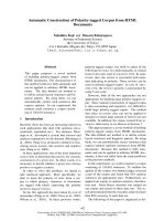
Figure 1 Immunohistochemical detection of HMGB1 protein in
different gastric tissues. A: Normal rectal sample. The low-expression
of HMGB1 was detected in epithelial and stromal cells B: Gastric ade-
nocarcinoma sample with well differentiation. The immunohis-
tochemical staining showed strong positive signal (+ + +) in the cancer
cells, and low-expression was detected in the stromal cells, which lo-
calized in the nucleus. C: Gastric adenocarcinoma sample with poor-
moderate differentiation. The immunohistochemical staining showed
strong positive signal (+ + +), which mainly localized in the nucleus. D:
In rare cases, the strong staining was detected in the nucleus and cy-
toplasm of the cancer cells. Original magnification × 200.
Bao et al. World Journal of Surgical Oncology 2010, 8:52
/>Page 4 of 7
ing to the Kaplan-Meier method for the patients with
HMGB1 expression status and stage. Tumor stage had a
significant effect on cancer-free survival for stage I+II
tumors compared with stage III + IV tumors(P = 0.022).
The expected survival time was 19.0000 ± 7.35247 m for
Stage I + II tumors (95% CI = 16.4743 - 21.5257), 16.4186
± 8.69108 m for stage III + IV tumors (95% CI = 13.7439 -
19.0933).
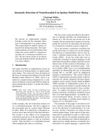
Furthermore, survival analysis revealed that HMGB1
overexpression affected cancer-free survival. There was
significant difference in cancer-free survival between
groups with HMGB1 overexpression and with its low-
level expression (P = 0.023, Figure 2). Multivariate analy-
sis showed that the expected cancer-free survival time
was 20.4375 ± 7.28648 m for tumors with HMGB1 over-
expression (95% CI = 17.8104 - 23.0646), 15.5870 ±
8.23158 m for tumors with HMGB1 no-and low-level
expression (95% CI = 13.1425 - 18.0314). HMGB1 overex-
pression was an independent predictor of cancer-free
survival for patients with resectable gastric adenocarci-
nomas. Furthermore, we analyzed the characteristics of
the patients with HMGB1 overexpression who died dur-
ing the follow-up period. We found the most of the cases
had a relatively late disease (Table 4).
Discussion
The occurrence and development of GC correlated with
various molecular and genetic incidents. To investigate
Table 2: Expression of HMGB1 in cancerous tissues and correlated non-cancerous tissues.
Variables All cases(n) Positive expression Significance Overexpression Significance
(n=) (%)
(P)
(n=) (%)
(P)
Cancer 78 69 88.5 0.202 32 41.0 < 0.001
Non-cancer 78 61 78.2 7 9.0
Table 3: Expression of HMGB1 in correlation with clinicopathologic variables.
Variables cases(n) HMGB1 low < 9 HMGB1 high(≥ 9) Significance(P)
(n=) (%) (n=) (%)
Total 784659.03241.0
Gender
Male 55 29 52.7 26 42.3 0.129
Female 23 17 73.9 6 26.1
Age at surgery
≤ 60 44 29 65.9 15 34.1 0.111
> 60 34 16 47.1 18 52.9
Tumor differentiation
G1 9 1 11.1 8 88.9 0.012
G2 29 21 72.4 8 27.6
G3 27 17 67.0 10 37.0
G4 13 7 53.8 6 46.2
Tumor stage
Stage I + II 35 23 65.7 12 34.3 0.356
Stage III + IV 43 23 53.5 20 46.5
pT stage
pT1-2 12 9 75.0 3 25.0 0.340
pT3-4 663756.12943.9
Nodal status
pN0 34 23 67.6 11 32.4 0.246
pN1~3 44 23 52.3 21 47.7
Bao et al. World Journal of Surgical Oncology 2010, 8:52
/>Page 5 of 7
the significance of the molecular expression in GC may
help us to identify potential treatment target and(or) pre-
dictive marker of prognosis and treatment response.
Overexpression as well as cytoplasmic localization of
HMGB1, particularly in conjunction with its receptor for
advanced glycation end products (AGEs), is associated
with the proliferation and metastasis of many tumor
types [14-16]. Furthermore, HMGB1 secreted from pri-
mary tumors decreased the number of macrophages to
attenuate the anti-metastatic defense in patients with col-
orectal cancers, through inducing growth inhibition and
apoptosis in macrophages[17,18]. HMGB1 can also influ-
ence a variety of important cell types within the tumor
microenvironment, including fibroblasts, leukocytes, and
vascular cells[19]. So, targeting the HMGB1 ligand or its
receptor represents an important potential application in
cancer therapeutics [20].
But, HMGB1 may play a controversial role in the occur-
rence and progression of cancers. Riuzzi F et al. reported
that the HMGB1-RB interaction perhaps induced the
HMGB1-mediated transcriptional repression, cell growth
inhibition, G1 cell cycle arrest, apoptosis induction, and
tumor growth suppression[21]. Furthermore, the func-
tional inactivation of RAGE in myoblasts results in
reduced myogenesis, increased proliferation, and tumor
formation in vivo [22]. On the other hand, the tumor cell
death triggered by chemotherapy or radiotherapy initi-
ates an immunoadjuvant pathway that contributes to the
success of cytotoxic treatments. After DNA-alkylating
damage, the activation of PARP regulates the transloca-
tion of HMGB1 from the nucleus to the cytosol[23]. The
interaction of HMGB1 protein released from dying tumor
cells with TLR4 on DCs was required for the cross-pre-
sentation of tumor antigens and the promotion of tumor
specific cytotoxic T-cell responses[10,11,24], which are
selectively involved in the cross-priming of anti-tumor T
lymphocytes in vivo [25,26]. The controversy indicates
that HMGB1 may affect the treatment response of can-
cers, and HMGB1 may affect the prognosis through com-
plicated pathways.
Of course, the main stream of the study on HMGB1 is
that it has the positive correlation with the occurrence,
progression, and metastasis of cancers. HMGB1
expressed and secreted by cancer cells are associated with
increased metastasis and poorer outcomes in a wide vari-
ety of tumors. HMGB1 levels are related with the clinico-
pathologic characteristics in many cancers. Cheng et al.
reported the serum HMGB1 protein levels in hepatocel-
lular carcinoma was significantly higher than those in
chronic hepatitis, liver cirrhosis and healthy control, and
positive correlation were found between HMGB1 and
Figure 2 Kaplan-Meier curves of cancer-free survival for HMGB1
overexpression-positive(1) and -negative(0) gastric cancer cases.
Table 4: The clinical and histopathologic characteristics of the patients with HMGB1 overexpression who died during the
follow-up period.
No. Gender Age Survival Time(m) pTNM Stage Grade
1 male 75 15 T4N1M1 G1
2 male 69 23 T3N1M0 G2
3 female 48 12 T3N1M0 G3
4 male 66 12 T3N3M0 G3
5 male 65 11 T3N3M0 G3
6 female 51 9 T4N1M0 G4
7 male 61 10 T4N1M0 G4
8 female 58 7 T3N2M0 G2
9 male 68 8 T3N2M0 G2
10 male 52 11 T3N2M0 G4
11 male 69 10 T4N1M0 G3
12 female 75 14 T4N2M0 G4
Bao et al. World Journal of Surgical Oncology 2010, 8:52
/>Page 6 of 7
alpha-fetoprotein, and between HMGB1 and the size of
tumor. HMGB1 were significant differences among
Edmondson grade, TNM stage and Cancer of the Liver
Italian Program score[27]. The similar results were also
obtained in the study on GC [28].
The study on the correlation of between HMGB1
expression and gastrointestinal cancers can be found
recently. Akaike et al. reported the expression of HMGB1
in GC cells with the intestinal type was significantly
increased compared to that in the diffuse type, which was
positively correlated with the degree of macrophage infil-
tration inside the tumor microenvironment. And the
prognosis of the low HMGB1 group was significantly
poorer than that of the high HMGB1 group [29]. Völp et
al. reported HMGB1 gene was overrepresented in one
third of colon cancers. Correspondingly, HMGB1 protein
levels were significantly elevated in 90% of the 60 colon
carcinomas tested compared with corresponding normal
tissues evaluable from the same patients [30]. HMGB1
overexpression was significantly associated with tumor
invasion, lymph node metastasis, distant metastasis and
Duke's stage, and inversely associated with overall sur-
vival [31].
In the present study, the expression of HMGB1 was
detected in most of the gastric adenocarcinoma samples,
as well as the borderline and normal epithelial cells. But
the increased expression of HMGB1 protein was found in
cancer samples, compared with the borderline and nor-
mal (distant) tissues. As we have found, the positive stain-
ing signals mainly detected in nucleus of gastric
adenocarcinoma cells and stromal cells of cancerous tis-
sues. In rare cases, the strong staining was detected in the
nucleus and cytoplasm of the cancer cells. In our another
study, there was a higher rate of cytoplasm staining in
colorectal cancer cells(data not shown here). The mecha-
nism and the significance need further study.
In the study, the rate of HMGB1 overexpression tended
to increase correlated with invasion depth, tumor stage,
and lymph node. But no statistical difference was found,
which had acceptable difference with the currently
reported results. It perhaps indicates that more sensitive
and stable methods are needed for the further study. But
it was confirmed that gastric adenocarcinoma showed a
high rate of HMGB1 overexpression (total expression
score ≥ 9). In the group of patients, 32/78(41.0%) showed
the overexpression of HMGB1.
Tumor stage is the current marker of prognosis of GC.
In the group of patients, survival analysis showed that
tumor stage inversely correlated with cancer-free sur-
vival. Furthermore, the survival analysis showed that
HMGB1 overexpression positively associated with the
cancer-free survival of patients with resectable gastric
adenocarcinoma. For GC patients with HMGB1 overex-
pression, they might have more chance to have a long
recurrence-free survival time after curative resection fol-
lowed adjuvant chemotherapy with ECF regimen.
In conclusion, the high-level expression of HMGB1
protein was detected in gastric adenocarcinoma cells. It
consisted with the other researchers' reports. In many
gastric adenocarcinomas, the overexpression of HMGB1
was found. The overexpression of HMGB1 was positively
correlated with the prognosis of the patients given cura-
tive resection and adjuvant chemotherapy.
Competing interests
The authors declare that they have no competing interests.
Authors' contributions
GB supervised research project, participated in the data collection, drafted the
manuscript. QQ participated in the data collection, supervised ICH. HZ carried
out the operation. XH carried out the operation, acted as corresponding
author and did the revisions. All authors read and approved the final manu-
script.
Acknowledgements
This study was supported in part by a grant from National Natural Science
Foundation of China (No. 30700810) and Shaanxi Department of Science and
Technology(No.2007K12-02(18)). The authors would like to thank Dr Yi
Wan(Department of medical statistics, FMMU, China) for his help with statistical
work and Dr Haichao Wang(Chief, Basic Science Research Program, Depart-
ment of Emergency Medicine, NSUH-NYU School of Medicine, Manhasset, NY)
for linguistic revision of the manuscript.
Author Details
Department of general surgery, Tangdu Hospital, The Fourth Military Medical
University, Xi'an 710038, China
References
1. Meyer A, Staratschek-Jox A, Springwald A, Wenk H, Wolf J, Wickenhauser
C, Bullerdiek J: Non-Hodgkin lymphoma expressing high levels of the
danger-signalling protein HMGB1. Leuk Lymphoma 2008, 49:1184-9.
2. Wang H, Bloom O, Zhang M, Vishnubhakat JM, Ombrellino M, Che J,
Frazier A, Yang H, Ivanova S, Borovikova L, Manogue KR, Faist E, Abraham E,
Andersson J, Andersson U, Molina PE, Abumrad NN, Sama A, Tracey KJ:
HMG-1 as a late mediator of endotoxin lethality in mice. Science 1999,
285:248-51.
3. Lange SS, Mitchell DL, Vasquez KM: High mobility group protein B1
enhances DNA repair and chromatin modification after DNA damage.
Proc Natl Acad Sci USA 2008, 105:10320-5.
4. Mollnes TE: High mobility group box-1 protein - one step closer to the
clinic? Crit Care 2008, 12:168.
5. Coffelt SB, Scandurro AB: Tumors sound the alarmin(s). Cancer Res 2008,
68:6482-5.
6. Bartling B, Fuchs C, Silber RE, Simm A: Fibroblasts mediate induction of
high mobility group box protein 1 in lung epithelial cancer cells by
diffusible factors. Int J Mol Med 2007, 20:217-24.
7. van Beijnum JR, Buurman WA, Griffioen AW: Convergence and
amplification of toll-like receptor (TLR) and receptor for advanced
glycation end products (RAGE) signaling pathways via high mobility
group B1 (HMGB1). Angiogenesis 2008, 11:91-9.
8. Campana L, Bosurgi L, Rovere-Querini P: HMGB1: a two-headed signal
regulating tumor progression and immunity. Curr Opin Immunol 2008,
20:518-23.
9. Hagemann T, Balkwill F, Lawrence T: Inflammation and cancer: a double-
edged sword. Cancer Cell 2007, 12:300-1.
10. Apetoh L, Tesniere A, Ghiringhelli F, Kroemer G, Zitvogel L: Molecular
interactions between dying tumor cells and the innate immune system
determine the efficacy of conventional anticancer therapies. Cancer
Res 2008, 68:4026-30.
Received: 21 February 2010 Accepted: 26 June 2010
Published: 26 June 2010
This article is available from: 2010 Bao et al; licensee BioMed Central Ltd. This is an Open Access article distributed under the terms of the Creative Commons Attribution License ( which permits unrestricted use, distribution, and reproduction in any medium, provided the original work is properly cited.World Journal of Surgical Oncology 2010, 8:52
Bao et al. World Journal of Surgical Oncology 2010, 8:52
/>Page 7 of 7
11. Dong Xda E, Ito N, Lotze MT, Demarco RA, Popovic P, Shand SH, Watkins S,
Winikoff S, Brown CK, Bartlett DL, Zeh HJ: High mobility group box I
(HMGB1) release from tumor cells after treatment: implications for
development of targeted chemoimmunotherapy. J Immunother 2007,
30:596-606.
12. Rauvala H, Rouhiainen A: RAGE as a receptor of HMGB1 (Amphoterin):
roles in health and disease. Curr Mol Med 2007, 7:725-34.
13. Mollnes TE: High mobility group box-1 protein one step closer to the
clinic? Crit Care 2008, 12:168.
14. Ellerman JE, Brown CK, de Vera M, Zeh HJ, Billiar T, Rubartelli A, Lotze MT:
Masquerader: high mobility group box-1 and cancer. Clin Cancer Res
2007, 13:2836-48.
15. Allmen EU, Koch M, Fritz G, Legler DF: V domain of RAGE interacts with
AGEs on prostate carcinoma cells. Prostate 2008, 68:748-58.
16. Tang D, Kang R, Zeh HJ, Lotze MT: High-mobility group box 1 and
cancer. Biochimica et Biophysica Acta 2010, 1799:131-40.
17. Moriwaka Y, Luo Y, Ohmori H, Fujii K, Tatsumoto N, Sasahira T, Kuniyasu H:
HMGB1 attenuates anti-metastatic defense of the lymph nodes in
colorectal cancer. Pathobiology 2010, 77(1):17-23.
18. Kuniyasu H, Yano S, Sasaki T, Sasahira T, Sone S, Ohmori H: Colon cancer
cell-derived high mobility group 1/amphoterin induces growth
inhibition and apoptosis in macrophages. Am J Pathol 2005,
166:751-60.
19. Logsdon CD, Fuentes MK, Huang EH, Arumugam T: RAGE and RAGE
ligands in cancer. Curr Mol Med 2007, 7:777-89.
20. Lotze MT, DeMarco RA: Dealing with death: HMGB1 as a novel target for
cancer therapy. Curr Opin Investig Drugs 2003, 4:1405-9.
21. Jiao Y, Wang HC, Fan SJ: Growth suppression and radiosensitivity
increase by HMGB1 in breast cancer. Acta Pharmacol Sin 2007,
28:1957-67.
22. Riuzzi F, Sorci G, Donato R: RAGE expression in rhabdomyosarcoma cells
results in myogenic differentiation and reduced proliferation,
migration, invasiveness, and tumor growth. Am J Pathol 2007,
171:947-61.
23. Ditsworth D, Zong WX, Thompson CB: Activation of poly(ADP)-ribose
polymerase (PARP-1) induces release of the pro-inflammatory
mediator HMGB1 from the nucleus. J Biol Chem 2007, 282:17845-54.
24. Apetoh L, Mignot G, Panaretakis T, Kroemer G, Zitvogel L:
Immunogenicity of anthracyclines: moving towards more
personalized medicine. Trends Mol Med 2008, 14:141-51.
25. Apetoh L, Ghiringhelli F, Tesniere A, Criollo A, Ortiz C, Lidereau R, Mariette
C, Chaput N, Mira JP, Delaloge S, André F, Tursz T, Kroemer G, Zitvogel L:
The interaction between HMGB1 and TLR4 dictates the outcome of
anticancer chemotherapy and radiotherapy. Immunol Rev 2007,
220:47-59.
26. Apetoh L, Ghiringhelli F, Tesniere A, Obeid M, Ortiz C, Criollo A, Mignot G,
Maiuri MC, Ullrich E, Saulnier P, Yang H, Amigorena S, Ryffel B, Barrat FJ,
Saftig P, Levi F, Lidereau R, Nogues C, Mira JP, Chompret A, Joulin V, Clavel-
Chapelon F, Bourhis J, André F, Delaloge S, Tursz T, Kroemer G, Zitvogel L:
Toll-like receptor 4-dependent contribution of the immune system to
anticancer chemotherapy and radiotherapy. Nat Med 2007, 13:1050-9.
27. Cheng BQ, Jia CQ, Liu CT, Lu XF, Zhong N, Zhang ZL, Fan W, Li YQ: Serum
high mobility group box chromosomal protein 1 is associated with
clinicopathologic features in patients with hepatocellular carcinoma.
Dig Liver Dis 2008, 40:446-52.
28. Chung HW, Lee SG, Kim H, Hong DJ, Chung JB, Stroncek D, Lim JB: Serum
high mobility group box-1(HMGB1) is closely associated with the
clinical and pathologic features of gastric cancer. J Transl med 2009,
7:38.
29. Akaike H, Kono K, Sugai H, Takahashi A, Mimura K, Kawaguchi Y, Fujii H:
Expression of high mobility group box chromosomal protein-1 (HMGB-
1) in gastric cancer. Anticancer Res 2007, 27:449-57.
30. Völp K, Brezniceanu ML, Bösser S, Brabletz T, Kirchner T, Göttel D, Joos S,
Zörnig M: Increased expression of high mobility group box 1 (HMGB1)
is associated with an elevated level of the antiapoptotic c-IAP2 protein
in human colon carcinomas. Gut 2006, 55:234-42.
31. Yao X, Zhao G, Yang H, Hong X, Bie L, Liu G: Overexpression of high-
mobility group box 1 correlates with tumor progression and poor
prognosis in human colorectal carcinoma. J Cancer Res Clin Oncol 2010,
136:677-84.
doi: 10.1186/1477-7819-8-52
Cite this article as: Bao et al., Prognostic value of HMGB1 overexpression in
resectable gastric adenocarcinomas World Journal of Surgical Oncology 2010,
8:52