báo cáo khoa học: "Primary testicular necrotizing vasculitis clinically presented as neoplasm of the testicle: a case report" ppt
Bạn đang xem bản rút gọn của tài liệu. Xem và tải ngay bản đầy đủ của tài liệu tại đây (466.88 KB, 5 trang )
CAS E REP O R T Open Access
Primary testicular necrotizing vasculitis clinically
presented as neoplasm of the testicle: a case
report
Anton Maričić
1
, Sanja Štifter
2
, Maksim Valenčić
1
, Gordana Ðorđević
2
, Dean Markić
1*
, Josip Španjol
1
,
Stanislav Sotošek
1
and Željko Fučkar
1
Abstract
We present a case of necrotizing vasculitis with the testicle as the isolated affected organ. A 25-year-old man,
pretreated for epididymo-orchitis, presented with a presumed testicular neoplasm. Radical orchiectomy was
performed and diagnosis of necrotizing vasculitis was established. In the absence of any other sign of systemic
disease, the diagnosis of isolated necrotizing vasculitis of the testis was confirmed. Two years after the operation,
the patient showed no symptoms of systemic disease.
Keywords: Necrotizing vasculitis, testicular neoplasm, radical orchiectomy, ultrasound
Background
Symptomatic vasculitis confined to the testis without
clinical or laboratory evidence of systemi c disease is not
a common finding [1-10] . It is difficult to diagnose this
condition clinically or using noninvasive methods. Ther-
apy for this condition remains controversial. We
describe a case with an unusual presentation simulating
a testicular neoplasm.
Case presentation
A 25-year-old Caucasian m an went to a general practi-
tioner because of right testicular swelling and was trea-
ted with oral antibiot ics for presumed epididymo-
orchitis. Over the next 10 days, swelling increased, the
testis became painful, body temperature increased to 38°
C, and the patient was referred for urological assess-
ment. The patient was admitted to the hospital for par-
enteral therapy, because peroral antibiotic therapy
(ciprofloxacin) was not effective.
Upon physical examination, the right testicle was
enlarged and painful on palpation, and the skin of the
right hemiscrotal region was red and warm. Pain
increased gradually and worsened slightly with time, but
this type of pain was not typical of the presumed
diagnosis. A palpable mass was found in the lower part
of the testicle. Structures of the funiculus were painless.
Prehn’s sign was negative. Laboratory findings demon-
strated a leukocytes count of 11.4 × 10
9
/L, an erythro-
cyte sedimentation rate of 30 mm/h, C reactive protein
(CRP) level of 61.7 mg/L and normal serum tumor mar-
ker levels. The results of a full blood count, serum elec-
trolyte measurements and liver function tests were
normal as were chest radiography findings. Urinalysis
and culture results were negative. The initial clinical
diagnosis was epididymo-orchitis, and parenteral anti-
biotic therapy was started (combined amoxicillin/clavu-
lanic acid 1.2 g three times daily and gentamicin 160
mg once daily). On the second day of hospitalization,
the patient became afebrile, but after 10 days of therapy,
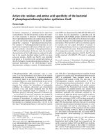
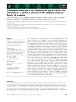
no improvement was observed. Scrotal ultrasound
examination revealed an abnormal right testis with a
focal lesion (2.5 × 2 cm) in the lower part. The lesion
was hypoechogenic compared with the surrounding tes-
ticular tissue, and suggested the existence of a tumor
mass (Figure 1). Left testis and epididymis were sono-
graphically normal. Doppler ultrasound examination
revealed well vascularized right and left testes. The focal
lesion of the right testis was also vasculari zed, similar to
the surrounding normal testicular parenchyma. This
finding practically excluded testicular torsio n, segmental
testicular infarction and orchitisaspossiblediagnoses.
* Correspondence:
1
Department of Urology, University Hospital Rijeka, Rijeka, Croatia
Full list of author information is available at the end of the article
Maričić et al. World Journal of Surgical Oncology 2011, 9:63
/>WORLD JOURNAL OF
SURGICAL ONCOLOGY
© 201 1 Mariččićć et al; licensee BioMed Central Ltd. This is an Open Access article distributed under the terms of the Creative
Commons Attribution License ( y/2.0), which permits unrestricted use, distribution, and
reproduction in any medium, provided the original work is properly cited.
Because ultrasound findings of the right testicle were
highly indicative of testicular neoplasm, right radical
orchiectomy was performed via an inguinal incision.
Histopathological findings
The testicle measured 4 × 3.5 × 2.5 cm and contained
well-demarcated areas of hemorrhage, 3 cm in diameter.
The epididymis, investing membranes and spermatic
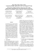
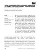
cord appeared grossly normal. Microscopy showed the
presence of a patchy, necrotizing vasculitis affecting
medium-sized and small-sized arteries of the testicle
(Figure 2). Several vessels showed fibrinoid necrosis of
their walls and muscular layer detachment with or with-
out a transmural infiltrate composed of polymorphonuc-
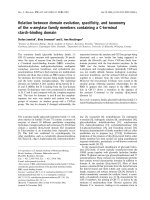
lear leukocytes and lymphocytes. Immunofluorescence
staining for fibrin was also performed, and positive
fibrin deposits were identified in arterial walls affected
with fibrinoid necrosis (Figure 3).
Follow-up
The postoperative course was uneventful. After the
operation, extensive clinical evaluation was performed to
exclude other systemic diseases characterized by vasculi-
tis. This included: complete blood count; erythrocyte
sedimentation rate; CRP; urinalysis; immunoglobulin
serum level; immunological blood tests, such as rheuma-
toid factor, antinuclear antibody tests and anti-neutro-
phil cytoplasmic autoantibody test; complement tests;
human leukocyte antigen tissue typing tests; ultrasound
of the abdomen; endoscopic examination of the ear,
nose and throat; chest X-ray; and ophthalmologist
examin ation. All test results were normal. There was no
sign of systemic disease. Two years after the diagnosis,
systemic disease had not developed.
Discussion
The most common appearance of testicular vasculitis is
as part of a multiorgan or systemic disease. Involvement
of the testicles is seen less frequently in Wegener’s gran-
ulomatosis, Henoch-Schönlein purpura, giant cell arteri-
tis, and rheumatoid arthritis, whereas testicle
involvement is commonly associated with polyarteritis
nodosa [3-5]. The microscopicall y observed changes are
almost identical in all vasculitis seen in other systemic
disorders. The results of postmortem studies suggest
that the testis is involved in 38-86% of cases of polyar-
teritis nodosa. At the same time, less than 18% of these
cases are symptomatic, and most will show other mani-
festations of polyarteritis nodosa [6,7].
Isolated testicular vasculitis is not a common condi-
tion [10-13]. It is usually found in young people, as in
our patient [12]. F rom the present literature findings, it
remains unclear whether such cases represent truly iso-
lated vasculitis or solely an unusual primary presenta-
tion site. The pathogenesis of isolated organ vasculitis is
unknown, as is why only one organ may be affected.
Additionally, it is unknown whether such cases carry the
risk of subsequent progression, a nd if so, the risk
remains to be determined. It is not known whether iso-
lated vasculitis has a better prognosis than does systemic
Figure 1 Ultrasound: hypoechogenic focal lesion in the lower pole of right testicle.
Maričić et al. World Journal of Surgical Oncology 2011, 9:63
/>Page 2 of 5
Figure 3 Immunofluorescence staining detected deposits of fibrin in testicular vessels affected with fibrinoid necrosis. This represents a
morphological hallmark of necrotizing vasculitis (200x).
Figure 2 Hematoxylin eosin (HE) staining showing the medium-sized artery in the testicular parenchyma showing fibrinoid necrosis
and segmental involvement of moderate inflammatory cell infiltrate and perivascular inflammation (200x).
Maričić et al. World Journal of Surgical Oncology 2011, 9:63
/>Page 3 of 5
disease. This is important because the classic form of
polyarteritis nodosa carries significant risks of mortality
and morbidity, even with treatment, and has a high rate
of relapse [9].
The conditions presenting as pain in the testicle or epi-
didymitis have been previously reported, but presentation
with clinical features suggestive of testicular neoplasm is
even more exceptional [7,10]. In the majority of reported
cases, clinical or laboratory evidence of disease in other
organ systems on presentation was present or developed
subsequently within a short time period [5].
Testicular necrotizing vasculitis is impossible to diag-
nose without tissue analysis. Our patient first presented
with symptoms and signs in favor of inflammation
(orchitis as concomitant disease cannot be excluded).
However, when ultrasound with Doppler was performed,
it was obvious that inflammation was not the cause of
this lesion. Additionally, because the lesion was well vas-
cularized, testicular torsion and segmental testicular
infarction were excluded. Testicu lar neoplasm remained
the most probable diagnosis. After orchiectomy, histo-
pathological findings were used to investigate the exis-
tence of necrotizing vasculitis. Histopathological
characteristics observed in necrotizing vasculitis are
mainly restricted to blood vessels. Fibrinoid necrosis is
the morphological hallmark of the disease. The walls of
small and medium-sized testicular arteries are affected,
as shown in t his case report. Notably, hemorrhagic
necrosis occurs in other pathological conditions, such as
testicular torsion, infarction and inflammation.
Serological markers such as CRP and von Willebrand
factor are possible indicators of endothelial injury in sys-
temic vasculitis but may not reflect the activity in iso-
lated organ disease. Ultrasound examination may fail to
show any abnormality but can also demonst rate the
existence of a hypoechogenic mass, as in our patient
[12]. Magnetic resonance imaging is a more sensitive
technique that can demonstrate focal testicular infarc-
tion, but, at present, the only “diagnostic tool” for vascu-
litis is histological confirmation.
To treat our patient with potentially toxic immunosup-
pressive therapy with the added risk of sterility, despite
the lack of clinical and objective laboratory evidence of
systemic disease, presents a difficult clinical dilemma.
Waterfield et al. reported on isolated t esti cular vasculitis
treated by immunosuppressive medications. Despite ther-
apy, the remaining testis became a ffected one year later
[10]. That patient responded well to an i ncrease in
immunosuppressive therapy. McGuirre et al. recom-
mended close surveillance without additional therapy
[12]. Because of his young age, we elected to perform
close follow-up of ou r patient, instead o f immunosup-
pressive therapy. Two years after diagnosis, the patient is
still without symptoms of systemic disease. This is the
longest asymptomatic period in a case of testicular vascu-
litis reported in the literature. In view of the high relapse
rate associated with polyarteritis no dosa, long-t erm fol-
low up for these patients is essential. However, the
absence of serological markers of disease activity makes
monitoring of any future relapse quite difficult.
Conclusion
Prima ry testicular manifestation of necrotizing vasculitis
is not a common finding. It is very important for pathol-
ogists and clinicians to know that such an entity can
initially present as a testicular mass. Follow-up of these
patients is recommended due to the risk of relapse;
however, due to the rarity of the con dition, the appro-
priate strategies for treatment and follow-up remain to
be determined.
Consent
Written informed consent was obtained from the patient
for publication of this Case report and any accompanying
images. A copy of the written consent is available for
review by the Editor-in-Chief of this journal.
Abbreviations
CRP: C reactive protein.
Acknowledgements
None
Author details
1
Department of Urology, University Hospital Rijeka, Rijeka, Croatia.
2
Department of Pathology, School of Medicine , University of Rijeka, Rijeka,
Croatia.
Authors’ contributions
MA, SS and MD tracked the clinical data and drafted the manuscript. FŽ,VM
and ŠJ participated in the design of the study and coordination and helped
to draft the manuscript. GÐ and SŠ provided the pathological technique.
All authors read and approved the final manuscript.
Competing interests
The authors declare that they have no competing interests.
Received: 10 March 2011 Accepted: 14 June 2011
Published: 14 June 2011
References
1. Raj GV, Ellington KS, Polascik TJ: Autoimmune testicular vasculitis. Urology
2003, 61:1035.
2. Susanto CR, Fedder G, Looijen-Salamon MG: Acute, painful, and swollen
testicle as the presenting feature in polyarteritis nodosa. Eur J Intern Med
2003, 14:441-443.
3. Huisman TK, Collins WT Jr, Voulgarakis GR: Polyarteritis nodosa
masquerading as a primary testicular neoplasm; a case report and
review of the literature. J Urol 1990, 144:1236-1238.
4. Belville WD, Insalaco SJ, Dresner ML, Buck AS: Benign testis tumors. J Urol
1982, 128:1198-1200.
5. Lee LM, Moloney PJ, Wong HC, Magil AB, McLoughlin MG: Testicular pain:
an unusual presentation of polyarteritis nodosa. J Urol 1983,
129:1243-1244.
6. Gondos B, Wong TW: Non-neoplastic diseases of the testis and
epididymis. In Urological pathology. Edited by: Murphy W. Philadelphia. WB
Saunders; 1989:249-313.
Maričić et al. World Journal of Surgical Oncology 2011, 9:63
/>Page 4 of 5
7. Shurbaji MS, Epstein JI: Testicular vasculitis: implications for systemic
disease. Hum Pathol 1988, 19:186-189.
8. Womack C, Ansell ID: Isolated arteritis of the epididymis. J Clin Pathol
1985, 38:797-800.
9. Gordon M, Luqmani RA, Adu D, Greaves I, Richards N, Michael J, Emery P,
Howie AJ, Bacon PA: Relapses in patients with a systemic vasculitis. QJ
Med 1993, 86:779-789.
10. Warfield AT, Lee SJ, Phillips SM, Pall AA: Isolated testicular vasculitis
mimicking a testicular neoplasm. J Clin Pathol 1994, 47:1121-1123.
11. Atis G, Memis OF, Güngör HS, Arikan O, Saglican Y, Caskurlu T: Testicular
polyarteritis nodosa mimicking testicular neoplasm. ScientificWorldJournal
2010, 10:1915-1918.
12. McGuire BB, O’Brien MF, Akhtar M, Fitzpatrick JM: Testicular vasculitis
mimicking a testicular neoplasm. Ir Med J 2006, 99:27-28.
13. Joudi FN, Austin JC, Vogelgesang SA, Jensen CS: Isolated testicular
vasculitis presenting as a tumor-like lesion. J Urol 2004, 171:799.
doi:10.1186/1477-7819-9-63
Cite this article as: Maričić et al.: Primary testicular necrotizing vasculitis
clinically presented as neoplasm of the testicle: a case report. World
Journal of Surgical Oncology 2011 9:63.
Submit your next manuscript to BioMed Central
and take full advantage of:
• Convenient online submission
• Thorough peer review
• No space constraints or color figure charges
• Immediate publication on acceptance
• Inclusion in PubMed, CAS, Scopus and Google Scholar
• Research which is freely available for redistribution
Submit your manuscript at
www.biomedcentral.com/submit
Maričić et al. World Journal of Surgical Oncology 2011, 9:63
/>Page 5 of 5