Báo cáo y học: "CD25brightCD4+ regulatory T cells are enriched in inflamed joints of patients with chronic rheumatic disease" potx
Bạn đang xem bản rút gọn của tài liệu. Xem và tải ngay bản đầy đủ của tài liệu tại đây (346.71 KB, 12 trang )
Open Access
Available online />R335
Vol 6 No 4
Research article
CD25
bright
CD4
+
regulatory T cells are enriched in inflamed joints of
patients with chronic rheumatic disease
Duojia Cao, Ronald van Vollenhoven, Lars Klareskog, Christina Trollmo and Vivianne Malmström
Rheumatology Unit, Department of Medicine at Karolinska Hospital, Karolinska Institutet, Stockholm, Sweden
Corresponding author: Vivianne Malmström,
Received: 25 Dec 2003 Revisions requested: 28 Jan 2003 Revisions received: 1 Apr 2004 Accepted: 7 May 2004 Published: 7 Jun 2004
Arthritis Res Ther 2004, 6:R335-R346 (DOI 10.1186/ar1192)
http://arthr itis-research.com/conte nt/6/4/R335
© 2004 Cao et al.; licensee BioMed Central Ltd. This is an Open Access article: verbatim copying and redistribution of this article are permitted in
all media for any purpose, provided this notice is preserved along with the article's original URL.
Abstract
CD25
+
CD4
+
regulatory T cells participate in the regulation of
immune responses. We recently demonstrated the presence of
CD25
bright
CD4
+
regulatory T cells with a capacity to control T
cell proliferation in the joints of patients with rheumatoid arthritis.
Here, we investigate a possible accumulation of these
regulatory T cells in the inflamed joint of different rheumatic
diseases including rheumatoid arthritis. The studies are also
extended to analyze whether cytokine production can be
suppressed by the regulatory T cells. Synovial fluid and
peripheral blood samples were obtained during relapse from 36
patients with spondyloarthropathies, 21 adults with juvenile
idiopathic arthritis and 135 patients with rheumatoid arthritis,
and the frequency of CD25
bright
CD4
+
T cells was determined.
Of 192 patients, 182 demonstrated a higher frequency of
CD25
bright
CD4
+
T cells in synovial fluid than in peripheral blood.
In comparison with healthy subjects, the patients had
significantly fewer CD25
bright
CD4
+
T cells in peripheral blood.
For functional studies, synovial fluid cells from eight patients
were sorted by flow cytometry, and the suppressive capacity of
the CD25
bright
CD4
+
T cells was determined in in vitro
cocultures. The CD25
bright
CD4
+
T cells suppressed the
production of both type 1 and 2 cytokines including interleukin-
17, as well as proliferation, independently of diagnosis. Thus,
irrespective of the inflammatory joint disease investigated,
CD25
bright
CD4
+
T cells were reduced in peripheral blood and
enriched in the joint, suggesting an active recruitment of
regulatory T cells to the affected joint. Their capacity to suppress
both proliferation and cytokine secretion might contribute to a
dampening of local inflammatory processes.
Keywords: autoimmunity, cytokines, human, immune homeostasis, interleukin-17
Introduction
Relapses and intermittent remission phases characterize
the disease course in rheumatic diseases and other chronic
inflammatory conditions. This waxing and waning probably
corresponds to a regulation of the disease, in which the
immune system is believed to play a major role by balancing
pro- and anti-inflammatory immune reactions.
CD25
+
CD4
+
regulatory T cells constitute one cell popula-
tion involved in maintaining this homeostatic balance,
because a lack of cells with this phenotype has been dem-
onstrated to be associated with autoimmune disease in ani-
mals. Early experiments showed that neonatal thymectomy
of mice led to the development of various organ-specific
autoimmune manifestations, such as gastritis, oophoritis,
orchitis and thyroiditis [1]. Later, it was demonstrated that
CD25 identified these regulatory CD4
+
T cells [2], and
despite extensive research, so far no other cell surface
marker has been found to be generally applicable for these
cells. The transcription factor Foxp3 has been shown to be
specific for these cells [3-5]; however, its intracellular
expression does not yet allow live sorting of cells for func-
tional assays. In the peripheral immune system of naive
mice and in any thymus, the expression of CTLA-4, GITR
(glucocorticoid induced tumor necrosis factor [TNF] recep-
tor) and CD62L are correlated with CD25
+
CD4
+
regula-
tory T cells [6-8]. However, because the expression of
these molecules is also altered after T cell activation, they
are not informative markers of CD25
+
regulatory T cells in
antigen-experienced animals or humans [9-11]. We instead
APC = antigen-presenting cell; CBA = cytometric bead array; CRP = C-reactive protein; ELISA = enzyme-linked immunosorbent assay; FACS =
fluorescence-activated cell sorting; IFN = interferon; IL = interleukin; JIA = juvenile idiopathic arthritis; PsA = psoriatic arthritis; RA = rheumatoid arthri-
tis; RF = rheumatoid factor; R
SF
= responder cells of synovial fluid origin; SFMC = synovial fluid mononuclear cells; SpA = spondyloarthopathies; TNF
= tumor necrosis factor.
Arthritis Research & Therapy Vol 6 No 4 Cao et al.
R336
subgroup the CD25
+
T cells according to the level of CD25
expression. This has been demonstrated to roughly divide
activated (intermediate CD25 expression) from regulatory
T cells (high CD25 expression) in the peripheral blood of
healthy subjects [12]. We have recently shown that
patients with rheumatoid arthritis (RA) have an enrichment
of CD25
bright
CD4
+
T cells in their inflamed joints [13]. Here,
owing to the accumulation of activated cells in the target
organ of the disease, the gate for inclusion of regulatory T
cells is even more restricted than in peripheral blood (bright
CD25 expression). Given the animal data in which cell
transfer of regulatory T cells into animals can prevent the
development of chronic inflammation, as reviewed in [14],
one would expect humans with an autoimmune disease to
have smaller numbers of regulatory T cells, but surprisingly,
despite counting only CD4
+
T cells with the highest level of
CD25 expression, an enrichment of regulatory T cells was
seen. Thus, the simple extrapolation of the animal data into
future therapeutic strategies that aim at reconstituting this
population does not seem to hold true in human organ-spe-
cific autoimmune diseases. Our results obtained from
patients with RA [13] therefore raised several questions:
Do these cells exist in inflamed joints of rheumatic patients
irrespective of diagnosis? Are they accumulated at the site
of inflammation? Can these regulatory T cells from synovial
fluid suppress cytokine production, which is an important
feature of the chronically inflamed joint? Also, with a large
patient cohort can we address the question of possible cor-
relations of frequency of regulatory T cells and clinical
parameters such as disease duration, severity of disease,
and treatment?
To address these questions we decided to analyze adult
patients with juvenile idiopathic arthritis (JIA) and patients
with spondyloarthropathies, diseases in which peripheral
joint inflammation occurs, and to compare them with results
from patients with RA. Despite different clinical features of
these disorders, the knee inflammation has similar charac-
teristics: an infiltration of inflammatory cells; an increase in
volume of synovial fluid; and a local production of proinflam-
matory cytokines. Interleukin-17 (IL-17) is a T cell-derived
cytokine with similar proinflammatory properties to IL-1 and
TNF in the inflamed joint [15]. We recruited 192 patients
with spondyloarthropathies, JIA or RA to study regulatory
CD25
bright
CD4
+
T cells in the inflamed joints. Our results
clearly demonstrate an enrichment of CD25
bright
CD4
+
reg-
ulatory T cells in the inflamed joints in comparison with
peripheral blood. This cell population suppressed both
cytokine production and proliferation of other T cells and
can therefore be regarded as containing regulatory T cells.
However, the frequency in the inflamed joint could not be
associated with disease duration, disease severity or
treatment.
Materials and methods
Sample material
Thirty-six patients with spondyloarthropathies, 21 with JIA
(as defined by the International League of Associations for
Rheumatology criteria [16,17]) and 135 with RA (as
defined by the American College of Rheumatology criteria
[18]) were recruited from the Rheumatology Clinic at the
Karolinska Hospital, Stockholm, Sweden. Within the group
of patients with spondyloarthropathies, 26 were diagnosed
with psoriatic arthritis (PsA) and the other 10 were diag-
nosed with ankylosing spondylitis or undifferentiated
spondyloarthropathies (SpA). The patients with JIA were all
adults and had a polyarticular disease. Of the 135 patients
with RA, 26 were seronegative for rheumatoid factor (RF).
The synovial samples were obtained from the patients
when excess fluid was removed from swollen joints before
glucocorticoid was injected as part of the clinical routine
procedure. Paired peripheral blood samples were obtained
from 166 of these 192 patients. Peripheral blood samples
were also obtained from 29 healthy donors. This study was
performed after human subject approval from the Karolin-
ska Hospital. Informed consent was obtained from all con-
tributing individuals. Table 1 provides a summary of the
patients and healthy controls included in the frequency
study.
Cell separation and flow cytometry
Mononuclear cells were prepared from peripheral blood
and synovial fluid by Ficoll separation (Ficoll-Paque; Phar-
macia, Sweden). For frequency determinations cells were
stained with anti-CD3-FITC (clone SK7), anti-CD4-PerCP
(SK3) and anti-CD25-APC (2A3) (all from Becton Dickin-
son [BD], Franklin Lakes, NJ, USA). The stained cells were
analyzed by flow cytometry on a FACSCalibur (BD). For
functional studies, synovial fluid mononuclear cells (SFMC)
expressing CD3 on their surface were sorted into
CD25
bright
CD4
+
T cells and CD25
-
CD4
+
T cells, also
referred to as responder cells (R
SF
). The sorting gate for
CD25
bright
CD4
+
T cells was adjusted to contain CD4
+
T
cells that expressed CD25 more brightly than activated
CD25
+
CD8
+
T cells. SFMC not expressing CD3 were
sorted as antigen-presenting cells (APCs). The sorting was
performed on a fluorescence-activated cell sorting (FACS)
Vantage SE cell sorter (BD) or on a MoFlo cell sorter (Cyto-
mation, Fort Collins, CO). After sorting, the purity of the
sorted populations was determined by FACS reanalysis of
an aliquot of cells, and was 90% on average (data not
shown). Small dying and large activated T cells were
excluded from the sorting gates.
Proliferation assays and enzyme-linked immunosorbent
assay (ELISA)
Coculture experiments were set up with 2 × 10
4
autolo-
gous APCs, 5 × 10
3
CD25
-
CD4
+
R
SF
, and varying num-
bers of CD25
bright
CD4
+
cells in plate-bound anti-CD3-
Available online />R337
coated wells (OKT-3; 1 µg/ml). The cell culture medium
was based on RPMI with 100 units/ml penicillin-streptomy-
cin, 2 mM glutamine, 10 mM HEPES buffer (all from Gibco
BRL, Invitrogen Corporation) and 5% human pooled serum
(Blood bank, Karolinska Hospital, Stockholm, Sweden). To
detect cytokines, some experiments were set up with
20,000 responder T cells per well (indicated in Table 2).
The sorted autologous CD3-depleted SFMC were irradi-
ated (33 Gy) and used as APCs. Cells were incubated at
37°C for 6 days, the last 15–18 h in the presence of
[
3
H]thymidine. Standard sandwich ELISAs and human
Th1/Th2 cytokine cytometric bead array (CBA) were per-
formed to determine the concentrations of interferon (IFN)-
γ, TNF, IL-2, IL-17, IL-10, IL-13 and IL-4 in culture superna-
tants after 5 days of culture. The antibodies used for ELISA
were bought from MABTECH AB (Sweden) (IFN-γ),
Pharmingen (IL-10 and IL-13) and R&D (IL-17). The CBA
kit was bought from BD.
Statistical analysis
The frequencies of CD25-expressing cells from peripheral
blood and synovial fluid were compared by the Mann–
Whitney test. The Kruskal–Wallis test was used for com-
parison of the frequencies of synovial and peripheral blood
CD25
bright
CD4
+
cells between groups of patients with dif-
ferent diagnoses. Regression analyses were performed to
Table 1
Summary of rheumatic patients included in the frequency study
Diagnosis,
subdiagnosis
Number Sex (F/M) Age (years)
Median (range)
CD25
bright
CD4
+
in PB
(%) Median (range)
CD25
bright
CD4
+
in SF
(%) Median (range)
Fold increase, SF/PB
a
Median (range)
b
Spondyloarthropathies 36 20/16 46 (21–83) 0.7 (0.2–2.9) 2.7 (0.4–9.1) 4.8 (0.7–20.7)***
PsA 26 14/12 50 (24–83) 0.6 (0.2–2.9) 2.6 (1.0–9.1) 4.7 (0.9–20.7)
SpA
c
10 6/4 33 (21–58) 1.2 (0.4–2.4) 3.4 (0.4–6.8) 5.0 (0.7–17.6)
JIA 21 18/3 30 (18–50) 0.4 (0.1–1.8) 3.7 (0.3–6.7) 6.7 (3.1–16.3)***
Rheumatoid arthritis 135 107/28 57 (22–85) 0.7 (0.04–2.9) 2.3 (0.2–19.9) 3.7 (0.4–56.8)***
RF
+
109 88/21 57 (25–81) 0.7 (0.04–2.9) 2.3 (0.3–19.9) 3.5 (0.4–56.8)
RF
-
26 19/7 56 (22–85) 0.9 (0.2–2.4) 2.4 (0.7–19.7) 4.3 (0.6–19.9)
Healthy controls 29 25/4 51 (23–63) 1.2 (0.3–2.4) na na
JIA, juvenile idiopathic arthritis; na, not analyzed; PB, peripheral blood, SF, synovial fluid.
a
Calculated as percentage of CD25
bright
CD4
+
cells in SF
divided by percentage of CD25
bright
CD4
+
cells in PB.
b
A significant enrichment of CD25
bright
CD4
+
T cells in SF over that in PB was found: ***P <
0.0001.
c
Ankylosing spondylitis and undifferentiated spondyloarthropathies. Excluded are patients with reactive arthritis and gastrointestinal
inflammation.
Table 2
Cytokine production and proliferation of CD25
-
CD4
+
T cells in in vitro cultures
Patient IFN-γ(pg/ml) IL-10 (pg/ml) IL-13 (pg/ml) TNF (pg/ml) Proliferation (c.p.m.)
PsA 1
a
6656 459 952 na 67,331
PsA 2
b
4775 67 na 468 42,662
SpA 1
a
879 bdl bdl na 13,086
SpA 2
b
3681 bdl na 806 42,469
JIA 1
b
1924 53 na 355 105,210
JIA 2
b
1656 30 na 530 12,621
RA 1
a
5677 385 293 na 106,269
RA 2
b
608 bdl na 109 103,724
Production of cytokines and proliferation of responder cells in patients with psoriatic arthritis (PsA), spondyloarthopathies (SpA), juvenile
idiopathic arthritis (JIA) and rheumatoid arthritis (RA) are shown. Changes in cytokine and proliferation levels after coculture with CD25
bright
CD4
+
T cells are shown in Fig. 5. bdl, below detection limit; IFN, interferon; IL, interleukin; na, not analyzed; TNF, tumor necrosis factor.
a
Enzyme-linked
immunosorbent assay; detection limit is 50 pg/ml; 20,000 cells per well in coculture.
b
Cytometric bead array; detection limit is 3 pg/ml; 5000 cells
per well in coculture.
Arthritis Research & Therapy Vol 6 No 4 Cao et al.
R338
analyze possible correlations between the frequency of
CD25
bright
CD4
+
cells and disease duration or levels of C-
reactive protein (CRP) in the circulation.
Results
CD25
bright
CD4
+
T cells are reduced in the circulation and
enriched in the inflamed joints of patients with chronic
rheumatic diseases
In total, 192 patients with different chronic rheumatic dis-
eases were included in this study, in which synovial fluid
and peripheral blood samples were screened for the fre-
quency of CD25
bright
CD4
+
T cells by flow cytometry. Infor-
mation about the 26 patients with PsA, the 10 patients with
SpA, the 21 patients with JIA and the 135 patients with RA
are provided in Table 1. For comparison, peripheral blood
samples from 29 healthy subjects were also analyzed. The
flow cytometric analysis gates, for determining the fre-
quency of CD25
bright
CD4
+
T cells in peripheral blood and
synovial fluid of patients, were set in accordance with our
previous study on patients with RA, in which functional reg-
ulatory CD25
+
T cells were isolated [13]. The gate for
CD25
bright
CD4
+
T cells in synovial fluid was set higher than
for peripheral blood, as shown in Fig. 1a. This is necessary
because the joint fluid contains a larger proportion of highly
activated T cells [19,20].
A frequency analysis of CD25
bright
CD4
+
T cells in synovial
fluid demonstrated high frequencies of these cells in all
patient groups, with a median of 2.6% in PsA, 3.4% in SpA,
3.7% in JIA and 2.3% in RA (Fig. 1b and Table 1). A parallel
analysis of peripheral blood samples showed a significantly
lower frequency of these cells in the blood than in synovial
fluid, with a median of 0.6% in PsA, 1.2% in SpA, 0.4% in
JIA and 0.7% in RA (Fig. 1c and Table 1). To confirm this
enrichment of CD25
bright
CD4
+
T cells at the level of single
individuals, the relative increase in synovial fluid over that in
peripheral blood was calculated. In all 21 JIA patients, in 21
of 23 PsA patients, in 6 of 7 SpA patients and in 110 of
117 RA patients, increased frequencies were measured in
the joint, indicated by a fold increase of more than 1 (Fig.
1d and Table 1). The median increase for patients with PsA
was 4.7, for patients with SpA 5.0, for patients with JIA 6.4
and for patients with RA 3.7; P values are given in Table 1.
The median level of expression of CD25
bright
CD4
+
T cells in
peripheral blood of healthy subjects was 1.2% (Fig. 1c and
Table 1). A comparison of peripheral blood frequencies of
CD25
bright
CD4
+
T cells between patients and healthy sub-
jects showed significantly lower levels in the rheumatic
patients, indicating a selective recruitment of regulatory T
cells in the inflamed joint. Only in the seven patients with
SpA was the median frequency in peripheral blood equal to
that of healthy controls.
The frequencies of CD25
bright
CD4
+
T cells in inflamed
joints are similar between patients with different
diagnoses, and persist over time
To investigate whether the enrichment of CD25
bright
CD4
+
T cells is a general phenomenon of the inflamed joint, we
compared the frequencies of synovial CD25
bright
CD4
+
T
cells between the different patient groups. No statistically
significant differences were found (Fig. 1b). Thus, these
data indicate that CD25
bright
CD4
+
T cells accumulate in
inflamed synovial joints of patients with chronic rheumatic
diseases irrespective of diagnosis.
Several of the rheumatic patients had recurrent effusions in
their knee joints, from which synovial fluid was obtained.
This allowed a longitudinal study of the frequencies of
CD25
bright
CD4
+
T cells in eight patients with PsA, four with
SpA and eight with JIA (Fig. 2). In nine of these patients,
three or more samples were obtained from the same joint.
Although we had some variations, the frequencies did not
differ significantly over time.
The frequency of synovial CD25
bright
CD4
+
T cells is not
associated with clinical parameters
Clinical data were collected from 100% of our SpA and RA
patients, and from 60% of PsA and JIA patients, to deter-
mine whether the frequencies of CD25
bright
CD4
+
T cells in
the synovial fluid and peripheral blood could be correlated
with disease duration, severity of disease and degree of
inflammation. Because of the large number of patients
required for statistically reliable analyses when subdividing
patients, we here present the results of the investigations
on only the RA patients in graphic format. The other patient
groups were, however, also studied and the results are pre-
sented at the end of this section.
The first question we addressed was whether the accumu-
lation of CD25
bright
CD4
+
T cells in the inflamed joints is
dependent on the chronicity of the disease. We thus inves-
tigated whether disease duration was correlated with the
frequency of CD25
bright
CD4
+
T cells. As shown in Fig. 3a,
the number of years with disease had no impact on the fre-
quencies of regulatory T cells, either in synovial fluid or
peripheral blood.
Second, to study whether the severity of disease could be
correlated with a decreased frequency of regulatory T cells,
we chose to study the presence or absence of RF and ero-
sions. RF is a predicting factor for the development of a
more severe disease course with the erosion of cartilage
and bone [21]. The presence of erosions is clinically ana-
lyzed in the small joints of hands or feet, which are the joints
first affected in RA. Thus, the X-ray analysis clearly answers
the questions of whether the disease has progressed to an
erosive, more severe disease. We categorized the RA
patients with regard to the presence or absence of RF in
Available online />R339
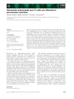
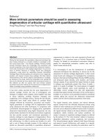
Figure 1
CD25
bright
CD4
+
T cells are enriched in the joint of patients with rheumatic diseases and are decreased in peripheral bloodCD25
bright
CD4
+
T cells are enriched in the joint of patients with rheumatic diseases and are decreased in peripheral blood. (a) Representative fluo-
rescence-activated cell sorting plots of paired peripheral blood (PB) mononuclear cells and synovial fluid (SF) mononuclear cells from one patient
with psoriatic arthritis (PsA). Numbers within the gates represent the percentage of CD25
bright
CD4
+
T cells of all CD4
+
T cells. (b) Frequency of
CD25
bright
CD4
+
T cells in synovial fluid of patients with PsA, spondyloarthopathies (SpA), juvenile idiopathic arthritis (JIA) and rheumatoid arthritis
(RA). Each triangle represents one individual. (c) The frequencies of CD25
bright
CD4
+
T cells in peripheral blood were compared between healthy
subjects and rheumatic patients. Significant differences between patient group and healthy subjects are indicated with asterisks: *** P < 0.0001; **
P = 0.001; * P = 0.02. Note that the scale is different from that in (b). (d) Relative increase of CD25
bright
CD4
+
T cells in synovial fluid in comparison
with that in peripheral blood (fold increase) analyzed in all patients from whom paired synovial fluid and peripheral blood samples had been obtained.
na, not applicable.
%CD25
bright
CD4
+
T cells in SF
%CD25
bright
CD4
+
T cells in PB
Fold increase
(a)
(b)
(c)
(d)
10
1
10
0
10
3
10
4
10
2
0.5%
9%
PB SF
CD25
CD4
HC PsA SpA JIA RA
0
5
10
15
2
0
(135)(10)(26)
(21)
na
HC PsA SpA JIA RA
0
1
2
3
(29) (23) (7) (117)(19)
*
**
**
*
HC PsA SpA JIA RA
1
10
100
(7)
(117)(23) (19)
na
Arthritis Research & Therapy Vol 6 No 4 Cao et al.
R340
serum (Fig. 3b) and with regard to the presence or absence
of erosions (Fig. 3c), and compared the frequencies of
CD25
bright
CD4
+
T cells in both synovial fluid and peripheral
blood. The range of CD25
bright
CD4
+
T cells in the inflamed
joint in the patient groups with or without RF was compara-
ble, with medians of 2.3% in the RF-positive group and
2.4% in the RF-negative group (Fig. 3b), as it was in periph-
eral blood, with a median of 0.7% in the RF-positive group
and 0.8% in the RF-negative group (Fig. 3b). When com-
paring the patients with and without confirmed erosions, no
significant differences were found between the groups
(Fig. 3c). The range of CD25
bright
CD4
+
T cells in the
inflamed joint in the patient groups with or without erosions
was the same, with a median of 2.2% in both groups, as
was the range in peripheral blood with a median of 0.7%
(Fig. 3c). This clearly demonstrates that within this patient
material there are no correlations between severity of dis-
ease and frequency of regulatory T cells.
Third, to correlate the degree of inflammation in the
patients, CRP levels were compared with frequencies of
CD25
bright
CD4
+
T cells in both synovial fluid and peripheral
blood. The level of CRP was measured on the day of visit
or within 1 week before synovial fluid sampling. No correla-
tion with the size of regulatory T cell population was
observed (Fig. 3d).
Last, we took into account the local, intra-articular, treat-
ments that the patients were receiving. Only those patients
with documentation of intra-articular cortisone injection
within 3 months before sampling are depicted in Fig. 3e. As
can be seen, no difference could be detected in the fre-
quency of CD25
bright
CD4
+
T cells irrespective of whether
the patients had received corticosteroids during their previ-
ous bout. The range of CD25
bright
CD4
+
T cells in the
inflamed joint in the patients treated or not treated was the
same, with a median of 2.6% in both groups (Fig. 3e), as
was the range in peripheral blood, with a median of 0.6%
(Fig. 3e).
As mentioned above, we also investigated the SpA, PsA
and JIA patients with regard to the stated clinical
parameters. None of the parameters showed any correla-
tion with the size of the CD25
bright
CD4
+
T cell population,
nor were there any tendencies in the limited pool of patients
with these diagnoses.
Synovial CD25
bright
CD4
+
T cells from rheumatic patients
have regulatory functions in vitro
To investigate whether synovial CD25
bright
CD4
+
T cells
from patients with PsA, SpA or JIA contain a suppressive
population, two patients from each group were selected for
functional studies. Because the cytokine suppression pro-
file of patients with RA has not been analyzed previously,
we also included two patients from this group for these
functional studies. Patient characteristics for these eight
patients are presented in Fig. 4. The selection of patients
was based on both the frequency of CD25
bright
CD4
+
T
cells and the availability of large numbers of synovial cells.
CD25
bright
CD4
+
T cells and CD25
-
CD4
+
T cells from syno-
vial fluid were sorted according to the sorting gates shown
in Fig. 4. The FACS plots are gated via CD3
+
cells and the
sorting gate for CD25
bright
CD4
+
T cells includes all cells
with a brighter CD25 expression than the CD25
+
CD8
+
T
cells. The experiments were set up with variable number of
CD25
bright
CD4
+
T cells added to a constant number of
autologous CD25
-
CD4
+
R
SF
. As expected from regulatory
T cells, CD25
bright
CD4
+
T cells alone did not proliferate in
response to the anti-CD3 stimulation in any of the patients
analyzed, depicted in the figure as CD25
br
(Fig. 5). The
amount of proliferation of responder cells alone is shown in
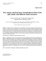
Figure 2
CD25
bright
CD4
+
T cell population persists over timeCD25
bright
CD4
+
T cell population persists over time. (a) Eight patients
with psoriatic arthritis (PsA), (b) four patients with spondyloarthopa-
thies (SpA) and (c) eight patients with juvenile idiopathic arthritis (JIA)
were followed longitudinally, and the frequency of synovial
CD25
bright
CD4
+
T cells was measured at each relapse from which syn-
ovial fluid was obtained. Open symbols depict patients who had two
relapses during the study period; filled symbols depict patients who
had three or more relapses. Time point zero corresponds to the first
time point at which synovial fluid was analyzed for the frequency of
CD25
bright
CD4
+
T cells. From one patient with PsA and one with SpA,
samples from both knees were obtained; arrows pointing left, left knee;
arrows pointing right, right knee.
0
5
10
15
20
0 3 6 9 12 15 18 21 24
0
5
10
15
20
0369
0
5
10
15
20
0369121518
months
JIA
n=8
SpA
n=4
PsA
n=8
% of CD25
bright
CD4
+
T cells% of CD25
bright
CD4
+
T cells% of CD25
bright
CD4
+
T cells
(a)
(b)
(c)
Available online />R341
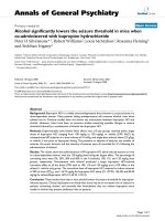
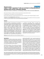
Figure 3
The frequency of CD25
bright
CD4
+
T cells of patients with rheumatoid arthritis is not associated with clinical parametersThe frequency of CD25
bright
CD4
+
T cells of patients with rheumatoid arthritis is not associated with clinical parameters. Peripheral blood (PB, left
column) and synovial fluid (SF, right column) were analyzed for the correlation with (a) disease duration, (b) the presence or absence of rheumatoid
factor (RF), (c) the presence or absence of erosions, (d) the level of C-reactive protein and (e) intra-articular cortisone treatment. In (a), (b) and (c)
each symbol represents one patient; that is, mean values of CD25
bright
CD4
+
T cells from the different visits. In (d) and (e) each symbol represents a
single visit; the number of symbols in each diagram is presented in brackets.
0 102030405060
0
1
2
3
Disease Duration (years
)
0102030405060
0
10
20
Disease Duration (years
)
erosion+ erosion-
0
1
2
3
(51) (1
7)
erosion+ erosion-
0
10
20
(59)
(21
)
0 50 100 150 200
0
1
2
3
CR
P
0 50 100 150 20
0
0
10
20
CR
P
RF+ RA RF- RA
0
1
2
3
(95)
(
22)
RF+ RA RF- RA
0
10
20
(109) (
26)
treated nontreated
0
10
20
(36)(51)
%CD2
bright
in PB
%CD25
bright
in PB%CD25
bright
in PB%CD25
bright
in PB%CD25
bright
in PB
%CD25
bright
in SF
%CD25
bright
in SF%CD25
bright
in SF%CD25
bright
in SF
%CD25
bright
in SF
PB
SF
disease
duration
rheumatoid
factor
erosion
c-reactive
protein
local
cortisone
injection
(a)
(b)
(c)
(d)
(e)
treated nontreated
0
1
2
3
(41)
(28)
Arthritis Research & Therapy Vol 6 No 4 Cao et al.
R342
Table 2. In all eight patients, the CD25
bright
CD4
+
T cell pop-
ulation was able to suppress the proliferation of responder
cells in a dose-dependent manner (Fig. 5), with suppres-
sion greater than 50% at a ratio of 3:1 of CD25
bright
CD4
+
T cells to responder T cells. At lower ratios the efficiency of
suppression showed high variability between patients (Fig.
5); this was also seen in our previous study on RA patients
[13].
The coculture supernatants were screened by ELISA or
CBA for the concentration of the T cell-produced cytokines
IFN-γ, IL-2, TNF, IL-17, IL-10, IL-13 and IL-4. In all patients,
IFN-γ was the major cytokine produced by the responder T
cells (Table 2). As expected, CD25
bright
CD4
+
T cells on
their own did not produce any of the cytokines investigated
(Fig. 5). However, these cells were able to significantly sup-
press the IFN-γ production by responder T cells in all
patients (Fig. 5). In addition, they suppressed the
production of TNF, IL-10 and IL-13 whenever the
responder T cells produced detectable amounts of these
cytokines (Fig. 5). Neither IL-2 nor IL-4 was detected under
these culture conditions. IL-17 was analyzed in one patient
with SpA and one with RA. Production of this cytokine was
also inhibited by the CD25
bright
CD4
+
T cells. Table 2 shows
the concentration of cytokines produced by the responder
cells alone. The amount of IL-17 produced by the
responder cells was 674 and 78 pg/ml, respectively.
In summary, CD25
bright
CD4
+
T cells isolated from synovial
fluid, irrespective of diagnosis, contain regulatory T cells
with a capacity to suppress T cell-driven immune
responses.
Discussion
This study demonstrates that the fluid from inflamed joints
of patients with PsA, SpA and JIA contains a population of
CD25
bright
CD4
+
T cells with a regulatory potential. These
results indicate that the presence of regulatory T cells is not
only a feature of an inflamed RA joint, either seropositive or
seronegative, but more generally one of chronic rheumatic
disease. We propose that these cells accumulate in the
joints, because in parallel with the enrichment in the joint a
decrease is observed in peripheral blood. In all three rheu-
matic diseases analyzed, the CD25
bright
CD4
+
T cells sup-
pressed not only proliferation but also cytokine production,
indicating a potential role for regulatory T cells to influence
the inflammatory processes in the joint. These features
were apparent in the vast majority of the patients despite
the different treatments they received, indicating that the
anti-rheumatic drugs that patients receive do not affect the
presence of CD25
bright
CD4
+
T cells in the joint.
The frequency of synovial CD25
bright
CD4
+
T cells was com-
parable between the different diseases. Also, a relative
increase of this population in the joint in comparison with
the circulation was observed in all patient groups. These
are interesting findings with regard to the clinical
differences between the diseases. First, the HLA associa-
tions differ: spondyloarthropathies are associated with
class I HLA antigens, mainly HLA-B27, whereas RA is
associated mostly with HLA-DR1 and HLA-DR4 of the
MHC class II alleles. Thus, the local accumulation of regu-
latory T cells to inflamed joints does not seem to be
dependent on specific MHC molecules despite the fact
that the generation of regulatory T cells in the thymus
seems to be antigen specific [22]. Indeed, it has been dem-
onstrated that the effector function, suppression, is not
antigen specific in the periphery [23].
Second, subdividing patients for the presence of RF or ero-
sions does not reveal any differences between the groups
with regard to frequency of CD25
bright
CD4
+
T cells. Our
data therefore indicate that the enrichment of regulatory T
cells in the joints is not correlated with disease severity.
Third, the different diseases analyzed here display differ-
ences with regard to the cellular assembly at the site of
inflammation. Both immunohistochemical and flow cyto-
metric analyses of T cells have shown a dominance of
CD4
+
T cells in RA, whereas CD8
+
T cells are more
frequent in inflamed joints of patients with PsA [24].
Despite these differences, the frequency of
CD25
bright
CD4
+
T cells is similar in the inflamed joints ana-
lyzed in this study. However, regulatory T cells have the
potential to suppress both CD4-driven and CD8-driven
immune responses [25] as well as innate immunity [26], so
this is perhaps not surprising. In summary, no significant dif-
ferences with regard to the frequencies of CD25
bright
CD4
+
T cells were found between the different rheumatic patients
analyzed.
In this study, all patients had chronic disease; however,
each joint was not necessarily continuously inflamed. The
synovial fluid samples were, however, always taken during
flares, which are the time points at which they can be
obtained. In our longitudinal samples from patients with
PsA, SpA and JIA, the frequency of CD25
bright
CD4
+
T cells
was found to be relatively stable over time. This parallels
our recent observations in RA patients [13]. This observa-
tion indicates that individual factors, as yet poorly under-
stood, determine the frequency of CD25
bright
CD4
+
T cells
that can be reached in the inflamed site.
Our data support trafficking from peripheral blood to the
site of inflammation, because a decreased frequency of
peripheral blood CD25
bright
CD4
+
T cells was observed in
the patients in comparison with healthy controls. Again, this
was true for the three groups of rheumatic diseases inves-
tigated so far, confirming the trend seen in our previous
study with a limited number of RA patients [13]. This finding
Available online />R343
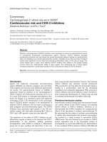
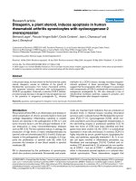
Figure 4
Sorting gates and disease characteristics for patients included in the functional studiesSorting gates and disease characteristics for patients included in the functional studies. Each row represents one patient. The sorting gates for iso-
lation of CD25
bright
(25
bright
) and CD25
-
(R) T cells are indicated in the fluorescence-activated cell sorting plots, which are gated on CD3
+
cells.
Under treatment only so-called disease-modifying anti-rheumatic drugs (DMARDs) are presented. All patients also received non-steroidal anti-inflam-
matory drugs.
Age (y)
% of
CD25
bright
R 25
bright
CD4
CD25
Diagnosis
psoriatric
arthritis 1
ankylosing
spondylitis 1
juvenile
idiopathic
arthritis 1
41
33
29
59
26
Disease
Duration
(years)
DMARD
a)
Treatment
psoriatric
arthriti
s 2
3 Methotrexate
5none
5 Chloroquine
14 none
juvenile
idiopathic
arthritis 2
21 13 Methotrexate
4.2
3.1
4.6
5.7
6.0
3.4
ankylosing
spondylitis 2
rheumatoid
arthritis 1
(RF-)
rheumatoid
arthritis 2
(RF-)
49 7.2
31 2.4
28
14
5 Methotrexate
CPH82
b)
Methotrexate
10
0
10
1
10
2
10
3
10
4
10
0
10
1
10
2
10
3
10
4
10
0
10
1
10
2
10
3
10
4
10
0
10
1
10
2
10
3
10
4
10
0
10
1
10
2
10
3
10
4
10
0
10
1
10
2
10
3
10
4
10
0
10
1
10
2
10
3
10
4
10
0
10
1
10
2
10
3
10
4
a)
Disease modifying anti-rheumatic drug
b)
An investigational broadly immunosuppressive agent
Arthritis Research & Therapy Vol 6 No 4 Cao et al.
R344
suggests that the regulatory T cells home to inflammatory
sites. Such a model is in line with the similar findings in
rodents, in which a selective recruitment of CD25
+
CD4
+
T
cells to the skin of mice infected with Leishmania major
was demonstrated [27]. Even though cell trafficking has
not been addressed in our present study, it is tempting to
speculate that the accumulation of CD25
bright
CD4
+
T cells
to the inflamed knee joints is due to selective recruitment.
However, we cannot formally exclude the possibility that
CD25
bright
CD4
+
T cells with a regulatory function can also
Figure 5
CD25
bright
CD4
+
T cells suppressed both proliferation and cytokine secretion of synovial responder cellsCD25
bright
CD4
+
T cells suppressed both proliferation and cytokine secretion of synovial responder cells. CD25
bright
CD4
+
T cells and CD25
-
CD4
+
T
cells (R
SF
) from two patients with psoriatic arthritis (PsA), two with spondyloarthopathies (SpA), two with juvenile idiopathic arthritis (JIA), and two
with rheumatoid arthritis (RA) were sorted by flow cytometry. Increasing numbers of CD25
bright
CD4
+
T cells were added to a fixed number of autolo-
gous R
SF
in coculture. Proliferation was measured after 6 days of culture with anti-CD3 stimulation (filled symbols). Culture supernatants were ana-
lyzed for cytokine content (open symbols). A response of 100% equals a proliferation/cytokine secretion of CD4
+
R
SF
on their own. IFN, interferon;
IL, interleukin; TNF, tumor necrosis factor.
0
50
100
PsA 1
0
50
100
SpA 1 SpA 2
JIA 2JIA 1
IL-10
TNF
IFN-
IL-13
PsA 2
% response of R
SF
ratio R:25
bright
R
1
:
0
.
1
1
:
1
1:
0
.
0
1
1
:3
2
5
br
RA 1 RA 2
100
50
0
100
50
0
% response of R
SF
RA2
IL-17
Available online />R345
expand locally. Nevertheless, at least in patients with JIA it
has been reported that T cells in synovial fluid are not in
active cell cycle [28].
This study is the first to demonstrate that CD25
bright
CD4
+
T cells from patients with spondyloarthropathies, PsA and
SpA, and JIA have a suppressive capacity on proliferation.
We also showed that these cells can suppress cytokine
production, which has not previously been reported for
patients with rheumatic diseases. All cytokines that were
produced by the responder T cells were suppressed by the
regulators, irrespective of whether they were type 1 or type
2 derived cytokines. The suppression of cytokine produc-
tion parallels the suppression of proliferation. This is in
agreement with several other studies [12,29,30]. However,
the suppressive function in vitro might not be as efficient in
vivo in the inflamed joints of the patients, where the
inflammation is perpetuated despite the enrichment of reg-
ulatory T cells. In rheumatic diseases the innate immune
system has clearly been shown to have a major role, but the
regulatory T cells might not efficiently suppress TNF pro-
duction by macrophages. However, this would be in con-
trast to the data by Maloy and colleagues [26], who
demonstrated that the immune suppressive properties of
murine CD25
+
CD4
+
T cells are not limited to T cells'
responses but also include inhibition of immune pathology
mediated by cells from the innate immune system. Another
possible explanation for the lack of efficiency of regulatory
T cells in the inflamed joint could be the presence of IL-6. It
was recently shown that IL-6 produced by innate immune
cells abrogated the suppression of CD25
+
CD4
+
T cells
[31]. This cytokine is well represented in the inflamed joint,
thus providing a possible way for the immune cells of the
joint to avoid being regulated. In addition, low levels of IL-2
in the joint could influence the function of the regulatory T
cells [32]. Further studies are therefore needed to under-
stand why the regulatory T cells do not efficiently halt the
chronic inflammation in the rheumatic joint. It has been
demonstrated that a dissociation between suppression in
vitro and in vivo can indeed occur [33].
The efficiency of suppression varied between patients. The
reason for this remains speculative until CD25
+
CD4
+
regu-
latory T cells can be uniquely identified, but it is likely that
different degrees of 'contaminating' activated T cells also
expressing CD25 account for at least some of the
observed variability. This is a major difference from animal
experiments, which are performed in naive animals with
only a few activated T cells. In humans, owing to a large
exposure of antigens, a large pool of activated memory
cells is always present. Here we partly circumvent this
problem by using CD25
bright
cells. The sorting gates
include only CD4
+
T cells expressing more CD25 than the
activated CD8
+
T cells; in this way the contamination of
activated non-regulatory CD25
+
T cells is reduced. In addi-
tion, the patients were receiving different combinations of
treatments, but our previous study on RA patients also
showed a high variability in the degree of suppression,
despite their receiving the same treatments.
Conclusion
Our data demonstrating an enrichment of CD25
bright
CD4
+
T cells in the joint of patients with rheumatic disorders sug-
gest that the immune system is actively attempting to con-
trol the inflammatory responses by recruiting regulatory T
cells. However, because of the complex cytokine environ-
ment, possibly leading to inactive regulatory T cells in the
local environment, the inflammation is not naturally
resolved.
Competing interests
None declared.
Acknowledgements
We thank Annika van Vollenhoven and Birgitta Wester for excellent cell
sorting, Ola Börjesson for clinical characterization of the patients, and all
the patients and personnel at the Rheumatology Clinic for providing
samples. This study was supported financially by the Alex and Eva Wall-
ström, Börje Dahlin, Tore Nilsson, Magn. Bergvall, Nanna Svartz' and
Åke Wiberg Foundations, the Swedish Association against Rheuma-
tism, the Swedish Medical Association, the King Gustaf the V:s 80 year
Foundation, and the Swedish Research Council.
References
1. Nishizuka Y, Sakakura T: Thymus and reproduction: sex-linked
dysgenesia of the gonad after neonatal thymectomy in mice.
Science 1969, 166:753-755.
2. Sakaguchi S, Sakaguchi N, Asano M, Itoh M, Toda M: Immuno-
logic self-tolerance maintained by activated T cells expressing
IL-2 receptor alpha-chains (CD25). Breakdown of a single
mechanism of self-tolerance causes various autoimmune
diseases. J Immunol 1995, 155:1151-1164.
3. Fontenot JD, Gavin MA, Rudensky AY: Foxp3 programs the
development and function of CD4
+
CD25
+
regulatory T cells.
Nat Immunol 2003, 4:330-336.
4. Hori S, Nomura T, Sakaguchi S: Control of regulatory T cell
development by the transcription factor Foxp3. Science 2003,
299:1057-1061.
5. Khattri R, Cox T, Yasayko SA, Ramsdell F: An essential role for
Scurfin in CD4
+
CD25
+
T regulatory cells. Nat Immunol 2003,
4:337-342.
6. Read S, Malmstrom V, Powrie F: Cytotoxic T lymphocyte-associ-
ated antigen 4 plays an essential role in the function of
CD25
+
CD4
+
regulatory cells that control intestinal
inflammation. J Exp Med 2000, 192:295-302.
7. Shimizu J, Yamazaki S, Takahashi T, Ishida Y, Sakaguchi S: Stim-
ulation of CD25
+
CD4
+
regulatory T cells through GITR breaks
immunological self-tolerance. Nat Immunol 2002, 3:135-142.
8. Itoh M, Takahashi T, Sakaguchi N, Kuniyasu Y, Shimizu J, Otsuka
F, Sakaguchi S: Thymus and autoimmunity: production of
CD25+CD4+ naturally anergic and suppressive T cells as a
key function of the thymus in maintaining immunologic self-
tolerance. J Immunol 1999, 162:5317-5326.
9. Taams LS, Smith J, Rustin MH, Salmon M, Poulter LW, Akbar AN:
Human anergic/suppressive CD4+CD25+ T cells: a highly dif-
ferentiated and apoptosis-prone population. Eur J Immunol
2001, 31:1122-1131.
10. Li Z, Mahesh SP, Kim BJ, Buggage RR, Nussenblatt RB: Expres-
sion of glucocorticoid induced TNF receptor family related
protein (GITR) on peripheral T cells from normal human
donors and patients with non-infectious uveitis. J Autoimmun
2003, 21:83-92.
Arthritis Research & Therapy Vol 6 No 4 Cao et al.
R346
11. Ermann J, Fathman CG: Costimulatory signals controlling regu-
latory T cells. Proc Natl Acad Sci USA 2003, 100:15292-15293.
12. Baecher-Allan C, Brown JA, Freeman GJ, Hafler DA:
CD4+CD25high regulatory cells in human peripheral blood. J
Immunol 2001, 167:1245-1253.
13. Cao D, Malmstrom V, Baecher-Allan C, Hafler D, Klareskog L,
Trollmo C: Isolation and functional characterization of regula-
tory CD25brightCD4+ T cells from the target organ of patients
with rheumatoid arthritis. Eur J Immunol 2003, 33:215-223.
14. Shevach EM: Regulatory T cells in autoimmmunity. Annu Rev
Immunol 2000, 18:423-449.
15. Miossec P: Interleukin-17 in rheumatoid arthritis: if T cells were
to contribute to inflammation and destruction through
synergy. Arthritis Rheum 2003, 48:594-601.
16. Fink CW: Proposal for the development of classification crite-
ria for idiopathic arthritides of childhood. J Rheumatol 1995,
22:1566-1569.
17. Petty RE, Southwood TR, Baum J, Bhettay E, Glass DN, Manners
P, Maldonado-Cocco J, Suarez-Almazor M, Orozco-Alcala J, Prieur
AM: Revision of the proposed classification criteria for juvenile
idiopathic arthritis: Durban, 1997. J Rheumatol 1998,
25:1991-1994.
18. Arnett FC, Edworthy SM, Bloch DA, McShane DJ, Fries JF, Cooper
NS, Healey LA, Kaplan SR, Liang MH, Luthra HS, Medsger TA Jr,
Mitchell DM, Neustadt DH, Pinals RS, Schaller JG, Sharp JI,
Wilder RL, Hunder GG: The American Rheumatism Association
1987 revised criteria for the classification of rheumatoid
arthritis. Arthritis Rheum 1988, 31:315-324.
19. Dauphinee MJ, Dang H, Flescher E, Wilson-Burris K, Galarza D,
Hempel K, Talal N: Characterization of the IL-2-receptor on
rheumatoid arthritis synovial fluid T cells. J Autoimmun 1989,
2:813-824.
20. Waalen K, Sioud M, Natvig JB, Forre O: Spontaneous in vivo
gene transcription of interleukin-2, interleukin-3, interleukin-4,
interleukin-6, interferon-gamma, interleukin-2 receptor
(CD25) and proto-oncogene c-myc by rheumatoid synovial T
lymphocytes. Scand J Immunol 1992, 36:865-873.
21. van der Heide A, Remme CA, Hofman DM, Jacobs JW, Bijlsma JW:
Prediction of progression of radiologic damage in newly diag-
nosed rheumatoid arthritis. Arthritis Rheum 1995,
38:1466-1474.
22. Jordan MS, Boesteanu A, Reed AJ, Petrone AL, Holenbeck AE,
Lerman MA, Naji A, Caton AJ: Thymic selection of CD4
+
CD25
+
regulatory T cells induced by an agonist self-peptide. Nat
Immunol 2001, 2:301-306.
23. Thornton AM, Shevach EM: Suppressor effector function of
CD4+CD25+ immunoregulatory T cells is antigen nonspecific.
J Immunol 2000, 164:183-190.
24. Costello P, Bresnihan B, O'Farrelly C, FitzGerald O: Predomi-
nance of CD8+ T lymphocytes in psoriatic arthritis. J
Rheumatol 1999, 26:1117-1124.
25. Kursar M, Bonhagen K, Fensterle J, Kohler A, Hurwitz R, Kamradt
T, Kaufmann SH, Mittrucker HW: Regulatory CD4+CD25+ T
cells restrict memory CD8+ T cell responses. J Exp Med 2002,
196:1585-1592.
26. Maloy KJ, Salaun L, Cahill R, Dougan G, Saunders NJ, Powrie F:
CD4+CD25+ T(R) cells suppress innate immune pathology
through cytokine-dependent mechanisms. J Exp Med 2003,
197:111-119.
27. Belkaid Y, Piccirillo CA, Mendez S, Shevach EM, Sacks DL:
CD4
+
CD25
+
regulatory T cells control Leishmania major per-
sistence and immunity. Nature 2002, 420:502-507.
28. Black AP, Bhayani H, Ryder CA, Gardner-Medwin JM, Southwood
TR: T-cell activation without proliferation in juvenile idiopathic
arthritis. Arthritis Res 2002, 4:177-183.
29. Ng WF, Duggan PJ, Ponchel F, Matarese G, Lombardi G, Edwards
AD, Isaacs JD, Lechler RI: Human CD4
+
CD25
+
cells: a naturally
occurring population of regulatory T cells. Blood 2001,
98:2736-2744.
30. Wing K, Lindgren S, Kollberg G, Lundgren A, Harris RA, Rudin A,
Lundin S, Suri-Payer E: CD4 T cell activation by myelin oli-
godendrocyte glycoprotein is suppressed by adult but not
cord blood CD25+ T cells. Eur J Immunol 2003, 33:579-587.
31. Pasare C, Medzhitov R: Toll pathway-dependent blockade of
CD4+CD25+ T cell-mediated suppression by dendritic cells.
Science 2003, 299:1033-1036.
32. Nelson BH: IL-2, regulatory T cells, and tolerance. J Immunol
2004, 172:3983-3988.
33. Nakamura K, Kitani A, Fuss I, Pedersen A, Harada N, Nawata H,
Strober W: TGF-beta1 plays an important role in the mecha-
nism of CD4+CD25+ regulatory T cell activity in both humans
and mice. J Immunol 2004, 172:834-842.