Báo cáo y học: "Airways Disease: Phenotyping Heterogeneity Using Measures of Airway Inflammation" pptx
Bạn đang xem bản rút gọn của tài liệu. Xem và tải ngay bản đầy đủ của tài liệu tại đây (285.57 KB, 10 trang )
REVIEW ARTICLE
Airways Disease: Phenotyping Heterogeneity Using
Measures of Airway Inflammation
Salman Siddiqui, MRCP and Christopher E. Brightling, MRCP, PhD
Despite asthma and chronic obstructive pulmonary disease being widely regarded as heterogeneous diseases, a consensus for an
accurate system of classification has not been agreed. Recent studies have suggested that the recognition of subphenotypes of
airway disease based on the pattern of airway inflammation may be particularly useful in increasing our understanding of the
disease. The use of non-invasive markers of airway inflammation has suggested the presence of four distinct phenotypes:
eosinophilic, neutrophilic, mixed inflammatory and paucigranulocytic asthma. Recent studies suggest that these subgroups may
differ in their etiology, immunopathology and response to treatment. Importantly, novel treatment approaches targeted at specific
patterns of airway inflammation are emerging, making an appreciation of subphenotypes particularly relevant. New developments in
phenotyping inflammation and other facets of airway disease mean that we are entering an era where careful phenotyping will lead
to targeted therapy.
Key words: asthma, COPD, eosinophil, inflammation, neutrophil
A
sthma and chronic obstructive pulmonary disease
(COPD) are the commonest respiratory diseases
managed by pulmonologists. The incidence of asthma
and COPD continues to rise.
1
By 2020, COPD is expected
to be the third largest cause of global mortality and
currently accounts for 3.5% of global disability-adjusted
life-years.
2
Exacerbations of airway disease, particularly
those that lead to hospital admissions, result in consider-
able morbidity and mortality as well as an enormous
economic burden within health care systems.
Asthma and COPD are characterized by the presence of
symptoms of cough, wheeze, and breathlessness with airflow
obstruction and underlying airway inflammation.
Traditionally, they are distinguished by the presence of
variable airflow obstruction, reversibility, and airway
hyperresponsiveness (AHR) in asthma and fixed airflow
obstruction in COPD. However, neither is specific, and
considerable overlap exists, with fixed airflow obstruction a
feature in some patients with severe asthma and partial
reversibility a frequent feature of COPD. Both diseases are
composed of a variety of different domains, for example,
airflow obstruction (fixed, reversible), AHR, atopy, and
airway inflammation. Each patient with airways disease has
elements from each domain that contributes to the disease.
Within an individual, features from different domains may
be associated and change together in response to treatment
but may also be dissociated. For example, inflammation is
often dissociated from the degree of airway responsiveness in
asthma or degree of airflow obstruction in COPD, and a
similar disparity may be observed with symptoms.
3,4
For
these reasons, it is important to characterize patients using a
composite of measures that describe an individual patient.
In this review article, we concentrate on airway
inflammation as a distinct disease domain in asthma and
COPD and highlight the clinicopathologic importance of
defining phenotypes of disease based on airway inflamma-
tion. We also describe new techniques that attempt to
combine outcomes from different domains to define
patients more accurately and how this may impact on
future disease classification and treatment.
New Era of Inflammometry
The ability to obtain an induced sputum sample using
hypertonic saline
5
has been a major advance in airways
disease. Sputum induction is a well-tolerated, safe, and
repeatable procedure even in patients with severe disease.
6,7
A number of other techniques, including the measurement
Salman Siddiqui and Christopher E. Brightling: Institute of Lung
Health, Leicester, England.
Correspondence to: Dr. Christopher E. Brightling, Institute for Lung
Health, University Hospitals of Leicester, Groby Road, Leicester, LE3
9QP, UK; e-mail:
DOI 10.2310/7480.2007.00005
60 Allergy, Asthma, and Clinical Immunology, Vol 3, No 2 (Summer), 2007: pp 60–69
of exhaled gases such as nitric oxide (eNO), as well as
inflammatory markers in exhaled breath condensates, have
been used to characterize airway inflammation in asthma
and COPD; however, the clinical utility of these techniques
remains to be proven. Measuring airway inflammation has
led to the recognition of new asthma phenotypes, identified
patients who respond best to corticosteroids, and, most
importantly, can reduce exacerbation frequency by targeting
anti-inflammatory treatment.
Induced Sputum Eosinophilia Predicts Response to
Inhaled and Oral Corticosteroids in Asthma and
COPD
Inhaled corticosteroids (ICSs) have been advocated in all
international guidelines for asthma and COPD, with
overwhelming evidence for improvement in lung function
and symptom scores/quality of life, as well as a reduction
in exacerbation frequency.
8–13
However, despite regular
use of ICSs, a large number of patients with asthma
continue to have persistent symptoms
14
and exacerbate
symptoms without prior deterioration in day-to-day
symptoms. Furthermore, the long-term use of high-dose
ICSs in asthma and COPD is associated with clinically
important side effects, such as a reduction in bone mineral
density and adrenal suppression.
15,16
Therefore, a strategy
aimed at identifying both physiologic and clinical
responders to ICSs is clinically important.
In asthma, sputum eosinophilia is associated with a
good response to corticosteroids.
17,18
Little and colleagues
demonstrated that a sputum eosinophilia of . 4% had a
positive predictive value of 68% for predicting a . 15%
forced expiratory volume in 1 second (FEV
1
) response to a
2-week oral corticosteroid trial.
19
Furthermore, a sputum
eosinophilia correlates positively with the degree of
improvement to inhaled and oral corticosteroids and
seems to be more closely associated with clinical response
than eNO or sputum/peripheral blood eosinophilic
cationic protein.
17
Even with so-called refractory asthma,
it is questionable whether patients with eosinophilic
inflammation have a real corticosteroid resistance.
Indeed, a double-blind, placebo-controlled study of
intramuscular triamcinolone in severe asthmatics on
high-dose inhaled and oral corticosteroids revealed that
after 2 weeks of triamcinolone, the sputum eosinophil
count was markedly attenuated from a median of 12.6 to
0.2% (p , .001). Within the triamcinolone group, changes
in sputum eosinophilia correlated strongly with improve-
ment in postbronchodilator FEV
1
and reduced use of
rescue medication.
20
A number of clinical studies have demonstrated that
sputum eosinophilia predicts a response to corticosteroids
in COPD. In a single-blind, sequential, placebo-controlled
study, treatment with a short-term prednisolone trial
had no effect on markers of neutrophilic inflammation
(sputum neutrophils, supernatant myeloperoxidase/
elastase); however, a marked reduction in sputum eosino-
phil count and supernatant eosinophilic cationic protein
(ECP) was observed. A subgroup with sputum eosinophils
. 3% had the greatest improvement in FEV
1
and quality
of life scores.
21
A randomized, placebo-controlled, double-
blind, crossover trial comparing a 2-week course of
prednisolone with placebo demonstrated a significant
sixfold reduction in the sputum eosinophil count after
prednisolone. Stratification of the baseline eosinophil
count into tertiles in this study revealed that postbronch-
odilator FEV
1
and symptom scores improved progressively
compared with placebo from the lowest to highest
eosinophil tertile.
22
These findings have been confirmed
with ICSs in a randomized, double-blind, crossover trial of
inhaled mometasone in stable COPD.
23
Although no
treatment benefit was observed overall in terms of
symptom scores, a reduction in sputum eosinophilia, or
postbronchodilator FEV
1
, after stratification into tertiles
according to the baseline sputum eosinophil count,
postbronchodilator FEV
1
increased progressively com-
pared with placebo from the least to the most eosinophilic
tertile. In contrast, Leigh and colleagues demonstrated that
4 weeks of treatment with inhaled budesonide in patients
with moderate to severe airflow obstruction and stable
COPD at a more potent beclomethasone dipropionate
(BDP)-equivalent dose (2,000 mg/d) normalized sputum
eosinophilia compared with placebo and led to significant
improvements in dyspnea, postbronchodilator lung func-
tion, and quality of life.
24
Therefore, induced sputum eosinophilia may be used
to predict the clinical and physiologic responses to inhaled
and oral corticosteroids in asthma and COPD.
Induced Sputum Eosinophilia: Preventing
Exacerbations in Asthma and COPD
Exacerbations represent an enormous health care challenge
in asthma and COPD. Corticosteroid reduction studies
have consistently shown that induced sputum eosinophilia
precedes asthma exacerbations,
25–27
suggesting that stra-
tegies targeting sputum eosinophilia can effectively reduce
exacerbations.
Three clinical studies have compared symptom- and
guideline-based asthma management to a sputum eosino-
Siddiqui and Brightling, Phenotyping Heterogeneity Using Measures of Airway Inflammation 61
phil–based strategy.
28–30
Green and colleagues conducted a
randomized, placebo-controlled study in which 74 patients
with moderate to severe asthma were assigned to standard
clinical management according to national guidelines or a
sputum-based strategy group with treatment targeted at
normalizing the sputum eosinophil count.
28
Patients in the
sputum management group had significantly fewer asthma
exacerbations compared with the guideline management
group (35 vs 109; p 5 .01) and significantly fewer patients
were admitted to hospital (1 vs 6; p 5 .047). Furthermore,
the average daily dose of inhaled or oral corticosteroids did
not differ between the two groups primarily owing to the
identification of a group of patients with noneosinophilic
asthma (NEA) in whom corticosteroids were reduced
without evidence of deterioration in asthma control.
Chlumsky and colleagues conducted a prospective, rando-
mized, controlled study of sputum-based management
targeting eosinophils versus standard clinical asthma
management in 55 patients with moderate to severe
persistent asthma.
30
Targeting eosinophilia led to a
significant reduction in exacerbations (defined as a
doubling in symptom frequency/bronchodilator use)
compared with the control group (0.22/patient/yr vs
0.78; p 5 .013). Furthermore, lung function (FEV
1
/forced
vital capacity) was significantly improved in the sputum
group compared with the control group at the end of the
18-month study period. There was no difference between
the two groups in ICS use over the study duration. In 117
subjects, Jayaram and colleagues conducted a 2-year,
follow-up, multicentre, randomized, parallel-group effec-
tiveness study.
29
Treatment directed at normalizing the
sputum eosinophil count also led to a reduction in
exacerbations (79 vs 47; p 5 .04) and increased the time to
first exacerbation by 213 days. This benefit was not at the
expense of increased therapy in the intervention group. In
this study, the inflammatory phenotype of the exacerba-
tions was characterized, and in the sputum guidelines
group, eosinophilic, but not noneosinophilic, exacerba-
tions were reduced. Interestingly, the noneosinophilic
exacerbations were more common (56%). The reduction
in exacerbations was more apparent in those with
moderate to severe disease. This suggests that it is probably
most appropriate to apply this technique to the manage-
ment of difficult-to-treat or refractory asthma but that its
use may not be applicable to a primary care population of
milder asthmatic patients.
COPD has been traditionally associated with neutro-
philic and CD8
+
T cell–mediated inflammation at all levels
of the airway tree.
31,32
However, eosinophilic inflamma-
tion has been observed in 20 to 40% of patients with stable
COPD.
21–23,33,34
Furthermore, during acute exacerbations,
the number of eosinophils in bronchial biopsies increases
by a factor of 30-fold, with only a 3-fold increase in
neutrophils.
33
The presence of sputum eosinophilia and
not neutrophilia or neutrophil elastase has been associated
with the presence of emphysema and high-resolution
computed tomography (HRCT) emphysema scores in
stable COPD.
35,36
However, neutrophilic inflammation
was associated with small airway changes assessed by
HRCT.
36
Siva and colleagues conducted a randomized trial of
traditional British Thoracic Society (BTS) guideline-based
management of COPD versus an induced sputum–based
strategy, based on eosinophilic airway inflammation.
37
Eighty-two patients, ages ranging from 45 to 82 years, with
a mean (SD) percent predicted FEV
1
of 38.2 (15.3), were
randomized. The frequency of severe exacerbations
(requiring hospital admission) in the sputum management
group was significantly less than that in the guideline
management group (0.2 exacerbations/patient/yr vs 0.5),
with a mean reduction of 60% (95% confidence interval
[CI 5]: 72%; p 5 .04). The average dose of ICS used
cumulatively did not differ between study groups,
suggesting that the reduction in exacerbation frequency
was not simply related to treatment alone. Further
prospective studies are awaited to confirm these findings.
Targeting sputum eosinophilia in secondary care is
therefore a key strategy in preventing exacerbations in
asthma and COPD and is a cost-effective measure for
health care providers.
28
NEA: A Distinct Clinicopathologic Disease Entity
NEA is defined by clinical symptoms of asthma and AHR
in the absence of sputum eosinophilia,
38,39
defined by a
sputum eosinophil count of , 1.01% (95th percentile
value of a healthy population). Noneosinophilic inflam-
mation extends across the entire spectrum of asthma
severity, and the phenotype is unlikely to be simply related
to corticosteroid treatment.
40–43
Corticosteroids appear to
have limited efficacy in NEA.
18
A recent double-blind,
placebo-controlled, crossover trial of inhaled mometasone
400 mg once daily in eosinophilic asthma (EA) versus NEA
demonstrated that patients with EA had a significant 5.5
doubling-dose improvement in the concentration of
methacholine required to cause a 20% fall in forced
expiratory volume in 1 second (PC
20
FEV1) after 8 weeks
of mometasone compared with placebo versus a 0.5
doubling-dose improvement in patients with NEA.
43
Furthermore, in the EA group, there was a net 1.0
62 Allergy, Asthma, and Clinical Immunology, Volume 3, Number 2, 2007
improvement in the juniper asthma quality of life score
(minimal clinically important difference 0.5) compared
with placebo versus a 0.2 improvement in the NEA group
(p , .05). A parallel pathologic analysis of endobronchial
biopsies revealed that patients with EA had increased
submuscosal tissue eosinophilia and thicker lamina
reticularis and reticular basement membranes compared
with patients with EA. Interestingly, the number of mast
cells within the airway smooth muscle did not differ
between the two groups but was significantly greater than
in matched healthy controls, suggesting that mast cell
smooth muscle myositis is fundamental to AHR, a finding
that has been borne out by previous pathologic studies in
asthma.
44
Asthmatic smokers have been shown to have reduced
eosinophils and increased neutrophils and interleukin-8
(IL-8) in sputum compared with asthmatic nonsmokers,
45
features similar to those observed in NEA. However, the
majority of studies that have assessed patients with NEA
have excluded cigarette smokers with a . 10-pack-year
history, and there is no difference in the proportion of ex-
smokers or never-smokers between NEA and EA in most
studies.
42
Few studies have examined the stability of NEA
in stable disease or during an exacerbation. However, the
limited data available suggest that NEA is a stable
phenotype. Using two sputum samples over a 6-week
period, there was moderate agreement between samples
(kappa statistic [95% CI] 0.64 [0.4–0.88]).
46
Perhaps more
compelling is that in a long-term reproducibility study that
examined seven NEA patients over a mean of 5.3 years, six
of seven remained noneosinophilic, indicating substantial
long-term reproducibility (kappa 0.77 [0.57–0.97]).
46
With asthma exacerbations, a subgroup did not develop
eosinophilic inflammation.
47
In occupational asthma,
Anees and colleagues assessed the short reproducibility of
NEA, collected duplicate sputum samples after 1 week, and
reported no change in asthma classification.
48
Therefore,
NEA represents a reproducible asthma phenotype across
the entire spectrum of asthma severity, which is cortico-
steroid nonresponsive. NEA can be further divided based
on the neutrophil count into those with neutrophilic
asthma or paucigranulocytic asthma in those subjects with
a normal eosinophil and neutrophil count (Figure 1).
Neutrophilic Inflammation in Asthma and COPD
The diagnostic criteria for significant neutrophilic inflam-
mation in induced sputum are . 61% based on the 95th
percentile value in a healthy population
46
or . 77.7%
based on +2 SD from a healthy population mean.
49
The
differential neutrophil count in induced sputum increases
according to age,
50
highlighting the importance of disease
groups well matched for age in clinical trials. The
diagnostic criterion for sputum neutrophilia based on
total counts is . 8.0 3 10
6
cells/g based on +2 SD from a
healthy population mean.
49
The total neutrophil count is
also an important marker of neutrophilic inflammation as
the neutrophil is a labile cell and neutrophil numbers are
increased by a variety of stimuli. Total neutrophil numbers
have been shown to be significantly increased in asthmatic
smokers,
51
in response to bacterial infection with common
pathogens in cystic fibrosis
52
and in response to lipopol-
ysaccharide inhalation in normal subjects.
53
Neutrophilic inflammation is a potentially important
clinical marker in patients with asthma. An isolated sputum
neutrophilia was associated with a poor ‘response’ in terms
of FEV
1
improvement and doubling-dose improvement in
PC
20
in a 2-week trial of ICSs in steroid-naive asthmatics.
28
Furthermore, the clinical profile of patients with isolated
neutrophilic inflammation differs, with patients being
predominantly older, female, and more likely to be
nonatopic but otherwise having clinical and physiologic
features similar to those of other asthmatics.
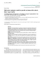
Figure 1. Sputum cytospins from different subjects with asthma
illustrate the heterogeneity of the airway inflammation. In the upper
left panel, the predominant cells are macrophages with a normal
neutrophil and eosinophil count; this cytospin cannot be distinguished
from a sample from a healthy control (paucigranulocytic asthma 3100
original magnification); the upper right panel shows combined
neutrophilic and eosinophilic inflammation (3400 original magnifi-
cation); the lower left panel shows neutrophilic inflammation (3400
original magnification); and the lower right panel shows eosinophilic
inflammation (3400 original magnification). Adapted from
Brightling.
98
(Romanowsky stain.)
Siddiqui and Brightling, Phenotyping Heterogeneity Using Measures of Airway Inflammation 63
In neutrophilic asthma, there is evidence of neutrophil
activation with increased neutrophil elastase and IL-8 in
induced sputum.
54
Importantly, there is associated activa-
tion of the innate immune response with increased
expression of Toll-like receptors 2 and 4 and CD14.
55
These changes are similar to those observed in bronch-
iectasis, suggesting that exposure to infection or endotoxin
may be important in the pathogenesis of neutrophilic
asthma. This is supported by the finding that endotoxin
levels were increased in neutrophilic asthma.
55
Cigarette smoking may be an important modulator of
neutrophilic inflammation in asthma. Smoking induces
neutrophilic airway inflammation, which correlates directly
with the number of pack-years smoked and inversely with
postbronchodilator FEV
1
.
45
Smoking cessation in asthma
leads to a reduction in neutrophilic inflammation.
56
In COPD, a variety of studies have demonstrated
neutrophilic inflammation in sputum,
57–61
bronchoalveo-
lar lavage (BAL),
62,63
and biopsies in COPD.
64–66
Neutrophilic inflammation in sputum has been associated
with both airflow obstruction and FEV
1
decline in
COPD.
67
Cigarette smoking is associated with neutrophilic
inflammation in COPD, but inflammation persists after
smoking cessation. Both inhaled and oral corticosteroids
have also been shown to have little effect in modulating
neutrophilic inflammation in sputum in stable COPD.
68,69
Bacterial colonization is also associated with neutrophilic
airway lumen inflammation in COPD independently from
cigarette smoking, suggesting that disordered host defense
is an integral driver of neutrophilic inflammation in
COPD.
70
Neutrophilic inflammation is not very susceptible to
current anti-inflammatory therapy, and new treatments
are required. In recent years, selective phosphodiesterase
(PDE) inhibitors (cilomilast, roflumilast) have been
developed to selectively block type 4 PDE, which is
expressed abundantly in inflammatory leukocytes, includ-
ing neutrophils.
71,72
PDE4 inhibitors have a variety of anti-
inflammatory effects on neutrophils, including inhibition
of chemotaxis,
73
suppression of proteolytic enzyme release,
inhibition of proinflammatory cytokine release, particu-
larly IL-8 and leukotriene B
4
,
74,75
and inhibition of CD11b
integrin expression.
73
In a placebo-controlled trial of 1,411
patients with stable COPD, roflumilast 500 mg once daily
was shown to reduce exacerbations by 34% and signifi-
cantly improve postbronchodilator FEV
1
compared with
placebo.
76
Furthermore, the drug was well tolerated, other
than class-specific side effects such as nausea, headache,
and diarrhea. Cilomilast has been shown to improve
symptoms, postbronchodilator lung function, and the
percentage of exacerbation-free weeks compared with
placebo in stable COPD
77
and reduces the submucosal,
but not sputum, neutrophil count.
78
Macrolides may also modulate neutrophilic inflamma-
tion,
62
but there are conflicting data, with one study
showing a reduction in sputum total cell count and IL-8
63
and the other showing no effect.
79
Neutrophilic inflammation is an important prognostic
marker in asthma and COPD; it may exist independently of
cigarette smoking and contribute toward FEV
1
decline and
airflow obstruction. Therefore, neutrophilic inflammation
identifies an important inflammatory phenotype, and
identification of a sputum neutrophilia will be able to direct
future therapies targeted at neutrophilic inflammation.
eNO: Utility in Predicting Eosinophilia, Preventing
Exacerbations, and Predicting Response to
Treatment
Assessment of eNO has the appeal of being a simple and
repeatable investigation to assess lower airway inflamma-
tion,
80,81
with the additional advantage of being easy to
perform and quicker than induced sputum analysis. eNO
may have utility in supporting the diagnosis of asthma. An
eNO value of . 16 ppb at a flow rate of 200 mL/s has a
specificity and positive predictive value of . 90% for
predicting asthma (defined as a PC
20
, 8 mg/mL and
bronchodilator reversibility of . 12%).
82
However, the
utility of eNO in asthma diagnosis in primary care based
on asthma symptoms and peak flow variability has not
been assessed.
Although eNO seems to correlate closely with eosino-
philic airway inflammation in sputum and mucosal tissue,
a raised eNO has little utility in predicting a clinically
significant sputum eosinophilia . 3%.
83,84
There are a
number of possible explanations for the discordance
between eNO and sputum eosinophilia. It may be possible
that neutrophilic inflammation modulates eNO; further-
more, nasal contamination of bronchial eNO (the levels of
eNO are 100-fold higher in the upper airways) output may
be a confounder despite the traditional notion that
bronchial eNO values are obtained with a closed glottis.
The use of eNO to guide response to inhaled and oral
corticosteroids is also far from convincing. Smith and
colleagues examined the use of eNO versus a conventional
symptom-based asthma management strategy to assess the
frequency of exacerbations and efficacy of ICS reduction
based on the two management regimens in a single-blind,
placebo-controlled study of 97 patients.
85
Management
with an eNO-based strategy did not affect the frequency of
64 Allergy, Asthma, and Clinical Immunology, Volume 3, Number 2, 2007
exacerbations compared with the symptom management
group. The study did report a significant reduction in the
use of ICSs in the eNO group versus conventional
management (370 mg/d vs 641 mg/d; p 5 .003).
However, these results should be interpreted with caution
as the study design did not allow ICS dose reduction in the
follow-up phase (phase 2) and the mean dose of ICS in the
control group at the end of the treatment optimizing phase
(phase 1) of the study was significantly higher than in the
eNO group (567 mg/d vs 292 mg/d; p 5 .003), fixing the
control group at a higher daily dose of ICS at the onset of
the follow-up phase. Furthermore, eNO was unable to
predict significant sputum eosinophilia in approximately
one-third of patients. Two further studies, one in adults
with mild to moderate asthma
86
and another in children,
87
also failed to demonstrate a reduction in asthma exacer-
bations with corticosteroid therapy targeted at reducing
eNO.
Therefore, current evidence does not support the use of
eNO to target anti-inflammatory treatment. However,
studies investigating the utility of eNO in patients with
COPD and in those with severe asthma are eagerly
awaited. In addition, the role of measuring other exhaled
gases and mediators in exhaled breath condensate in the
phenotyping and management of airways disease is
unknown.
A More Complex Approach to a Complex Problem:
Generation ‘Omics’
One of the limitations of current clinical markers of
inflammation in both asthma and COPD is that they fail to
capture the complexity and diversity of the inflammatory
cascade. As a consequence, significant heterogeneity exists
in response to treatments that modulate inflammation.
An emerging approach in recent years to address this
problem has been to try to generate phenotype-specific
fingerprints of the inflammatory cascade or its genetic
regulation. Omics-based technologies—genomics, proteo-
mics, and metabolomics—offer a potential solution to the
problem of capturing inflammatory diversity in indivi-
duals with airways disease.
Genomics
With the development of complementary deoxyribonu-
cleic acid (DNA) microarrays, it has become possible to
gain information on the level of gene expression for
thousands of genes. This opens a new era of biomarker
discovery and has the potential to further develop specific
expression profiles associated with certain features of
airways disease, to predict response to treatment and
disease progression. This approach has been applied to
cancer, and whether it has applications in airways disease is
awaited.
Proteomics
A vast number of proteins mediate both the normal and
aberrant host inflammatory response. Identifying which
aspects of the proteome are associated with different
patterns of disease expression will allow us to develop
effective and selective drugs to target the inflammatory
cascade.
Surface-enhanced laser desorption ionization time-of-
flight mass spectrometry (SELDI-TOF MS) and matrix-
assisted laser desorption ionization time-of-flight mass
spectrometry (MALDI-TOF-MS), together with new
developments in more traditional two-dimensional gels,
have emerged as powerful tools to examine the proteome
and discover potentially novel biomarkers in a variety of
airway diseases.
88–90
SELDI-TOF MS is a combination of
miniaturized chromatographic prefractionation on a
protein chip followed by MALDI-TOF analysis of
subfractions. The process allows capture of proteins in
biologic fluids such as BAL or induced sputum super-
natants on an immobilized chip that is designed to capture
different physicochemical aspects of protein biochemistry
(eg, hydrophobicity, metal ion affinity, cationic/anionic
properties).
91
SELDI-TOF offers a variety of advantages;
outputs can be generated from very small amounts of
biologic fluid at a very high throughput.
A proteomic study of human BAL fluid from smokers
with COPD combining SELDI-TOF with mass spectro-
metry profiling demonstrated that defensins 1 and 2 and
calgranulins A and B were elevated compared with
asymptomatic smokers.
88
Alpha-defensins are major con-
stituents of neutrophil azurophilic granules, whereas beta-
defensins are expressed in airway epithelial cells and could
contribute to the pathogenesis of COPD by amplifying
cigarette smoke–induced and infection-induced inflam-
matory reactions, leading to lung injury.
92
Calgranulins
may have an important role in neutrophil chemotaxis to
the airway and neutrophil elastase–mediated tissue damage
seen in COPD.
Large studies using SELDI-TOF-based techniques in
well-characterized cohorts of patients with COPD and
asthma are eagerly awaited and are likely to play a
significant role in drug discovery and biomarker identifi-
cation in the future. In particular, proteomic approaches
Siddiqui and Brightling, Phenotyping Heterogeneity Using Measures of Airway Inflammation 65
will enable the development of specific panels of mediators
that can be assessed using new multiplex systems, such as
Luminex or Meso-Scale, that may be particularly helpful in
predicting response to treatment and prognosis.
Metabolomics
Metabolomics and the related term metabonomics can be
defined as the attempt to dynamically measure the
metabolic output within a cell, tissue, or organism in
response to interventions or changes in their environment.
Like proteomics, metabolomics offers promise in the
analysis of global inflammation from biologic fluids in
asthma and COPD and the possibility of generating a
fingerprint metabotype.
93,94
Multidimensional Phenotyping in Asthma and
COPD
This review has focused on the current and potential
future use of measuring airway inflammation in pheno-
typing airway disease. However, it is important to
recognize that this encompasses a single domain of these
complex diseases. Both asthma and COPD are character-
ized by a variety of clinicopathologic domains. Airway
physiology (variable vs fixed airflow obstruction), airway
inflammation, systemic inflammation (COPD), symptoms
and quality of life, genetic predisposition, and environ-
mental/occupational triggers all contribute to the patho-
genesis of both diseases. Furthermore, each domain is
characterized by a number of measurable variables. The
number of variables varies considerably between domains;
for example, a large number of candidate genes modulate
genetic predisposition, whereas a much more finite
number of clinical parameters (eg, FEV
1
,PC
20
, peak flow)
define airway physiology.
95
Most clinical studies predefine
asthma and COPD based on a single dimension, for
example, variable airflow obstruction in asthma or fixed
airflow obstruction in COPD. However, these disease
definitions are limiting and do not fully capture the
complexity of the disease or acknowledge the multi-
dimensional nature of the disease.
A variety of studies in asthma and COPD have
demonstrated that important clinical domains show
significant dissociation. Haldar and colleagues examined
271 patients with refractory asthma attending a difficult
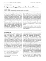
Figure 2. Airway diseases are composed of a number of domains that can be assessed by several outcome measures. The combination of outcome
measures allows for phenotyping the heterogeneity, which impacts on clinical management and research. AHR 5 airway hyperresponsiveness; BD
5 bronchodilator; BMI 5 body mass index; CRP 5 C-reactive protein; HRCT 5 high-resolution computed tomography, PEFR 5 peak flow
reading; PFTs 5 pulmonary function tests.
66 Allergy, Asthma, and Clinical Immunology, Volume 3, Number 2, 2007
asthma clinic in the United Kingdom.
96
A data reduction
technique known as factor analysis minimized 17 variables
into five distinct domains: (1) symptom scores, (2) allergy,
(3) psychosocial, (4) inflammation, and (5) variable
airflow obstruction. This suggests that asthma comprises
a number of distinct factors and that the relative
contribution of one or more of these factors in a patient
determines individual phenotype. Lappere and colleagues
studied disease heterogeneity in 114 patients with mild to
moderate COPD using factor analysis.
3
Considerable
dissociation was demonstrated between airway function,
AHR, and airway inflammation assessed by induced
sputum, suggesting that these are discrete, nonoverlapping
disease dimensions.
Progress in the study of airways disease may require
deviation from the traditional definitions of asthma and
COPD.
97
Furthermore, standardized, nonobjective mea-
surements of different disease-specific variables across
domains, within a network of collaborating centres,
followed by data mining and data reduction are more
likely to allow us to define important disease phenotypes
that relate to clinically important outcomes as well as
tailoring treatment toward individual patients (Figure 2).
Conclusions
The measurement of airway inflammation by induced
sputum is a useful technique in identifying important
clinicopathologic outcomes in asthma and COPD.
However, a variety of other parameters capturing the
complexity of the inflammatory cascade can now be
readily measured, and a collaborative approach between
centres with a specialist interest in airways disease
combined with advanced data mining is likely to further
our understanding of disease phenotypes in the future.
References
1. Braman SS. The global burden of asthma. Chest 2006;130(1
Suppl):4S–12S.
2. Lopez AD, Mathers CD, Ezzati M, et al. Global and regional
burden of disease and risk factors, 2001: systematic analysis of
population health data. Lancet 2006;367:1747–57.
3. Lapperre TS, Snoeck-Stroband JB, Gosman MM, et al. Dissociation
of lung function and airway inflammation in chronic obstructive
pulmonary disease. Am J Respir Crit Care Med 2004;170:499–504.
4. Rosi E, Scano G. Association of sputum parameters with clinical
and functional measurements in asthma. Thorax 2000;55:235–8.
5. Pin I, Gibson PG, Kolendowicz R, et al. Use of induced sputum cell
counts to investigate airway inflammation in asthma. Thorax 1992;
47:25–9.
6. Pizzichini E, Pizzichini MM, Leigh R, et al. Safety of sputum
induction. Eur Respir J Suppl 2002;37:9s–18s.
7. Efthimiadis A, Spanevello A, Hamid Q, et al. Methods of sputum
processing for cell counts, immunocytochemistry and in situ
hybridisation. Eur Respir J Suppl 2002;37:19s–23s.
8. Juniper EF, Kline PA, Vanzieleghem MA, et al. Long-term effects of
budesonide on airway responsiveness and clinical asthma severity
in inhaled steroid-dependent asthmatics. Eur Respir J 1990;3:1122–
7.
9. Juniper EF, Kline PA, Vanzieleghem MA, et al. Effect of long-term
treatment with an inhaled corticosteroid (budesonide) on airway
hyperresponsiveness and clinical asthma in nonsteroid-dependent
asthmatics. Am Rev Respir Dis 1990;142:832–6.
10. Haahtela T, Jarvinen M, Kava T, et al. Comparison of a beta 2-
agonist, terbutaline, with an inhaled corticosteroid, budesonide, in
newly detected asthma. N Engl J Med 1991;325:388–92.
11. Adams NP, Bestall JB, Malouf R, et al. Inhaled beclomethasone
versus placebo for chronic asthma. Cochrane Database Syst Rev
2005;(1):CD002738.
12. Alsaeedi A, Sin DD, McAlister FA. The effects of inhaled
corticosteroids in chronic obstructive pulmonary disease: a
systematic review of randomized placebo-controlled trials. Am J
Med 2002;113:59–65.
13. Man SF, Sin DD. Inhaled corticosteroids in chronic obstructive
pulmonary disease: is there a clinical benefit? Drugs 2005;65:579–
91.
14. Beasley R. The burden of asthma with specific reference to
the United States. J Allergy Clin Immunol 2002;109(5 Suppl):
S482–9.
15. Mortimer KJ, Tata LJ, Smith CJ, et al. Oral and inhaled
corticosteroids and adrenal insufficiency: a case-control study.
Thorax 2006;61:405–8.
16. Mortimer KJ, Harrison TW, Tattersfield AE. Effects of inhaled
corticosteroids on bone. Ann Allergy Asthma Immunol 2005;94:
15–21.
17. Meijer RJ, Postma DS, Kauffman HF, et al. Accuracy of eosinophils
and eosinophil cationic protein to predict steroid improvement in
asthma. Clin Exp Allergy 2002;32:1096–103.
18. Pavord ID, Brightling CE, Woltmann G, Wardlaw AJ. Non-
eosinophilic corticosteroid unresponsive asthma. Lancet 1999;353:
2213–4.
19. Little SA, Chalmers GW, MacLeod KJ, et al. Non-invasive markers
of airway inflammation as predictors of oral steroid responsiveness
in asthma. Thorax 2000;55:232–4.
20. ten Brinke A, Zwinderman AH, Sterk PJ, et al. ‘‘Refractory’’
eosinophilic airway inflammation in severe asthma: effect of
parenteral corticosteroids. Am J Respir Crit Care Med 2004;170:
601–5.
21. Pizzichini E, Pizzichini MM, Gibson P, et al. Sputum eosinophilia
predicts benefit from prednisone in smokers with chronic
obstructive bronchitis. Am J Respir Crit Care Med 1998;158(5 Pt
1):1511–7.
22. Brightling CE, Monteiro W, Ward R, et al. Sputum eosinophilia
and short-term response to prednisolone in chronic obstructive
pulmonary disease: a randomised controlled trial. Lancet 2000;356:
1480–5.
23. Brightling CE, McKenna S, Hargadon B, et al. Sputum eosinophilia
and the short term response to inhaled mometasone in chronic
obstructive pulmonary disease. Thorax 2005;60:193–8.
Siddiqui and Brightling, Phenotyping Heterogeneity Using Measures of Airway Inflammation 67
24. Leigh R, Pizzichini MM, Morris MM, et al. Stable COPD:
predicting benefit from high-dose inhaled corticosteroid treatment.
Eur Respir J 2006;27:964–71.
25. Leuppi JD, Salome CM, Jenkins CR, et al. Predictive markers of
asthma exacerbation during stepwise dose reduction of inhaled
corticosteroids. Am J Respir Crit Care Med 2001;163:406–12.
26. Jatakanon A, Lim S, Barnes PJ. Changes in sputum eosinophils
predict loss of asthma control. Am J Respir Crit Care Med 2000;
161:64–72.
27. Pizzichini MM, Pizzichini E, Clelland L, et al. Prednisone-
dependent asthma: inflammatory indices in induced sputum. Eur
Respir J 1999;13:15–21.
28. Green RH, Brightling CE, McKenna S, et al. Asthma exacerbations
and sputum eosinophil counts: a randomised controlled trial.
Lancet 2002;360:1715–21.
29. Jayaram L, Pizzichini MM, Cook RJ, et al. Determining asthma
treatment by monitoring sputum cell counts: effect on exacerba-
tions. Eur Respir J 2006;27:483–94.
30. Chlumsky J, Striz I, Terl M, Vondracek J. Strategy aimed at
reduction of sputum eosinophils decreases exacerbation rate in
patients with asthma. J Int Med Res 2006;34:129–39.
31. O’Shaughnessy TC, Ansari TW, Barnes NC, Jeffery PK.
Inflammation in bronchial biopsies of subjects with chronic
bronchitis: inverse relationship of CD8+ T lymphocytes with FEV1.
Am J Respir Crit Care Med 1997;155:852–7.
32. Saetta M, Di Stefano A, Turato G, et al. CD8+ T-lymphocytes in
peripheral airways of smokers with chronic obstructive pulmonary
disease. Am J Respir Crit Care Med 1998;157(3 Pt 1):822–6.
33. Saetta M, Di Stefano A, Maestrelli P, et al. Airway eosinophilia in
chronic bronchitis during exacerbations. Am J Respir Crit Care
Med 1994;150(6 Pt 1):1646–52.
34. Confalonieri M, Mainardi E, Della PR, et al. Inhaled corticoster-
oids reduce neutrophilic bronchial inflammation in patients with
chronic obstructive pulmonary disease. Thorax 1998;53:583–5.
35. O’Donnell RA, Peebles C, Ward JA, et al. Relationship between
peripheral airway dysfunction, airway obstruction, and neutro-
philic inflammation in COPD. Thorax 2004;59:837–42.
36. Boschetto P, Quintavalle S, Zeni E, et al. Association between
markers of emphysema and more severe chronic obstructive
pulmonary disease. Thorax 2006;61:1037–42.
37. Siva R, Green R, Brightling CE, et al. Modulation of eosinophilic
airway inflammation in COPD. Eur Respir J 2005;26 Suppl
49:441s.
38. O’Donnell RA, Frew AJ. Is there more than one inflammatory
phenotype in asthma? Thorax 2002;57:566–8.
39. Douwes J, Gibson P, Pekkanen J, Pearce N. Non-eosinophilic
asthma: importance and possible mechanisms. Thorax 2002;57:
643–8.
40. Gibson PG, Simpson JL, Saltos N. Heterogeneity of airway
inflammation in persistent asthma: evidence of neutrophilic
inflammation and increased sputum interleukin-8. Chest 2001;
119:1329–36.
41. Wenzel SE, Schwartz LB, Langmack EL, et al. Evidence that severe
asthma can be divided pathologically into two inflammatory
subtypes with distinct physiologic and clinical characteristics. Am J
Respir Crit Care Med 1999;160:1001–8.
42. Godon P, Boulet LP, Malo JL, et al. Assessment and evaluation of
symptomatic steroid-naive asthmatics without sputum eosinophi-
lia and their response to inhaled corticosteroids. Eur Respir J 2002;
20:1364–9.
43. Berry MA, Morgan A, Green RH, et al. Clinical and pathological
features on non-eosinophilic asthma: a distinct asthma phenotype
associated with corticosteroid resistance. Thorax 2005;60 Suppl
2:ii4.
44. Brightling CE, Bradding P, Symon FA, et al. Mast-cell infiltration
of airway smooth muscle in asthma. N Engl J Med 2002;346:1699–
705.
45. Chalmers GW, MacLeod KJ, Little SA, et al. Influence of cigarette
smoking on inhaled corticosteroid treatment in mild asthma.
Thorax 2002;57:226–30.
46. Simpson JL, Scott R, Boyle MJ, Gibson PG. Inflammatory subtypes
in asthma: assessment and identification using induced sputum.
Respirology 2006;11:54–61.
47. Turner MO, Hussack P, Sears MR, et al. Exacerbations of asthma
without sputum eosinophilia. Thorax 1995;50:1057–61.
48. Anees W, Huggins V, Pavord ID, et al. Occupational asthma due to
low molecular weight agents: eosinophilic and non-eosinophilic
variants. Thorax 2002;57:231–6.
49. Belda J, Leigh R, Parameswaran K, et al. Induced sputum cell
counts in healthy adults. Am J Respir Crit Care Med 2000;161(2 Pt
1):475–8.
50. Thomas RA, Green RH, Brightling CE, et al. The influence of age
on induced sputum differential cell counts in normal subjects.
Chest 2004;126:1811–4.
51. Chalmers GW, MacLeod KJ, Thomson L, et al. Smoking and
airway inflammation in patients with mild asthma. Chest 2001;120:
1917–22.
52. Sagel SD, Kapsner R, Osberg I, et al. Airway inflammation in
children with cystic fibrosis and healthy children assessed by
sputum induction. Am J Respir Crit Care Med 2001;164(8 Pt 1):
1425–31.
53. Nightingale JA, Rogers DF, Hart LA, et al. Effect of inhaled
endotoxin on induced sputum in normal, atopic, and atopic
asthmatic subjects. Thorax 1998;53:563–71.
54. Simpson JL, Scott RJ, Boyle MJ, Gibson PG. Differential
proteolytic enzyme activity in eosinophilic and neutrophilic
asthma. Am J Respir Crit Care Med 2005;172:559–65.
55. Simpson JL, Grissell TV, Douwes J, et al. Innate immune activation
in neutrophilic asthma and bronchiectasis. Thorax 2007;62(3):211–
8.
56. Chaudhuri R, Livingston E, McMahon AD, et al. Effects of
smoking cessation on lung function and airway inflammation in
smokers with asthma. Am J Respir Crit Care Med 2006;174:127–
33.
57. Peleman RA, Rytila PH, Kips JC, et al. The cellular composition of
induced sputum in chronic obstructive pulmonary disease. Eur
Respir J 1999;13:839–43.
58. Turato G, Di Stefano A, Maestrelli P, et al. Effect of smoking
cessation on airway inflammation in chronic bronchitis. Am J
Respir Crit Care Med 1995;152(4 Pt 1):1262–7.
59. Keatings VM, Collins PD, Scott DM, Barnes PJ. Differences in
interleukin-8 and tumor necrosis factor-alpha in induced sputum
from patients with chronic obstructive pulmonary disease or
asthma. Am J Respir Crit Care Med 1996;153:530–4.
60. Beeh KM, Beier J, Kornmann O, et al. Long-term repeatability of
induced sputum cells and inflammatory markers in stable,
moderately severe COPD. Chest 2003;123:778–83.
68 Allergy, Asthma, and Clinical Immunology, Volume 3, Number 2, 2007
61. Rutgers SR, Postma DS, ten Hacken NH, et al. Ongoing airway
inflammation in patients with COPD who do not currently smoke.
Chest 2000;117(5 Suppl 1):262S.
62. Tamaoki J. The effects of macrolides on inflammatory cells. Chest
2004;125(2 Suppl):41S–50S.
63. Basyigit I, Yildiz F, Ozkara SK, et al. The effect of clarithromycin
on inflammatory markers in chronic obstructive pulmonary
disease: preliminary data. Ann Pharmacother 2004;38:1400–5.
64. Hogg JC, Chu F, Utokaparch S, et al. The nature of small-airway
obstruction in chronic obstructive pulmonary disease. N Engl J
Med 2004;350:2645–53.
65. Pesci A, Majori M, Cuomo A, et al. Neutrophils infiltrating
bronchial epithelium in chronic obstructive pulmonary disease.
Respir Med 1998;92:863–70.
66. Baraldo S, Turato G, Badin C, et al. Neutrophilic infiltration within
the ASM in patients with COPD. Thorax 2004;59:308–12.
67. Stanescu D, Sanna A, Veriter C, et al. Airways obstruction, chronic
expectoration, and rapid decline of FEV1 in smokers are associated
with increased levels of sputum neutrophils. Thorax 1996;51:267–
71.
68. Culpitt SV, Maziak W, Loukidis S, et al. Effect of high dose inhaled
steroid on cells, cytokines, and proteases in induced sputum in
chronic obstructive pulmonary disease. Am J Respir Crit Care Med
1999;160(5 Pt 1):1635–9.
69. Keatings VM, Jatakanon A, Worsdell YM, Barnes PJ. Effects of
inhaled and oral glucocorticoids on inflammatory indices in
asthma and COPD. Am J Respir Crit Care Med 1997;155:542–8.
70. Sethi S, Maloney J, Grove L, et al. Airway inflammation and
bronchial bacterial colonization in chronic obstructive pulmonary
disease. Am J Respir Crit Care Med 2006;173:991–8.
71. Hatzelmann A, Schudt C. Anti-inflammatory and immunomodu-
latory potential of the novel PDE4 inhibitor roflumilast in vitro. J
Pharmacol Exp Ther 2001;297:267–79.
72. Bundschuh DS, Eltze M, Barsig J, et al. In vivo efficacy in airway
disease models of roflumilast, a novel orally active PDE4 inhibitor.
J Pharmacol Exp Ther 2001;297:280–90.
73. Spoelstra FM, Berends C, Dijkhuizen B, et al. Effect of theophylline
on CD11b and L-selectin expression and density of eosinophils and
neutrophils in vitro. Eur Respir J 1998;12:585–91.
74. Au BT, Teixeira MM, Collins PD, Williams TJ. Effect of PDE4
inhibitors on zymosan-induced IL-8 release from human neu-
trophils: synergism with prostanoids and salbutamol. Br J
Pharmacol 1998;123:1260–6.
75. Cortijo J, Villagrasa V, Navarrete C, et al. Effects of SCA40 on
human isolated bronchus and human polymorphonuclear leuko-
cytes: comparison with rolipram, SKF94120 and levcromakalim. Br
J Pharmacol 1996;119:99–106.
76. Rabe KF, Bateman ED, O’Donnell D, et al. Roflumilast—an oral
anti-inflammatory treatment for chronic obstructive pulmonary
disease: a randomised controlled trial. Lancet 2005;366:563–71.
77. Rennard SI, Schachter N, Strek M, et al. Cilomilast for COPD:
results of a 6-month, placebo-controlled study. Chest 2006;129:56–
66.
78. Gamble E, Grootendorst DC, Brightling CE, et al. Anti-
inflammatory effects of the phosphodiesterase-4 inhibitor cilomi-
last (Ariflo) in chronic obstructive pulmonary disease. Am J Respir
Crit Care Med 2003;168:976–82.
79. Banerjee D, Honeybourne D, Khair OA. The effect of oral
clarithromycin on bronchial airway inflammation in moderate-to-
severe stable COPD: a randomized controlled trial. Treat Respir
Med 2004;3:59–65.
80. Kharitonov SA, Yates D, Robbins RA, et al. Increased nitric oxide
in exhaled air of asthmatic patients. Lancet 1994;343:133–5.
81. Alving K, Weitzberg E, Lundberg JM. Increased amount of nitric
oxide in exhaled air of asthmatics. Eur Respir J 1993;6:1368–70.
82. Dupont LJ, Demedts MG, Verleden GM. Prospective evaluation of
the validity of exhaled nitric oxide for the diagnosis of asthma.
Chest 2003;123:751–6.
83. Berry MA, Shaw DE, Green RH, et al. The use of exhaled nitric
oxide concentration to identify eosinophilic airway inflammation:
an observational study in adults with asthma. Clin Exp Allergy
2005;35:1175–9.
84. Payne DN, Adcock IM, Wilson NM, et al. Relationship between
exhaled nitric oxide and mucosal eosinophilic inflammation in
children with difficult asthma, after treatment with oral pred-
nisolone. Am J Respir Crit Care Med 2001;164(8 Pt 1):1376–81.
85. Smith AD, Cowan JO, Brassett KP, et al. Use of exhaled nitric
oxide measurements to guide treatment in chronic asthma. N Engl
J Med 2005;352:2163–73.
86. Shaw DE, Berry MA, Thomas M, et al. Asthma exacerbations and
exhaled nitric oxide: a randomised controlled trial. Eur Respir J
2006;28 Suppl 50:572s.
87. Pijnenburg MW, Bakker EM, Hop WC, de Jongste JC. Titrating
steroids on exhaled nitric oxide in children with asthma: a
randomized controlled trial. Am J Respir Crit Care Med 2005;172:
831–6.
88. Merkel D, Rist W, Seither P, et al. Proteomic study of human
bronchoalveolar lavage fluids from smokers with chronic obstruc-
tive pulmonary disease by combining surface-enhanced laser
desorption/ionization-mass spectrometry profiling with mass
spectrometric protein identification. Proteomics 2005;5:2972–80.
89. Sloane AJ, Lindner RA, Prasad SS, et al. Proteomic analysis of
sputum from adults and children with cystic fibrosis and from
control subjects. Am J Respir Crit Care Med 2005;172:1416–26.
90. Kriegova E, Melle C, Kolek V, et al. Protein profiles of
bronchoalveolar lavage fluid from patients with pulmonary
sarcoidosis. Am J Respir Crit Care Med 2006;173:1145–54.
91. Wulfkuhle JD, Liotta LA, Petricoin EF. Proteomic applications for
the early detection of cancer. Nat Rev Cancer 2003;3:267–75.
92. Wallace AM, He JQ, Burkett KM, et al. Contribution of alpha- and
beta-defensins to lung function decline and infection in smokers:
an association study. Respir Res 2006;7:76.
93. Nicholson JK, Holmes E, Wilson ID. Gut microorganisms,
mammalian metabolism and personalized health care. Nat Rev
Microbiol 2005;3:431–8.
94. Nicholson JK, Lindon JC, Holmes E. ‘Metabonomics’: under-
standing the metabolic responses of living systems to pathophy-
siological stimuli via multivariate statistical analysis of biological
NMR spectroscopic data. Xenobiotica 1999;29:1181–9.
95. Wardlaw AJ, Silverman M, Siva R, et al. Multi-dimensional
phenotyping: towards a new taxonomy for airway disease. Clin Exp
Allergy 2005;35:1254–62.
96. Haldar P, Green RH, Berry M, et al. Categorising the asthma
phenotype: results of a factor analysis. Thorax 2006;60 Suppl 2:ii53.
97. Hargreave FE, Parameswaran K. Asthma, COPD and bronchitis are
just components of airway disease. Eur Respir J 2006;28:264–7.
98. Brightling CE. Clinical applications of induced sputum. Chest
2006;129:1344–8.
Siddiqui and Brightling, Phenotyping Heterogeneity Using Measures of Airway Inflammation 69