UNDERSTANDING THE COMPLEXITIES OF KIDNEY TRANSPLANTATION Part 7 pptx
Bạn đang xem bản rút gọn của tài liệu. Xem và tải ngay bản đầy đủ của tài liệu tại đây (1.62 MB, 58 trang )
ABO-Incompatible Kidney Transplantation
339
In conventional plasmapheresis, smaller proteins such as albumin are also removed in
addition to pathogenic molecules, antibody or high molecular weight proteins. In general,
plasma separated with a plasma separator is discarded and replaced with the same volume
of replacement fluid such as fresh frozen plasma or albumin solution. There are several
options of plasmapheresis, which separate blood components more selectively.
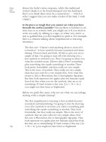
Double filtration plasmapheresis (DFPP) uses two filters which have different pore sizes. In
the first filter, blood is separated as plasma and cell components, and plasma is further
separated by the second filter. Large molecular-weight proteins including immunoglobulins
such as anti-donor isoagglutinins are removed, while smaller molecular-weight substances
such as albumin are returned to the patient’s circulation. In this procedure, need of
replacement is decreased compared with conventional plasmapheresis, thus adverse effects
related to the replacement fluid can be reduced (Fig. 4) (Genberg et al., 2010; Tanabe, 2007b).
In the immunoadsorption, specialized adsorption column selectively adsorbs a specific
substance such as immunoglobulin or low-density lipoprotein. This process removes the
element of interest specifically and the remaining elements are returned to the patients.
Many kinds of immunoadsorption devices for the removal of various types of components
are commercially available but generally expensive. For the removal of anti-A and -B
antibody, AB antigen-specific carbohydrate columns (Glycosorb AB, Glycorex
Transplantation AB, Lund, Sweden) were developed (Tyden et al., 2005) and have been
widely used in more than 400 cases of ABO-I kidney transplantation (Genberg et al., 2010;
Tyden et al., 2005; Winters et al., 2004). This procedure could decrease the complications
associated with plasma exchange such as coagulopathy and transfusion reactions.
Fig. 4. Schematic presentation of double filtration plasmapheresis (DFPP). In DFPP, plasma
separated with a plasma separator (1
st
filter) passes through the plasma component
separator with a small pore size (2
nd
filter). Molecules that are larger than the pore size such
as immunoglobulins are removed, and smaller molecules such as albumin are returned to
the patient.
Understanding the Complexities of Kidney Transplantation
340
4. Determination of isoagglutinin titer
To reduce isoagglutinin titers prior to ABO-I kidney transplantation, preparative regimens
including plasmapheresis, DFPP, or immunoadsorption and immunosuppressive therapy
have been used. The clinical significance of isoagglutinin titer in ABO-I kidney
transplantation is not entirely clear (Tobian et al., 2011). The goal of isoagglutinin titer to
prevent hyperacute rejection is variable across transplantation centers, ranging from ≤ 1:8 to
≤ 1:32 before transplantation (Crew & Ratner, 2010). However, minimal research has been
performed to determine the optimal pretransplant titer. The possibility of AMR would
decrease as anti-donor antibody titer decreases. In our institution, the titer is lowered to ≤
1:4 before transplantation. The measurement of isoagglutinin is known to be essential in the
assessment of the efficacy of antibody removal, and the prediction of AMR (Kobayashi &
Saito, 2006). Although most recipients with AMR had an elevated titer, the positive
predictive value of a high titer for AMR was poor (Tobian et al., 2010). Thus, posttransplant
titers should be monitored, but must be combined with the other factors assessing AMR.
Accurate measurement of isoagglutinin titer is an important aspect for successful ABO-I
kidney transplantation. If the isoagglutinin titer is underestimated compared to the actual
titer of patient, we could consider a patient as safe for transplantation and it could lead to
rejection or short duration of allograft survival (Crew & Ratner, 2010). IgM antibody mediates
complement activation and endothelial damage in AMR, and it is more rapidly removed by
plasmapheresis than IgG. However, IgG titers are more emphasized for patient eligibility,
rejection risk, and plasmapheresis guidance. Reporting both IgM and IgG titers has been
recommended by a working group from US centers (Montgomery et al., 2004). Importantly,
measured titers are method-dependent and considerably variable according to assays.
Tube method Column
agglutination
Flow cytometry
A column ingredient Not needed Sephadex gel
or glass bead
Not needed
Use of RBC Yes Yes Yes
Antihuman globulin Yes Yes No
Secondary antibody No No Yes
Deletion of IgM DTT or 2ME DTT or 2ME Not needed
Interpretation Agglutination Agglutination Fluorescence detection
Result Titer Titer MFIR or titer
Instrument Not needed Not needed Needed
Cost Low Intermediate Relatively high
Assay time 30 - 60 min 30 - 60 min 1- 2 hours
DTT, dithiothreitol; 2ME, 2-mercaptoethanol; MFIR, mean fluorescence intensity ratio.
Table 1. Various assays for measurement of isoagglutinin titer
ABO-Incompatible Kidney Transplantation
341
There are several options for the measurement of isoagglutinin titers: conventional tube
method, gel or bead column agglutination method, and flow cytometry (Krishnan et al.,
2008; Stussi et al., 2005). These three methods are summarized in Table 1. In addition,
enzyme-linked immunosorbent assay technique (Lindberg et al., 2011; Rieben et al., 1991),
surface plasmon resonance (Kimura et al., 2005; Yurugi et al., 2007), and KODE technology
(Frame et al., 2007) were developed, although these methods are not routinely available in
most institutions.
4.1 Conventional tube method
The conventional tube method has been used in most institutions for the semiquantitative
measurement of isoagglutinin titers. IgG and IgM can be measured together, and if
dithiothreitol or antiglobulin reagents are used, they can be measured separately. In general,
recipient serum is serially diluted and incubated with RBC aliquots of the appropriate blood
type in a test tube for about 10 minutes at room temperature. After the mixture is
centrifuged, macroscopic agglutinations of RBCs are checked for IgM detection. For IgG
detection, additional testing with antihuman globulin is performed to check the
agglutination. Titers are determined as the highest dilution that produces 1+ macroscopic
agglutination. However, technical variables greatly affect the results, and care should be
taken to achieve the most uniform practice (Roback, 2008). Considerable inter-examiner
variability may occur, because the titer is determined mainly by visual observation of
agglutinated RBCs in tubes. Inter-institutional difference can also occur possibly due to
variations in procedures and lack of assay standardizations.
A recent study reported the results of isoagglutinin titers from 26 different labs using sera
from six patients of different blood groups (Kobayashi & Saito, 2006). In this report, inter-
institutional variation between maximum and minimum value reached as much as 32-fold
in IgM and 256-fold in IgG. These variations seemed to be due to different techniques
between laboratories, but considerable variation was still noted after standardization of
techniques. Another report also showed a large variation of isoagglutinin titers (a median
three-fold difference) among three centers performing ABO-I kidney transplants in
Germany and Sweden (Kumlien et al., 2007). In this report, gel hemagglutination technique
significantly decreased inter-center difference (a median one titer difference) compared with
tube methods.
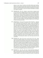
4.2 Gel or bead column agglutination
In gel or bead column agglutination method, a cassette (or card) containing gels or beads is
used. Commercially available assays include DiaMed ID Micro Typing system (Bio-Rad,
Hercules, CA, USA), BioVue System (Ortho Clinical Diagnosis, Raritan, NJ, USA), or
Olympus ID-Micro Typing System (Olympus Co., Tokyo, Japan). In these assays, plasma
from the patient is stepwise diluted 1:2 with normal saline or phosphate buffered saline and
packed RBCs are used to make a suspension with cell stabilization solution. In each
incubation well, recommended cell suspension is mixed with diluted plasma. After
incubation and centrifugation, agglutination is observed in card or cassette. In column
agglutination method, negative (unagglutinated) test cells pellet to the bottom of the
column, and positive (agglutinated) cells are captured at the top of or within the body of
column (Fig. 5). The gel or bead particles trap the RBC agglutinates as a filter during
centrifugation. The agglutination is graded from 0 to 4 +, and inverted value of the highest
plasma dilution that gives a 1+ agglutination reaction is interpreted as the titer (Kumlien et
al., 2007).
Understanding the Complexities of Kidney Transplantation
342
Fig. 5. Interpretation of column agglutination method. The agglutination is graded from
0 to 4+.
4.3 Flow cytometry
In flow cytometry method, quantifications of anti-A/B IgG and IgM are performed using
fluorescence conjugated, anti-human IgG and IgM as secondary antibodies. A mixture of
RBC suspension and recipient serum is transferred into the test tube and incubated (at 37°C
in a CO
2
incubator for IgG antibody; and at room temperature, for IgM antibodies). After
washing, fluorescence conjugated, anti-human IgG and IgM secondary antibodies are added
in test tube. After incubation and washing steps, binding of anti-A/B antibody is measured
by flow cytometry. Human AB serum, which is further depleted by incubation with highly
concentrated A and B RBCs, can be used as a negative control, and human serum of blood
group O is used as a positive control. Commercially available O RBCs with information of
antigen expression are also helpful for the detection of irregular antibodies (Stussi et al.,
2005).
Using undiluted serum, quantification of anti-A/B antibody can be determined by
calculation of the geometric mean fluorescence intensity ratio (MFIR). This value is
calculated by dividing the geometric mean fluorescence intensity of test sera with that of
negative control. One study reported that the correlation coefficient between MFIR using
flow cytometry and isoagglutinin titer was 0.870 for IgM and 0.783 for IgG (Stussi et al,
2005). For determination of titer using flow cytometry, recipient serum is serially diluted
with normal saline solution (2% bovine serum albumin, 0.1% azide). After incubation and
washing, secondary antibody is added. After reaction, binding of antibody is determined by
flow cytometry. A gated value above assigned cut-off (5% for example) is regarded as
positive serum dilution. In a study comparing the reproducibility of the results performed
by various assays, flow cytometry showed excellent reproducibility and no measurement
deviation was noted, whereas gel column agglutinin assay and tube technique showed two-
fold and four-fold differences, respectively (Tanabe, 2007b). However, flow cytometry assay
needs the flow cytometry instrument, and the reagents are relatively expensive.
ABO-Incompatible Kidney Transplantation
343
5. Conclusion
The ABO blood group barrier is now being crossed in the field of transplantation, and ABO-
I kidney transplantation is becoming more common worldwide. Removing the ABO barrier
can expand the donor pool and increase the availability of organs for transplantation.
Moreover, it can decrease the time on the organ waiting list, and eventually facilitate the
timely transplantation before comorbid conditions develop in the patients. Currently
observed long-term results of ABO-I kidney transplantation are similar to those of ABO-
compatible kidney transplantation. With the application of adequate antibody reducing
strategies, future results would be more promising. To promote accomodation and to
prevent acute complement-mediated graft injury, methods for preventing and treating AMR
are still needed. Researches for the insights into the mechanism of accomodation will
provide us a scientific basis for the development of innovative approaches for the better
outcome of ABO-I kidney transplantation.
As the number of ABO-I transplantation increases, there is a need of the optimal methods
for ABO isoagglutinin titer for the effective monitoring of ABO-I transplanted patients.
Compared with the conventional test tube method, gel card or flow cytometric
measurement can provide more accurate and objective results. However, reproducibility,
interpretation, and standardization of isoagglutinin titration methods are still unsatisfactory,
and further researches should be performed to determine the optimal method for ABO
antibody titer assessment. There are also several promising techniques under development,
focused on the endothelium, enzymes, or blocking antibodies. Ongoing improvement of
promising modalities could make more successful transplantation outcomes in this field.
6. Acknowledgment
The authors appreciate Professor Jin Q Kim, The President of Konkuk University, Korea, for
his critical review and valuable comments on this work.
7. References
Alexandre, G.P.J.; De Bruyere, M.; Squifflet, J.P.; Moriau, M.; Latinne, D. & Pirson, Y. (1985).
Human ABO-incompatible living donor renal homografts. The Netherlands Journal
of Medicine, Vol. 28, No. 6, (August 1985), pp. 231-234, ISSN 0300-2977
Alexandre, G.P.J.; Squifflet, J.P.; De Bruyere, M.; Latinne, D.; Moriau, M.; Carlier, M.; Pirson,
Y. & Lecomte, C. (1986). ABO-incompatible related and unrelated living donor
renal allografts. Transplantation Proceedings, Vol. 18, No. 3, (May-June 1986), pp.
452-455, ISSN 0041-1345
Alexandre, G.P.J.; Squifflet, J.P.; De Bruyere, M.; Latinne, D.; Moriau, M. & Ikabu, N. (1985).
Splenectomy as a prerequisite for successful human ABO-incompatible renal
transplantation. Transplantation Proceedings, Vol. 17, No. 1 Pt. I, (January-February
1985), pp. 138-143, ISSN 0041-1345
Alexandre, G.P.J.; Latinne, D.; Gianello, P. & Squifflet, J.P. (1991). Preformed cytotoxic
antibodies and ABO-incompatible grafts. Clinical Transplantation, Vol. 5, No. 6 Pt. II,
(December 1991), pp. 583-594, ISSN 0902-0063
Bach, F.H.; Ferran, C.; Hechenleitner, P.; Mark, W.; Koyamada, N.; Miyatake, T.; Winkler,
H.; Badrichani, A.; Candinas, D. & Hancock, W.W. (1997). Accommodation of
Understanding the Complexities of Kidney Transplantation
344
vascularized xenografts: expression of "protective genes" by donor endothelial cells
in a host Th2 cytokine environment. Nature Medicine, Vol. 3, No. 2, (February 1997),
pp. 196-204, ISSN 1078-8956
Cardella, C.J.; Pei, Y. & Brady, H.R. (1987). ABO blood group incompatible kidney
transplantation: a case report and review of the literature. Clinical Nephrology, Vol.
28, No. 6, (December 1987), pp. 295-299, ISSN 0301-0430
Colvin R. B. & Smith R.N. (2005). Antibody mediated organ allograft rejection. Nature Review
Immunology, Vol. 5, No. 10, (October 2005), pp. 807-817, ISSN 1474-1733
Crew, R.J. & Ratner, L.E. (2010). ABO-incompatible kidney transplantation: current practice
and the decade ahead. Current Opinion in Organ Transplantation, Vol. 15, No. 4,
(August 2010), pp. 526-530, ISSN 1531-7013
Frame, T.; Carroll, T.; Korchagina, E.; Bovin, N. & Henry, S. (2007). Synthetic glycolipid
modification of red blood cell membranes. Transfusion, Vol. 47, No. 5, (May 2007),
pp. 876-882, ISSN 0041-1132
Genberg, H.; Kumlien, G.; Wennberg, L. & Tyden, G. (2010). Isoagglutinin adsorption in
ABO-incompatible transplantation. Transfusion and Apheresis Science, Vol. 43, No. 2,
(October 2010) pp. 231-235, ISSN 1473-0502
Haidinger, M.; Schmaldienst, S.; Körmöczi, G.; Regele, H.; Soleiman, A.; Schwartz, D.;
Derfler, K.; Steininger, R.; Mühlbacher, F. & Böhmig, G.A. (2009). Vienna
experience of ABO-incompatible living-donor kidney transplantation. Wiener
klinische Wochenschrift, Vol. 121, No. 7-8, (June 2009), pp. 247-255, ISSN 0043-5325
Hume, D.L.; Merrill, J.P.; Miller, B.F. & Thorn, G.W. (1955). Experiences with renal
homotransplantation in the human: report of nine cases. The Journal of Clinical
Investigation, Vol. 34, No. 2, (February 1955), pp. 327-382, ISSN 0021-9738
Ishida, H.; Miyamoto, N.; Shirakawa, H.; Shimizu, T.; Tokumoto, T.; Ishikawa, N.;
Shimmura, H.; Setoguchi, K.; Toki, D.; Iida, S.; Teraoka, S.; Takahashi, K.; Toma, H.;
Yamaguchi, Y. & Tanabe, K. (2007). Evaluation of immunosuppressive regimens in
ABO-incompatible living kidney transplantation single center analysis. American
Journal of Transplantation, Vol. 7, No. 4, (April 2007), pp. 825-831, ISSN 1600-6135
Jeon, B.J.; Kim, I.G.; Seong, Y.K. & Han, B.H. (2010). Analysis of the results of ABO-
incompatible kidney transplantation: in comparison with ABO-compatible kidney
transplantation. Korean Journal of Urology, Vol. 51, No. 12, (December 2010), pp. 863-
869, ISSN 2005-6745
Kenmochi, T.; Saigo, K.; Maruyama, M.; Akutsu, N.; Iwashita, C.; Otsuki, K.; Ito, T.; Suzuki,
A. & Miyazaki, M. (2008). Results of kidney transplantation from ABO-
incompatible living donors in a single institution. Transplantation Proceedings, Vol.
40, No. 7, (September 2008), pp. 2289-2291, ISSN 0041-1345
Kimura, S.; Yurugi, K.; Segawa, H.; Kuroda, J.; Sato, K.; Nogawa, M.; Yuasa, T.; Egawa, H.;
Tanaka, K. & Maekawa, T. (2005). Rapid quantitation of immunoglobulin G
antibodies specific for blood group antigens A and B by surface plasmon
resonance. Transfusion, Vol. 45, No. 1, (January 2005), pp. 56-62, ISSN 0041-1132
Kissmeyer-Nielsen, F.; Olsen, S.; Petersen, V.P. & Fjeldborg, O. (1966). Hyperacute rejection
of kidney allografts associated with pre-existing humoral antibodies against donor
cells. Lancet, Vol. 2, No. 7465, (September 1966), pp. 662-665, ISSN 0140-6736
ABO-Incompatible Kidney Transplantation
345
Kobayashi, T. & Saito, K. (2006). A series of surveys on assay for anti-A/B antibody by
Japanese ABO-incompatible Transplantation Committee. Xenotransplantation, Vol.
13, No. 2, (March 2006), pp. 136-140, ISSN 0908-665X
Krishnan, N. S.; Fleetwood, P.; Higgins, R. M.; Hathaway, M.; Zehnder, D.; Mitchell, D.;
Hamer, R.; Fletcher, S.; Lam, F. T.; Kashi, H.; Tan, L. C.; Imray, C. & Briggs, D.
(2008). Application of flow cytometry to monitor antibody levels in ABO
incompatible kidney transplantation. Transplantation, Vol. 86, No. 3, (August 2008),
pp. 474-477, ISSN 0041-1337
Kumlien, G.; Wilpert, J.; Safwenberg, J. & Tyden, G. (2007). Comparing the tube and gel
techniques for ABO antibody titration, as performed in three European centers.
Transplantation, Vol. 84, No. 12 Suppl, (December 2007), pp. S17-S19, ISSN 0041-
1337
Lindberg, L.; Johansson, S.M.; Liu, J.; Grufman, P. & Holgersson, J. (2011). Is there a clinical
need for a diagnostic test allowing detection of chain type-specific anti-A and anti-
B. Transfusion, Vol. 51, No. 3, (March 2011), pp. 494-503, ISSN 1537-2995
Lynch, R.J. & Platt, J.L. (2008). Accommodation in organ transplantation. Current Opinion in
Organ Transplantation, Vol. 13, No. 2, (April 2008), pp. 165-170, ISSN 1531-7013
Lynch, R.J. & Platt, J.L. (2010). Accommodation in renal transplantation: unanswered
questions. Current Opinion in Organ Transplantation, Vol. 15, No. 4, (August 2010),
pp. 481-485, ISSN 1531-7013
Marionneau, S.; Cailleau-Thomas, A.; Rocher, J.; Le Moullac-Vaidye, B.; Ruvoën, N.;
Clément, M. & Le Pendu, J. (2001). ABH and Lewis histo-blood group antigens, a
model for the meaning of oligosaccharide diversity in the face of a changing world.
Biochimie, Vol. 83, No. 7, (July 2001), pp. 565-573, ISSN 0300-9084
Montgomery, R.A.; Hardy, M.A.; Jordan, S.C.; Racusen, L.C.; Ratner, L.E.; Tyan, D.B.;
Zachary, A.A. & Antibody Working Group on the diagnosis, reporting, and risk
assessment for antibody-mediated rejection and desensitization protocols. (2004).
Consensus opinion from the antibody working group on the diagnosis, reporting,
and risk assessment for antibody-mediated rejection and desensitization protocols.
Transplantation, Vol. 78, No. 2, (July 2004), pp. 181-185, ISSN 0041-1337
Moon, H.W.; Yun, Y.M.; Hur, M.; Park, J.H.; Lee, H.W.; Chang, S.H. & Yun, I.J. (2009). An
experience of ABO-incompatible kidney transplantation using plasmapheresis and
anti-CD20 monoclonal antibody. Korean Journal of Laboratory Medicine, Vol. 29, No.
6, (December 2009), pp. 585-588, ISSN 1598-6535
Murray, J.E.; Merrill, J.P.; Dammin, G.J.; Dealy, J.B. Jr.; Walter, C.W.; Brooke, M.S. & Wilson,
R.E. (1960). Study on transplantation immunity after total body irradiation: clinical
and experimental investigation. Surgery, Vol. 48, (July 1960), pp. 272-284, ISSN
0039-6060
Oettl, T.; Halter, J.; Bachmann, A.; Guerke, L.; Infanti, L.; Oertli, D.; Mihatsch, M.; Gratwohl,
A.; Steiger, J. & Dickenmann, M. (2009). ABO blood group-incompatible living
donor kidney transplantation: a prospective, single-centre analysis including serial
protocol biopsies. Nephrology, Dialysis, Transplantation, Vol. 24, No. 1, (January
2009), pp. 298-303, ISSN 1460-2385
Ogawa, H.; Mohiuddin, M.M.; Yin, D.P.; Shen, J.; Chong, A.S. & Galili, U (2004). Mouse-
heart grafts expressing an incompatible carbohydrate antigen. II. Transition from
Understanding the Complexities of Kidney Transplantation
346
accommodation to tolerance. Transplantation, Vol. 77, No. 3, (February 2004), pp.
366–373, ISSN 0041-1337
Orlin, J.B. & Berkman, E.M. (1980). Partial plasma exchange using albumin replacement:
removal and recovery of normal plasma constituents. Blood, Vol. 56, No.6,
(December 1980), pp. 1055-1059, ISSN 0006-4971
Park, W.D.; Grande, J.P.; Ninova, D.; Nath, K.A.; Platt, J.L.; Gloor, J.M. & Stegall, M.D.
(2003). Accommodation in ABO-incompatible kidney allografts, a novel
mechanism of self-protection against antibody-mediated injury. American Journal of
Transplantation, Vol. 3, No. 8, (August 2003), pp. 952-960, ISSN 1600-6135
Platt, J.L.; Vercellotti, G.M.; Dalmasso, A.P., Matas, A.J., Bolman R.M., Najarian, J.S. & Bach,
F.H. (1990). Transplantation of discordant xenografts: a review of progress.
Immunology Today, Vol. 11, No. 12, (December 1990), pp. 450-456, ISSN 0167-5699
Reding, R.; Squifflet, J.P.; Latinne, D.; de Bruyere, M.; Pirson, Y. & Alexandre, G.P.J. (1987).
Early postoperative monitoring of natural anti-A and anti-B isoantibodies in ABO-
incompatible living donor renal allografts. Transplantation Proceedings, Vol. 19, No.
1 Pt 3, (Febrary 1987), pp. 1989-1990, ISSN 0041-1345
Rieben, R.; Buchs, J. P.; Fluckiger, E. & Nydegger, U. E. (1991) Antibodies to histo-blood
group substances A and B: agglutination titers, Ig class, and IgG subclasses in
healthy persons of different age categories. Transfusion, Vol. 31, No. 7, (September
1991), pp. 607-615, ISSN 0041-1132
Roback, J. D. (Ed.). (2008). Technical Manual. 16th ed. American Association of Blood Banks,
ISBN 978-1563952609 (1563952602), Bethesda, USA.
Segev, D.L.; Simpkins, C.E.; Warren, D.S.; King, K.E.; Shirey, R.S.; Maley, W.R.; Melancon,
J.K.; Cooper, M.; Kozlowski, T. & Montgomery, R.A. (2005). ABO incompatible
high-titer renal transplantation without splenectomy or anti-CD20 treatment.
American Journal of Transplantation, Vol. 5, No. 10, (October 2005), pp. 2570-2575,
ISSN 1600-6135
Squifflet, J.P.; De Meyer, M.; Malaise, J.; Latinne, D.; Pirson, Y. & Alexandre, G.P. (2004).
Lessons learned from ABO-incompatible living donor kidney transplantation: 20
years later. Experimental and Clinical Transplantation, Vol. 2, No. 1, (June 2004), pp.
208-213, ISSN 1304-0855
Slapak, M.; Naik, R.B. & Lee, H.A. (1981). Renal transplant in a patient with major donor-
recipient blood group incompatibility: reversal of acute rejection by the use of
modified plasmapheresis. Transplantation, Vol. 31, No. 1, (January 1981), pp. 4-7,
ISSN 0041-1337
Slapak, M.; Digard, N.; Ahmed, M.; Shell, T. & Thompson, F. (1990). Renal transplantation
across the ABO barrier - a 9 year experience. Transplantation Proceedings, Vol. 22,
No. 4, (August 1990), pp. 1425-1428, ISSN 0041-1345
Starzl, T.E.; Marchioro, T.L.; Holmes, J.H.; Hermann, G.; Brittain, R.S.; Stonington, O.H.,
Talmage, D.W. & Waddell, W.R. (1964). Renal homografts in patients with major
donor-recipient blood group incompatibilities. Surgery, Vol. 55, (Febrary 1964), pp.
195-200, ISSN 0039-6060
Stussi, G.; Huggel, K.; Lutz, H.U.; Schanz, U.; Rieben, R. & Seebach, J.D. (2005). Isotype-
specific detection of ABO blood group antibodies using a novel flow cytometric
method. British Journal of Haematology, Vol. 130, No. 6, (September 2005), pp. 954-
963, ISSN 0007-1048
ABO-Incompatible Kidney Transplantation
347
Szczepiorkowski, Z.M.; Winters, J.L.; Bandarenko, N.; Kim, H.C.; Linenberger, M.L.;
Marques, M.B. ; Sarode, R.; Schwartz, J.; Weinstein, R. & Shaz, B.H. (2010).
Guidelines on the use of therapeutic apheresis in clinical practice evidence-based
approach from the Apheresis Applications Committee of the American Society for
Apheresis. Journal of Clinical Apheresis, Vol. 25, No.3, (June 2010), pp. 83-1217, ISSN
0733-2459
Takahashi, K. (2007). Recent findings in ABO-incompatible kidney transplantation:
classification and therapeutic strategy for acute antibody-mediated rejection due to
ABO-blood-group-related antigens during the critical period preceding the
establishment of accommodation. Clinical and Experimental Nephrology, Vol. 11, No.
2, (June 2007), pp. 128-141, ISSN 1342-1751
Tanabe, K. (2007). Japanese experience of ABO-incompatible living kidney transplantation.
Transplantation, Vol. 84, No. 12 Suppl, (December 2007), pp. S4-S7, ISSN 0041-1337
Tanabe, K. (2007). Interinstitutional variation in the measurement of anti-A/B antibodies:
the Japanese ABO-Incompatible Transplantation Committee survey.
Transplantation, Vol. 84, No. 12 Suppl, (December 2007), pp. S13-S16, ISSN 0041-
1337
Tanabe, K.; Ishida, H.; Shimizu, T.; Omoto, K.; Shirakawa, H. & Tokumoto, T. (2009).
Evaluation of two different preconditioning regimens for ABO-incompatible living
kidney donor transplantation. A comparison of splenectomy vs. rituximab-treated
non-splenectomy preconditioning regimens. Contributions to Nephrology, Vol. 162,
(Febrary 2009), pp. 61-74, ISSN 0302-5144
Tang, A.H. & Platt, J.L. (2007). Accommodation of grafts: implications for health and
disease. Human Immunology, Vol. 68, No. 8, (August 2007), pp. 645-651, ISSN 0198-
8859
Thielke, J.; Kaplan, B. & Benedetti, E. (2007). The role of ABO-incompatible living donors in
kidney transplantation: state of the art. Seminars in Nephrology, Vol. 27, No. 4, (July
2007), pp. 408-413, ISSN 0270-9295
Tobian, A.A.; Shirey, R.S. & King, K.E. (2011) ABO antibody titer monitoring for
incompatible renal transplantation. Transfusion, Vol. 51, No. 3, (March 2011), pp.
454-457, ISSN 1537-2995
Tobian, A.A.; Shirey, R.S.; Montgomery, R.A.; Cai, W.; Haas, M.; Ness, P.M. & King, K.E.
(2010). ABO antibody titer and risk of antibody-mediated rejection in ABO-
incompatible renal transplantation. American Journal of Transplantation, Vol. 10, No.
5, (May 2010), pp. 1247-1253, ISSN 1600-6143
Tobian, A.A.; Shirey, R.S.; Montgomery, R.A.; Ness, P.M. & King, K.E. (2008). The critical
role of plasmapheresis in ABO-incompatible renal transplantation. Transfusion, Vol.
48, No. 11, (November 2008), pp. 2453-2460, ISSN 1537-2995
Tobian, A.A.; Shirey, R.S.; Montgomery, R.A.; Tisch, D.J.; Ness, P.M. & King, K.E. (2009).
Therapeutic plasma exchange reduces ABO titers to permit ABO-incompatible
renal transplantation. Transfusion, Vol. 49, No. 6, (June 2009), pp. 1248-1254, ISSN
1537-2995
Tyden, G.; Kumlien, G.; Genberg, H.; Sandberg, J.; Lundgren, T. & Fehrman, I. (2005). ABO
incompatible kidney transplantations without splenectomy, using antigen-specific
immunoadsorption and rituximab. American Journal of Transplantation, Vol. 5, No.1,
(January 2005), pp. 145-148, ISSN 1600-6135
Understanding the Complexities of Kidney Transplantation
348
Valli, P.V.; Puga, Y.G.; Fehr, T.; Schulz-Huotari, C.; Kaup, N.; Güngör, T.; Ambühl, P.;
Weber, M.; Schanz, U.; Seebach, J.D. & Stussi, G. (2009). Changes of circulating
antibody levels induced by ABO antibody adsorption for ABO-incompatible
kidney transplantation. American Journal of Transplantation, Vol. 9, No. 5, (May
2009), pp. 1072-1080, ISSN 1600-6143
Winters, J.L.; Gloor, J.M.; Pineda, A.A.; Stegall, M.D. & Moore, S.B. (2004). Plasma exchange
conditioning for ABO-incompatible renal transplantation. Journal of Clinical
Apheresis, Vol. 19, No.2, (2004), pp. 79-85, ISSN 0733-2459
Yamamoto F. (2004). Review: ABO blood group system ABH oligosaccharide antigens,
anti-A and anti-B, A and B glycosyltransferases, and ABO genes. Immunohematology
/ American Red Cross, Vol. 20, No. 1, (March 2004), pp. 3-22, ISSN 0894-203X
Yurugi, K.; Kimura, S.; Ashihara, E.; Tsuji, H.; Kawata, A.; Kamitsuji, Y.; Hishida, R.;
Takegawa, M.; Egawa, H. & Maekawa, T. (2007). Rapid and accurate measurement
of anti-A/B IgG antibody in ABO-unmatched living donor liver transplantation by
surface plasmon resonance. Transfusion Medicine, Vol. 17, No. 2, (April 2007), pp.
97-106, ISSN 0958-7578
16
Combined Liver and Kidney Transplantation
Cláudia Fagundes and Mónica Guevara
*
Liver Unit/Hospital Clinic Barcelona
Spain
1. Introduction
Combined liver and kidney transplant (CLKT) is the procedure of choice for patients with
both liver and kidney end-stage-disease. In addition, patients with polycystic liver or kidney
disease or with hyperoxaluria, or those with cirrhosis and acute renal failure, including
hepatorenal syndrome receiving hemodialysis (HD) for more than two months, may also
benefit of CLKT.
The decision to transplant both, the liver and kidney, is more difficult in cases when kidney
dysfunction may be temporary. Hepatorenal syndrome is a potentially reversible renal
failure caused by advance liver disease. Currently, the treatment of choice of hepatorenal
syndrome is liver transplant alone and not a combined liver/kidney transplant.
The model for end-stage liver disease (MELD) replaced the United Network for Organ
Sharing status classification for the allocation of liver organs. Due to the heavily weighted
serum creatinine value in the calculation of the MELD score, candidates with renal failure
have received organs more rapidly. As a result there has been considerable increase in
number of combined liver-kidney transplants in the past few years.
The reason to propose both liver and kidney transplant for patients with cirrhosis and renal
failure relays on the negative impact that renal failure has on patients submitted to liver
transplant alone (LTA). Results of several studies show that renal failure in patients with
chronic liver disease is associated with high mortality and morbidity after liver transplant
alone. Nevertheless, it’s very hard to identify a cut-off point of renal dysfunction that
determines those patients who may benefit from combined liver and kidney transplant
instead of liver transplant alone.
In this chapter, we will review the main points to be considered when evaluating candidates
for combined liver kidney transplant, as well as some concerns that have not been yet
clarified.
2.Assessment of renal function and evaluating of CLKT in patients with end
stage liver disease
Renal failure in cirrhotic patients is associated with poor prognosis. It is well known that
cirrhotic patients with renal failure have decreased survival when compared to patients
*
corresponding author. Associate Investigator. IDIBAPS
Understanding the Complexities of Kidney Transplantation
350
with normal renal function. This negative effect is also evident when these patients undergo
liver transplantation, as shown by reduced graft and patient survival.
Ideally, patients with a high probability of developing end stage renal disease after liver
transplantation alone should receive a combination of liver and kidney transplant.
However, is still a great challenge to identify these patients who are at higher risk.
The presence and the severity of pretransplant kidney failure are factors independently
associated with postoperative sepsis, need for renal replacement therapy and poor graft and
patient outcomes.
In addition to the degree of renal dysfunction, duration and cause of renal failure should
also be considered when evaluating candidates for liver transplantation alone or combined
liver kidney transplantation.
Patients with pretransplant renal dysfunction (defined as pretransplant Scr > 1.5mg/dL) for
a period longer than 12 weeks showed higher probability of progression to end-stage renal
disease at 3 years post transplant. However in this study the etiology of renal dysfunction
was not specified, mainly due to the authors concern of potential bias in classifying renal
failure in absence of kidney biopsy.
Renal failure is usually defined by a reduction in glomerular filtration rate (GFR) that can be
acute when it occurs in hours to weeks or chronic when it occurs gradually over time.
Currently, serum creatinine remains the most widely used method to assess renal function
in cirrhotic patients.
However, patients with liver dysfunction have reduced creatinine production secondary to
loss of muscle mass, and therefore, in those patients serum creatinine usually overestimates
renal function. As the Cockroft-Gauld and MDRD (Modification of Diet in Renal Disease)
formulas are based on serum creatinine concentration, adjusted by race, age, sex and weight,
they also overestimate renal function in patients with cirrhosis and should not be used in
clinical settings.
In this context, cystatin C has emerged as an option for evaluate renal function since its level
is not influenced by muscle mass. Nevertheless, its value has not been well established and
is not available as standart test.
More accurate methods, such as determination of inulin clearance or radionuclide markers,
represent the gold standard for measuring glomerular filtration rate. Indeed, its use in daily
attendance is not feasible, because of its complexity, making repeated measurements that these
patients often require difficult. These gold standard methods should be indicated for selected
patients when there is a need to accurately assess renal function to decide between performing
liver transplantation alone or CKLT. Their routinary use, however, is not mandatory.
Beyond the degree of renal function, the etiology of renal failure should be assessed, as
prognosis varies according to the cause of renal failure. In a recent study with a large
population of hospitalized patients with cirrhosis, the most common cause of renal failure
was due to bacterial infections (46%), followed by hypovolemia (32%), hepatorenal
syndrome (13%) and intrinsic nephropathy (9%). Patients with HRS and bacterial infections
had lower 3-month survival compare to patients with intrinsic nephropathy. Even though
patients with intrinsic nephropathy present better survival among all causes of renal failure
in cirrhosis, its chronic form of renal failure has a non-reversible character and are most
likely to receive CKLT.
The diagnostic diagram of etiology of renal failure include a complete medical history and
physical examination, searching for presence of diabetes and/or hypertension as well as any
other evidence of organ damage. Laboratory evaluation should include urinalysis to seek for
Combined Liver and Kidney Transplantation
351
signs of intrinsic nephropathy, like hematuria, pyuria, cell and granular casts, and 24h urine
collection to assess protein excretion.
In addition to urine test, a renal ultrasonography, is useful in evaluating preexisting renal
disease. Findings such as alteration of renal echogenicity and reductions in the size of the
kidneys indicate the existence of chronic kidney disease.
Finally, a definitive diagnostic may require the realization of a renal biopsy, which may also
give prognostic information. In patients with intrinsic nephropathy, marked
tubulointerstitial injury is associated with progression to end stage renal disease, even if the
primary disease is a glomerulopathy. Among histological findings, the degree of tubular
interstitial fibrosis is the most powerful predictor of subsequent progression of renal
impairment. There are very limited data on renal biopsies findings in cirrhotic patients. A
study evaluated 23 kidney biopsies performed in liver transplant candidates with renal
failure of unknown etiology or persisted HRS (> 4 weeks) demonstrated a variety of
pathologic findings. These included menbranoproliferative glomerulopathy, IgA
nephropathy, diabetes nephropathy and acute tubular necrosis. Of note, 4 patients showed
normal histology. In this study CLKT was recommended for 10 of 26 patients with > 40%
global glomeruloesclerosis, > 30% of interstitial fibrosis or severe glomerular
ischemia/injury. Although these histological criteria have not been evaluated in further
studies in patients with cirrhosis, it suggests that renal histopathology changes may alter
therapeutic management, including the need for combined liver and kidney transplant.
Therefore according to a recent consensus, a renal biopsy should be performed in patients
with an estimated glomerular filtration rate less than 30ml/min with a chronic course .The
decision to perform a transjugular or percutaneous renal biopsy should take into account
professional experience and patient’s clinical conditions, mostly platelet count and
coagulation parameters.
Hepatorenal syndrome is a form of kidney failure that is secondary to a severe circulatory
disorder in patients with cirrhosis. This particular complication of liver disease can be
potentially reversible with the combination of systemic vasoconstrictors and intravenous
albumin. Even though the definite treatment of this severe condition remains liver
transplantation, the importance of pre-liver transplantation treatment should not be
underestimate. Patients with HRS treated with systemic vasoconstrictors and albumin
before liver transplantation and pretransplant serum creatinine inferior to 1.5 mg/dL had a
three year survival similar to patients transplanted with normal renal function.
Finally, the current criteria to perform CLKT according to the consensus conference is
shown in table 1.
1. Evidence of chronic kidney disease and renal biopsy demonstrating more than 30% of
glomeruloesclerosis or 30% of interstitial fibrosis.
2. If the biopsy is not possible, the decision is made based on National Kidney Foundation
criteria for chronic kidney disease, which is an eGFR less than 30ml/min for more than 3
months.
3. Patients with end stage renal disease in renal replacement therapy
4. Patients with hepatorenal syndrome or acute kidney injury with creatinine greater or
equal to 2.0 mg/dL and on dialysis for more than 8 weeks.
Table1. Indications for combined liver kidney transplant in patients with end stage liver
disease.
Understanding the Complexities of Kidney Transplantation
352
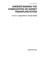
3. Evaluation of candidates for CLKT in patients with end stage renal
Disease (ESRD)
The benefit of combined liver kidney transplantation is not well established for patients
with compensated cirrhosis and ESRD. The decision to perform CLKT or only a liver
transplant is matter of debate. In a study of patients with chronic hepatitis C on RRT who
underwent kidney transplantation alone, the degree of liver fibrosis correlated with
patient and graft survival at 3 years .It is recommended that patients with chronic liver
disease and ESRD who are candidates for kidney transplantation should be sought for the
presence of significant liver fibrosis and cirrhosis. These patients should be submitted to
transjugular liver biopsy with assessment of hepatic venous pressure gradient(HVPG).
Patients with cirrhosis and/or clinical significant portal hypertension, determined by an
HVPG greater than 10mmHg should be referred to CLKT. The option of kidney
transplantation alone should be offered for those patients without these characteristics.
Even though most of the data regarding these situations comes from patients with
cirrhosis due to hepatitis C, the recommendations are generally applied to all patients
irrespective of etiology of cirrhosis.
Combined Liver Kidney Transplantation
Portal Hypertension
> 10 mmHg
Kidney Transplant Alone
Normal
`
Wedge Hepatic Vein Pressure
Cirrhosis
Kidney Transplant Alone
No Cirrhosis
Liver Biopsy
Asymptomatic Liver Disease
Combined Liver and Kidney Transplantation
Symptomatic or Evidence of Portal Hypertension
End Stage Renal Disease
with
Liver Disease
Fig. 1. Diagram for End Stage Renal Disease and Liver Disease (adapted from Consensus
Conference on Simultaneous Liver Kidney Transplantation).
4. Outcomes in combined liver and kidney transplantation
Cirrhosis may not be the only indication for CKLT. In a large series of 3520 patients
evaluated between 1984-2008, the main indications for combined liver kidney
transplantation were: hiperoxaluria type 1 (42.7%), liver cirrhosis and chronic renal failure
(23.5%), polycystic liver and kidney disease (15.5%), liver cirrhosis with hepatorenal
syndrome (7.1%) and end stage liver disease with renal failure of unknown cause (6%).
Hence, prognosis and outcomes of combined liver kidney transplantation are not well
known because most of the data came from series that include patients treated with CLKT
Combined Liver and Kidney Transplantation
353
not only with end stage liver disease but also patients with inherited diseases without
cirrhosis.
In recent years, MELD score has increasingly been used for liver allocation. Due to the
presence of serum creatinine in the formula of MELD score, candidates with renal failure are
more likely to receive a liver graft. Although pre transplant renal failure is associated with
poor outcomes in liver transplantation settings, this modification on organ allocation system
was not followed by changes in survival. The 3-year survival of liver transplant recipients
remained almost unchanged when compared pre and pos-MELD era (81% vs. 80%,
respectively).
A large case-control study compared the outcomes of patients submitted to liver transplant
alone with or without renal failure to combined liver kidney transplants (CKLT) between
1987 and 2006. After adjusting for multiple donor (age, race, cause of death) and recipient
(MELD, dialysis status at time of transplant) characteristic’s, recipients of CLKT had a
similar one-year survival compared to liver transplant alone (82 vs. 81.8%). However, the
degree of renal failure in both groups was not described. The only subgroup in which CLKT
had benefit on survival was in patients on long-term pre transplant hemodialysis (defined as
a period equal to or greater than 12 weeks). In this subgroup, CKLT recipients had a higher
survival than those submitted to liver transplantation alone (84.5% vs. 70.8%, P=0.008).
Another study demonstrated that patients on hemodialysis prior to transplantation had a
significantly higher 1-year survival for CLKT group when compared to LT alone (79.4% vs.
73.7%, p=0.004). This difference, however, was not observed when only patients with renal
failure (defined by serum creatinine ≥ 2.5 mg/dL) not on dialysis where analyzed. In this
subgroup, 1-year survival was similar for patients who received CLKT or liver transplant
alone (81% vs.78.8%, p= n.s.). An important issue to highlight is that patients receiving
CLKT, either on hemodialysis or not, had better liver function at the time of transplant
compared to those receiving liver transplantation alone. Mean MELD score of patients
receiving LTA or CKLT was 36 vs. 31 for recipients on hemodialysis, and 34 vs. 28 for those
with renal failure (serum creatinine >2.5 mg/dl) but not on hemodialysis (p<0.01 for both
comparisons).
Most studies of survival in combined liver kidney transplantation analyzed a very
heterogeneous population respect to the etiology of liver transplantation. Though, a recent
study that only included patients with cirrhosis and chronic kidney disease, showed a 1-
year survival lower for patients treated with CKLT compared to liver transplant alone group
(80 vs. 97%, p=0.014). The probability of survival at 3 years was also lower in the CLKT
group, but the difference between both groups did not reach statistical significance (75%
and 88%, respectively). The incidence of complications was also higher for CKLT. Patients
with CLKT had a higher incidence of bacterial infections and transfusions requirements
compared to LTA group. Nevertheless, the comparison group (liver transplant alone) did
not present renal failure at the time of transplant (mean serum creatinine value of 0.96±0.27
mg/dL), because all patients with cirrhosis and advanced chronic kidney disease (defined
by a glomerular filtration rate below 30ml/min) were considered candidates for CLKT.
Another important point is the potential reversibility of renal failure after liver
transplantation. As mentioned previously, patients with HRS should be treated to reverse
the renal failure before liver transplantation. Many of these patients, however, do not
respond to treatment and eventually undergo CKLT. Only a few single-center series had
described outcomes of patients with hepatorenal syndrome submitted to CLKT. One of
them compared the results of patients with HRS on hemodialysis who received CLKT (n=22,
Understanding the Complexities of Kidney Transplantation
354
median time of pretransplant hemodialysis of 41 days) to those with HRS on hemodialysis
who received liver transplant alone (n=80, pretransplant hemodialysis time inferior to 30
days). The one-year survival for patients undergoing CLKT or LTA was similar (72% vs.
66%, respectively, p=0.88). Most of the benefit of performing CKLT was observed in patients
on hemodialysis for more than 8 weeks pre transplant. This group had higher survival than
those receiving CLKT on hemodialysis for a period inferior than 8 weeks (88% vs.66%,
respectively). Among patients receiving liver transplantation alone, recovery of renal
function was achieved in 90% of patients at one-month, even though most of them required
hemodialysis at post transplant period.
The possible benefit of CLKT on LTA in patients with hepatorenal syndrome was also
evaluated in a study comparing patients submitted to CLKT to patients with HRS submitted
to LTA. Survival at 5 years was similar for CLKT recipients (48.1%) and patients with HRS
receiving LTA (67.1%) (p=ns).
Some recent data on patients who received CLKT (n=75) over a 23 year-period show
excellent 1-, 3- and 5- year patients survival (81%, 73% and 67%, respectively). However,
short-term mortality (< 90 days) was especially high because of sepsis/infection on
postoperative period. In addition, there was no difference in patient survival based on
whether or not a recipient was on dialysis pre-transplantion. Nevertheless, the need of post
transplant renal replacement therapy was significantly associated with poor prognosis
(p=0.0012).
Regarding graft survival, it seems that the liver graft has an immune protective effect on
kidney graft when both organs came from the same donor. A study comparing renal
allograft outcomes of patients who undergone CLKT to kidney after liver transplantation
(KALT) demonstrated a higher incidence of chronic rejection in KALT group than CLKT
group (4.6 vs. 1.2%, P < 0.001). One and three-year rejection-free renal graft survival of
KALT was lower than CLKT group (77% and 67% KALT vs. 85% and 78% CLKT,
respectively; P < 0.001). Renal half-life of KALT allograft was shorter than CLKT group
(6.6+/-0.9 vs. 11.7+/-1.3 years, P < 0.001). It has been speculated that this effect is secondary
to the secretion of soluble HLA antigens by the liver and to phagocytosis of these reactive
antibodies by kupffer cells.
Although many theories have been described to explain the possible hepatic protection on
renal graft, some recent findings have questioned this statement. A case report of acute
humoral rejection in kidney allograft in an ABO compatible CLKT was described. Even
treating, the humoral rejection with plasmapheresis, intravenous immunoglobulin and
rituximab, the kidney required 3 months to recovery function and finally progressed to
chronic allograft nephropathy.
5. Combined liver and kidney transplantation in special conditions
Polycystic kidney diseases (PKD) compass a group of inherited diseases that causes an
irreversible decline in kidney function. Autosomal dominant polycystic kidney disease
(ADPKD) is associated with cysts in the kidneys and, in many cases, cysts in the liver and
pancreas. The autosomal dominant form (ADPKD) is the most common genetic cause of
chronic kidney disease .As survival with dialysis or transplant increase, incidence of liver
disease will also increase. When cysts are diffused, fenestration/resection procedures are
not successful and LKA offers a good survival option. For combined liver and kidney
transplantation one- and two-year patient survival rates were similar to combined
Combined Liver and Kidney Transplantation
355
transplantation for other indications. For patients with acceptable renal function at time of
transplantation, solitary liver transplantation has an excellent outcome.
Primary hyperoxaluria (PHO) is a rare metabolic disorder with autosomal recessive
inheritance. PHO is induced by one of two enzymatic defects, both of which result in
markedly enhanced conversion of glyoxalate to poorly soluble oxalate which is then
excreted in the urine. Combined liver-kidney transplantation is probably the treatment of
choice for children with type 1 PHO with progressive renal disease. The liver provides the
missing enzyme, thereby lowering oxalate production to the normal range. The outcome
may be best if transplantation is performed when the GFR falls to 25 mL/min per 1.73 m2
and prior to marked tissue oxalate deposition. Isolated liver transplantation has been
proposed for patients with rapidly progressive disease who still have a glomerular filtration
rate above 30 mL/min per 1.73 m
2
.
6. Conclusion
Since implementation of MELD score as an organ allocation system, a crescent number of
cirrhotic patients with renal failure has been submitted to CLKT. Due to increase shortage of
organ donors, is of outstanding importance to define which are the patients who benefit
most of this procedure.
The decision to perform orthotopic transplant alone or combined kidney-liver
transplantation is still challenging, mainly because there is not enough data on factors that
can predict renal function recovery. In patients with possible reversible causes of kidney
dysfunction, including those with hepatorenal syndrome and acute renal failure, it is
difficult to precise the boundaries between functional and irreversible damage. Therefore, in
these cases kidney biopsy should be encouraged in order to evaluate interstitial and
glomerular injury.
Combined liver kidney transplantation seems to be an adequate treatment in patients with
end stage liver disease and chronic kidney disease on renal replacement therapy, as well as
for those with inherited disease. The survival advantage in others subsets of patients is not
well established and more studies are needed.
7. Acknowledgment
Supported in part by grants from Fondo de Investigación Sanitaria FIS070443 and 080108.
Centro de investigaciones en red de enfermedades hepaticas y digestivas. CIBEREHD is
supported by the Instituto de Salud Carlos III. Claudia Fagundes is supported by a grant of
Fundación Renal Reina Sofía
8. References
Baccaro, ME. (2010). Combined liver–kidney transplantation in patients with cirrhosis and
chronic kidney disease. Nephrology Dialysis Transplantation, Vol. 25, No. 7, pp.
2356-63, ISSN 0931-0509
Bahirwani, R. (2008). Transplantation: impact of pretransplant renal insufficiency. Liver
Transplantation, Vol. 14, No. 5, pp. 665-71, ISSN 1527-6473
Understanding the Complexities of Kidney Transplantation
356
Barua M. (2009). Family history of renal disease severity predicts the mutated gene in
ADPKD. Journal of American Society of Nephrology, Vol. 20, No. 8, pp. 1833-8, ISSN
1533-3450
Brinkert, F. (2009). Transplantation procedures in children with primary hyperoxaluria type
1: outcome and longitudinal growth. Transplantation, Vol. 87, No. 9, pp. 1415-21,
ISSN 1534-0608
Campbell, MS. (2005).Renal Function after Orthotopic Liver Transplantation is Predicted by
Duration of Pretransplantation Creatinine elevation. Liver Transplantation, Vol. 11,
No. 9,pp. 1048-1055, ISSN 1527-6465
Caregaro, L. (1994). Limitations of serum creatinine level and creatinine clearance as
filtration markers in cirrhosis. Archives of Internal Medicine, Vol. 154, No. 2, pp. 201-
205, ISSN 0003-9926
Cochat, P. (1993). Should liver transplantation be performed before advanced renal
insufficiency in primary hyperoxaluria type 1?. Paediatric Nephrology, Vol. 7, No. 2,
pp. 212-8, ISSN 0931-041X.
Davis, CL. (2007).Simultaneous liver-kidney transplantation: evaluation to de decision
making. American Journal of Transplantation,Vol.7, No. 7,pp. 1702-9, ISSN 1600-
6135
D'Amico, G. (1992).Influence of clinical and histological features on actuarial renal
survival in adult patients with idiopathic IgA nephropathy, membranous
nephropathy, and membranoproliferative glomerulonephritis: survey of the
recent literature. American Journal of Kidney Disease, Vol.20, No. 4,pp.315-23, ISSN
0272-6386
D’Amico, G. (2006).Natural history and prognostic indicators of survival in cirrhosis: a
systematic review of 118 studies. Journal of Hepatology, Vol.44, No. 1,pp.217-
231,ISSN 0168-8278
De Eason, JD. (2008). Proceedings of consensus conference on simultaneous liver kidney
transplantation (SLK). American Journal of Transplantation, Vol.8, No. 11,pp. 2243-
51,ISSN 1600-6135
Ginès P, Schrier RW. (2009). Renal failure in cirrhosis. New England Journal of Medicine,
Vol.13, No. 361, pp.1279-1290, ISSN 0028-4793
Gonwa, TA. (1995). Impact of pretransplant renal function on survival after liver
transplantation. Transplantation,Vol.59, No. 3,pp.361-365, ISSN 0041-1337
Gonwa, TA. (2006).Continued influence of preoperative renal function on outcome of
orthotopic liver transplant (OLTX) in the US: where will MELD lead us? American
Journal of Transplantation, Vol.6, No. 11,pp.2651-2659,ISSN 1600-6135
Jeyarajah, DR. (1997). Hepatorenal syndrome: combined liver kidney transplants versus
isolated liver transplant. Transplantation, Vol. 64, No. 12, pp. 1760-5, ISSN 1534-
0608.
Jeyarajah, DR. (1997).Hepatorenal syndrome : combined liver kidney transplants versus
isolated liver transplant. Transplantation,Vol.64, No. 12,pp.1760-1765. ISSN 0041-
1337
Combined Liver and Kidney Transplantation
357
Locke, JE. (2008). Declining outcomes in simultaneous liver-kidney transplantation in the
MELD era: ineffective usage of renal allografts. Transplantation, Vol. 85, No. 7,
pp.935-42, ISSN 1534-0608
Maluf, DG. (2007). Hepatitis C virus infection and kidney transplantation: predictors of
patient and graft survival. Transplantation, Vol. 83, No. 7, pp. 853-7, ISSN 1534-
0608
Martin-Llahí, M. (2010).Prognostic importance of the cause of renal failure in patients with
cirrhosis. Gastroenterology,Vol.140, No. 2,pp. 488-496, ISSN 0016-5085
McMillan, RW. (1997). Soluble fraction of class I human histocompatibility leukocyte
antigens in the serum of liver transplant recipients. Clinical Transplantation, Vol. 11,
No. 2, pp. 98-103, ISSN 0902-0063
Mehrabi, A. (2009). A single center experience of combined liver kidney transplantation.
Clinical transplantation, Vol. 23, Suppl. 21, pp. 102-114, ISSN 0902-0063
Millan MT. (2003). One hundred percent patient and kidney allograft survival with
simultaneous liver and kidney transplantation in infants with primary
hyperoxaluria: a single-center experience. Transplantation,. Vol. 76, No. 10, pp. 1458-
63, ISSN 1534-0608.
Nair, S. (2002).Pretransplant renal function predicts survival in patients undergoing
orthotopic liver transplantation. Hepatology ,Vol.35, No. 5,pp.1179-85, Online ISSN
1527-3350
Nath, KA. (1992).Tubulointerstitial changes as a major determinant in the progression of
renal damage. American Journal of Kidney Disease, Vol.20, No. 1,pp.1-17. ISSN 0272-
6386
Pöge, U. (2006).Calculation of glomerular filtration rate based on cystatin C in cirrhotic
patients. Nephrology Dial Transplan,.Vol.21, No. 3, pp.660-664, ISSN 0931-0509
Reichmann, TW. (2009). Acute humoral rejection in an ABO compatible combined liver-
kidney transplant-the kidney is not always protected, Vol. 9, No. 8, pp1957-60,
ISSN 1600-6143
Ruiz, R. (2006). Long-term analysis of combined liver and kidney transplantation at a single
center. Archives of Surgery, Vol. 141, No. 8, pp. 735-41, ISSN 1435-2451
Ruiz, R. (2010). Indications for combined liver and kidney transplantation: propositions after
a 23-yr experience. Clinical Transplantation, Vol. 24, No. 6, pp. 807-11, ISSN 0902-
0063
Schmitt, TM. (2009). Combined liver-kidney and liver transplantation in patients with renal
failure outcomes in the MELD era. Transplantation International, Vol. 22, No. 9, pp.
876-83, ISSN 1432-2277
Sherman, DS. (2003). Assessing renal function in cirrhotic patients: problems and
pitfalls. American Journal of Kidney Diseases, Vol. 41, No.2, pp. 269-78, ISSN 0272-
6386.
Skulzacek, PA. (2003). Prediction of GFR in liver transplant candidates. American Journal of
Kidney Diseases, Vol. 42, No. 6, pp. 1169-76, ISSN 0272-6386.
Simpson, N. (2006). Comparison of renal allograft outcomes in combined liver-kidney
transplantation versus subsequent kidney transplantation in liver transplant
Understanding the Complexities of Kidney Transplantation
358
recipients: Analysis of UNOS Database. , Vol. 82, No. 10, pp. 1298-303, ISSN 1534-
0608
Ueno,T. (2006). Liver and kidney transplantation for polycystic liver and kidney-renal
function and outcome. Transplantation, Vol. 82, No. 4, pp. 501-7, ISSN 1534-0608.
17
Transplantation for the Complex
Patient with Hepatitis C and End
Stage Renal Disease: A Review
Jorge Ortiz, Jason Andre, Kamran Khanmoradi and Victor Araya
Albert Einstein Medical Center, Philadelphia PA
USA
1. Introduction
Hepatitis C (HCV) and End Stage Renal Disease (ESRD) are two major health issues
affecting millions worldwide. The diagnosis of HCV in the dialysis patient has significant
prognostic indications and specific interventions are necessary in order to evaluate the
extent of liver disease and the feasibility of medical treatment or the need for organ
replacement therapy. For the transplant candidate, unique issues with respect to
immunosuppressive agents and the appropriate use of HCV positive donors may be
particularly vexing. Prior reviews have focused on issues classically limited to nephrology
or hepatology, this update will address transplantation issues as well.
2. Epidemiology
The Hepatitis C virus (HCV) is a member of the Flaviviridae family. Approximately 150
million people are infected by this single stranded RNA virus, 5 million of whom live in the
United States. It is estimated that 85% of patients will develop chronic infection, which is
defined as the presence of HCV RNA for six months after presumed onset. Subsequent
spontaneous clearing of the virus is rare. Approximately 10-30% will develop cirrhosis. In
the renal dialysis population, the incidence of de novo infection is 3-7% per year. The
prevalence ranges from 10-20% and may be underestimated due to cases of low viral
load.
1,2,3
Factors associated with virus acquisition in this patient population include the number of
blood units transfused (which has decreased with the advent of erythropoietin alpha and
blood bank screening), the length of dialysis therapy and the type of renal replacement
therapy. Patients on hemodialysis are at higher risk compared to those on peritoneal
dialysis.
4
There are at least six genotypes and many subtypes. HCV accounts for 30-50% of
liver transplantation procedures performed and is also associated with many extra hepatic
manifestations,
5
(Table 1) most importantly diabetes. The mechanisms underlying the
diabetogenicity of HCV likely involve insulin resistance, diminished hepatic glucose uptake
and the directly injurious effect of the virus on beta cells of the pancreas.
6
In the kidney,
HCV is strongly associated with membranoproliferative glomerulonephritis (MPGN),
membranous glomerulonephritis, focal segmental glomerulosclerosis, mesangial
proliferative glomerulonephritis
7
and albuminuria.
8
Clinically silent immune complex
Understanding the Complexities of Kidney Transplantation
360
glomerulonephritis was commonly seen in biopsies of patients with end stage HCV liver
disease undergoing liver transplantation.
9
Antiphospholipid s
y
ndrome
Aplastic Anemia
Autoimmune hemolytic anemia
Autoimmune thyroiditis
Chronic fatigue syndrome
Behcet’s Syndrome
Carotid atherosclerosis
CRST syndrome
Dermatomyositis
Diabetes
Fibromyalgia
Guillain-Barré syndrome
Hypertrophic cardiomyopathy
Hypocholesterolemia
Idiopathic pulmonary fibrosis
Idiopathic thrombocytopenic purpura
IgA deficiency
Lichen planus
MALToma
Mooren corneal ulcers
Multiple myeloma
Non-Hodgkins lymphoma
Neurocognitive impairment
Pancreatitis Polyarteritis nodosa
Polymyositis Porphyria cutanea tarda
Rheumatoid arthritis
Sialadenitis
Sjogren’s syndrome
Systemic lupus erythematosis
Uveitis
Waldenstrom’s macroglobuminemia
Table 1. Extrahepatic disease manifestations with HCV infection
3. Evaluation for treatment and kidney transplantation
Evaluation of the potential kidney transplant recipient with HCV involves a careful history
and physical examination. Patients with encephalopathy, variceal bleeding, ascites and
muscle wasting have significant risk of continued deterioration and should be considered
for liver (and kidney) transplantation. The presence of hepatocellular carcinoma within the
Milan or UCSF criteria
10
should also be considered an indication for combined liver and
kidney transplantation.
False positives (and negatives (0.23%)) are not uncommonly seen with the current
generation of ELISA blood tests and therefore a confirmatory PCR should be ordered
11
. The
Transplantation for the Complex Patient with
Hepatitis C and End Stage Renal Disease: A Review
361
mean time from detection of HCV RNA to the appearance of antibody may be as long as six
months
12
. Nevertheless, screening with PCR is not recommended. A negative PCR in a
previously positive patient should be repeated because frequent variations in the viral load
can be seen. The genotype of the virus may determine its susceptibility to interferon
treatment. However, early studies in patients with renal replacement therapy failed to
demonstrate that HCV genotype is a factor in interferon responsiveness
13
. Additionally,
HCV genotype does not seem to influence survival in renal transplant recipients
14
.
4. Biopsy
Liver function tests are not sensitive enough to determine whether there is significant
inflammation or even cirrhosis
15
. Liver biopsies are therefore indicated in all HCV positive
candidates being considered for kidney transplantation and possible treatment. Studies
indicate that advanced fibrosis is a common finding despite normal aminotransferase
levels
16
. Histologic features of chronic hepatitis will be seen in 100% of ESRD patients with
HCV. 60-80% of patients will have significant fibrosis and 10-12% will have cirrhosis
17
.
Established cirrhosis was found to be the most important predictor of death after renal
transplantation and is considered a relative contraindication to isolated renal
transplantation
18
. If the liver biopsy shows cirrhosis mandatory screening for hepatocellular
carcinoma must be instituted
19
.
Regarding the biopsy technique, obtaining tissue via the transjugular route may be safer
than the percutaneous method especially if the patient has ascites, disorders of the
coagulation system or undergoes peritoneal dialysis. An additional advantage of the
transjugular approach is the determination of portal pressure gradients which may help to
diagnose sub clinical portal hypertension. Radiologic imaging or upper endoscopy (another
important screening tool) may demonstrate obvious cirrhosis and varices perhaps obviating
the need for this particular intervention. In the absence of cirrhosis, biopsies should be
performed at five year intervals. Surrogate serum markers for fibrosis and cirrhosis have
been investigated but are not yet the standard of care
20
.
5. Hepatocellular carcinoma
The incidence of hepatocellular carcinoma (HCC) is increasing in the general population
21
and is higher in patients with ESRD. The prognosis is also worse for patients with ESRD
22
.
Screening is crucial as prognosis after the onset of symptoms is dismal while patients with
small expeditiously treated lesions reap a significant survival advantage. The yearly risk of
HCC in patients with HCV is highest in those with established cirrhosis (about 2-8% per
year). HCV infected patients who do not have cirrhosis have a lower risk of developing
HCC. Based on current knowledge all patients with HCV and cirrhosis should undergo
surveillance. This should entail a radiologic exam (CT scan, MRI or ultrasound) and alpha
fetoprotein monitoring. These screens should be performed (in cirrhotics) at 6-12 month
intervals. If HCC is found, metastatic workup includes bone scans and chest CT scans.
Surgical resection can be safely performed for patients with ESRD and preserved liver
function
23
. For patients with decompensated cirrhosis and small solitary HCC or early
multifocal disease (up to three lesions, total tumor burden less than 6.5cm) the best option is
liver (and kidney) transplantation
24
. Other modalities used to treat HCC include
chemoembolization, alcohol infusion, radiofrequency ablation,Y-90, and acetic acid infusion.
Understanding the Complexities of Kidney Transplantation
362
Systemic chemotherapy is not associated with improvements in patient survival. Sorafenib
(Nexavar) may be associated with survival improvements in untransplantable patients.
6. Anti viral therapy
Antiviral therapy before transplantation with the objective of eradicating the virus is the
current standard of care. Secondary benefits may include the prevention of hepatic
decompensation and hepatocellular carcinoma. In dialysis patients, the only recommended
treatment is Alpha Interferon monotherapy. The average virological response is 40% and is
independent of genotype. Interferon therapy interruption, seen in up to 60% of patients, is
due to side effects. The most common of which are flu like symptoms, neurologic symptoms
and gastrointestinal symptoms. Sustained viral response (SVR) may be durable (22 months
average) post transplantation in those patients successfully treated before surgery. Of the
sixteen patients studied in one report, HCV viral counts remained negative in all.
25
Immunosuppressive issues remain troublesome in this complex patient population. Others
have also indicated that successfully treated dialysis patients may have an improved graft
survival and lower incidence of HCV related kidney disease
26
and new onset post transplant
diabetes.
The higher rate of SVR after interferon therapy may result from higher levels of interferon in
patients with renal failure. The dose of interferon is 3 million units one to three times a
week. Pegylated interferons, although commonly used, are not yet recommended. From a
pharmacokinetic standpoint dose adjustments would probably be unnecessary in patients
with renal impairment
27
. Absorption may vary with a patient on dialysis
28
. One study
reported 87.5% viral clearance in 8 patients after 12 weeks of therapy. All of the 6 patients
who completed 48 weeks of therapy achieved a biochemical response
28
. In another report,
two of six genotype 1 patients completed a 24 week course of Pegylated Interferon and
achieved a SVR
30
. The appropriate dose of Pegylated Interferon Alpha-2 is probably 135
micrograms a week, this gives similar serum levels as 180 micrograms per week in patients
with preserved renal function. Pegylated Interferon Alpha-2 should probably be dosed
between 0.5-1.0 micrograms/kg (as opposed to 1.0-1.5 ug/kg)
31
.
Ribavirin is contraindicated, alone and with interferon, because of the hemolytic anemia
associated with it. However, some groups have shown that it can be used in combination
with interferon at reduced dosages with plasma monitoring and erythropoietin and iron
supplementation
32
. These studies did not prove that ribavirin in low doses, in this
population, improved response rates. It is very important to note that if hemolysis results
in anemia that necessitates blood transfusion, the patient may be rendered
untransplantable because of increased immune reactivity. Amantadine has not proven
beneficial.
In kidney transplant recipients, interferon treatment is contraindicated because of the
increased risk of acute cellular and antibody meditated rejection
33
. An exception is the
patient with fibrosing cholestatic hepatitis (FCH). FCH is characterized by cholestasis with
only mild to moderate elevation of transaminases and a rapid deterioration in liver
function
34
. Some investigators believe that after combined liver and kidney transplantation,
the liver protects the kidney from rejection and interferon can therefore safely be
administered.
Ribavirin monotherapy may improve serum aminotransferases and proteinuria, but its
effect on liver histology is controversial. Chronic hemolysis may prevent its safe use. Some
Transplantation for the Complex Patient with
Hepatitis C and End Stage Renal Disease: A Review
363
have recommended that ribavirin be dose adjusted for those renal transplant recipients with
HCV who have developed significant proteinuria
35
.
7. Prognosis
HCV infection in renal failure patients is usually asymptomatic. The virus seems to have a
lower impact on the liver histology of dialysis patients than on the histology of the HCV-
positive immunocompetent patients with normal renal function
36
. It would appear that
histological progression of liver injury after transplantation is minimal in HCV positive
kidney recipients. In fact, fibrosis might regress in some patients
37
. Nevertheless, it is a
negative prognostic indicator for survival on dialysis and after kidney transplantation. HCV
may intensify oxidative stress in patients with uremia, leading to cardiovascular
compromise
38
. Diabetes and cardiovascular disease were statistically significantly associated
with patient death (while on dialysis) in one study
39
. Those patients with cirrhosis have a
35% higher death rate than noncirrhotic counterparts.
40,41
In another report, HCC was a
statistically significantly more common cause of death in HCV positive dialysis patients
42
.
Overall survival in these patients is improved after kidney transplantation compared to
remaining on dialysis, despite the theoretical risk of accelerating virus replication with
immunosuppression
43
, but worse than HCV negative counterparts. This might be related to
an increased risk of cardiovascular disease, posttransplant diabetes mellitus, sepsis
44,45,46
,
and rejection
47
. Thrombotic microangiopathy, MPGN and proteinuria are also associated
with HCV infection and may result in lower rates of patient and graft survival
48
. The most
common cause of proteinuria post transplant is still chronic allograft nephropathy, and a
biopsy is crucial for the diagnosis
49
. All cause hospitalizations are significantly higher in
HCV positive kidney recipients compared to HCV negative ones. HCV positive kidney
transplant recipients are more likely to be African American, male, older, and have a higher
rate of alcohol abuse, experience extended time on dialysis, malnutrition (as measured by
serum albumin) and prior transplantation. Those patients with concomitant hepatitis B
infection do particularly poorly in terms of patient and graft survival
50
. As do patients with
HIV co-infection
51
.
8. HCV and Tacrolimus
As stated, HCV infection is associated with pre transplant and de novo post transplant
diabetes. This is seen more commonly with Tacrolimus compared to Cyclosporine.
Nevertheless, the U.S. FK506 multicenter trial demonstrated higher patient survival in those
HCV positive patients who received Tacrolimus compared to Cyclosporine. According to a
recent query of the UNOS database (Tables 2-3), 1,3,5 year graft survival for HCV positive
recipients of HCV negative organs was 89.7%, 76.7% and 61.6% for those patients treated
with cyclosporine. 1,3,5 year graft survival with Tacrolimus immunosuppression was 92.2%,
80.6% and 63.3%. If the donor were HCV positive, 1,3,5 year graft survival for HCV positive
recipients was 92.7%, 76% and 56.3% for cyclosporine treated recipients and 89.6%, 74.6%
and 52.5% for Tacrolimus treated patients. Patient survival at 1,3, and 5 years for HCV
negative donor organs was 94.8%, 88.8% and 80.5% with cyclosporine 95.6%, 89.4% and
79.7% with Tacrolimus. If the donor were HCV positive, patient survival at 1,3 and 5 years
was 98%, 91.1% and 82% for cyclosporine and 93.9%, 87% and 75.8% for Tacrolimus.
52
The
mechanisms behind diminished graft and patient survival with HCV positive donors and
Tacrolimus immunosuppression are not entirely clear.