Báo cáo khoa học: "Phytotherapeutic effects of Echinacea purpurea in gamma-irradiated mice" pot
Bạn đang xem bản rút gọn của tài liệu. Xem và tải ngay bản đầy đủ của tài liệu tại đây (573.06 KB, 11 trang )
JOURNAL OF
Veterinary
Science
J. Vet. Sci. (2007), 8(4), 341
351
*Corresponding author
Tel: +20-233371211 ext. 2468; Fax: +20-233370931
E-mail:
Phytotherapeutic effects of Echinacea purpurea in gamma-irradiated
mice
Amira M. K. Abouelella
1
, Yasser E. Shahein
2,
*
, Sameh S. Tawfik
3
, Ahmed M. Zahran
1
1
Radiation Biology Department, National Centre for Radiation Research and Technology (NCRRT), Cairo, Egypt
2
Molecular Biology Department, National Research Centre, Cairo, Egypt
3
Health Radiation Research Department, National Centre for Radiation Research and Technology (NCRRT), Cairo, Egypt
Echinacea (E.) purpurea herb is commonly known as the
purple coneflower, red sunflower and rudbeckia. In this
paper, we report the curative efficacy of an Echinacea ex-
tract in
γ
-irradiated mice. E. purpurea was given to male
mice that were divided into five groups (control, treated,
irradiated, treated before irradiation & treated after irra-
diation) at a dose of 30 mg/kg body weight for 2 weeks be-
fore and after irradiation with 3 Gy of
γ
-rays. The results
reflected the detrimental reduction effects of
γ
-rays on pe-
ripheral blood hemoglobin and the levels of red blood
cells, differential white blood cells, and bone marrow cells.
The thiobarbituric acid-reactive substances (TBARs) lev-
el, Superoxide dismutase (SOD) and glutathione perox-
idase (GSPx) activities and DNA fragmentation were also
investigated. FT-Raman spectroscopy was used to explore
the structural changes in liver tissues. Significant changes
were observed in the microenvironment of the major con-
stituents, including tyrosine and protein secondary struc-
tures. E. purpurea administration significantly amelio-
rated all estimated parameters. The radio-protection ef-
fectiveness was similar to the radio-recovery curativeness
in comparison to the control group in most of the tested
parameters. The radio-protection efficiency was greater
than the radio-recovery in hemoglobin level during the
first two weeks, in lymphoid cell count and TBARs level at
the fourth week and in SOD activity during the first two
weeks, as compared to the levels of these parameters in the
control group.
Key words: Echinacea purpurea, γ-rays, immunostimulant,
radio-protection, radio-recovery
Introduction
Antioxidants protect against radiation-induced oncogenic
transformation in experimental systems [9]. Many natural
and synthetic compounds have been investigated for their
efficacy to protect against irradiation damage [35].
Previous studies developed radio-protective and radio-re-
covery agents to protect from the indirect effects of radia-
tion by eliminating free radicals produced in response to
radiation [54] and immunostimulants to counteract im-
mune suppression [61]. Supplementary phytochemicals,
including polyphynols, flavonoids, sulfhydryl com-
pounds, plant extracts and immunomodulators, are anti-
oxidants and radioprotective in experimental systems [55].
A potential treatment strategy for radiation exposure might
be to strengthen the immune system [19].
The recent use of numerous herbal products as dietary
supplements places them in a unique category of food to
drugs (nutraceuticals) that are used for their therapeutic
value. The realistic distinction between foods, dietary sup-
plements, and drugs is often based on their future uses [10].
Echinacea (E.) purpurea was used to treat dizziness,
snake bites and as an anti-infective agent until the advent of
modern antibiotics [27]. Its recent resurgence as a treat-
ment for recurrent genital herpes [57] and acute upper res-
piratory tract infections [46] has placed Echinacea among
the most widely used herbs in the United States and
Europe. In addition, Echinacea is also used as a pre-
operative herbal remedy [2], and it has anti-tumor [7] and
anti-inflammatory [41] activities.
E. purpurea contains large amounts of chicoric acid and
caftaric acid, which are largely recognized in the inhibition
of hyaluronidase which is secreted by streptococci and oth-
er bacteria to enable penetration into tissue, has been dem-
onstrated with Echinacea plant juice [31]. It also controls
candidiasis infestation [22], enhances resistance to influ-
enza viruses [49] and vesicular stomatitis virus [6] and en-
342 Amira M. K. Abouelella et al.
hances phagocytosis when administered orally to mice
[12] and humans [26]. This phagocytic enhancement is at-
tributed to its isobutylamide content, which inhibits the
pro-inflammatory metabolite production induced by lip-
oxygenase [22] and is responsible for the local anaesthetic
effects applied to relieve oral pain, such as toothaches and
sore throats [37].
The "immune stimulation" by E. purpurea observed
in-vitro and after parenteral administration has not been
confirmed after oral intake in rats [52] and humans [47],
and its preparations were immunologically inactive, even
though they did show antioxidant and anti-inflammatory
activities [42]. Other studies concerning the management
of sinusitis in adults have demonstrated the efficacy of E.
purpurea in the stimulation of the immune system, thereby
reducing the incidence, duration and severity of respiratory
infections [56]. The efficacy of E. purpurea has also been
demonstrated in supportive treatment of urinary infections
and for the external treatment of wounds and chronic ar-
thritis [8]. New investigations have also shown that macro-
phage stimulation and the induction of cytokines are major
parts of the mode of action [5]. Additionally, root extracts
of E. purpurea were found to contain anti-oxidant com-
pounds [39], to be capable of scavenging hydroxyl radicals
and to suppress the oxidation of human low-density lip-
oprotein [21].
Most of the E. purpurea-related studies did not involve a
thorough structural exploration of tissue proteins, partic-
ularly at the molecular level. Therefore, we used near-in-
frared Fourier transform Raman spectroscopy to study the
structural changes of major liver constituents in irradiated
mice.
The preventive and therapeutic properties of the im-
munomodulator and immunonutrient E. purpurea against
radiation were reviewed by evaluating the changes in bone
marrow and peripheral blood cell count and peripheral
blood antioxidant activity.
Materials and Methods
Administration of E. purpurea
Standardized dried powder extract from E. purpurea
(Echinacin; Madaus AG, Germany) at a dose of 30 mg/kg
body wt/day, was suspended in 1.0 ml of saline and gav-
aged to each animal for 2 weeks as previously described by
Di Carlo [13]. The dried powder extract from E. purpurea
includes caffeic acid derivatives (primarily echinocoside),
flavonoids, essential oils, polyacetylenes, alkylamides and
polysaccharides [26].
Animals
Male Swiss albino mice aged 10 ± 1 weeks with an aver-
age weight of 21 ± 2 g were obtained from the Holding
Company for Biological Products and Vaccines, Cairo,
Egypt. The animals were kept under good ventilation, at a
temperature of 22 ± 3°C, 60% humidity, and suitable illu-
mination conditions (light/dark cycle of 14/10 h) and al-
lowed maintenance nutrients and fresh water ad libitum.
Irradiation
A
137
Cesium-γ-irradiator was provided by NCRRT, Egypt
and was manufactured by Atomic Energy of Canada Ltd.
The dose rate was 0.6 Gy/min of exposure.
Animal groups
A total of 120 mice were randomly divided into five
groups of 24 animals each. In addition, each group was fur-
ther divided according to the time of sacrifice.
Group A - Control group: Untreated and non-irradiated
animals were given 1.0 ml normal saline/mouse/day for 2
weeks.
Group B - E. purpurea-treated group: Each mouse was
given an appropriate dose of E. purpurea suspension/day
for 2 weeks.
Group C - Irradiated group: Animals were subjected to
one shot of whole body γ-rays (3 Gy) and then given 1.0 ml
of normal saline/mouse/day for 2 weeks.
Group D - E. purpurea-treated and irradiated group
(radio-protected group): Each mouse was given E. purpur-
ea dosage/day for 2 weeks, and the animal was subjected to
whole body γ-rays (3 Gy) at one hour after the last dose.
Group E - Irradiated and E. purpurea-treated group
(radio- recovery group): Animals were subjected to whole
body γ-rays (3 Gy), and each mouse was then given E. pur-
purea dosage/day for 2 weeks.
After the animals in the experimental group had been giv-
en all of the treatments, at intervals of 1, 2 & 4 weeks, the
animals were sacrificed by cervical dislocation. Since leu-
kocytes and erythrocytes have a relatively short life span of
about 4 weeks in mice [54], the selected intervals were be-
lieved to reflect the hematological changes preceding irra-
diation [33].
Analytical methods
All hematological and biochemical parameters were per-
formed according to standard laboratory methods using
pure chemical materials from Sigma-Aldrich Co, USA.
Peripheral blood and bone marrow cell count
Peripheral blood samples were drawn from mice at ex-
perimental intervals of 1, 2 and 4 weeks. The hemoglobin
(Hb) level, erythrocyte (RBC) count, total leukocyte
(WBC) count and differential leukocytes (lymphocytes,
neutrophils and monocytes) were investigated using an au-
tomated blood counter (Coulter Model T-450; Contronics,
UK). On the same time intervals, femur bone marrow cells
were prepared as described by Goldberg et al. [17]. Briefly,
femoral bone was exposed under aseptic conditions, cells
Phytotherapeutic effects of Echinacea purpurea in gamma-irradiated mice 343
Tabl e 1. Effect of E. purpurea administration on hemoglobin (Hb) content and the numbers of erythrocytes (RBCs) and total leukocyte
s
(WBCs) in γ-irradiated mice
Groups Hb (g/ dl) RBCs (× 10
6
/ ml) WBCs (/ ml)
Control
1 week
2 weeks
4 weeks
12.2 ± 0.88
12.3 ± 0.57
12.6 ± 0.91
8.1 ± 0.57
8.1 ± 0.68
8.2 ± 0.49
7,633 ± 289.2
7,641 ± 436.1
7,732 ± 464.3
E. purpurea
1 week
2 weeks
4 weeks
12.3 ± 0.11
12.1 ± 0.35
12.2 ± 0.89
8.3 ± 0.38
8.1 ± 0.54
8.2 ± 0.82
7,654 ± 312.5
7,649 ± 401.4
7,782 ± 376.3
γ-Irradiated (3Gy)
1 week
2 weeks
4 weeks
9.2 ± 0.84
a,b
10.2 ± 0.96
a,b
A,B
11.4 ± 0.71
a,b
5.3 ± 0.37
a,b
6.4 ± 0.73
a,b
6.5 ± 0.46
a,b
3,984 ± 586.4
a,b
4,402 ± 456.6
a,b
4,478 ± 521.4
a,b
E. purp. + Irrad.
1 week
2 weeks
4 weeks
12.1 ± 0.75
c
12.2 ± 0.82
c
12.3 ± 0.87
c
7.4 ± 0.53
a,b,c
7.7 ± 0.32
c
7.9 ± 0.28
c
5,495 ± 487.2
a,b,c
6,154 ± 523.4
a,b,c
A
6,975 ± 346.9
a,b,c
Irrad. + E. purp.
1 week
2 weeks
4 weeks
11.2 ± 0.75
a,b,c,d
11.5 ± 0.82
a,b,c.d
12.3 ± 0.87
c
7.2 ± 0.61
a,b,c
7.6 ± 0.66
c
7.7 ± 0.42
c
5,184 ± 617.8
a,b,c
6,051 ± 553.3
a,b,c
A,B
6,876 ± 469.7
a,b,c
A
Significantly different from value at 1week.
B
Significantly different from value at 2 weeks.
a
Significantly different from control group.
b
Significantly different from E. purpurea group.
c
Significantly different from irradiated (3Gy) group.
d
Significantly different from irradiatio
n
+ E. purpurea group. p < 0.05.
were washed with 199-medium (Sigma, USA), suspended
by a syringe with a needles of various diameters, and wash-
ed again 2-3 times with 199 medium by repeated cen-
trifugation at 150 × g for 10 min between each washing
step. Smears of the cells were drawn on clean slides, fixed
with methanol for 10 min and stained with May-
Granwald-Giemsa (Sigma, USA). At least 1,000 cells were
scored from each animal to determine the total myelokar-
yocyte count and differential elements (lymphoid & eryth-
roid cells).
Peripheral blood antioxidant activities
Lipid peroxidation in plasma was determined as thio-
barbituric acid-reactive substances (TBARs) as described
by Yoshioka et al. [62]. Superoxide dismutase (SOD) and
glutathione peroxidase (GSPx) activities [32,38] were de-
termined in fresh blood samples obtained from mice.
DNA fragmentation
Liver tissues (100 mg) were treated with 100 mM
Tris-HCl, 5 mM EDTA, 150 mM sodium chloride and
0.5% sarkosyl, pH 8.0, at 4
o
C for 10 min. Samples were in-
cubated with ribonuclease (50 µg/ml) and proteinase K
(100 µg/ml) for 2 h at 37
o
C for 45 min. DNA was obtained
by phenol:chloroform:isoamyl alcohol (25 : 24 : 1) (Sigma,
USA) extraction, and precipitated with 0.3 M sodium
chloride and cold isopropanol (1 : 1) at -20
o
C for 12 h.
DNA was recovered by centrifugation of the sample at
20,800 × g at 4
o
C for 10 min. Thereafter, the precipitate
was washed with 70% ethanol, dried and resuspended in
Tris containing EDTA (10 mM Tris, 1 mM EDTA) at pH
8.0. Samples (100 µg DNA) were analyzed on a 1.5% agar-
ose gel with ethidium bromide (0.5 µg/ml).
Raman measurement
Resonance Raman-spectroscopy was used as a fast and
non-invasive optical method for measuring protein struc-
tural characterization in liver cells. FT-Raman with multi-
plex and high-throughput properties is able to obtain
high-quality structural information at the molecular level.
In the current study, near-infrared FT-Raman was used to
detect the structural changes in the mouse liver following γ
-irradiation and liver protection with E. purpurea.
FT-Raman spectra of liver tissues from the five groups
344 Amira M. K. Abouelella et al.
Table 2. Effect of E. purpurea administration on the numbers of differential leukocytes in γ-irradiated mice
Groups Lymphocytes (/ml) Neutrophils (/ml) Monocytes (/ml)
Control
1 week
2 weeks
4 weeks
5,536 ± 289.6
5,489 ± 268.9
5,571 ± 301.2
1,547 ± 113.5
1,488 ± 104.7
1,592 ± 119.4
405 ± 40.2
398 ± 36.9
412 ± 42.7
E. purpurea
1 week
2 weeks
4 weeks
5,677 ± 323.5
5,553 ± 271.3
5,668 ± 312.7
1,604 ± 123.1
1,523 ± 111.6
1,606 ± 120.3
409 ± 44.9
401 ± 38.8
388 ± 47.6
Irradiated (3Gy)
1 week
2 weeks
4 weeks
2,713 ± 432.4
a,b
2,909 ± 367.8
a,b
3,068 ± 287.3
a,b
1,089 ± 86.5
a,b
1,052 ± 67.3
a,b
1,043 ± 72.4
a,b
187 ± 32.5
a,b
224 ± 36.6
a,b
A
259 ± 42.4
a,b
E. purp. + Irrad.
1 week
2 weeks
4 weeks
4,467 ± 821.4
a,b,c
4,404 ± 768.3
a,b,c
5,344 ± 687.2
c
1,321 ± 124.4
a,b,c
1,264 ± 112.2
a,b,c
1,398 ± 103.4
a,b,c
349 ± 28.6
a,b,c
351 ± 25.6
a,b,c
379 ± 21.5
c
Irrad. + E. purp.
1 week
2 weeks
4 weeks
4,388 ± 876.5
a,b,c
4,391 ± 919.3
a,b,c
4,958 ± 649.7
c
1,299 ± 155.5
a,b,c
1,282 ± 116.4
a,b,c
1,385 ± 123.1
a,b,c
341 ± 31.1
a,b,c
348 ± 29.8
a,b,c
370 ± 23.3
c
A
Significantly different from value at 1week.
B
Significantly different from value at 2 weeks.
a
Significantly different from control group.
b
Significantly different from E. purpurea group.
c
Significantly different from irradiated (3Gy) group.
d
Significantly different from irradiatio
n
+ E. purpurea group. p < 0.05.
were obtained using a Nicolet 670 spectrometer with the
Nicolet Raman module 940 (Thermo Nicolet, USA) and
Nd
3+
laser operating at 1,064 nm with a maximum power of
2 W. The system was equipped with an InGaAs (Indium-
Gallium Arsenide) detector, XT-KBr beam-splitter with
180-reflective optics, and a fully motorized sample posi-
tion adjustment feature. A laser output power of 2 W was
used and was low enough to prevent possible laser-induced
sample damage and a high signal to noise ratio. Data were
collected at 8 cm
-1
resolution with 256 scans. Spectra were
obtained in the Raman shift range between 400 and 3,700
cm
-1
. The system was operated using the OMNIC 5.3 soft-
ware and the experiments were replicated three times. The
intensity ratio of Raman bands 855-832 cm
-1
(I
855/832
) was
used to evaluate the microenvironment of tyrosine. Each
numerical calculation of the Raman intensity ratio was
based on the average of triplicate measurements.
Statistical analysis
The data were presented as mean ± SD of 8 mice in each
group. Comparison between groups was carried out by
two-way ANOVA “F” test according to Mclauchlan and
Gowenlok [29], p-values were considered to be significant
at 5% and determined by Duncan's multiple-range test
[28].
Results
There were no significant differences between control
groups and E. purpurea-treated groups in peripheral blood,
bone marrow cell count, and peripheral blood antioxidant
activities at any of the three time intervals. The time inter-
vals of sacrificing also had no effect on the above-men-
tioned parameters within the five study groups (Tables
1-4).
As shown in Table 1, the Hb levels and RBC and WBC
counts in the γ-irradiated groups showed significant de-
creases in comparison with the control and E. purpur-
ea-treated groups. However, there were significant differ-
ences in Hb level at week 4 as compared with weeks 1 and
2.
In the E. purpurea-treated group followed by irradiation
(radio-protected group) and irradiated group followed by
E. purpurea-treatment (radio-recovery group), Hb levels
during the three time intervals increased significantly in
comparison to the control group, but the difference in Hb
Phytotherapeutic effects of Echinacea purpurea in gamma-irradiated mice 345
Table 3. Effect of E. purpurea administration on total myelokaryocyte, lymphoid and erythroid in
γ
-irradiated mice
Groups
Myelokaryocyte
(× 10
3
/femur)
Lymphoid
(× 10
3
/femur)
Erythroid
(× 10
9
/femur)
Control
1 week
2 weeks
4 weeks
105 ± 37.3
109 ± 41.4
107 ± 39.2
75 ± 21.4
77 ± 24.3
74 ± 19.8
1.11 ± 0.003
1.23 ± 0.005
1.22 ± 0.005
E. purpurea
1 week
2 weeks
4 weeks
120 ± 39.5
120 ± 37.6
123 ± 36.1
82 ± 28.6
(a)
95 ± 27.8
(a)
93 ± 25.7
(a)
1.24 ± 0.006
1.25 ± 0.005
1.24 ± 0.004
Irradiated (3Gy)
1 week
2 weeks
4 weeks
50 ± l9.7
a,b
A
73 ± 29.4
a,b
A,B
86 ± 31.9
a,b
37 ± 11.8
a,b
A
51 ± 15.3
a,b
A,B
60 ± 18.4
a,b
0.61 ± 0.008
a,b
0.73 ± 0.007
a,b
A
0.76 ± 0.006
a,b
E. purp. + Irrad.
1 week
2 weeks
4 weeks
61 ± 17.7
a,b,c
A
82 ± 33.4
a,b
A
93 ± 34.6
b,c
44 ± 16.7
a,b,c
61 ± 17.3
a,b,c
68 ± 22.2
b,c
1.02 ± 0.002
c
1.12 ± 0.001
c
1.12 ± 0.009
c
Irrad. + E. purp.
1 week
2 weeks
4 weeks
59 ± 18.8
a,b,c
A
79 ± 32.5
a,b
A
88 ± 29.9
a,b
42 ± 17.3
a,b,c
58 ± 19.1
a,b,c
65 ± 24.4
a,b
0.99 ± 0.004
c
1.10 ± 0.003
c
1.11 ± 0.005
c
A
Significantly different from value at 1week.
B
Significantly different from value at 2 weeks.
a
Significantly different from control group.
b
Significantly different from E. purpurea group.
c
Significantly different from irradiated (3Gy) group.
d
Significantly different from irradiation
+
E
. purpurea group. p < 0.05.
level in the radio-recovery group, E. purpurea treated
group and that of the control and radio-protected groups
became insignificant at week 4.
The RBC count in both the radio-protected and radio-re-
covery groups increased significantly in comparison with
the irradiated group at each of the three time intervals. The
RBC count tended to increase towards the counts in both
the control group and E. purpurea-treated group at weeks
2 and 4, in both the radio-protected and radio-recovery
groups. In addition, the total WBC count increased sig-
nificantly in both the radio-protected and radio-recovery
groups in comparison with the irradiated group at each of
the three time intervals. There were significant differences
between the total WBC count at the 4th week as compared
with its level at the 1st week in the radio-protected group
while the differences in total WBC count were significant
in the radio-recovery groups at week 4 in comparison with
its level during both the 1st and 2nd weeks (Table 1).
As shown in Table 2, the γ-irradiated groups showed sig-
nificant decreases in the numbers of lymphocytes, neu-
trophils and monocytes as compared with the control group
and E. purpurea-treated groups. In both the radio-pro-
tected and radio-recovery groups, the differential leuko-
cyte counts tended to increase towards the normal levels at
all three time intervals.
As shown in Table 3, the γ-irradiated groups showed
marked and significant decreases in the numbers of myelo-
karyocyte, lymphoid and erythroid cells as compared with
the control and E. purpurea-treated groups. The time inter-
vals had effects on the numbers of myelokaryocyte and
lymphoid cells at weeks 1 and 2 and in the three types of
cells at all three time intervals. In both the radio-protected
and radio-recovery groups, the counts of the three types of
cells increased significantly compared to the irradiated
group until the difference in monotype cells between the
radio-protected and radio-recovery groups and the control
and E. purpurea-treated groups became insignificant at the
three time intervals (Table 3).
As shown in Table 4, the γ-irradiated groups showed
marked and significant augmentation in TBARs levels at
the three time intervals as compared with its level in both
the control and E. purpurea-treated groups. There was a
significant difference between the TBARs values at weeks
1 and 4 in the γ-irradiated groups. In contrast, there were
346 Amira M. K. Abouelella et al.
Tabl e 4 . Effect of E. purpurea administration on lipid peroxidation (TBARs), superoxide dismutase (SOD) and glutathione peroxidas
e
(GSPx) in γ-irradiated mice
Groups TBARs (n mol/ dl) SOD (µ mol/ ml) GSPx (µ mol/ ml)
Control
1 week
2 weeks
4 weeks
4.5 ± 0.39
4.4 ± 0.37
4.4 ± 0.41
4.2 ± 0.06
4.2 ± 0.05
4.3 ± 0.04
0.39 ± 0.018
0.39 ± 0.04
0.38 ± 0.014
E. purpurea
1 week
2 weeks
4 weeks
4.4 ± 0.33
4.4 ± 0.29
4.3 ± 0.27
4.5 ± 0.12
4.4 ± 0.15
4.3 ± 0.09
0.36 ± 0.024
0.37 ± 0.021
0.35 ± 0.019
Irradiated (3Gy)
1 week
2 weeks
4 weeks
8.3 ± 0.78
a,b
7.8 ± 0.68
a,b
A
7.2 ± 0.66
a,b
2.6 ± 0.24
a,b
2.8 ± 0.21
a,b
A
3.1 ± 0.17
a,b
0.23 ± 0.031
a,b
0.25 ± 0.027
a,b
A
0.28 ± 0.018
a,b
E. purp. + Irrad.
1 week
2 weeks
4 weeks
5.3 ± 0.33
a,b.c
5.1 ± 0.31
a,b.c
4.6 ± 0.22
c
3.9 ± 0.18
c
4.1 ± 0.22
c
4.1 ± 0.16
c
0.34 ± 0.017
a,b,c
0.35 ± 0.0
c
0.35 ± 0.013
c
Irrad. + E. purp.
1 week
2 weeks
4 weeks
A
6.4 ± 0.53
a,b.c.d
6.1 ± 0.21
a,b.c.d
5.8 ± 0.23
a,b.c.d
3.4 ± 0.12
a,b.c
3.6 ± 0.14
a,b,c
A
4.0 ± 0.13
c
0.33 ± 0.11
a,b,c
0.36 ± 0.017
c
0.36 ± 0.015
c
A
Significantly different from value at 1week.
B
Significantly different from value at 2 weeks.
a
Significantly different from control group.
b
Significantly differ
-
ent from E. purpurea group.
c
Significantly different from irradiated (3Gy) group.
d
Significantly different from irradiation + E. purpurea group. p < 0.05.
Fig. 1. Effect of E. purpurea administration on DNA fragmenta-
tion in mouse liver cells. Lane 1 : control group, Lane 2 : E. pur-
p
urea-treated group, Lane 3 : radio-protected group, Lane 4 : γ
-irradiated group, Lane 5 : radio-recovered group. The absorb-
ance of a representative band of DNA fragmentation was meas-
ured in each sample. The image is representative of the 4-wee
k
time interval of the experiment.
significant decreases in SOD and GSPx activities as com-
pared to the control and E. purpurea-treated groups. There
were also significant differences in TBARs value at weeks
1 and 4 in the γ-irradiated groups.
The TBARs levels in both the radio-protected and ra-
dio-recovery groups decreased significantly as compared
with the γ-irradiated groups, but the decrease in the ra-
dio-protected groups was greater than that in the radio-re-
covery groups at the three time intervals. The activities of
SOD and GSP increased significantly in both the ra-
dio-protected and radio-recovery groups as compared with
the γ-irradiated groups (Table 4).
The administration of E. purpurea before γ-exposure re-
duced apoptosis as measured by DNA fragmentation (Fig.
1). In our experiments, the DNA fragmentation in the
mouse liver cells could not be recovered by the admin-
istration of E. purpurea after γ-irradiation.
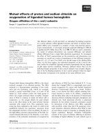
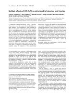
Fig. 2 shows the Raman spectra, which ranged from 400
to 3,700 cm
-1
, of mouse livers in the control group (A), E.
purpurea-treated group (B), 3 Gy gamma-irradiated group
(C), radio-protected group (D) and radio-recovery group
(E). The secondary structure information, primarily seen as
antiparallel β-pleated sheets, was indicated by the vibra-
Phytotherapeutic effects of Echinacea purpurea in gamma-irradiated mice 347
Fig. 2. A: Raman spectra in the 400-3,700 cm
1
region of control mouse liver (A), E. purpurea-treated mice (B), 3 Gy γ-irradiated mic
e
(C), radio-protected mice (D), and radio-recovered mice (E). The spectra represent the samples at the 4-week time interval of the
experiment. B: The expanded spectral region for amide I, amide III and tyrosine.
tional stretch of amide I (∼1,670 cm
1
) and amide III (∼
1,241 cm
1
) only in Groups A and B. The secondary struc-
ture of the protein in the mouse liver was not stable in C and
amide I was shifted to ∼1,590 cm
1
while amide III was
stable. In Group D, the secondary structure of the protein in
liver cells was stable enough to resist changes in the
spectra. The comparison of the Raman spectra of the ra-
dio-recovery group (E) in Fig. 2 and the control group (A)
showed that the vibrational stretch of amide I was shifted to
∼1,620 cm
1
. The vibrational stretch of amide III in Group
C could not be detected by Raman spectra, while the amide
III in Group E was shifted to 1,180 cm
1
.
The tyrosine residues were detected at the doublet Raman
shift of 855 and 832 cm
1
. The ratio of both doublets in-
dicated the hydrogen bonding environment in the liver.
The intensity ratios (I
855/832
) for A, B, C, D, and E Group
were 0.48, 0.47, 1.25, 0.62 and 1.17, respectively. In other
words, the tyrosine residues in Group C were greatly af-
fected by radiation. The tyrosine residues in Group D were
more susceptible to E. purpurea treatment before radiation
than the E. purpurea treatment after radiation (Group E).
Discussion
E. purpurea has generally been considered to be safe and
without significant toxicity, significant herb-drug inter-
actions, contraindications, or adverse side effects [8,23,
28].
The hematopoietic system is known to be one of the most
radiosensitive systems, and its damage may play lead to the
development of hematopoietic syndrome and result in
death. Survival after irradiation actually results from the
recovery of several target systems, such as the bone mar-
row, gastrointestinal tract, skin and hemostatic systems
[59]. Death from the so-called hematopoietic syndrome re-
sults from infection due to the impairment of the immune
system [11]. Various mechanisms, such as the prevention
of damage through the inhibition of free radical generation
or its intensified scavenging, enhancement of DNA and
membrane repair, replacement of dead hematopoietic and
other cells and the stimulation of immune-cells activities,
are considered to be important approaches for radio-pro-
tection and radio-recovery [36].
In the present study, the reduction in both Hb level and
RBC count at each of the three time intervals in the irradi-
ated groups were attributed to the impairment of cell divi-
sion, obliteration of blood-forming organs, alimentary
tract injury [14], depletion of factors needed for erythro-
blast differentiation and reticulocyte release from the bone
marrow [18] and the loss of cells from the circulation by
hemorrhage or leakage through capillary walls and/or the
direct destruction of mature circulating cells [53]. Reco-
very of both Hb level and RBC count was evident in both
the protected and recovered groups, but the recovery of the
Hb parameter was more distinct in the radio-protection
group than in the radio-recovery group. In contrast, the
RBC counts in the radio-protection and radio-recovery
groups were the same as those of the control, E. purpurea-
348 Amira M. K. Abouelella et al.
treated and irradiated groups.
The present work describes the marked decrease in WBC
count in mice subjected to irradiation at three time
intervals. Irradiation-induced leucopoenia has likewise
been reported in γ-ray irradiated mice [33]. It seems appa-
rent that the leucopoenia observed in these mice was a di-
rect consequence of the lymphopenia and neutropenia that
occurred following irradiation. An obvious degree of ei-
ther radio-protection or radio-recovery was obtained using
E. purpurea. These results agree with the findings of
Barrett [3] and Widel [59], who reported that Echinacea
preparations influenced the leukocyte count, stimulated
the phagocytic activity and/or increased the release of
cytokines. It has been suggested that Echinacea is able to
stimulate innate immune responses, including those regu-
lated by macrophages and natural killer cells (white blood
cells). In addition, macrophages respond to purified poly-
saccharide and alkylamide preparations incorporated into
Echinacea. Treatment with ionizing radiation resulted in
cytokine-mediated cellular damage [30]. For patients un-
dergoing radiation and chemotherapy treatments, studies
have proven that E. purpurea, while boosting the immune
system, also produced additional white blood cells and
stimulated bone marrow production, which was dimin-
ished by chemotherapy [43]. However, the mechanisms of
stimulation for cells responsible for adaptive immunity
have not been fully elucidated for the other molecules pres-
ent in E. purpurea preparations [22].
Since the peripheral blood pattern observed during the en-
tire post-irradiation period was primarily a reflection of
processes occurring in hematopoietic organs [59], the sig-
nificant protective effects of E. purpurea against lymphoid
cell death in bone marrow can lead to their accelerated re-
covery in peripheral blood. In fact, the tendency to return to
the normal value of reduced blood leukocyte count
throughout the three time intervals was more rapid in the E.
purpurea-treated groups both before and after irradiation
than in the irradiated mice.
In this study, lymphocytes, neutrophils and monocytes
were significantly decreased throughout the three time
intervals. Mature lymphocytes are considered to be the
most sensitive type of blood cell [60], and the earliest blood
change following whole body irradiation is lymphopenia
[45]. Neutrophils have a half-life of only about 10-12 h
once they leave the marrow, a site that serves as a reservoir
for mature neutrophils [34]. These data agree with the find-
ings of Kafafy et al. [25]. The data showed that E. purpurea
administration has significant radio-protective and ra-
dio-recovery effects on the levels of lymphocytes, neu-
trophils and monocytes.
It has been reported that E. purpurea has an IFN-like ef-
fect, activating macrophages and inducing the production
of interleukin -1 (IL-1) and IFN [48]. In addition, Mishima
et al. [33] reported that the administration of E. purpurea
had a suppressive effect on radiation-induced leucopoenia,
especially on lymphocytes and monocytes, and resulted in
a faster recovery of the blood cell count in mice and rabbits
[24]. In addition, peripheral blood antioxidant activity was
increased by E. purpurea, which suggested a relationship
between the antioxidant effect and the suppressive effects
on radiation-induced leucopoenia. In contrast, Schwarz et
al. [48] reported that the oral administration of E. purpurea
for 2 weeks had only minor effects on 2 out of 12 lympho-
cyte subpopulations determined by flow-cytometry in a
double-blind, placebo-controlled cross-over study.
In the present study, irradiation caused remarkable in-
creases in the TBARs content and incredible decreases in
the activities of SOD and GSPx. Zahran et al. [63] and
Tawfik et al. [54] recently confirmed these finding. After
the administration of E. purpurea, the TBARs level and an-
tioxidant activities were attenuated in comparison to their
values in irradiated mice at each of the three time intervals.
The mechanisms of antioxidant activity in the extracts de-
rived from
Echinacea included free radical scavenging and
transition metal chelating properties [23].
Several experimental models have described the in vivo
and in vitro protection from liver injury induced by free
radicals [1,40]. They reported that prostaglandin (PGE1)
was able to reduce DNA fragmentation in rat hepatocytes
and that it protected against galactosamine (D-GalN)- in-
duced apoptosis. It is interesting to note that the admin-
istration of Echinacea also reduced the effects of gamma
irradiation on DNA fragmentation. In contrast, the admin-
istration of Echinacea after gamma exposure was not ef-
fective at reducing the apoptotic mechanisms induced by
gamma irradiation. The protection provided by Echinacea
against apoptosis induced by gamma irradiation may be as-
sociated with its ability to block the induction of internal
factors, such as inducible nitric oxide synthase (iNOS) and
nitric oxide (NO) production. In fact, Echinacea was able
to slightly enhance DNA fragmentation in control cells.
Nevertheless, more studies are needed in order to confirm
these findings.
The secondary structure of liver proteins is easily moni-
tored by observing the frequencies of amide I and amide III
originating from a peptide backbone [50]. The sharpening
of amide I peaks in A, B and D may indicate the uniformity
of hydrogen bonds whereas the flattening of amide I in the
gamma-irradiated mice (Group C) and its shifting to 1,590
cm
-1
may indicate the loss of uniformity in hydrogen
bonds.
In the liver, tyrosine is a key component of many en-
zymes, which may be inhibited through the oxidative mod-
ification of their tyrosine residues. Therefore, it is very im-
portant to probe the microenvironment of tyrosine. Shih et
al. [51] reported that the tyrosine doublet at 850
-1
and 830
cm
-1
was sensitive to the nature of the hydrogen bond of the
phenol hydroxyl group. If a tyrosine residue is on the sur-
Phytotherapeutic effects of Echinacea purpurea in gamma-irradiated mice 349
face of a protein in aqueous solution, the phenolic OH will
simultaneously act as an acceptor and donor of moderate to
weak H-bonds, and the doublet intensity ratio (I
850/830
) will
be about 1 : 0.8 (I = 1 : 25). If the phenolic oxygen is the ac-
ceptor atom in a strong H-bond, the intensity ratio will be
about 1 : 0.4 (I = 2 : 5). If the phenolic hydroxyl is the pro-
ton donor in a strong H-bond, the intensity ratio will be ap-
proximately 1 : 2 (I = 0.5). Accordingly, the current result
that the intensity ratio in the gamma-irradiated mice
(Group C) was about 1 : 0.8 might indicate that the phe-
nolic hydroxyl of tyrosine was on the surface of liver pro-
teins with a moderate to weak H-bond. On the other hand,
the doublet intensity ratio in the radio-protected mice
(Group D) was sensitive to the level of E. purpurea admin-
istration, as shown in Fig. 2b. However, the mechanisms
by which these antioxidative effects protect major liver
constituents, including thiol compounds, tyrosine, trypto-
phan, and water content, from oxidative insults remains to
be elucidated.
Weiss and Landauer [58] documented a protective effect
of polyphenols from Echinacea against free radical dam-
age and a class of specific antioxidants known as caffeoyl
derivatives in appreciable amounts. Furthermore, Sasagawa
et al. [44] reported that the alkylamides present in
Echinacea species inhibited IL action and hypothesized
that the constituents present in its dry extracts exert direct
immunomodulatory effects on the immune system [44]. In
addition, single X-ray irradiation causes considerable dis-
turbances to the liver. The administration of Echinacea
tinctures was assumed to induce their beneficial effects,
primarily by stimulating certain components of the non-
specific immune system. Previous studies have proven that
the most important pharmacological effects were the stim-
ulation of the phagocytic activity of polymorphonuclear
leucocytes and other phagocytes [3], as well as the activa-
tion of phagocytes to produce the pro-inflammatory cyto-
kines TNF-α, IL-1, IL-6 and other mediators [4].
E. purpurea was able to regulate the process of apoptosis
in-vivo. The splenic-lymphocytes from mice orally treated
with Echinacea for 14 days at a dose of level 30 mg kg
-1
per
day were shown to be significantly more resistant to apop-
tosis than those from mice treated only with the vehicle
[13]. Moreover, Gan et al. [16] demonstrated that
Echinacea extracts are potent activators of natural killer
(NK) cytotoxicity, augmented the frequency of NK target
conjugates and activated the programming for NK cell
lysis. The Echinacea extracts also enhanced the anti-
body-forming cell response and humeral immune re-
sponses as well as the innate immune responses in female
mice [15]. It also enhanced the nonspecific immune or cel-
lular immune systems (or both) in the AKR/J-mice [20]. It
also sensitized the immune cells and led to lifespan pro-
longation in mice [12].
Raso et al. [41] evaluated the anti-inflammatory activity
of E. purpurea in mice treated at doses of 30 and 100 mg
kg
-1
twice daily. Only the higher-dose treatment sig-
nificantly inhibited the formation of edema in a time-de-
pendent manner. Western blot analysis showed that in vivo
treatment with this extract could modulate lipo-poly-
saccharide and INF-γ-induced cyclooxygenase-2 (COX-
2) and iNOS expression in peritoneal macrophages. They
suggested that the anti-inflammatory effect of that partic-
ular extract could be in part related to its modulation of
COX-2 expression.
The mechanisms of the stimulatory effect observed in the
present study remain to be clarified. The authors suggest
that the factors that might be involved are changes in the in-
testinal absorption of immune stimulating-compounds
present in the Echinacea preparation caused by the
irradiation. Brinker [10] reported that the experimental
success of the oral administration of the immunostimulant
E. purpurea was probably due to the receptor binding of its
polymeric markers on mucosal- or gut-associated lym-
phoid tissues.
In conclusion, the immune stimulatory ability of E. pur-
purea extracts may have a therapeutic potential to regulate
the protection and recovery of immune responses as well as
the activation measures in irradiated mice.
Therefore, further studies are needed to clarify the mecha-
nism(s) that are responsible for the beneficial effect of
Echinacea preparations observed in this study, and future
research must also be conducted on the use of E. purpurea
as an immunonutrient and useful adjunct to conventional
cancer therapies because of its immune-stimulating pro-
perties.
Acknowledgments
The authors greatly appreciate Dr. Mohamed Samy
Soliman, Radiation Health Research Department, NCRRT
for his technical assistance with the bone marrow
examination. We thank Dr. Abdel Monem Abdalla,
Molecular Biology Department, National Research Centre
for performing the FT-Raman analysis.
References
1. Abou-Elella AMKE, Siendones E, Padillo J, Montero JL,
De la Mata M, Muntan
é
Relat J. Tumour necrosis factor-al-
pha and nitric oxide mediate apoptosis by D-galactosamine in
a primary culture of rat hepatocytes: exacerbation of cell death
by cocultured Kupffer cells. Can J Gastroenterol 2002, 16,
791-799.
2. Ang-Lee MK, Moss J, Yuan CS. Herbal medicines and peri-
operative care. JAMA 2001, 286, 208-216.
3. Barrett B. Medicinal properties of Echinacea: a critical
review. Phytomedicine 2003, 10, 66-86.
4. Barrett B. Echinacea: a safety review. HerbalGram 2003, 57,
36-39.
350 Amira M. K. Abouelella et al.
5. Bauer R. New knowledge regarding the effect and effective-
ness of Echinacea purpurea extracts. Wien Med Wochenschr
2002, 152, 407-411.
6. Binns SE, Hudson J, Merali S, Arnason JT
·
. Antiviral ac-
tivity of characterized extracts from Echinacea spp. (Helian-
theae: Asteraceae) against herpes simplex virus (HSV-I).
Planta Med 2002, 68, 780-783.
7. Block KI, Boyd DB, Gonzalez N, Vojdani A. Point-coun-
terpoint: the immune system in cancer. Integr Cancer Ther
2002, 1, 294-316.
8. Block KI, Mead MN. Immune system effects of echinacea,
ginseng and astragalus: a review. Integr Cancer Ther 2003, 2,
247-367.
9. Borek C. Antioxidants and radiation therapy. J Nutr 2004,
134, 3207S-3209.
10. Brinker F. Variations in effective botanical products.
HerbalGram 1999, 46, 36-50.
11. Chen YM, Lin SL, Chiang WC, Wu KD, Tsai TJ.
Pentoxifylline ameliorates proteinuria through suppression
of renal monocyte chemoattractant protein-1 in patients with
proteinuric primary glomerular diseases. Kidney Int 2006,
69, 1410-1415.
12. Currier NL, Miller SC. The effect of immunization with kil-
led tumor cells, with/without feeding of Echinacea purpurea
in an erythroleukemic mouse model. J Altern Complement
Med 2002, 8, 49-58.
13. Di Carlo G, Nuzzo I, Capasso R, Sanges MR, Galdiero E,
Capasso F, Carratelli CR. Modulation of apoptosis in mice
treated with Echinacea and St. John's wort. Pharmacol Res
2003, 48, 273-277.
14. El-Habit OH, Saada HN, Azab KS, Abdel-Rahman M,
El-Malah DF. The modifying effect of
β
-carotene on gamma
radiation-induced elevation of oxidative reactions and geno-
toxicity in male rats. Mutat Res 2000, 466, 179-186.
15. Freier DO, Wright K, Klein K, Voll D, Dabiri K, Cosulich
K, George R. Enhancement of the humoral immune response
by Echinacea purpurea in female Swiss mice. Immunophar-
macol Immunotoxicol. 2003, 25, 551-560.
16. Gan XH, Zhang L, Heber D, Bonavida B. Mechanism of
activation of human peripheral blood NK cells at the single
cell level by Echinacea water soluble extracts: recruitment of
lymphocyte-target conjugates and killer cells and activation
of programming for lysis. Int Immunopharmacol 2003, 3,
811-824.
17. Goldberg ED, Dygai AM, Shakhov VP. Methods of Tissue
Culture in Hematology. pp. 256-257, TGU Publishing House,
Tomsk, 1992.
18. Gridley DS, Pecaut MJ, Miller GM, Moyers MF, Nelson
GA. Dose and dose rate effects of whole-body gamma-irradi-
ation: II. Hematological variables and cytokines. In vivo
2001, 15, 209-216.
19. Gunsilius E, Clausen J, Gastl G. Palliative immunotherapy
of cancer. Ther Umsch 2001, 58, 419-424.
20. Hayashi I, Ohotsuki M, Suzuki I, Watanabe T. Effects of
oral administration of Echinacea purpurea (American herb)
on incidence of spontaneous leukemia caused by recombi-
nant leukemia viruses in AKR/J mice. Nihon Rinsho Meneki
Gakkai Kaishi 2001, 24, 10-20.
21. Hu C, Kitts DD. Studies on the antioxidant activity of
Echinacea root extract. J Agric Food Chem 2000, 48, 1466-
1472.
22. Hwang SA, Dasgupta A, Actor JK. Cytokine production by
non-adherent mouse splenocyte cultures to Echinacea ext-
racts. Clin Chim Acta 2004, 343, 161-166.
23. Izzo AA, Ernst E. Interactions between herbal medicines and
prescribed drugs: a systematic review. Drugs 2001, 61, 2163-
2175.
24. Jurk
š
tien
ė
V, Kondrotas AJ, K
ė
velaitis E. Compensatory
reactions of immune system and action of Purple Coneflower
(Echinacea purpurea (L.) Moench) preparations. Medicina
(Kaunas) 2004, 40, 657-662.
25. Kafafy YA, Roushdy HM, Abdel-Haliem M, Mossad MN,
Ashry OM, Salama SF. Green tea antioxidative potential in
irradiated pregnant rats. Eygpt J Radiat Sci Appl 2005, 18,
313-333.
26. Kim LS, Waters RF, Burkholder PM. Immunological ac-
tivity of larch arabinogalactan and Echinacea: a preliminary,
randomized, double-blind, placebo-controlled trial. Altern
Med Rev 2002, 7, 138-149.
27. Kligler B. Echinacea. Am Fam Physician 2003, 67, 77-80.
28. Knapp RG, Miller MC. Clinical Epidemiology and Bios-
tatistics. pp. 55-70, Williams & Wilkins, Baltimore, 1992.
29. McLauchlan DM, Gowenlock AH. Statistics. In: Gowen-
lock AH, McLauchlan DM, McMurray JR (eds.). Varely's
Practical Clinical Biochemistry. 6th ed. pp. 232-272,
Heinemann Medical Books, London, 1988.
30. Meky NH, Mansour MAE, Soliman MS, Tawfik E. Effects
of gamma irradiation on some linked processes between co-
agulation and inflammatory reactions. Egypt J Radiat Sci
Appl 2002, 15, 1-23.
31. Melchart D, Clemm C, Weber B, Draczynski T, Worku F,
Linde K, Weidenhammer W, Wagner H, Saller R.
Polysaccharides isolated from Echinacea purpurea herba cell
cultures to counteract undesired effects of chemotherapy a
pilot study. Phytother Res 2002, 16, 138-142.
32. Minami M, Yoshikawa H. A simplified assay method of su-
peroxide dismutase activity for clinical use. Clin Chem Acta
1979, 92, 337-342.
33. Mishima S, Saito K, Maruyama H, Inoue M, Yamashita
T, Ishida T, Gu Y. Antioxidant and immuno-enhancing ef-
fects of Echinacea purpurea. Biol Pharm Bull 2004, 27,
1004-1009.
34. Mollinedo F, Borregaard N, Boxer LA. Novel trends in
neutrophil structure, function and development. Immunol
Today 1999, 20, 535-537.
35. Nair CK, Salvi V, Kagiya TV, Rajagopalan R. Relevance
of radioprotectors in radiotherapy: studies with tocopherol
monoglucoside. J Environ Pathol Toxicol Oncol 2004, 23,
153-160.
36. N
ü
bel T, Damrot J, Roos WP, Kaina B, Fritz G. Lovastatin
protects human endothelial cells from killing by ionizing ra-
diation without impairing induction and repair of DNA dou-
ble-strand breaks. Clin Cancer Res 2006, 12, 933-939.
37. Osowski S, Rostock M, Bartsch HH, Massing U. Pharma-
ceutical comparability of different therapeutic Echinacea
preperations. Forsch Komplementarmed Klass Naturheilkd
2000, 7, 294-300.
38. Paglia DE, Valentine WN. Studies on the quantitative and
Phytotherapeutic effects of Echinacea purpurea in gamma-irradiated mice 351
qualitative characterization of erythrocyte glutathione pero-
xidase. J Lab Clin Med 1967, 70, 158-169.
39. Pellati F, Benvenuti S, Melegari M, Lasseigne T.
Variability in the composition of anti-oxidant compounds in
Echinacea species by HPLC. Phytochem Anal 2005, 16,
77-85.
40. Quintero A, Pedraza CA, Siendones E, Kamal ElSaid
AM, Colell A, Garc
í
a-Ruiz C, Montero JL, De la Mata M,
Fern
á
ndez-Checa JC, Mi
ñ
o G, Muntan
é
J. PGE1 pro-
tection against apoptosis induced by D-galactosamine is not
related to the modulation of intracellular free radical pro-
duction in primary culture of rat hepatocytes. Free Radical
Res 2002, 36, 345-355.
41. Raso GM, Pacilio M, Di Carlo G, Esposito E, Pinto L, Meli
R. In-vivo and in-vitro anti-inflammatory effect of Echinacea
purpurea and Hypericum perforatum. J Pharm Pharmacol
2002, 54, 1379-1383.
42. Rininger JA, Kichker S, Chigurupati P, McLean A,
Franck Z. Immuno-pharmacological activity of Echinacea
preparations following simulated digestion on murine macro-
phages and human peripheral blood mononuclear cells. J
Leukoc Biol 2000, 68, 503-510.
43. Rosenthal D, Ades T. Complementary and alternative
methods. CA Cancer J Clin 2001, 51, 316-320.
44. Sasagawa M, Cech NB, Graym DE, Elmer GW, Wenner
CA. Echinacea alkylamides inhibit interleukin-2 production
by Jurkat T cells. Int Immunopharmacol 2006, 6, 1214-1221.
45. Seddek MN, Abou Gabal HA, Salama SF, El-Kashef HS.
Effect of deltamethrin and
γ
-radiation on immunohemato-
logical elements of pregnant rats. J Egypt Ger Soc Zool 2000,
31, 171-182.
46. Schulten B, Bulitta M, Ballering-Br
ü
hl B, K
ö
ster U,
Sch
ä
fer M. Efficacy of Echinacea purpurea in patients with
a common cold. A placebo-controlled, randomized, dou-
ble-blind clinical trail. Arzneimittelforschung 2001, 51,
563-568.
47. Schwarz E, Metzler J, Diedrich JP, Freudenstein J, Bode
C, Bode JC. Oral administration of freshly expressed juice of
Echinacea purpurea herbs fail to stimulate the nonspecific
immune response in healthy young men: results of a dou-
ble-blind, placebo-controlled crossover study. J Immunother
2002, 25, 413-420.
48. Schwarz E, Parlesak A, Henneicke-von Zepelin HH, Bode
JC, Bode C. Effect of oral administration of freshly pressed
juice of Echinacea purpurea on the number of various sub-
populations of B- and T-lymphocytes in healthy volunteers:
results of a double-blind, placebo-controlled cross-over
study. Phytomedicine 2005, 12, 625-631.
49. Senchina DS, McCann DA, Asp JM, Johnson JA, Cunnick
JE, Kaiser MS, Kohut ML. Changes in immunomodulatory
properties of Echinacea spp. root infusions and tinctures stor-
ed at 4 degrees C for four days. Clin Chim Acta 2005, 355,
67-82.
50. Shi YB, Fang JL, Liu XY, Tang WX. Fourier transform IR
and Fourier transform Raman spectroscopy studies of metal-
lothionein-lll: amide l band assignments and secondary struc-
tural comparison with metallothioneins-l and -ll. Biopoly-
mers 2000, 65, 81-88.
51. Shih S, Weng YM, Chen S, Huang SL, Huang CH, Chen
W. FT-Raman spectroscopic investigation of lens proteins of
tilapia treated with dietary vitamin E. Arch Biochem Biophys
2003, 420, 79-86.
52. South EH, Exon JH. Multiple immune functions in rats fed
Echinacea extracts. Immunopharmacol Immunotoxicol 2001,
23, 411-421.
53. Tawfik SS. Efficiency of taurine usage as treatment for ex-
posure to ionizing radiation. Ph.D. Dissertation. Institute of
Environmental Studies and Researches, Ain Shams Universi-
ty, Cairo, 2003.
54. Tawfik SS, Abbady MI, Azab KhSh, Zahran AM. Anti-
clastogenic and haemodynamic efficacy of flavonoid mixture
challenging the oxidative stress induced by gamma rays in
male mice. Egypt J Rad Sci Applic 2006, 19, 195-210.
55. Tawfik SS, Elshamy E, Sallam MH
. Aged garlic extract
modulates the oxidative modifications induced by
γ
-rays in
mouse bone marrow and erythrocytes cells. Egypt J Rad Sci
Applic 2006, 19, 499-512.
56. Turner RB, Riker DK, Gangemi JD. Ineffectiveness of
Echinacea for prevention of experimental rhinovirus colds.
Antimicrob Agents Chemother 2000, 44, 1708-1709.
57. Vonau B, Chard S, Mandalia S, Wilkinson D, Barton SE.
Does the extract of the plant Echinacea purpurea influence
the clinical course of recurrent genital herpes? Int J STD
AIDS 2001, 12, 154-158.
58. Weiss JF, Landauer MR. Protection against ionizing radia-
tion by antioxidant nutrients and phytochemicals. Toxicolo-
gy 2003, 189, 1-20.
59. Widel M, Jedrus S, Lukaszczyk B, Raczek-Zwierzycka K,
Swierniak A. Radiation-induced micronucleus frequency in
peripheral blood lymphocytes is correlated with normal tis-
sue damage in patients with cervical carcinoma undergoing
radiotherapy. Radiat Res 2003, 159, 713-721.
60. Wintrobe MM, Lee GR, Foerster J, Lukens J, Paraskevas
F, Greer JP, Rodgers GM. Wintrobe's Clinical Hemato-
logy. 10th ed. Vol. 2. p.1852, Lippincott Williams & Wilkins,
Baltimore, 1999.
61. Yang K, Azoulay E, Attalah L, Zahar JR, Van de Louw A,
Cerf C, Soussy CJ, Duvaldestin P, Brochard L, Brun-
Buisson C, Harf A, Delclaux C. Bactericidal activity re-
sponse of blood neutrophils from critically ill patients to
in-vitro granulocyte colony-stimulating factor stimulation.
Intensive Care Med 2003, 29, 396-402.
62. Yoshioka T, Kawada K, Shimada T, Mori M. Lipid perox-
idation in maternal and cord blood and protective mechanism
against activated-oxygen toxicity in the blood. Am J Obstet
Gynecol 1979, 135, 372-376.
63. Zahran AM, Azab KhSh, Abbady MI. Modulatory role of
allopurinol on xanthine oxidoreductase system. Egypt J Rad
Sci Applic 2006, 19, 373-388.