Báo cáo khoa học: "Relative biological effectiveness of fast neutrons for apoptosis in mouse hair follicles" potx
Bạn đang xem bản rút gọn của tài liệu. Xem và tải ngay bản đầy đủ của tài liệu tại đây (902.17 KB, 6 trang )
JOURNAL OF
Veterinary
Science
J. Vet. Sci. (2007), 8(4), 335
340
*Corresponding author
Tel: +82-62-530-2837; Fax: +82-62-530-2841
E-mail:
Relative biological effectiveness of fast neutrons for apoptosis in mouse
hair follicles
Hae-June Lee
1
, Sung-Ho Kim
2,
*
1
Korea Institute of Radiological & Medical Science, Seoul 139-240, Korea
2
College of Veterinary Medicine, Chonnam National University, Gwangju 500-757, Korea
This study compared the effects of high linear energy
transfer (LET) fast neutrons on the induction of apoptosis
in the hair follicles of ICR mice with those of low LET
60
Co
γ
-rays. The changes that occurred from 0 to 24 h af-
ter exposing the mice to either 2 Gy of
γ
-rays (2 Gy/min)
or 0.8 Gy of neutrons (94 mGy/min, 35 MeV) were exa-
mined. The maximum frequency was found at 12 h (
γ
-
rays) or 8 h (neutrons) after irradiation. The mice that re-
ceived 0-8 Gy of
γ
-rays or 0-1.6 Gy of neutrons were exam-
ined 8 h after irradiation. The dose-response curves were
analyzed using the best-fit curve model. The dose-response
curves were linear-quadratic, and a significant relation-
ship was found between the frequency of apoptotic cells
and the dose. The morphological findings in the irradiated
groups were typical apoptotic fragments in the matrix re-
gion of the hair follicle, but the spontaneous existence of
apoptotic fragments was rarely observed in the control
group. In the presence of an apoptosis frequency between
2 and 14 per follicle, the relative biological effectiveness
values of neutrons in small and large follicles were 2.09
±
0.30 and 2.15
±
0.18, respectively.
Key words: apoptosis, biological effectiveness, fast neutrons,
gamma-rays, hair follicle
Introduction
The hair follicle and its hair have long been recognized as
potentially useful biological indicators for the quantitative
index of radiation injury in nuclear and medical radiation.
Hairs are located over much of the body surface and can
provide regional information. Therefore, the skin and its
appendages would appear to offer the only system in which
the dose distribution of radiation over the body surface
may be assessed by an estimate of the received dose on a
suitable time-scale for clinical intervention [5,12,31].
Apoptosis is a spontaneous or induced phenomenon that
can be observed in many cell types [17]. Radiation-in-
duced programmed cell death is a degradative and pro-
gressive process. The degradative process is initiated in the
target nucleus, ultimately resulting in the quantitative con-
version of the target genome into small DNA fragments.
Apoptosis is initiated not only by pathological conditions,
but is also triggered by factors such as cellular mechanisms
intrinsically or extrinsically regulated by physiological
stimuli. Radiation-induced apoptosis has mainly been cha-
racterized in lymphocytes in vitro, and appears to be re-
lated to the number of DNA strand breaks, the rate at which
they occur, and the rapidity and effectiveness of the DNA
repair mechanisms [2,9,22]. However, other results sug-
gest that DNA might not be the only target that induces an
apoptotic stimulus after irradiation that could mainly in-
volve cell membrane damage [20,27]. These cellular stud-
ies do not take into account cell-to-cell interactions and cell
differentiation processes that can play important roles dur-
ing the initiation and progression of apoptosis. These roles
can be examined using histological methods, and the few
available data have mostly come from extremely radia-
tion-sensitive tissues such as the adult gut [25] or the cen-
tral nervous system during histogenesis [7,16,18].
The biological effects of fast neutrons in normal tissues
and in tumors are of interest in relation to clinical radio-
therapy, for radiation protection purposes, and to aid in the
basic understanding of the radiation-induced inactivation
of cells, whether by low or high linear energy transfer
(LET) radiation. In general, the biological effects of
high-LET radiation are greater than those of low-LET
radiation. The variations in the relative biological effec-
tiveness (RBE) with dose, with oxygenation, with cell cy-
cle parameters, and from one tissue to another are well-
documented [3]. However, few data are available on apop-
tosis in hair follicles exposed to radiation at higher ionizing
density, such as neutrons. In this study, we used cyclo-
tron-derived fast neutrons with a peak energy of 35 MeV to
336 Hae-June Lee et al.
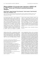
Fig. 1. Photomicrograph of small (A, C) and large (B, D) hair fol-
licles of mice sacrificed 8 h after irradiation. The apoptotic cells,
which occur predominantly in the matrix region of the follicle,
were easily recognized from the condensation of their cytoplas
m
and nuclear chromatin. A and B; H&E staining, C and D; TUNE
L
staining, ×400.
investigate how the energy of neutrons affects the bio-
logical processes. We evaluated the RBE for fast neu-
tron-induced apoptosis in the hair follicles using ICR mice
compared with the results of parallel experiments using γ
-rays.
Materials and Methods
Animals and irradiation
Male ICR mice were obtained from a specific patho-
gen-free colony (Oriental Bio, Korea), and were allowed 1
week of quarantine and acclimatization. The animals were
housed in a room that was maintained at 23 ± 2°C, with a
relative humidity of 50 ± 5%, artificial lighting from 08 : 00
to 20 : 00, and 13-18 air changes per hour. The animals
were housed four per stainless steel wire mesh cage, and
were given tap water and commercial rodent chow
(Samyang Feed, Korea) ad libitum. ICR mice between the
ages of 7 and 8 weeks were used. At this age, the skin of the
mice contains a synchronous resting population of hair fol-
licles (telogen phase). These resting follicles can be stimu-
lated into activity by the simple act of plucking a liquid
plastic dressing (Alteco-Ace; Alteco Korea, Korea), which
dries within 10 min after application, and can be removed
from the animals together with the embedded hairs. Ten
days after plucking, the follicles were in mid-anagen and
the animals were subjected to whole-body irradiation with
either γ-rays or fast neutrons. The neutrons were generated
from the KCCH cyclotron using the proton-beryllium
reaction. The estimated forward neutron spectra estab-
lished a peak energy of 35 MeV. The mean dose rate for
neutrons was 94 mGy/min. The contamination of γ-rays
was estimated as 14.2% of the neutron dose. Exposure to
137
Cs-generated γ-rays was conducted with Gammacell
(Nordion, Canada). The mean dose rate of γ-rays was 2
Gy/min. Fifty-two mice were assigned to thirteen groups:
non-irradiated control, fast neutron (0.8 Gy), and γ-rays (2
Gy). The mice were sacrificed at various periods from 2 to
24 h after irradiation (four mice for each time interval).
Forty mice were exposed to 0, 0.2, 0.4, 0.8, or 1.6 Gy of
neutrons or 0.5, 1.0, 2.0, 4.0, or 8.0 Gy of γ-rays, and were
sacrificed at 8 h after irradiation. The Institutional Animal
Care and Use Committee at Chonnam National University
approved the protocols used in this study, and the animals
were cared for in accordance with the Guidelines for
Animal Experiments.
Tissue preparation
Skin samples obtained from the mid-dorsum were fixed
with 10% neutral-buffered formalin. The skin was first
flattened onto a piece of paper to prevent it from curling
during fixation. Three micrometer sections were cut on a
plane parallel to the long axis of the animal rather than
across the animal. These provided longitudinal sections of
the follicles that were aligned parallel to the long axis. In
order to visualize the apoptotic cells, we used the
TdT-mediated dUTP-biotin nick end-labeling (TUNEL)
method of immunohistochemical staining with a commer-
cial kit (ApopTag Plus Peroxidase In Situ Apoptosis
Detection kit; Intergen, USA), and stained the cells with
hematoxylin and eosin (H&E).
Counting procedures
The follicles were selected for scoring of apoptotic cells if
they were good examples of longitudinal sections. In prac-
tice, this meant that they had to contain the developing hair
root and a full longitudinal section of the dermal papilla.
They were subjectively grouped into large or small fol-
licles, and 20 of each were scored using an oil immersion
(×1,000). Large follicles are most likely to be those respon-
sible for the three types of guard hairs, while small follicles
are responsible for the small underfur or zigzag hairs. All
analyses were performed using the Graph PAD In Plot
computer program (Graph Pad Software, USA).
Results
The apoptotic cells, which are primarily found in the ma-
trix region of the follicle, were easily recognized from the
condensation of their cytoplasm and nuclear chromatin.
The dead cells break up into several fragments. Not all of
the fragments necessarily contain fragments of the cell
nucleus. These cytoplasmic fragments can usually be rec-
ognized by their eosinophilic staining properties in H&E
stain. Apoptosis was easily recognized by the presence of
RBE of fast neutrons for apoptosis in hair follicles 337
Fig. 2. Variation in apoptotic cell frequency in small () or larg
e
(■) hair follicle with time after whole-body irradiation of IC
R
mice with 0.8 Gy of fast neutrons (A) and 2.0 Gy of γ-rays (B).
Results are presented as means ± SD from four mice in each
group.
Fig. 3. Dose-response for fast neutrons (■) and γ-rays (•) induce
d
apoptotic cells in small (A) or large (B) hair follicle. The lines
represent the results of a linear-quadratic fit through the data in-
dicated in the figure.
Tabl e 1 . Apoptotic cells in hair follicles 8 h after irradiation
Group Dose (Gy)
Apoptotic cells per follicle*
Small follicle Large follicle
Control 0 0.11 ± 0.017 0.21 ± 0.023
Neutrons 0.2 1.74 ± 0.077 2.57 ± 0.038
0.4 2.29 ± 0.089 3.42 ± 0.155
0.8 4.67 ± 0.323 9.80 ± 1.064
1.6 8.91 ± 0.635 17.62 ± 1.390
γ-rays 0.5 1.90 ± 0.085 3.82 ± 0.594
1.0 3.30 ± 0.481 6.92 ± 1.471
2.0 6.44 ± 0.113 10.72 ± 1.188
4.0 10.86 ± 0.651 17.62 ± 0.481
8.0 14.48 ± 1.358 24.54 ± 2.461
*100 follicles scored from four mice in each group. Values are mea
n
± SD.
whole apoptotic bodies showing peroxidase staining. In
the TUNEL-positive cells or bodies, the stained products
exactly correlated with the typical morphological charac-
teristics of apoptosis as seen at the light microscopic level
(Fig. 1). A small number of cells in the hair follicle ex-
hibited apoptosis in the sham-irradiated mice at the levels
of 0.10 (small) and 0.21 (large) per follicle. At 2 h after irra-
diation, there was an increase in the number of apoptotic
cells, and the maximal frequency was found at 12 h (γ-ray)
or 8 h (neutron) after irradiation (Fig. 2).
Table 1 shows the amount of cell death caused by apopto-
sis at each dose. Apoptotic cell death, which was occasion-
ally found in the control animals, was markedly enhanced
by irradiation. The dose-response curves were analyzed
using the best-fit curve model. The dose-response curves
were linear-quadratic, and a significant relationship was
found between the frequency of apoptotic cells and the
dose (Fig. 3). Taking the controls into account, the lines of
best-fit are as follows:
γ-rays:
small follicles: y = (3.573 ± 0.0356)D + (0.222 ± 0.00498)
D
2
+ (0.114 ± 0.0085), r
2
= 1.0
large follicles: y = (6.000 ± 0.2755)D + (0.372 ± 0.03848)D
2
+ (0.210 ± 0.0115), r
2
= 0.995 ;
Neutrons:
small follicles: y = (6.034 ± 0.5289)D + (0.342 ± 0.36884)
D
2
+ (0.114 ± 0.0085), r
2
= 0.995
large follicles: y = (10.979 ± 1.619)D + (0.00935 ± 1.1291)
D
2
+ (0.210 ± 0.0115), r
2
= 0.989;
338 Hae-June Lee et al.
Tabl e 2 . Empirical and theoretical values for the induced apoptotic cells in hair follicles by
γ
-rays (V
γ
), neutron-
γ
mixed radiation (V
n+γ
)
and neutrons (V
n
)
Apoptotic cells
per follicle
Required dose
(Gy) of
V
γ
(DV
γ
)*
Required dose
(Gy) of
V
n+γ
(DV
n+γ
)*
Required dose
(Gy) of
V
n
(DV
n
)*
RBE
(DV
γ
/DV
n+γ
)(DV
γ
/DV
n
)
Small follicle
2.0 0.546 0.318 0.298 1.72 1.83
6.0 1.863 1.036 0.966 1.80 1.93
10.0 3.550 1.828 1.697 1.94 2.09
14.0 6.561 2.721 2.600 2.41 2.52
Large follicle
2.0 0.304 0.163 0.155 1.87 1.96
6.0 1.031 0.528 0.497 1.95 2.07
10.0 1.842 0.892 0.835 2.07 2.21
14.0 2.776 1.257 1.169 2.21 2.37
*Calculated from best fitting linear-quadratic model.
where y is the number of apoptotic cells per follicle and D
is the irradiation dose in Gy.
Since the neutrons cause mixed neutron-γ radiation, the
rate of induction by neutrons (V
n+γ
) can be approximated by
V
n+γ
= pV
n
+ (1 }p)V
γ
, where p is the fraction of the neutron
dose contributing to the total dose of fast neutrons, V
n
is the
value induced by neutrons, and V
γ
is the value induced by
γ-rays. V
n
+ γ = pV
n
+ (1 }p)V
γ
can be rewritten as V
n
= V
γ
+
(V
n+γ
V
γ
) ÷ p. When analyzed by the linear-quadratic mod-
el, the lines of best-fit of the theoretical dose-response to
neutrons are as follows:
small follicles: y = (6.44135 ± 0.6164)D + (0.3627 ± 0.4299)
D
2
+ (0.114 ± 0.0085), r
2
= 0.994;
large follicles: y = (11.5469 ± 1.464)D + (0.216648 ± 2.099)
D
2
+ (0.210 ± 0.0115), r
2
= 0.985.
In order to determine the RBE of neutrons compared with
γ-rays, the equation, y = aD + bD
2
+ c was transformed as
D = [a ± √ {a
2
}4b(c }y)}] ÷ 2b. The RBEs of the neutrons
were obtained from this equation. In the presence of an
apoptosis frequency between 2 and 14 per follicle, the
RBEs of the neutrons in the small and large follicles were
2.09 ± 0.30 and 2.15 ± 0.18, respectively (Table 2).
Discussion
The recognition that apoptotic cell death can be a major
component of radiation damage, particularly in rapidly
proliferating cell populations, has important implications
in radiobiological studies. Since hair loss following ex-
posure to radiation is a well-recognized phenomenon, the
hair follicle has been shown to be a radiosensitive organ.
However, there have been relatively few studies of the pa-
rameters related to the dose-response relationships for ra-
diation-induced damage [19,24,30].
The data obtained in this study indicate that there is a
quantitative change in apoptotic cells that are produced by
various doses of radiation. The morphological findings for
the apoptotic cells showed chromatin condensation into
the crescent caps at the nuclear periphery, along with nu-
clear disintegration, a decrease in nuclear size, a reduction
of the cell volume, and an increase in cell density in the hair
follicles. The location of the target cells against radia-
tion-induced programmed cell death in the hair follicles
has not been adequately elucidated in previous studies, but
data has shown that most of the cells that are killed by
apoptosis are found in the lower regions of the follicle ger-
minal matrix. These findings indicate that some of the fol-
licle stem cells are sensitive to this programmed cell death.
The quantification of the apoptotic fragments is a more
sensitive and accurate assay than the other scoring systems
based on visual observations [19,26,30].
The hairs and their follicles are readily accessible, easily
sampled, and cover most of the skin surface. As such, they
represent the only system that can be used to estimate the
local doses or dose distributions of radiation over the body
surface. Therefore, an examination of the whole follicle
would be a more sensitive means of detecting radiation
damage than other biological indicators, particularly be-
cause radiation-induced cell damage in the growing hair
matrix can usually be detected within a few hours in sec-
tioned follicles. This effect is similar in appearance to that
observed in crypt cells of the small intestine. This index is
known to be one of the most sensitive radiobiological end-
points [14,15].
The present study was the first to show a dose-response
relationship for neutron-induced apoptosis in hair follicles.
The dose-response curve for the neutron-irradiation
groups was much steeper than that for the γ-irradiated
RBE of fast neutrons for apoptosis in hair follicles 339
groups. The yield of the cells undergoing apoptosis appears
to show a linear-quadratic relationship to the dose. It is
generally known that the dose-effect relationship in the cell
death induced by neutrons is best fit to a linear model,
while low-LET radiation-induced cell death fits a line-
ar-quadratic model. However, most of these data have been
derived from in vitro studies with acute high dose
irradiation. Several in vivo experiments that demonstrated
the dose-response curves of some neutron-induced tissue
injuries were fir to the linear-quadratic model, such as the
normal tissues reviewed by Broerse and Barendsen [4] and
IARC [13].
Although a wide range of RBE values has been reported
for fast neutrons [29], an RBE value near 1 was reported for
radiation-induced apoptosis in human lymphocytes ex-
posed to high-energy 14.5 MeV neutrons [33]. Due to the
spread in the measured RBE values in various tissues, it is
still difficult to estimate the RBE associated with this radi-
ation quality, which indicates the need for further research
to resolve this issue. Apoptosis is the most sensitive in-
dicator of the radiation response. Hendry et al. [10,11] cal-
culated an RBE of 4 for the apoptosis data in the mouse
small intestine irradiated with fast neutrons with an energy
of 14.7 MeV. Warenius et al. [34] reported an RBE of 1.0
for the apoptosis data of mouse thymocytes irradiated with
fast neutrons at 62.5 MeV. Here, we showed that the RBEs
of fast neutrons were 2.09 (small follicle) and 2.15 (large
follicle) in the presence of an apoptosis frequency between
2 and 14 per follicle. Therefore, it appears that the RBE for
apoptosis is tissue-dependent. On the other hand, Fujikawa
et al. [8] calculated an RBE of 4.6 for the apoptosis of thy-
mocytes in mice irradiated with fission neutrons. There-
fore, the small RBE value of thymocyte apoptosis reported
by Warenius et al. [34] could be ascribed to the large en-
ergy of neutrons.
The RBE estimated for fast neutrons in this study was
greater than unity. This means that the apoptosis assay in
mouse hair follicles is sensitive to a difference in radiation
quality. The reported studies of DNA damage induced by
radiation of different qualities have generally shown a rela-
tively higher fraction of non-rejoining DNA double-strand
breaks (DSBs) after high-LET radiation [1,6,28,32]. In ad-
dition, high-LET radiations and gamma-rays have been
shown to produce initial DSBs, although they are of differ-
ent quality, with similar efficiency in cultured rodent cells
[21,23]. Overall, it is believed that DSB repair in the hair
follicle is involved as a determinant of the RBE of
high-LET radiation for induced apoptotic cell formation in
hair follicles.
In summary, this study determined the time-response re-
lationships of apoptotic cell formation in the hair follicles
of ICR mice for fast neutrons and γ-rays, and established a
linear-quadratic dose-effect relationship for both types of
radiation. Based on the dose-response data, the RBE values
of fast neutrons were estimated to be 2.09 for small fol-
licles, and 2.15 for large follicles. Further mechanistic
studies on the effects of neutron-induced apoptosis in the
hair follicle will be needed to extrapolate the experimental
data for protection against radiation in humans.
Acknowledgments
This work was supported by a Korea Science and
Engineering Foundation (KOSEF) Grant funded by the
Government (MOST), Korea.
References
1. Bl
ӧ
cher D. DNA double-strand break repair determines the
RBE of
α
-particles. Int J Radiat Biol 1988, 54, 761-771.
2. Bonicalzi ME, Haince JF, Droit A, Poirier GG. Regulation
of poly(ADP-ribose) metabolism by poly(ADP-ribose) gly-
cohydrolase: where and when? Cell Mol Life Sci 2005, 62,
739-750.
3. Britten RA, Peters LJ, Murray D. Biological factors influ-
encing the RBE of neutrons: implications for their past, pres-
ent and future use in radiotherapy. Radiat Res 2001, 156,
125-135.
4. Broerse JJ, Barendsen GW. Relative biological effective-
ness of fast neutrons for effects on normal Tissues. Curr Top
Radiat Res Q 1973, 8, 305-350.
5. Denekamp J, Joiner MC, Maughan RL. Neutron RBEs for
mouse skin at low doses per fraction. Radiat Res 1984, 98,
317-331.
6. Fox JC, McNally NJ. The rejoining of DNA double-strand
break following irradiation with
238
Pu
α
-particles: evidence
for a fast component of repair as measured by neutral filter
elution. Int J Radiat Biol 1990, 57, 513-521.
7. Fritsch P, Richard-Le Naour H, Denis S, M
é
n
é
trier F.
Kinetics of radiation-induced apoptosis in the cerebellum of
14-day-old rats after acute or during continuous exposure. Int
J Radiat Biol 1994, 66, 111-117.
8. Fujikawa K, Hasegawa Y, Matsuzawa S, Fukunaga A,
Itoh T, Kondo S. Dose and dose-rate effects of X rays and
fission neutrons on lymphocyte apoptosis in p53(+/+) and
p53(-/-) mice. J Radiat Res (Tokyo) 2000, 41, 113-127.
9. Hale AJ, Smith CA, Sutherland LC, Stoneman VE,
Longthorne VL, Culhane AC, Williams GT. Apoptosis:
molecular regulation of cell death. Eur J Biochem 1996, 236,
1-26.
10. Hendry JH, Potten CS, Chadwick C, Bianchi M. Cell death
(apoptosis) in the mouse small intestine after low doses: ef-
fects of dose-rate, 14.7 MeV neutrons, and 600 MeV (maxi-
mum energy) neutrons. Int J Radiat Biol Relat Stud Phys
Chem Med 1982, 42, 611-620.
11. Hendry JH, Potten CS, Merritt A. Apoptosis induced by
high- and low-LET radiations. Radiat Environ Biophys 1995,
34, 59-62
12. Hopewell JW. The skin: its structure and response to ionizing
radiation. Int J Radiat Biol 1990, 57, 751-773.
13. IARC. Neutrons. IARC Monogr Eval Carcinog Risks Hum
340 Hae-June Lee et al.
2000, 75, 401-409.
14. Ijiri K, Potten CS. Response of intestinal cells of differing
topographical and hierarchical status to ten cytotoxic drugs
and five sources of radiation. Br J Cancer 1983, 47, 175-185.
15. Ijiri K, Potten CS. Further studies on the response of in-
testinal crypt cells of different hierarchical status to eighteen
different cytotoxic agents. Br J Cancer 1987, 55, 113-123.
16. Ishida Y, Ohmachi Y, Nakata Y, Hiraoka T, Hamano T,
Fushiki S, Ogiu T. Dose-response and large relative bio-
logical effectiveness of fast neutrons with regard to mouse fe-
tal cerebral neuron apoptosis. J Radiat Res 2006, 47, 41-47.
17. Kerr JF, Wyllie AH, Currie AR. Apoptosis: a basic bio-
logical phenomenon with wide-ranging implications in tissue
kinetics. Br J Cancer 1972, 26, 239-257.
18. Kim SH, Chung CY, Son CH. Cell death by apoptosis in the
neonatal mouse cerebellum following gamma-irradiation.
Anticancer Res 1998, 18, 1629-1632.
19. Kim SH, Kim SR, Lee HJ, Oh H, Ryu SY, Lee YS, Kim
TH, Jo SK. Apoptosis in growing hair follicles following
gamma-irradiation and application for the evaluation of ra-
dioprotective agents. In Vivo 2003, 17, 211-214.
20. Konings AW. Dose-rate effects on lymphocyte survival. J
Radiat Res (Tokyo) 1981, 22, 282-285.
21. Kysela BP, Arrand JE, Michael BD. Relative contribution
of levels of initial damage and repair of double-strand breaks
to the ionizing radiation-sensitive phenotype of the Chinese
hamster cell mutant, XR-V15B. Part II. Neutrons. Int J Radiat
Biol 1993, 64, 531-538.
22. Maity A, McKenna WG, Muschel RJ. The molecular basis
for cell cycle delays following ionizing radiation: a review.
Radiother Oncol 1994, 31, 1-13.
23. Newman HC, Prise KM, Folkard M, Michael BD. DNA
double-strand break distributions in X-ray and
α
-particle irra-
diated V79 cells: evidence for non-random breakage. Int J
Radiat Biol 1997, 71, 347-363.
24. Potten CS. Biological dosimetry of local radiation accidents
of skin: possible cytological and biochemical methods. Br J
Radiol Suppl 1986, 19, 82-85.
25. Potten CS. A comprehensive study of the radiobiological re-
sponse of the murine (BDF1) small intestine. Int J Radiat Biol
1990, 58, 925-973.
26. Potten CS, Geng L, Taylor P. Hair medullary cell counts: a
simple and sensitive indicator of radiation exposure. Int J
Radiat Biol 1990, 57, 13-21.
27. Ramakrishnan N, McClain DE, Catravas GN. Membranes
as sensitive targets in thymocyte apoptosis. Int J Radiat Biol
1993, 63, 693-701.
28. Ritter MA, Cleaver JE, Tobias CA. High-LET radiations
induce a large proportion of non-rejoining DNA breaks.
Nature 1977, 266, 653-655.
29. Ryan LA, Wilkins RC, McFarlane NM, Sung MM,
McNamee JP, Boreham DR. Relative biological effective-
ness of 280 keV neutrons for apoptosis in human lympho-
cytes. Health Phys 2006, 91, 68-75.
30. Sieber VK, Sugden EM, Alcock CJ, Belton RR. Reduction
in the diameter of human hairs following irradiation. Br J
Radiol 1992, 65, 148-151.
31. Song S, Lambert PF. Different responses of epidermal and
hair follicular cells to radiation correlate with distinct pat-
terns of p53 and p21 induction. Am J Pathol 1999, 155, 1121-
1127.
32. Van der Sachans GP, Paterson MC, Cross WG. DNA
strand break and rejoining in cultured human fibroblasts ex-
posed to fast neutrons or gamma-rays. Int J Radiat Biol Relat
Stud Phys Chem Med 1983, 48, 75-85.
33. Vral A, Cornelissen M, Thierens H, Louagie H, Philipp
é
J, Strijckmans K, De Ridder L. Apoptosis induced by fast
neutrons versus
60
Co gamma-rays in human peripheral blood
lymphocytes. Int J Radiat Biol 1998, 73, 289-295.
34. Warenius HM, Down JD. RBE of fast neutrons for apoptosis
in mouse thymocytes. Int J Radiat Biol 1995, 68, 625-629.