Báo cáo sinh học: "Nuclear localization is required for Dishevelled function in Wnt/ -catenin signaling" pps
Bạn đang xem bản rút gọn của tài liệu. Xem và tải ngay bản đầy đủ của tài liệu tại đây (1.56 MB, 12 trang )
Research article
Nuclear localization is required for Dishevelled function in
Wnt/

-catenin signaling
Keiji Itoh*, Barbara K Brott*, Gyu-Un Bae*, Marianne J Ratcliffe* and
Sergei Y Sokol*
†
Addresses: *Department of Microbiology and Molecular Genetics, Harvard Medical School, and Beth Israel Deaconess Medical Center,
Boston, MA 02215, USA.
†
Current address: Department of Molecular, Cell and Developmental Biology, Mount Sinai School of Medicine,
Box 1020, One Gustave L. Levy Place, New York, NY 10029, USA.
Correspondence: Sergei Y Sokol. E-mail:
Abstract
Background: Dishevelled (Dsh) is a key component of multiple signaling pathways that are
initiated by Wnt secreted ligands and Frizzled receptors during embryonic development.
Although Dsh has been detected in a number of cellular compartments, the importance of its
subcellular distribution for signaling remains to be determined.
Results: We report that Dsh protein accumulates in cell nuclei when Xenopus embryonic
explants or mammalian cells are incubated with inhibitors of nuclear export or when a specific
nuclear-export signal (NES) in Dsh is disrupted by mutagenesis. Dsh protein with a mutated
NES, while predominantly nuclear, remains fully active in its ability to stimulate canonical Wnt
signaling. Conversely, point mutations in conserved amino-acid residues that are essential for
the nuclear localization of Dsh impair the ability of Dsh to activate downstream targets of
Wnt signaling. When these conserved residues of Dsh are replaced with an unrelated SV40
nuclear localization signal, full Dsh activity is restored. Consistent with a signaling function for
Dsh in the nucleus, treatment of cultured mammalian cells with medium containing Wnt3a
results in nuclear accumulation of endogenous Dsh protein.
Conclusions: These findings suggest that nuclear localization of Dsh is required for its
function in the canonical Wnt/-catenin signaling pathway. We discuss the relevance of these
findings to existing models of Wnt signal transduction to the nucleus.
BioMed Central
Journal
of Biology
Journal of Biology 2005, 4:3
Open Access
Published: 15 February 2005
Journal of Biology 2005, 4:3
The electronic version of this article is the complete one and can be
found online at />Received: 29 June 2004
Revised: 30 November 2004
Accepted: 22 December 2004
© 2005 Itoh et al.; licensee BioMed Central Ltd.
This is an Open Access article distributed under the terms of the Creative Commons Attribution License ( />which permits unrestricted use, distribution, and reproduction in any medium, provided the original work is properly cited.
Background
The specification of cell fates during embryonic develop-
ment frequently depends on inductive interactions, which
involve transmission of extracellular signals from the cell
surface to the nucleus. In the transforming growth factor 
(TGF) signal transduction pathway, Smad proteins that are
initially associated with TGF receptors move to the nucleus
to regulate target genes [1]. Another example of a direct link
between the cell surface and the nucleus during embryonic
development is the proteolytic cleavage and nuclear translo-
cation of the cytoplasmic fragment of the Notch receptor
[2]. In contrast, multiple steps appear to be required for a
Wnt signal to reach the nucleus. In this molecular pathway,
signals from Frizzled receptors are transduced to Dishev-
elled (Dsh), followed by inactivation of the -catenin degra-
dation complex that includes the adenomatous polyposis
coli protein (APC), Axin and glycogen synthase kinase 3
(GSK3) [3,4]. Stabilization of -catenin is thought to
promote its association with members of the T-cell factor
(Tcf) transcription factor family in the nucleus, resulting in
the activation of target genes [5,6]. As well as the canonical
-catenin-dependent pathway, Frizzled receptors also activate
small GTPases of the Rho family, protein kinase C and Jun-
N-terminal kinases (JNKs) to regulate planar cell polarity in
Drosophila and convergent extension cell movements and
tissue separation in Xenopus [7-12]. Thus, the Wnt/Frizzled
pathway serves as a model for molecular target selection
during signal transduction.
Dsh is a common intracellular mediator of several pathways
activated by Frizzled receptors and is composed of three con-
served regions that are known as the DIX, PDZ and DEP
domains [13]. Different domains of Dsh are engaged in spe-
cific interactions with different proteins, thereby leading to
distinct signaling outcomes [13]. Daam, a formin-related
protein, promotes RhoA activation by Dsh [9], whereas
Frodo, another Dsh-binding protein, is required for Wnt/
-catenin signaling in the nucleus [14]. These interactions
may take place in various cellular compartments, linking spe-
cific activities of Dsh to its distribution inside the cell. Dsh is
found in a complex with microtubules and with the actin
cytoskeleton [15-17]. Dsh is also associated with cytoplasmic
lipid vesicles, and this localization was shown to require the
DIX domain [7,16,18]. Overexpressed Frizzled receptors can
recruit Dsh to the cell membrane in Xenopus ectoderm, and
this redistribution requires the DEP domain [7,18,19]. The
DIX domain is essential for the Wnt/-catenin pathway,
whereas the DEP domain plays a role in the planar cell polar-
ity pathway [7,8,16,18,20,21]. Thus, the specific subcellular
localization of Dsh may be crucial for local signaling events.
The current study was based on our initial observation that
a Dsh construct lacking the carboxy-terminal DEP domain
was found in cell nuclei. We have now identified a nuclear
export signal in the deleted region and also discovered that
Dsh proteins accumulate in the nuclei of Xenopus ectodermal
cells and mammalian cells upon inhibition of nuclear
export. Dsh also accumulated in the nuclei after stimulation
of mammalian cells with Wnt3a-containing culture medium.
By analyzing various mutant Dsh constructs in Xenopus ecto-
derm, we show that the signals responsible for Dsh nuclear
localization reside in a novel domain and that the nuclear
translocation of Dsh is essential for its ability to activate
Wnt/-catenin signaling.
Results and discussion
A nuclear export signal in Dsh is responsible for the
cytoplasmic localization of Dsh
We studied the subcellular distribution of fusions of Dsh
with green fluorescent protein (GFP) in Xenopus ectodermal
cells. In contrast to Dsh-GFP, which is localized in punctate
structures within the cytoplasm [7,18], the Ds2 construct,
lacking the carboxy-terminal region, accumulates in the
nucleus (Figure 1a-c), indicating that the carboxyl terminus
contains sequences for nuclear export. Indeed, we found a
potential leucine-rich nuclear export signal (NES) in Dsh at
positions 510-515, corresponding to the conserved consen-
sus M/LxxLxL (single letter amino-acid code, where x is any
amino acid) [22,23]. When leucines 513 and 515 in this
putative NES were substituted with alanines, the mutated
Dsh fusion construct, DsNESm, was localized predomi-
nantly in the nucleus (Figure 1a,d), demonstrating that the
sequence is a functional nuclear export signal.
To examine whether inhibition of nuclear export abrogates
Dsh activity, we compared the abilities of DsNESm and
wild-type Dsh-GFP to induce secondary axes in frog
embryos. Although the molecular mechanism operating
during axis induction remains to be elucidated, this assay
faithfully reflects the biological activity of Dsh in the canon-
ical Wnt/-catenin pathway [14,16,18,24]. DsNESm, which
was expressed at similar levels to the wild-type Dsh-GFP
(data not shown), induced secondary axes at least as effi-
ciently as Dsh-GFP (Table 1). Induced axes contained pro-
nounced head structures with eyes and cement glands
(Figure 1e-g). These results suggest that Dsh may function in
the nucleus to trigger dorsal axial development.
Nuclear localization of Dsh in cells treated with
nuclear export inhibitors
Accumulation of DsNESm in the nucleus implies that the
wild-type Dsh shuttles between the nucleus and the cyto-
plasm. We therefore studied the subcellular distribution of
Dsh in Xenopus embryonic cells under conditions in which
nuclear export is blocked. When ectodermal cells expressing
3.2 Journal of Biology 2005, Volume 4, Article 3 Itoh et al. />Journal of Biology 2005, 4:3
Dsh-GFP were incubated with N-ethylmaleimide (NEM),
an inhibitor of the nuclear export receptor CRM1/exportin
[25,26], Dsh-GFP was detected predominantly in the
nucleus, compared to the punctate cytoplasmic pattern of
Dsh-GFP in untreated cells (Figure 2a,b). This effect was
specific to full-length Dsh-GFP, as Ds3, a Dsh construct
that lacks 48 amino acids adjacent to the PDZ domain
(Figure 1a), did not accumulate in the nucleus after NEM
treatment (Figure 2e,f). The nuclear retention of Dsh-GFP
was also observed using leptomycin B (LMB), another
inhibitor of CRM1-dependent nuclear export [22,23]
(Figure 2c,d). These results indicate that Dsh shuttles
between the cytoplasm and the nucleus, and that its
abundance in the cytoplasm is due to highly efficient
nuclear export.
To ensure that the Dsh-GFP fusion behaves similarly to the
endogenous Dsh protein, we examined the localization of
endogenous Dvl2, a mammalian homolog of Dsh, in
human and rat tissue culture cells. Human embryonic
kidney (HEK) 293 cells treated with LMB accumulated Dvl2
in the nucleus, contrasting with the cytoplasmic localization
of Dvl2 in untreated cells (Figure 3a-c). We also evaluated
the subcellular localization of endogenous Dvl2 in Rat-1
fibroblasts, which are known to respond to Wnt signaling.
Fractionation of cells into nuclear and cytoplasmic protein
Journal of Biology 2005, Volume 4, Article 3 Itoh et al. 3.3
Journal of Biology 2005, 4:3
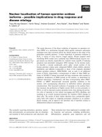
Figure 1
Nuclear export of Dsh is not critical for its activity. (a) The Dsh constructs used to analyze nuclear export. (b-d) RNAs encoding Dsh-GFP, Ds2
and DsNESm (0.5 ng each) were injected into two animal blastomeres of 4-8-cell embryos. Animal-cap explants were excised at stage 10, fixed and
examined for GFP fluorescence. (b) Wild type Dsh-GFP localized in punctate structures of the cytoplasm, whereas (c) Ds2 and (d) DsNESm
accumulated in the nucleus of animal pole cells. (e,f) One ventral vegetal blastomere of 8-cell embryos was injected with 1 ng Dsh-GFP or DsNESm
RNA as indicated. Complete secondary axes were induced in both cases. (g) Uninjected sibling embryos.
GFPDEPPDZDIX
B
Dsh-GFP
Ds2
Ds3
LSL ASA
DsNESm
(a)
Dsh-GFP DsNESm Uninjected
(b) (c) (d)
(e) (f) (g)
Dsh-GFP Ds2 DsNESm
pools revealed only a small amount of endogenous Dvl2 in
intact nuclei, whereas after NEM treatment, Dvl2 was local-
ized predominantly in the nuclear fraction (Figure 3d). The
efficiency of subcellular fractionation was controlled for by
staining with antibodies to glyceraldehyde phosphate dehy-
drogenase (GAPDH) and nuclear lamins. These proteins
remained exclusively cytoplasmic or nuclear, respectively, in
both untreated and NEM-treated cells (Figure 3d). Thus, our
data show that Dsh translocates into the nucleus and is
actively exported into the cytoplasm of both Xenopus ecto-
dermal cells and mammalian fibroblasts.
Identification of sequences responsible for Dsh
nuclear localization
To identify specific amino-acid sequences that direct the
transport of Dsh to the nucleus, we studied the subcellular
distribution of mutated Dsh-GFP fusion constructs
(Figure 4a). The removal of the DIX and PDZ domains
(Ds1) did not eliminate nuclear translocation in response to
NEM or LMB (Figure 4a-d), indicating that these two
domains are not required for the nuclear import. Similarly,
the DEP domain is not required for Dsh nuclear localiza-
tion (Ds2; Figure 1a,c). Comparison of Ds1 and Ds2 (see
Figure 4a), both capable of nuclear localization, reveals a
short stretch of shared amino acids located between the
PDZ and DEP domains. Strikingly, the removal of just this
48 amino-acid region abrogated nuclear import of Dsh in
the presence of NEM or LMB (Ds3; Figures 2e,f and 4a).
Together these experiments identify amino acids 333-381 as
the region required for nuclear localization of Dsh.
Although this short sequence is highly conserved in all Dsh
homologs from Hydra to humans (Figure 4j), it does not
bear detectable similarity to nuclear localization signals
characterized in other proteins [27]. This sequence may
interact directly with components of the nuclear import
machinery or bind to a protein that itself binds a karyo-
pherin/importin and mediates the nuclear import of Dsh by
a piggyback mechanism. Interestingly, this region overlaps a
novel proline-rich domain identified by mutational analysis
of Dsh in Drosophila [28]. To define further the specific
amino acids necessary for nuclear localization, a panel of
Dsh constructs with point mutations spanning the con-
served region was examined (data not shown). Nuclear
import was eliminated with the substitution of three amino
acids, converting IVLT into AVGA (DsNLSm; Figure 4a,e-g,j),
indicating that these three amino acids are critical.
3.4 Journal of Biology 2005, Volume 4, Article 3 Itoh et al. />Journal of Biology 2005, 4:3
Table 1
Axis induction by Dsh constructs
Total number Complete Partial
of injected secondary secondary
Injected RNA embryos axes (%) axes (%)
Experiment 1
Dsh-GFP 150 46.6 25.3
DsNESm-GFP 194 54.6 30.4
Experiment 2
Dsh-GFP 144 28.5 45.1
DsNLSm-GFP 149 0.7 39.5
DsSNLS-GFP 137 24.0 42.3
Embryos were injected as described in Figure 1e,f. Partial secondary
axes are defined by a morphologically visible ectopic neural tube up to
the hindbrain level. Complete axes are defined by the presence of the
secondary head structures, including eyes and cement glands. The
frequency of secondary axes in uninjected embryos was less than 1%.
Data pooled from several independent experiments are shown.
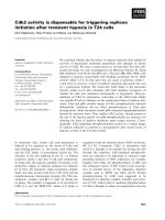
Figure 2
Accumulation of Dsh in the nucleus in the absence of nuclear export.
(a-d) Dsh-GFP RNA (0.7 ng) was injected into two animal blastomeres
of 4-8 cell embryos. Animal caps were excised at stage 10 and then left
(a) untreated or (b) treated with 10 mM NEM or (c,d) 50 ng/ml
leptomycin B (LMB), fixed and examined for GFP fluorescence. (a) Dsh-
GFP is mainly localized to vesicular structures in the cytoplasm. In the
presence of (b) NEM or (c) LMB, Dsh-GFP accumulates in the nucleus,
as supported by (d) DAPI staining of nuclei in the same field as in (c).
Nuclear staining is marked by arrowheads (c,d). (e,f) The Ds3
construct, lacking amino acids 334-381, remained in the cytoplasm in
the (e) absence or (f) presence of NEM.
(a) (b)
(c) (d)
(e) (f)
Dsh Untreated Dsh NEM
DAPI LMB
Ds3 NEMDs3 Untreated
Dsh LMB
Dsh nuclear translocation is crucial for its function in
the -catenin pathway
To determine whether nuclear localization of Dsh is
required for its activity, we compared the abilities of
DsNLSm and wild-type Dsh to induce secondary axes in
frog embryos. We also assessed activation of a luciferase
reporter construct for Siamois [29], an immediate target of
Wnt/-catenin signaling. DsNLSm had impaired ability to
induce secondary axes and to activate the Siamois reporter
when compared with wild-type Dsh (Figure 5a,b; Table 1).
Furthermore, DsNLSm failed to stabilize -catenin
(Figure 5c). This difference was not due to differences in
protein expression, as both constructs were present in
embryo lysates at similar levels (Figure 5c). Thus, these find-
ings indicate that the nuclear localization of Dsh is critical
for its functional activity in the -catenin pathway.
Not only was the function of DsNLSm in the -catenin
pathway impaired, but we found that this construct
behaved as a dominant inhibitor of Wnt signaling and pre-
vented the activation of the Siamois reporter by Xwnt3a and
Xwnt8 RNAs (Figure 6a,b). Consistent with these observa-
tions, another construct lacking the region responsible for
the nuclear localization (Ds3; see Figure 4a) also suppressed
Wnt signaling (Figure 6b). Despite these inhibitory proper-
ties, dorsally injected DsNLSm RNA, like Xdd1, a dominant
negative deletion mutant [24], did not suppress primary
axis formation (data not shown).
Impaired activity of the DsNLSm construct may be due to its
inability to translocate to the nucleus, or due to a coinciden-
tal elimination of a binding site for an essential cofactor that
functions together with Dsh in the cytoplasm. To exclude the
latter possibility, the IVLT sequence of Dsh NLS was replaced
with KKKRK, an unrelated NLS from SV40 T antigen [27].
This construct, DsSNLS, relocated to the nucleus even in the
absence of nuclear export inhibitors (Figure 4a,i). Notably,
all activities of wild-type Dsh, including induction of com-
plete secondary axes, activation of the Siamois promoter and
-catenin stabilization were significantly restored in DsSNLS
(Figure 5a-c; Table 1). In contrast to DsNLSm, DsSNLS did
not inhibit the ability of Wnt ligands to activate pSia-Luc
(Figure 6b), consistent with its being a positive regulator of
the Wnt pathway. We note that the signaling activity of
DsSNLS was not enhanced compared to wild-type Dsh, sug-
gesting that the rate of the nuclear translocation of Dsh
rather than its steady state levels in the nucleus is critical for
target gene activation. It is also possible that other nuclear
components, rather than Dsh, become rate-limiting for sig-
naling. Overall, the simplest interpretation of our data is that
the nuclear import of Dsh is essential for its activity.
We next examined the ability of DsNLSm to bind critical
Wnt signaling components, such as casein kinase 1
(CK1), a positive regulator of the -catenin pathway
[30,31], or Axin, a negative regulator [20,32-36], both of
which are known to bind Dsh. Both DsSNLS, enriched in
the nucleus, and DsNLSm and Ds3, which do not enter the
nucleus, bound CK1 and XARP, a Xenopus Axin-related
protein [20] (Figure 7). Thus, these mutated Dsh constructs
retain the ability to associate with critical components of
the Wnt/-catenin pathway, arguing that defective nuclear
translocation of DsNLSm is likely to be responsible for its
inability to activate -catenin signaling.
Suppression of Dsh nuclear import does not affect
noncanonical signaling
Besides the -catenin pathway, Dsh also functions in a
planar cell polarity (PCP) pathway, which involves Rho
GTPase and JNK and controls morphogenetic movements in
Journal of Biology 2005, Volume 4, Article 3 Itoh et al. 3.5
Journal of Biology 2005, 4:3
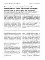
Figure 3
Endogenous Dsh shuttles between the cytoplasm and nucleus.
Immunofluorescent staining of HEK293 cells with anti-Dvl2 antibodies
reveals different subcellular localization of Dvl2 (a) without or (b) with
LMB treatment. (c) DAPI staining shows the location of nuclei in the
same field as (b); the arrowheads indicate corresponding nuclei in (b)
and (c). (d) Distribution of endogenous Dvl2 recognized by anti-Dvl2
antibodies in the nuclear and the cytoplasmic fractions of Rat-1
fibroblasts. In the absence of NEM, Dvl2 is localized mainly in the
cytoplasm (C), while after NEM treatment Dvl2 is exclusively localized
in the nuclei (N). W, whole cell lysate. Antibodies to lamin and GAPDH
show the separation of the nuclear and cytoplasmic fractions.
− NEM + NEM
-
98
119
52
Anti-Dvl2
Anti-lamin
Anti-GAPDH
-
-
MW
WCNWC N
(a) (b)
(d)
(c)
Dvl2 Untreated Dvl2 LMB DAPI LMB
3.6 Journal of Biology 2005, Volume 4, Article 3 Itoh et al. />Journal of Biology 2005, 4:3
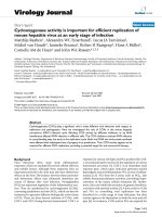
Figure 4
Mapping nuclear localization signals in Dsh. (a) The Dsh constructs used to study nuclear transport and their localization to the nucleus after NEM
or LMB treatment; the DIX, PDZ and DEP domains are shown as in Figure 1a; B is the basic region and nd denotes not done. (b-i) Subcellular
localization of Dsh-GFP constructs in the absence or presence of NEM or LMB. Embryos were injected with 0.5 ng of each mRNA, and GFP analysis
was carried out as in Figure 1b-d. (b-d) Ds1, (e-g) DsNLSm, (h) Dsh, (i) DsSNLS. (b,e,i) no NEM treatment; (c,f) after NEM treatment; (d,g,h) after
LMB treatment. (j) Comparison of conserved amino-acid sequences that are required for Dsh nuclear localization; X denotes the Xenopus protein,
m the mouse and h the human. Amino-acid residues mutated in DsNLSm are indicated by asterisks.
GFPDEPPDZDIX B
IVLT AVGA
IVLT
KKKRK
Xdsh
***
mDvl1
mDvl2
mDvl3
hDsh2
Dsh
Hydra Dsh
(a)
(j)
Dsh-GFP
Ds1
Ds3
DsNLSm
DsSNLS
Nuclear localization
+ NEM
++/−
+
+
+ LM B
+
++/−
+
−
Ds2
nd
+
+
nd
−
−
−
−
−
−
Ds1 Untreated
DsNLSm NEM DsNLSm LMB Dsh LMB
DsNLSm Untreated
DsSNLS Untreated
Ds1 NEM Ds1 LMB
(b) (c) (d) (e)
(f) (g) (h) (i)
P
P
P
P
P
P
P
I
I
I
I
I
I
I
V
S
V
T
V
K
M
L
L
L
L
L
L
L
T
T
T
T
T
V
T
V
V
V
V
V
V
V
A
A
A
A
A
A
A
K
K
K
K
K
K
K
C
C
C
C
C
C
C
W
W
W
W
W
W
W
D
D
G
D
D
D
D
P
P
P
P
P
P
P
S
T
S
S
S
N
N
P
P
P
P
P
P
P
Q
R
Q
R
Q
K
K
G
S
A
G
A
G
G
Y
Y
Y
C
Y
Y
Y
F
F
F
F
F
F
F
T
T
T
T
T
T
T
L
I
L
L
L
I
V
P
P
P
P
P
P
P
R
R
R
R
R
R
R
N
A
N
S
N
T
N
E
D
E
E
E
E
D
P
P
P
P
P
P
V
I
V
I
I
I
V
T
H
R
Q
R
Q
R
R
P
P
P
P
P
P
P
I
I
I
I
I
I
I
D
D
D
D
D
D
D
P
P
P
P
P
P
P
A
A
A
A
A
G
A
A
A
A
A
A
A
A
W
W
W
W
W
W
W
V
L
V
V
V
V
M
S
S
S
S
S
A
Q
H
H
H
H
H
H
H
S
T
S
T
S
T
S
A
A
A
A
A
Q
E
A
A
A
A
A
A
A
L
L
L
M
L
L
V
early embryos [8,9,37-39]. We asked whether mutations in
DsNLSm influence the -catenin pathway exclusively or
affect the PCP pathway as well. First, we observed that both
Dsh-GFP and DsNLSm-GFP were efficiently recruited to the
cell membrane by overexpressed Xfz8, a Frizzled family
member [40] (Figure 8a). As Dsh relocalization to the cell
membrane in response to Frizzled is associated with its
ability to signal in the PCP pathway [7,8], this observation
suggests that DsNLSm can respond to Frizzled signaling
independent of -catenin.
In Xenopus, the PCP pathway involving Dsh is implicated in
the control of convergent extension movements [24,41,42].
Overexpression of the Xdd1 deletion mutant leads to the
development of short embryos when expressed in dorsal
marginal cells ([24]; Figure 8b). Severe convergent extension
defects (Figure 8b) were observed in 22%, and mild defects
were observed in 28% of the embryos injected with Xdd1
RNA (N = 35). In contrast, only mild morphogenetic
defects were observed in embryos coinjected with Dsh
(15%; N = 40) or DsNLSm RNA (18%; N = 39), indicating
that both Dsh and DsNLSm partially rescued the effect of
Xdd1. This indicates that DsNLSm is active in noncanonical
PCP-like signaling. We also examined whether DsNLSm
activates c-Jun N-terminal kinase (JNK), which is thought to
function downstream of Dsh in the PCP pathway [8,37-39].
Both DsNLSm and Dsh activated JNK at equivalent levels
(Figure 8c), suggesting that nuclear localization of Dsh is
not required for its function in noncanonical signaling.
Nuclear accumulation of Dsh following Wnt3a
stimulation
Our findings are consistent with a scenario in which Wnt
signaling may cause nuclear translocation of Dsh followed
Journal of Biology 2005, Volume 4, Article 3 Itoh et al. 3.7
Journal of Biology 2005, 4:3
(a)
(b)
(c)
Relative luciferase units (x 10
3
)
Dsh
DsNLSm
DsSNLS
∆RGS-Axin
−
Flag-β-catenin
Uninjected
Anti-β-tubulin
Anti-Xdsh
Anti-flag
+++++−
Dsh DsNLSm
DsSNLS Uninjected
0
20
40
60
80
100
120
140
Dsh
DsSNLS
No RNA
DsNLSm
Figure 5
Activation of the Wnt/-catenin pathway requires nuclear localization
of Dsh. (a) Axis-inducing activity of Dsh constructs. One ventral
vegetal blastomere of 8-cell embryos was injected with 1 ng Dsh-GFP,
DsNLSm, or DsSNLS mRNA as indicated. Uninjected sibling embryos
are also shown. (b) Activation of the Siamois reporter gene. The
reporter -833pSia-Luc plasmid (20 pg) was coinjected with Dsh-GFP,
DsNLSm or DsSNLS mRNA (0.5 ng each) into a single animal ventral
blastomere of 8-cell embryos. Injected embryos were lysed at stage
10+ for luciferase activity determination. Results are shown in relative
light units as the mean +/- standard deviation from triplicate samples.
(c) Requirement for Dsh NLS for the stabilization of -catenin. Flag--
catenin mRNA (0.4 ng) was coinjected with Dsh, DsNLSm, DsSNLS or
⌬RGS-Axin mRNA (2 ng each) into four animal blastomeres of 4-8-cell
embryos. Levels of -catenin and Dsh constructs were assessed in
stage 10 embryo lysates with anti-Flag antibodies and anti-Xdsh
antibodies; -tubulin serves as a loading control. Dsh and DsSNLS, but
not DsNLSm, are able to stabilize -catenin. ⌬RGS-Axin was used as a
control for an activator of the Wnt pathway.
by formation of a stable -catenin/Tcf3 complex and tran-
scriptional activation of target genes. In support of this
hypothesis, Dsh was reported to move to the nucleus in
response to Wnt signaling in primary embryonic kidney
cells [17]. In Rat-1 cells, we did not detect a significant
change in Dsh distribution in response to Wnt signals (data
not shown), possibly due to highly efficient nuclear export
of Dsh in these cells. But immunofluorescence staining for
Dvl2 revealed the nuclear accumulation of the protein in
HEK293 and MCF7 cells after 3-6 h stimulation with
Wnt3a-containing medium (Figure 9a, and data not
shown). The effect was quantified by measuring nuclear to
cytoplasmic (N/C) ratios of fluorescence intensity. The N/C
ratio averaged 28% after 6 h treatment with the control
medium, but increased to 91% after stimulation with
Wnt3a-conditioned medium (Figure 9b). These observa-
tions are consistent with the view that Dsh regulates Wnt-
dependent gene targets in the nucleus.
A role for Dsh in the nucleus
In the current view, Wnt signaling causes inactivation of the
-catenin degradation complex, leading to stabilization and
nuclear translocation of -catenin [3]. Given that Dsh is
genetically upstream of the -catenin degradation complex
[3,4] and that -catenin degradation is thought to occur in
the cytoplasm [43], Dsh nuclear import is unexpected. Never-
theless, our data demonstrate that Dsh shuttles between the
cytoplasm and the nucleus and that its presence in the
nucleus is critical for signaling. One explanation of these
results is that -catenin degradation may occur in the
nucleus. Consistent with this possibility, APC, Axin and
GSK3, components of the -catenin degradation complex,
have also recently been found to shuttle between the cyto-
plasm and the nucleus [22,23,44-47]. Moreover, Frat/GBP,
a positive regulator of -catenin, has been reported to expel
GSK3 from the nucleus [47]. We show that the ability of
Dsh constructs to enter the nucleus correlates with their
ability to stabilize -catenin (Figure 5c). These observations
indicate that Wnt/-catenin signaling may depend on the
nuclear localization of pathway components.
Alternatively, nuclear localization of Dsh may affect
-catenin stability indirectly, by regulating protein interac-
tions that sequester -catenin in the nucleus, thereby pre-
venting its cytoplasmic degradation [48]. Although we did
not detect a significant change in nuclear import of
-catenin-GFP in Xenopus ectoderm cells overexpressing
Dsh (data not shown), this process may be cell-context-
dependent. On the other hand, we recently showed that
Frodo, a nuclear Dsh-interacting protein, associates with
Tcf3 and influences Tcf3-dependent transcription [49]. It
is thus possible that Frodo links Tcf3 and Dsh to regulate
3.8 Journal of Biology 2005, Volume 4, Article 3 Itoh et al. />Journal of Biology 2005, 4:3
Figure 6
Dominant inhibition of Wnt-dependent transcription by Dsh mutants.
Eight-cell embryos were injected (a) in one animal ventral blastomere
or (b) in one vegetal ventral blastomere with -833pSia-Luc DNA (20
pg), mRNAs encoding Xwnt3a (5 pg) or Xwnt8 (2 pg), and Dsh-GFP,
DsNLSm, Ds3 or DsSNLS mRNA (0.5 ng) as indicated. Luciferase
activity was measured as described in Figure 5b.
(a)
Relative luciferase units (x 10
3
)
Relative luciferase units (x 10
3
)
(b)
Xwnt3a
Xwnt3a + Dsh
Xwnt3a + DsNLSm
No RNA
Xwnt8 + DsNLSm
Xwnt8 + DsSNLS
Xwnt8 + Ds3
Xwnt8
1000
2000
3000
4000
0
400
800
1200
1600
0
Figure 7
Dsh mutants retain the ability to bind CK1 and XARP. Four-cell
embryos were injected in four sites in the animal hemisphere with
CK1, HA-XARP, Myc-tagged Dsh, DsNLSm, Ds3 or DsSNLS RNA
alone (2 ng each) or in combinations as indicated. The embryonic
lysates were collected at stage 10.5 for immunoprecipitation with anti-
Myc antibodies. Co-immunoprecipitated (a) CK1 or (b) HA-XARP
was probed with anti-CK1 or anti-HA antibodies; -tubulin served as
a loading control.
IP: Anti-Myc Lysates
IP: Anti-Myc Lysates
Blot:
Anti-CK1ε
Anti-Myc
Anti-β-tubulin
Blot:
Anti-HA
Anti-Myc
Anti-β-tubulin
HA-XARP
Myc-DsNLSm
Myc-DsSNLS
Myc-Ds3
CK1ε
MycDsh
MycDsNLSm
MycDsSNLS
+
−
−
−
+
+
−
−
+
−
+
−
+
−
−
+
−
−
−
−
+
−
−
−
+
+
−
−
+
−
+
−
+
−
−
+
−
−
−
−
+
−
−
−
+
+
−
−
+
−
+
−
+
−
−
+
−
−
−
−
+
−
−
−
+
+
−
−
+
−
+
−
+
−
−
+
(a)
(b)
Wnt target genes. Future studies should examine molecular
components critical for the nuclear function of Dsh.
Materials and methods
DNA constructs
GFP-tagged Dsh constructs were all derived from the
DshGFP-RN3 plasmid that encodes the Xdsh protein fused
at amino acid 724 to the first amino acid of GFP (Figures
1a, 4a). Ds1 lacks the first 332 amino-terminal amino acids.
Ds2 is the carboxy-terminal deletion of Xdsh, starting with
amino acid 383. Ds3 lacks amino acids 334-381. In
DsNLSm, the IVLT residues at positions 334-337 were
replaced with AVGA, whereas in DsSNLS the same region is
replaced with KKKRK, the SV40 T antigen NLS [27]. In
DsNESm, L513 and L515 were substituted for alanines.
To generate these constructs, DshGFP-pRN3 was used as a
template. The in-frame deletion in Ds3 was made by PCR.
Other GFP fusion constructs were synthesized with specific
primers and PfuI DNA polymerase followed by DpnI diges-
tion of the template [50]. The following primers were used:
5’-GTCCATAAACCGGGGCCCGCAGTCGGCGCCGTGGCC-
AAATGCTGG-3’ for DsNLSm; 5’-ACACTAGGCCGCAGAATG-
CCCATTGTCCTGACCGTG-3’ for Ds1; 5’-TCCATAAACCGG-
GGCCAAAGAAGAAGCGAAAGGTGGCCAAATGCTGGGA-3’
for DsSNLS; 5’-TTCCCAGTGTACCCCGGGGCCATGGTGA-
GCAAGGGC-3’ for Ds2, and 5’-GAGAACTATGACCAAC-
GCTAGCGCGAATGACAACGATGGAT-3’ for DsNESm. All
constructs were verified by sequencing. Myc-tagged Dsh
mutant constructs were made by replacing mutated regions
with corresponding regions of Myc-Dsh [24]. Cloning
details are available as an Additional data file with the
online version of this article.
Journal of Biology 2005, Volume 4, Article 3 Itoh et al. 3.9
Journal of Biology 2005, 4:3
Anti-phospho-c-Jun
Anti-GST
Anti-Dvl2
Dsh
DsNLSm
Uninjected
Xdd1 Xdd1 + Dsh
Xdd1 + DsNLSm Uninjected
DsNLSm + Fz8DsNLSm
Anti-β-tubulin
(a)
(b)
(c)
Dsh + Fz8Dsh
Figure 8
DsNLSm, defective in the -catenin pathway, is active in noncanonical
signaling. (a) Fz8-dependent recruitment of Dsh-GFP constructs to the
cell membrane. Dsh-GFP or DsNLSm RNA (0.5 ng) was injected alone
or with Fz8 RNA (1 ng) into two animal blastomeres at the 4-8-cell stage.
GFP fluorescence was assessed in animal cap explants as in Figure 1b-d.
Both Dsh and DsNLSm are efficiently recruited to the cell membrane by
Fz8. Arrowheads point to cell membranes. (b) DsNLSm can rescue
convergent extension defects caused by Xdd1. Four-cell embryos were
injected with 0.6 ng Xdd1 RNA alone or together with 2 ng Dsh-GFP or
DsNLSm RNA into two vegetal dorsal blastomeres. The injected
embryos were allowed to develop until the sibling embryos reached
stage 32. (c) Activation of JNK by the Dsh nuclear import mutant. Four
animal blastomeres of four-cell embryos were each injected with 1 ng of
RNAs encoding Dsh-GFP or DsNLSm. Embryonic lysates were collected
at stage 10.5 for in vitro JNK activity assay using anti-phospho-specific c-
Jun antibodies. Total GST-c-Jun levels were assessed with anti-GST
antibodies. Dsh-GFP and DsNLSm were equally expressed, as monitored
with anti-Dvl2 antibodies; -tubulin served as a loading control.
Embryo culture, axis-induction assay and axis-
extension assay
In vitro fertilization, culture and microinjections of Xenopus
eggs were essentially as described previously [24]. Stages
were determined according to Nieuwkoop and Faber [51].
Axis induction was carried out by injecting mRNAs encoding
different Dsh constructs (1 ng) into a single vegetal ventral
blastomere at the 4-8-cell stage and assessed when the
injected embryos reached stage 36-40. To monitor axis
extension defects, 0.6 ng of Xdd1 RNA was injected alone or
with 2 ng of Dsh or DsNLSm RNA into two dorsovegetal
blastomeres of 4-cell embryos and the injected embryos were
allowed to develop until sibling embryos reached stage 32.
GFP fluorescence and luciferase assay
For subcellular localization of Dsh-GFP constructs, mRNAs
were injected into the animal pole region of 2-4-cell
embryos. Animal cap explants were dissected at stages 9-10.5,
incubated for 60 min in 10 mM N-ethylmaleimide (NEM;
Sigma, St Louis USA) in 0.8 ϫ MMR (Marc’s Modified
Ringer’s solution, 1 ϫ MMR: 100 mM NaCl, 2 mM KCl,
1 mM MgCl2, 2 mM CaCl2, 5 mM HEPES, pH 7.4), or in
control (0.8 ϫ MMR), then fixed in 4% paraformaldehyde in
phosphate-buffered saline (PBS) for 30-45 min, washed
three times in PBS, and mounted in 70% glycerol, 30% PBS
containing 25 mg/ml of diazabicyclo(2,2,2)-octane (Sigma).
Leptomycin B was used at 50 ng/ml in low-calcium medium
(76 mM NaCl, 1.4 mM KCl, 0.2 mM CaCl
2
, 0.1 mM MgCl
2
,
0.5 mM Hepes, 1.2 mM sodium phosphate, (pH 7.5),
0.6 mM NaHCO
3
and 0.06 mM EDTA) for one hour prior to
fixation. In some experiments, nuclei were stained by addi-
tion of 1 g/ml 4,6-diamidino-2-phenylindole (DAPI) to the
final PBS wash. For membrane localization studies, Xfz8
RNA was coinjected with RNAs encoding the Dsh constructs
in the animal-pole region; animal-cap explants were dis-
sected at stage 9-9.5 and mounted for observation. Fluores-
cence was visualized using a Zeiss Axiophot microscope.
For luciferase assays, pSiaLuc reporter plasmid (20-40 pg) was
coinjected with mRNAs encoding Xwnt3a [52] or Xwnt8 [53]
and different Dsh constructs into one or two animal-ventral
blastomeres or into one ventral-vegetal blastomere at the 4-8-
cell stage. Luciferase activity was measured as described [29].
Tissue culture, immunocytochemistry and
subcellular fractionation
Rat-1 fibroblasts, human embryonic kidney (HEK) 293 cells
and MCF7 human breast carcinoma cells were cultured in
1 ϫ Dulbecco’s Modified Eagle Medium (DMEM; Gibco/
Invitrogen, Carlsbad, USA) supplemented with 10% fetal
calf serum and 5 g/ml gentamicin. Conditioned medium
was prepared from L cells stably transfected with Wnt3a as
described [54], with the medium from untransfected L cells
serving as a control.
For immunocytochemistry, HEK293 cells were treated with
50 ng/ml LMB for 14 h while MCF7 cells were treated with
3.10 Journal of Biology 2005, Volume 4, Article 3 Itoh et al. />Journal of Biology 2005, 4:3
Figure 9
Nuclear translocation of Dvl2 upon Wnt3a treatment. (a) MCF7 cells
were treated either with Wnt3a-conditioned or control medium for 6 h,
fixed and immunostained with anti-Dvl2 antibodies. In control cells,
cytoplasmic and perinuclear staining is visible. Wnt3a-conditioned, but not
control, medium enhanced nuclear translocation of Dvl2. DAPI staining
indicates the position of cell nuclei. Corresponding cells are shown by
arrowheads. (b) Nuclear/cytoplasmic (N/C) ratios of fluorescence were
calculated for each panel in (a) as the mean +/- standard deviation.
(a)
Anti-Dvl2
Untreated
Anti-Dvl2 Wnt3a CM
Anti-Dvl2 Control CM
DAPI
Untreated
DAPI Wnt3a CM
DAPI Control CM
Untreated
Wnt3a CM
Control CM
0
20
40
N/C ratio of
fluorescence (%)
60
80
100
(b)
Wnt3a or control conditioned medium for 1, 3, 6 or 8 h.
Cells were fixed with 4% paraformaldehyde, immersed in
methanol, and incubated with anti-Dvl2 antibodies and then
Cy3-conjugated anti-rabbit IgG. Nuclei were stained by addi-
tion of 1 g/ml DAPI as described for animal-cap cells. Fluor-
escence was observed under the Zeiss Axiophot microscope;
10-15 cells from each group were randomly picked up for
measurement of the nuclear and cytoplasmic staining inten-
sity using Image-Gauge software (Fuji Film, Tokyo, Japan).
For subcellular fractionation, confluent cultures of Rat-1
cells were harvested by scraping plates and resuspended in
hypotonic lysis buffer containing 1 mM EGTA, 1 mM EDTA,
2 mM MgCl
2
, 10 mM KCl, 1 mM DTT, 10 mM -glycero-
phosphate, 1 mM sodium orthovanadate, 1 g/ml leu-
peptin, 1 g/ml aprotinin, and 1 g/ml pepstatin. Cells
were swollen for 30 min, and broken open with 25 strokes
in a tight fitting Dounce homogenizer. Lysates were layered
into tubes containing 1 M sucrose in hypotonic lysis buffer,
and spun at 1600 ϫ g for 10 min. Supernatant remaining
above the sucrose cushion was used as the cytoplasmic frac-
tion. The pellet, containing nuclei, was resuspended in an
equivalent volume of hypotonic lysis buffer.
Immunoprecipitation and western blotting
Immunoprecipitation and western analysis were carried out
with cell and embryo lysates as described [14]. To prepare
embryo lysates at stage 10+, four animal blastomeres of
4-8-cell embryos were injected with RNAs encoding different
forms of Dsh, ⌬RGS-Axin [32], Flag--catenin [55], CK1
[30] and HA-XARP [20]. To generate anti-Xdsh polyclonal
antibodies, rabbits were immunized with a carboxy-terminal
half of Xdsh (amino acids 301-736) fused to GST. First, GST
beads were used for purification of anti-GST antibodies.
Subsequently anti-Xdsh antibodies were affinity-purified on
GST-Xdsh (301-736) beads. Polyclonal anti-Dvl2 antibody
was generated in rabbits and affinity-purified on PVDF
membrane blotted with human Dvl2 (79-249) [56]. A small
aliquot of anti-human Dvl2 was obtained from M. Snyder
(Yale University, New Haven, USA). Anti-GAPDH antibody
was a gift from A. Stuart-Tilley and S. Alper (Beth Israel
Deaconess Medical Center, Boston, USA), anti-lamin anti-
body was from F. McKeon (Harvard Medical School,
Boston, USA). Anti--tubulin antibodies were from Bio-
Genex (San Ramon, USA), anti-Flag M2 antibody was from
Sigma and anti-CK1 antibodies were from BD Biosciences
(Palo Alto, USA). Anti-Myc and anti-HA monoclonal anti-
bodies are hybridoma supernatants of 9E10 and 12CA5
cells (Roche Applied Science, Indianapolis, USA).
JNK assay
Four-cell embryos were injected with 4 ng Dsh or
DsNLSm RNA into four animal blastomeres. Embryo
lysates were prepared at stage 10.5 and in vitro kinase
assays were carried out essentially as described [57],
except that phosphorylated c-Jun-GST was detected with
anti-phospho-c-Jun-specific antibodies (Cell Signaling
Technology, Beverly, USA) by western blotting rather than
with autoradiography.
Additional data files
The following is provided as an additional data file with the
online version of this article. Additional data file 1, contain-
ing cloning details of Dsh mutant constructs.
Acknowledgements
We thank S. Alper, F. McKeon and M. Snyder for antibodies, and X. He,
F. Costantini and J. Graff for plasmids, J. Kitajewsky for Rat-1 cells,
R. Nusse for L cells transfected with Wnt3a, and J. Martinez, Y. Yoneda
and M. Yoshida for leptomycin B. We also thank V. Krupnik and M.
Lisovsky for help with the generation of anti-Xdsh and anti-Dvl2 anti-
bodies. We are grateful to J. Green, V. Krupnik, B. Neel, N. Perrimon
and members of this laboratory for reading of the manuscript and useful
discussions. This work was supported by NIH grants to S.Y.S.
References
1. Massagué J, Wotton D: Transcriptional control by the
TGF-

/Smad signaling system. EMBO J 2000, 19:1745-1754.
2. Struhl G, Adachi A: Nuclear access and action of Notch
in vivo. Cell 1998, 93:649-660.
3. Peifer M, Polakis P: Wnt signaling in oncogenesis and
embryogenesis - a look outside the nucleus. Science 2000,
287:1606-1609.
4. Wodarz A, Nusse R: Mechanisms of Wnt signaling in devel-
opment. Annu Rev Cell Dev Biol 1998, 14:59-88.
5. Bienz M, Clevers H: Linking colorectal cancer to Wnt signal-
ing. Cell 2000, 103:311-320.
6. Gumbiner BM: Carcinogenesis: a balance between

-catenin
and APC. Curr Biol 1997, 7:R443-R446.
7. Axelrod JD, Miller JR, Shulman JM, Moon RT, Perrimon N: Differ-
ential recruitment of Dishevelled provides signaling speci-
ficity in the planar cell polarity and Wingless signaling
pathways. Genes Dev 1998, 12:2610-2622.
8. Boutros M, Paricio N, Strutt DI, Mlodzik M: Dishevelled acti-
vates JNK and discriminates between JNK pathways in
planar polarity and wingless signaling. Cell 1998, 94:109-118.
9. Habas R, Kato Y, He X: Wnt/Frizzled activation of Rho regu-
lates vertebrate gastrulation and requires a novel Formin
homology protein Daam1. Cell 2001, 107:843-854.
10. Sheldahl LC, Park M, Malbon CC, Moon RT: Protein kinase C is
differentially stimulated by Wnt and Frizzled homologs in
a G-protein-dependent manner. Curr Biol 1999, 9:695-698.
11. Sokol SY: A role for Wnts in morphogenesis and tissue
polarity. Nat Cell Biol 2000, 2:E124-E126.
12. Winklbauer R, Medina A, Swain RK, Steinbeisser H: Frizzled-7
signalling controls tissue separation during Xenopus gas-
trulation. Nature 2001, 413:856-860.
13. Boutros M, Mlodzik M: Dishevelled: at the crossroads of
divergent intracellular signaling pathways. Mech Dev 1999,
83:27-37.
14. Gloy J, Hikasa H, Sokol SY: Frodo interacts with Dishevelled
to transduce Wnt signals. Nat Cell Biol 2002, 4:351-357.
15. Ciani L, Krylova O, Smalley MJ, Dale TC, Salinas PC: A divergent
canonical WNT-signaling pathway regulates microtubule
dynamics: Dishevelled signals locally to stabilize micro-
tubules. J Cell Biol 2003, 164:243-253.
Journal of Biology 2005, Volume 4, Article 3 Itoh et al. 3.11
Journal of Biology 2005, 4:3
16. Capelluto DGS, Kutateladze TG, Habas R, Finkielstein CV, He X,
Overduin M: The DIX domain targets dishevelled to actin
stress fibres and vesicular membranes. Nature 2002,
419:726-729.
17. Torres MA, Nelson WJ: Colocalization and redistribution of
Dishevelled and Actin during Wnt-induced mesenchymal
morphogenesis. J Cell Biol 2000, 149:1433-1442.
18. Rothbächer U, Laurent MN, Deardorff MA, Klein PS, Cho KWY,
Fraser SE: Dishevelled phosphorylation, subcellular localiza-
tion and multimerization regulate its role in early
embryogenesis. EMBO J 2000, 19:1010-1022.
19. Yang-Snyder J, Miller JR, Brown JD, Lai C-J, Moon RT: A frizzled
homolog functions in a vertebrate Wnt signaling pathway.
Curr Biol 1996, 6:1302-1306.
20. Itoh K, Antipova A, Ratcliffe MJ, Sokol S: Interaction of Dishev-
elled and Xenopus Axin-related protein is required for
Wnt signal transduction. Mol Cell Biol 2000, 20:2228-2238.
21. Yanagawa S, van Leeuwen F, Wodarz A, Klingensmith J, Nusse R:
The Dishevelled protein is modified by Wingless signaling
in Drosophila. Genes Dev 1995, 9:1087-1097.
22. Henderson BR: Nuclear-cytoplasmic shuttling of APC regu-
lates

-catenin subcellular localization and turnover. Nat
Cell Biol 2000, 2:653-660.
23. Rosin-Arbesfeld R, Townsley F, Bienz M: The APC tumour sup-
pressor has a nuclear export function. Nature 2000,
406:1009-1012.
24. Sokol SY: Analysis of Dishevelled signalling pathways during
Xenopus development. Curr Biol 1996, 6:1456-1467.
25. Kudo N, Matsumori N, Taoka H, Fujiwara D, Schreiner EP, Wolff
B, Yoshida M, Horinouchi S: Leptomycin B inactivates
CRM1/exportin 1 by covalent modification at a cysteine
residue in the central conserved region. Proc Natl Acad Sci
USA 1999, 96:9112-9117.
26. Holaska JM, Paschal BM: A cytosolic activity distinct from
Crm1 mediates nuclear export of protein kinase
inhibitor in permeabilized cells. Proc Natl Acad Sci USA 1998,
95:14739-14744.
27. Jans DA, Xiao C-Y, Lam MHC: Nuclear targeting signal recog-
nition: a key control point in nuclear transport? BioEssays
2000, 22:532-544.
28. Penton A, Wodarz A, Nusse R: A mutational analysis of
dishevelled in Drosophila defines novel domains in the
Dishevelled protein as well as novel suppressing alleles of
axin. Genetics 2002, 161:747-762.
29. Fan MJ, Grüning W, Walz G, Sokol SY: Wnt signaling and tran-
scriptional control of Siamois in Xenopus embryos. Proc Natl
Acad Sci USA 1998, 95:5626-5631.
30. Peters JM, McKay RM, McKay JP, Graff JM: Casein kinase 1
transduces Wnt signals. Nature 1999, 401:345-350.
31. Sakanaka C, Leong P, Xu L, Harrison SD, Williams LT: Casein
kinase 1 in the Wnt pathway: regulation of -catenin
function. Proc Natl Acad Sci USA 1999, 96:12548-12552.
32. Zeng L, Fagotto F, Zhang T, Hsu W, Vasicek TJ, Perry WL, Lee JJ,
Tilghman SM, Gumbiner BM, Costantini F: The mouse Fused
locus encodes Axin, an inhibitor of the Wnt signaling
pathway that regulates embryonic axis formation. Cell
1997, 90:181-192.
33. Kishida S, Yamamoto H, Hino S, Ikeda S, Kishida M, Kikuchi A:
DIX domains of Dvl and Axin are necessary for protein
interactions and their ability to regulate -catenin stability.
Mol Cell Biol 1999, 19:4414-4422.
34. Smalley MJ, Sara E, Paterson H, Naylor S, Cook D, Jayatilake H,
Fryer LG, Hutchinson L, Fry MJ, Dale TC: Interaction of Axin
and Dvl2 proteins regulates Dvl-2-stimulated TCF-
dependent transcription. EMBO J. 1999, 18:2823-2835.
35. Li L, Yuan H, Weaver CD, Mao J, Far III GH, Sussman DJ, Jonkers
J, Kimelman D, Wu D: Axin and Frat1 interact with Dvl and
GSK, bridging Dvl to GSK in Wnt-mediated regulation of
LEF-1. EMBO J 1999, 18:4233-4240.
36. Salic A, Lee E, Mayer L, Kirschner MW: Control of -catenin
stability: reconstitution of the cytoplasmic steps of the Wnt
pathway in Xenopus egg extracts. Mol Cell 2000, 5:523-532.
37. Li L, Yuan H, Xie W, Mao J, Caruso AM, McMahon A, Sussman DJ,
Wu D: Dishevelled proteins lead to two signaling path-
ways. Regulation of LEF-1 and c-Jun N-terminal kinase in
mammalian cells. J Biol Chem 1999, 274:129-134.
38. Moriguchi T, Kawachi K, Kamakura S, Masuyama N, Yamanaka H,
Matsumoto K, Kikuchi A, Nishida E: Distinct domains of
mouse Dishevelled are responsible for the c-Jun N-termi-
nal kinase/stress-activated protein kinase activation and
the axis formation in vertebrates. J Biol Chem 1999,
274:30957-30962.
39. Habas R, Dawid IB, He X: Coactivation of Rac and Rho by
Wnt/Frizzled signaling is required for vertebrate gastrula-
tion. Genes Dev 2003, 17:295-309.
40. Itoh K, Jacob J, Sokol SY: A role for Xenopus Frizzled 8 in
dorsal development. Mech Dev 1998, 74:145-157.
41. Tada M, Smith JC: Xwnt11 is a target of Xenopus Brachyury:
regulation of gastrulation movements via Dishevelled, but
not through the canonical Wnt pathway. Development 2000,
127:2227-2238.
42. Wallingford JB, Rowning BA, Vogeli KM, Rothbächer U, Fraser SE,
Harland RM: Dishevelled controls cell polarity during
Xenopus gastrulation. Nature 2000, 405:81-85.
43. Wiechens N, Fagotto F: CRM1- and Ran-independent nuclear
export of -catenin. Curr Biol 2001, 11:18-27.
44. Wiechens N, Heinle K, Englmeier L, Schohl A, Fagotto F: Nucleo-
cytoplasmic shuttling of Axin, a negative regulator of the
Wnt--catenin pathway. J Biol Chem 2004, 279:5263-5267.
45. Cong F, Varmus H: Nuclear-cytoplasmic shuttling of Axin
regulates subcellular localization of -catenin. Proc Natl
Acad Sci USA 2004, 101:2882-2887.
46. Diehl JA, Cheng M, Roussel MF, Sherr CJ: Glycogen synthase
kinase-3 regulates cyclin D1 proteolysis and subcellular
localization. Genes Dev 1998, 12:3499-3511.
47. Franca-Koh J, Yeo M, Fraser E, Young N, Dale TC: The regula-
tion of glycogen synthase kinase-3 nuclear export by
Frat/GBP. J Biol Chem 2002, 277:43844-43848.
48. Lee E, Salic A, Kirschner MW: Physiological regulation of
-catenin stability by Tcf3 and CK1. J Cell Biol 2001,
154:983-993.
49. Hikasa H, Sokol SY: The involvement of Frodo in TCF-
dependent signaling and neural tissue development.
Development 2004, 131:4725-4734.
50. Makarova O, Kamberov E, Margolis B: Generation of deletion
and point mutations with one primer in a single cloning
step. BioTechniques 2000, 29:970-972.
51. Nieuwkoop PD, Faber J: Normal table of Xenopus laevis (Daudin).
2nd edition. Amsterdam: North Holland; 1967.
52. Wolda SL, Moody CJ, Moon RT: Overlapping expression of
Xwnt3A and Xwnt1 in neural tissue of Xenopus laevis
embryos. Dev Biol 1993, 155:46-57.
53. Sokol S, Christian JL, Moon RT, Melton DA: Injected Wnt RNA
induces a complete body axis in Xenopus embryos. Cell
1991, 67:741-752.
54. Willert K, Shibamoto S, Nusse R: Wnt-induced dephosphory-
lation of Axin releases -catenin from the Axin complex.
Genes Dev 1999, 13:1768-1773.
55. Liu C, Kato Y, Zhang Z, Do VM, Yanker BA, He X: -Trcp
couples -catenin phosphorylation-degradation and regu-
lates Xenopus axis formation. Proc Natl Acad Sci USA 1999,
96:6273-6278.
56. Semënov MV, Snyder M: Human Dishevelled genes consti-
tute a DHR-containing multigene family. Genomics 1997,
42:302-310.
57. Lysovsky M, Itoh K, Sokol SY: Frizzled receptors activate a
novel JNK-dependent pathway that may lead to apoptosis.
Curr Biol 2002, 12:53-58.
58. Sokol SY, Klingensmith J, Perrimon N, Itoh K: Dorsalizing and neu-
ralizing properties of Xdsh, a maternally expressed Xenopus
homolog of dishevelled. Development 1995, 121:1637-1647.
59. Lemaire P, Garrett N, Gurdon JB: Expression cloning of
Siamois, a Xenopus homeobox gene expressed in dorsal-
vegetal cells of blastulae and able to induce a complete
secondary axis. Cell 1995, 81:85-94.
3.12 Journal of Biology 2005, Volume 4, Article 3 Itoh et al. />Journal of Biology 2005, 4:3