Chapter 127. Treatment and Prophylaxis of Bacterial Infections (Part 2) potx
Bạn đang xem bản rút gọn của tài liệu. Xem và tải ngay bản đầy đủ của tài liệu tại đây (81.7 KB, 7 trang )
Chapter 127. Treatment and Prophylaxis
of Bacterial Infections
(Part 2)
Inhibition of Cell-Wall Synthesis
One major difference between bacterial and mammalian cells is the
presence in bacteria of a rigid wall external to the cell membrane. The wall
protects bacterial cells from osmotic rupture, which would result from the cell's
usual marked hyperosmolarity (by up to 20 atm) relative to the host environment.
The structure conferring cell-wall rigidity and resistance to osmotic lysis in both
gram-positive and gram-negative bacteria is peptidoglycan, a large, covalently
linked sacculus that surrounds the bacterium. In gram-positive bacteria,
peptidoglycan is the only layered structure external to the cell membrane and is
thick (20–80 nm); in gram-negative bacteria, there is an outer membrane external
to a very thin (1-nm) peptidoglycan layer.
Chemotherapeutic agents directed at any stage of the synthesis, export,
assembly, or cross-linking of peptidoglycan lead to inhibition of bacterial cell
growth and, in most cases, to cell death. Peptidoglycan is composed of (1) a
backbone of two alternating sugars, N-acetylglucosamine and N-acetylmuramic
acid; (2) a chain of four amino acids that extends down from the backbone (stem
peptides); and (3) a peptide bridge that cross-links the peptide chains.
Peptidoglycan is formed by the addition of subunits (a sugar with its five attached
amino acids) that are assembled in the cytoplasm and transported through the
cytoplasmic membrane to the cell surface. Subsequent cross-linking is driven by
cleavage of the terminal stem-peptide amino acid.
Virtually all the antibiotics that inhibit bacterial cell-wall synthesis are
bactericidal. That is, they eventually result in the cell's death due to osmotic lysis.
However, much of the loss of cell-wall integrity following treatment with cell
wall–active agents is due to the bacteria's own cell-wall remodeling enzymes
(autolysins) that cleave peptidoglycan bonds in the normal course of cell growth.
In the presence of antibacterial agents that inhibit cell-wall growth, autolysis
proceeds without normal cell-wall repair; weakness and eventual cellular lysis
occur.
Antibacterial agents act to inhibit cell-wall synthesis in several ways, as
described below.
Bacitracin
Bacitracin, a cyclic peptide antibiotic, inhibits the conversion to its active
form of the lipid carrier that moves the water-soluble cytoplasmic peptidoglycan
subunits through the cell membrane to the cell exterior.
Glycopeptides
Glycopeptides (vancomycin and teicoplanin) are high-molecular-weight
antibiotics that bind to the terminal D-alanine–D-alanine component of the stem
peptide while the subunits are external to the cell membrane but still linked to the
lipid carrier. This binding sterically inhibits the addition of subunits to the
peptidoglycan backbone.
β-Lactam Antibiotics
β-Lactam antibiotics (penicillins, cephalosporins, carbapenems, and
monobactams; Table 127-2) are characterized by a four-membered β-lactam ring
and prevent the cross-linking reaction called transpeptidation. The energy for
attaching a peptide cross-bridge from the stem peptide of one peptidoglycan
subunit to another is derived from the cleavage of a terminal D-alanine residue
from the subunit stem peptide. The cross-bridge amino acid is then attached to the
penultimate D-alanine by transpeptidase enzymes. The β-lactam ring of the
antibiotic forms an irreversible covalent acyl bond with the transpeptidase enzyme
(probably because of the antibiotic's steric similarity to the enzyme's D-alanine–D-
alanine target), preventing the cross-linking reaction. Transpeptidases and similar
enzymes involved in cross-linking are called penicillin-binding proteins (PBPs)
because they all have active sites that bind β-lactam antibiotics.
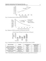
Table 127-2 Classification of β-Lactam Antibiotics
Route of Administration
Class Parenteral Oral
Penicillins
β-Lactamase–
susceptible
Narrow-spectrum Penicillin G Penicillin V
Enteric-active Ampicillin Amoxicillin,
ampicillin
Enteric-
active and
antipseudomonal
Ticarcillin,
piperacillin
None
β-Lactamase–
resistant
Antistaphylococcal
Oxacillin,
nafcillin
Cloxacillin,
dicloxacillin
Combined with β-
lactamase inhibitors
Ticarcillin plus
clavulanic acid,
ampicillin plus
sulbactam, piperacillin
plus tazobactam
Amoxicillin plus
clavulanic acid
Cephalosporins
First-generation Cefazolin,
cephalothin, cephapirin
Cephalexin,
cephradine, cefadroxil
Second-generation
Haemophilus-
active
Cefamandole,
cefuroxime, cefonicid,
ceforanide
Cefaclor, cefuroxime
axetil, ceftibuten
, cefdinir,
cefprozil,
cefpodoxime,
a
loracarbef
Bacteroides-active
Cefoxitin,
cefotetan, cefmetazole
None
Third-generation
Extended-spectrum
Ceftriaxone,
cefotaxime,
ceftizoxime
None
Extended-spectrum
and antipseudomonal
Ceftazidime,
cefepime
None
Carbapenems Imipenem-
cilastatin, meropenem,
None
ertapenem
Monobactams Aztreonam None
a
Some sources classify cefpodoxime as a third-
generation oral agent
because of a marginally broader spectrum.