Hepatocellular Carcinoma: Targeted Therapy and Multidisciplinary P23 docx
Bạn đang xem bản rút gọn của tài liệu. Xem và tải ngay bản đầy đủ của tài liệu tại đây (162.43 KB, 10 trang )
12 Laparoscopic Liver Resection for HCC 205
47. Torzilli G, Donadon M, Marconi M et al (2008) Hepatectomy for stage B and stage C hepato-
cellular carcinoma in the Barcelona Clinic Liver Cancer classification: results of a prospective
analysis. Arch Surg 143:1082–1090
48. Capussotti L, Ferrero A, Viganò L, Muratore A, Polastri R, Bouzari H (2006) Portal
hypertension: contraindication to liver surgery? World J Surg 30:992–999
49. Abdel-Atty MY, Farges O, Jagot P, Belghiti J (1999) Laparoscopy extends the indications for
liver resection in patients with cirrhosis. Br J Surg 86:1397–400
50. Jiao LR, Ayav A, Navarra G et al (2008) Laparoscopic liver resection assisted by the
laparoscopic Habib Sealer. Surgery 144:770–774
51. Ros A, Gustafsson L, Krook H et al (2001) Laparoscopic cholecystectomy versus mini-
laparotomy cholecystectomy: a prospective, randomized, single-blind study. Ann Surg
234:741–749
52. McMahon AJ, Russell IT, Baxter JN et al (1994) Laparoscopic versus minilaparotomy
cholecystectomy: a randomised trial. Lancet 343:135–138
53. Fleshman J, Sargent DJ, Green E et al (2007) Laparoscopic colectomy for cancer is not infe-
rior to open surgery based on 5-year data from the COST Study Group trial. Ann Surg 246:
655–662
54. Belghiti J, Hiramatsu K, Benoist S, Massault P, Sauvanet A, Farges O (2000) Seven hundred
forty-seven hepatectomies in the 1990s: an update to evaluate the actual risk of liver resection,
J Am Coll Surg 191:38
55. Lai EC, Fan ST, Lo CM, Chu KM, Liu CL, Wong J (1995) Hepatic resection for hepatocellular
carcinoma. An audit of 343 patients. Ann Surg 221:291–298
56. Huscher CG, Lirici MM, Chiodini S (1998) Laparoscopic liver resections. Semin Laparosc
Surg 5:204–210
57. Kaneko H, Takagi S, Otsuka Y, et al (2005) Laparoscopic liver resection of hepatocellular
carcinoma. Am J Surg 189:190–194
58. Dagher I, Lainas P, Carloni A et al (2008) Laparoscopic liver resection for hepatocellular
carcinoma. Surg Endosc 22:372–378
59. Santambrogio R, Aldrighetti L, Barabino M et al (2009) Laparoscopic liver resections for
hepatocellular carcinoma. Is it a feasible option for patients with liver cirrhosis? Langenbecks
Arch Surg 394:255–264
60. Lai EC, Tang CN, Ha JP, Li MK (2009) Laparoscopic liver resection for hepatocellular
carcinoma: ten-year experience in a single center. Arch Surg 144:143–147
61. Cai XJ, Yang J, Yu H et al (2008) Clinical study of laparoscopic versus open hepatectomy for
malignant liver tumors. Surg Endosc 22:2350–2356
62. Belli G, Fantini C, D’Agostino A, Belli A, Russolillo N (2004) Laparoscopic liver resections
for hepatocellular carcinoma (HCC) in cirrhotic patients. HPB 6:236–46
63. Morino M, Morra I, Rosso E, Miglietta C, Garrone C (2003) Laparoscopic vs open hepatic
resection: a comparative study. Surg Endosc 17:1914–1918
64. Troisi R, Montalti R, Smeets P et al (2008) The value of laparoscopic liver surgery for solid
benign hepatic tumors. Surg Endosc 22:38–44
65. Simillis C, Constantinides VA, Tekkis PP et al (2007) Laparoscopic versus open hepatic
resections for benign and malignant neoplasms – a meta-analysis. Surgery 141:203–211
66. Laurent A, Cherqui D, Lesurtel M, Brunetti F, Tayar C, Fagniez PL (2003) Laparoscopic liver
resection for subcapsular hepatocellular carcinoma complicating chronic liver disease. Arch
Surg 138:763–769
67. Tekkis PP, Senagore AJ, Delaney CP, Fazio VW (2005) Evaluation of the learning curve in
laparoscopic colorectal surgery: comparison of right-sided and left-sided resections. Ann Surg
242:83–91
68. Schlachta CM, Mamazza J, Seshadri PA, Cadeddu M, Gregoire R, Poulin EC (2001) Defining
a learning curve for laparoscopic colorectal resections. Dis Colon Rectum 44:217–222
69. Dulucq JL, Wintringer P, Stabilini C, Berticelli J, Mahajna A. (2005) Laparoscopic liver
resections: a single center experience. Surg Endosc 19:886–891
206 L. Viganò and D. Cherqui
70. Lesurtel M, Cherqui D, Laurent A, Tayar C, Fagniez PL (2003) Laparoscopic versus open left
lateral hepatic lobectomy: a case-control study. J Am Coll Surg 196:236–242
71. Abu Hilal M, McPhail MJ, Zeidan B et al (2008) Laparoscopic versus open left lateral hepatic
sectionectomy: A comparative study. Eur J Surg Oncol 34:1285–1288
72. Aldrighetti L, Pulitanò C, Catena M et al (2008) A prospective evaluation of laparoscopic
versus open left lateral hepatic sectionectomy. J Gastrointest Surg 12:457–462
73. Belli G, Fantini C, D’Agostino A, Belli A, Cioffi L, Russolillo N (2006) Laparoscopic left
lateral hepatic lobectomy: a safer and faster technique. J Hepatobiliary Pancreat Surg 13:
149–154
74. Robles R, Marín C, Abellán B, López A, Pastor P, Parrilla P (2008) A new approach to hand-
assisted laparoscopic liver surgery. Surg Endosc 22:2357–2364
75. Gayet B, Cavaliere D, Vibert E et al (2007) Totally laparoscopic right hepatectomy. Am J Surg
194:685–689
76. O’Rourke N, Fielding G (2004) Laparoscopic right hepatectomy: surgical technique.
J Gastrointest Surg 8:213–216
77. Dagher I, Caillard C, Proske JM, Carloni A, Lainas P, Franco D (2008) Laparoscopic right
hepatectomy: original technique and results. J Am Coll Surg 206:756–760
78. Fong Y, Brennan MF, Turnbull A et al (1993) Gallbladder cancer discovered during
laparoscopic surgery–potential for iatrogenic dissemination. Arch Surg 128:1054–1056
79. Johnstone PA, Rohde DC, Swartz SE, Fetter JE, Wexner SD (1996) Port site recurrences after
laparoscopic and thoracoscopic procedures in malignancy. J Clin Oncol 14:1950–1956
80. Cherqui D, Laurent A, Tayar C et al (2006) Laparoscopic liver resection for peripheral hepa-
tocellular carcinoma in patients with chronic liver disease: midterm results and perspectives.
Ann Surg 243:499–506
81. Belli G, Cioffi L, Fantini C et al (2009) Laparoscopic redo surgery for recurrent hepatocellular
carcinoma in cirrhotic patients: feasibility, safety, and results. Surg Endosc 23:1807–1811
82. Jaeck D, Bachellier P, Oussoultzoglou E, Weber JC, Wolf P (2004) Surgical resection of hep-
atocellular carcinoma. Post-operative outcome and long-term results in Europe: an overview.
Liver Transpl 10(2 Suppl 1):S58–S63
83. Fong Y, Sun RL, Jarnagin W, Blumgart LH (1999) An analysis of 412 cases of hepatocellular
carcinoma at a Western center. Ann Surg 229:790–799
84. Poon RT, Fan ST, Lo CM et al (2001) Improving survival results after resection of hepatocel-
lular carcinoma: a prospective study of 377 patients over 10 years. Ann Surg 234:63–70
85. Sala M, Fuster J, Llovet JM et al (2004) High pathological risk of recurrence after surgical
resection for hepatocellular carcinoma: an indication for salvage liver transplantation. Liver
Transpl 10:1294–1300
86. Scatton O, Zalinski S, Terris B et al (2008) Hepatocellular carcinoma developed on com-
pensated cirrhosis: resection as a selection tool for liver transplantation. Liver Transpl
14:779–88
87. Hoshida Y, Villanueva A, Kobayashi M et al (2008) Gene expression in fixed tissues and
outcome in hepatocellular carcinoma. N Engl J Med 359:1995–2004
88. Adam R, Azoulay D, Castaing D et al (2003) Liver resection as a bridge to transplantation for
hepatocellular carcinoma on cirrhosis: a reasonable strategy? Ann Surg 238:508–518
89. Laurent A, Tayar C, Andréoletti M, Lauzet JY, Merle JC, Cherqui D (2009) Laparoscopic l iver
resection facilitates salvage liver transplantation for hepatocellular carcinoma. J Hepatobiliary
Pancreat Surg 16:310–314
Chapter 13
Laparoscopic Liver Surgery for the
Management of Hepatocellular Carcinoma:
The American Perspective
Kadiyala V. Ravindra and Joseph F. Buell
Keywords Laparoscopic liver surgery · Laparoscopic liver resection ·
HCC · Hepatic resection · Patient selection
Despite better understanding and advances in oncology, the best available thera-
peutic option for the management of hepatocellular carcinoma (HCC) is surgical –
either liver transplantation or resection. Liver transplantation appears most attractive
since it treats the primary tumor and the field defect associated with the underly-
ing liver disease. However, this option i s feasible only when there are an adequate
number of organs available and when the disease and patient meet certain stringent
criteria. Most centers abide by the Milan criteria [1] to determine candidacy for
liver transplantation. These are a single tumor 5 cm, two or three tumors all <3 cm,
absence of major vascular invasion, and no extrahepatic disease. Unfortunately, only
a minority of hepatoma patients fit these morphological parameters. Many other cir-
rhotic patients do not fulfill the requirements for transplantation due to comorbidity
or psychosocial reasons. A few centers have attempted to expand transplantation
to patients with greater tumor burden. These criteria were developed by the UCSF
group and consist of solitary tumor ≤6.5 cm, or three or fewer nodules with the
largest lesion ≤4.5 cm, and total tumor diameter ≤8 cm, without gross vascular
invasion [2].
Hepatic resection should be considered for patients deemed unsuitable for
transplantation. However, proper selection of patients is required to avoid postop-
erative liver failure. On rare occasions, laparoscopic resection has been utilized
to select patients for liver transplantation – particularly when there is a ques-
tion of major vascular invasion arising in the presence of small tumors. When
patients are unable to undergo resection, they are then considered for ablative strate-
gies including radiofrequency ablation [3], cryoablation [4], percutaneous alcohol
injection [5], microwave ablation [6], laser ablation [7], chemoembolization [8],
chemotherapeutic beads, and infusion of yttrium microspheres [9].
K.V. Ravindra (B)
Department of Surgery, Duke University Medical Center, Durham, NC, USA
207
K.M. McMasters, J N. Vauthey (eds.), Hepatocellular Carcinoma,
DOI 10.1007/978-1-60327-522-4_13,
C
Springer Science+Business Media, LLC 2011
208 K.V. Ravindra and J.F. Buell
Hepatic resection poses several important challenges. In the setting of normal
parenchyma, resection maybe limited only by the presence of extrahepatic spread,
bi-lobar disease, or major vascular extension. These criteria serve only as relative
contraindications and should be considered on a case-by-case basis. Major liver
resection in a patient with normal parenchyma is tolerated down to a functional
liver remnant of only two or three segments. However, in the setting of a diseased
liver, resection is an entirely different proposition. A fibrotic or cirrhotic liver has
poor and unpredictable ability to regenerate with resultant liver failure. This is a
deterrent to major liver resection in hepatoma occurring against the background of
cirrhosis.
Various methods have been used to guide the extent of possible resection in this
situation. These include the Child’s status, ICG excretion test [10], and evidence of
portal hypertension (platelet count, wedged hepatic venous pressure gradient) [11]
(Table 13.1). Despite these tools, planning and executing liver resection in cirrho-
sis continues to be a serious undertaking. Recent advances in the care of cirrhotic
patients have enabled mortality rates as low as 3% [12].
Table 13.1 Selection criteria
for liver resection for
hepatocellular carcinoma in
chronic liver disease
For a major resection (≥ three segments)
Child-Pugh class A
Indocyanine green retention at 15 min <15%
No esophageal varices
Platelets >100,000/mm
3
Transaminases ≤ two times normal
Hypertrophy of liver after portal vein embolization
Functional residual liver volume > 50%
For a limited resection (<three segments)
Child-Pugh class A
Child-Pugh class B for a peripheral tumorectomy
Esophageal varices grade 2 maximum
With permission from Bryant et al. [11]
Liver surgery has evolved significantly over the last two decades. It is well stan-
dardized and has excellent results largely due to advances in the techniques of liver
surgery aided by knowledge of the segmental anatomy of liver, improved imaging
techniques, better intra- and postoperative management of these patients, particu-
larly cirrhotics. Almost concurrent with the advance in liver surgery, the field of
minimally invasive surgery exploded and caused a major surgical revolution. It was
inevitable that liver surgeons would apply these techniques. Large series of laparo-
scopic hepatic resections [13–15] have been reported and have encouraged wider
application of the technique even in patients with malignant neoplasms.
Selection of Patients for Surgery
The decision to operate on a patient with hepatoma is largely determined by the mor-
phological evaluation of the tumor on imaging and an evaluation of the functional
reserve of the liver. The indications for surgery are as follows:
13 Laparoscopic Liver Surgery for the Management of Hepatocellular Carcinoma 209
1) Diagnostic: while evaluating a hypervascular lesion in a cirrhotic – to differenti-
ate neoplasia from a regenerative nodule
2) Resection of the lesion with intent to cure
3) Assessment of histological features for transplant indications
4) Ablation of the lesion at surgery by use of RF or microwave energy
Imaging
Evaluation of lesions developing in a background of cirrhosis remains a challenge
when the lesions are small. A variety of techniques such as ultrasound, triple phase
CT scan, and MR imaging are essential in guiding therapy. The latter two are
comparable and must be chosen based on local expertise and equipment. CT imag-
ing characteristics that are diagnostic include intense enhancement on late arterial
phase, washout on portal/delayed phase, and a late capsule/pseudo capsule enhance-
ment. MR may be preferred in differentiating regenerative nodules from tumors
when the lesions are small. However, the presence of renal impairment (not uncom-
mon in cirrhotics) precludes the use of gadolinium. Lesions less than 1 cm are
difficult to characterize on imaging studies and may be followed by serial imag-
ing in 3–6 months to detect an increase in size. This information helps stage the
disease based on the number of lesions, size of individual lesions, presence of major
vascular invasion, and extrahepatic disease. Imaging of the lungs and bone scans are
routinely performed at most centers prior to planning surgical resection to rule out
metastatic disease.
Additionally, CT volumetry permits calculation of the residual liver volume
(the volume of liver remaining after resection). Preoperative volumetric analysis
is essential to ensure sufficient functional liver parenchyma remains. The functional
residual liver volume is calculated by the formula: volume of residual liver/volume
of total liver – volume of the tumor. Vauthey [16] demonstrated that a future liver
remnant of <25% was associated with increased complications following extended
liver resections in patients without underlying disease. However, in the presence of
parenchymal liver disease this figure may need to be higher than 50% [11].
Functional Reserve of Liver
This is an extremely important component of liver resection in cirrhotics. Many
methods have been utilized to estimate the functional reserve and guide the extent
of liver resection. Typically liver resections are contemplated only in Child’s
A or early B cirrhosis. But the assessment of the liver reserve based on syn-
thetic function of the liver (serum albumin, prothrombin time) has not been very
reliable. Dynamic assessment of complex liver functions such as clearance of sub-
stances (ICG – indocyanine green) or the formation of metabolites (lidocaine to
210 K.V. Ravindra and J.F. Buell
monoethylglycinexylidide [MEGX] or 14C-aminopyrine) has been used to more
accurately delineate the functional reserve in patients with liver disease.
In the Far East, the indocyanine green (ICG) retention test has been used with
success in selecting candidates for liver resection [17]. ICG is an infrared absorb-
ing fluorescent agent which is almost exclusively eliminated by the liver into the
bile. Following the intravenous injection of 0.5 mg/kg of ICG the rate of disap-
pearance from the plasma is calculated. A r etention of >15% of the injected dye at
15 min indicates poor liver reserve and predicts poor outcome with liver resections
involving three or more segments.
The ICG retention test proved to be the best discriminating preoperative test in
patients with hepatoma prior to hepatectomy [10]. However, this test has not been
widely used in the West to guide liver surgery. The guidelines listed in a recent
review summarize the criteria used by major centers to select patients for liver
resection in the presence of chronic liver disease [11]. A gross rule of thumb for
what would be considered possibly safe is lobar resection for Child-Pugh class A
patients, a 15% resection for class B, and a 5% resection for class C.
Laparoscopic Liver Resection for Hepatoma
The debate about the feasibility and safety of laparoscopic liver surgery is slowly but
surely being put to rest. Large series of liver resections performed laparoscopically
have been published and have matched the results of open surgery. Laparoscopic
methods have the potential to lower the stress posed by liver surgery. Whether a
significant reduction in morbidity actually is achieved has yet to be conclusively
determined.
History
A multicenter European study published in 2002 was the first to present the results of
laparoscopic liver resection for malignant liver tumors [18]. However, the retrospec-
tive study involving 11 centers contained only 10 patients with hepatoma, 9 of whom
were cirrhotic. The next significant data came from the Henri Mondor Hospital in
Paris in 2006 [19]. This single center prospective study included 27 patients who
were followed for a mean period of 2 years. The paper conclusively demonstrated
the feasibility and the midterm safety of laparoscopic resection. Subsequent papers
from Italy [20] and Taiwan [21] have corroborated this concept.
Technique
There are three different terminologies that have been used with regard to laparo-
scopic liver resections:
13 Laparoscopic Liver Surgery for the Management of Hepatocellular Carcinoma 211
1. Pure laparoscopic
2. Hand-assisted laparoscopic resection
3. Laparoscopic-assisted (hybrid) open resection
There is no clear advantage of one approach over the others. All aim to reduce
the surgical trauma by minimizing the length of surgical incision. An incision is
often required to extract the tumor specimen and one may as well make this incision
at the beginning if it will aid the dissection. Poon [22] has reported the following
advantages with the insertion of a hand port:
1. Palpation with the hand and the use of intraoperative ultrasonography through the
hand port improve the staging of tumor and permit better delineation of resection
margin
2. The hand is the best retractor
3. Manual compression in the event of major bleeding
4. Hand assistance in intracorporeal suturing
5. Specimen retrieval through the hand port
The position of the patient is supine when performing resections on the left
lobe segments. The French surgeons utilize the lithotomy position with the surgeon
standing between the legs during the surgery. The left lateral decubitus with a steep
reverse Trendelenburg position is ideal for lesions in the right lobe – particularly
those requiring mobilization of the right lobe to gain access to the posterior surface.
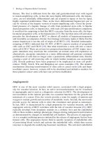
When a hand port is inserted, the location has varied in different series. It may be
placed in the midline close to the xiphoid with a lateral extension or in t he midclav-
icular line at or above the plane of the umbilicus [23] (Fig. 13.1). The exact position
varies with the individual anatomy, the size of the liver, and location of the tumor(s).
Fig. 13.1 Port placement and surgeon positioning during laparoscopic liver resection (right-sided
resection). With permission from Buell et al. [23]
212 K.V. Ravindra and J.F. Buell
Typically the procedure is initiated with the placement of a trocar inferior to
the umbilicus. Incisions through or above the umbilicus are not recommended in
order to avoid collaterals in the falciform ligament. After a preliminary examina-
tion, an ultrasound of the liver is performed. Laparoscopic ultrasound has been used
extensively in most series. This helps in confirming the site and size of the lesion,
detecting additional lesions, identifying the vascular structures in proximity to the
lesion, and guiding the placement of biopsy needle or radiofrequency probe.
The majority of liver resections performed for hepatoma in cirrhotics have
involved one or two segments or non-anatomical resections [24] (Table 13.2). In
Table 13.2 Findings and results from literature [24]
Variable Results
Index number of
analyzable patients
(% out of 300 patients)
Sex ratio M/F 132/58 190 (68%)
Mean age 61.8 (34–76) 175 (62%)
Liver cirrhosis 156 (78%) 201 (72%)
Child-Pugh classification: A/B/C 130/28/4 169 (60%)
Mean tumor size (mm) 33.6 (9–75) 188 (70%)
Location (Couinaud segments)
2/3 s 81 (38%)
4 s 38 (18%)
5/6 s 85 (39%)
7s 8(4%)
8s 3(1%)
LUS used 200 (83%) 240 (85%)
Type of resection 211 (75%)
Atypical 121 (57%)
Segmentectomy 35 (17%)
Left Lobectomy 40 (19%)
Left Hepatectomy 7 (3%)
Right Hepatectomy 4 (2%)
Mesohepatectomy 3 (1%)
Bisegementectomy (5/6 s) 1
Pringle maneuver 62 (38%) 162 (58%)
Pringle maneuver (mean duration) 50.6 (15–17)
Perioperative complications 17 (10%) 169 (60%)
Conversion to laparoscopy 24 (9%) 262 (93%)
Mean operative time (min) 216.8 (50–680) 175 (62%)
Mean blood losses (ml) 401 (0–1,700) 155 (55%)
Transfusion rate 17 (11%) 161 (57%)
Surgical margins (>1 cm) 77 (65%) 118 (42%)
Operative mortality 5 (1.7%) 281 (100%)
Postoperative complications 42 (20%) 211 (70%)
Reoperation 2 (0.9%) 214 (76%)
Mean postoperative hospital stay (days) 12.3 (2–76) 161 (57%)
Mean follow-up time (months) 19.6 102 (36%)
Alive without recurrence 86 (57%) 151 (54%)
With permission from Santambrogio et al. [24]
13 Laparoscopic Liver Surgery for the Management of Hepatocellular Carcinoma 213
a review of 300 undergoing laparoscopic hepatectomy for hepatoma in cirrhotics,
only 11 involved resection of an entire lobe. This illustrates the difficulty of major
liver resections in cirrhotics. The location of the hepatoma determines the feasibil-
ity and ease of laparoscopic resection. When the lesion i s located peripherally in the
anterior and inferior aspects (segments 2, 3, 4b, 5, or 6) surgical resection is easier.
Lesions located on the superior and posterior parts (segments 1, 4a, 7, or 8) pose a
challenge.
Depending on the location of the liver, the mobilization of the liver is performed –
the falciform and the appropriate triangular ligaments are divided. For lesions in
the posterior right lobe, the bare area of the liver will often have to be freed with
exposure of the retrohepatic inferior vena cava. The option of a Pringle maneuver
has been utilized by some centers to reduce bleeding during the resection. However,
this is not mandatory.
Transecting the liver parenchyma with minimal blood loss is a challenge, espe-
cially in the cirrhotic liver. This has led to development of a variety of devices
which utilize different types of energy to dissect the liver and seal the blood
vessels. These include the ultrasonic dissector (Harmonic Scalpel
TM
), bipolar
diathermy, water jet dissector (Helix Hydro-jet
TM
), dissecting sealer (Tissuelink
TM
and Aquamantys
TM
), Habib 4X
TM
, radiofrequency device. The caliber of the ves-
sels traversing the most superficial 2–3 cm of the parenchyma is small and hence
any of the above devices can be successfully employed. As the depth increases,
larger vessels (those associated with the Glissonian pedicle and the hepatic veins)
are encountered. We believe that these are most safely and expeditiously dealt with
by the use of a vascular stapler. As experience with vascular staplers has grown,
it is being widely used even to divide the parenchyma without isolation of major
blood vessels. As no single device has been shown superior to the others, it is best
to develop expertise depending on the devices available at each center. However, it
should be noted that the cirrhotic parenchyma poses specific challenges for laparo-
scopic resection related to the stiffness of the liver, which impairs mobility, and
fibrosis, which can limit the use of vascular staplers.
Preferred Technique at Our Center
The positioning of the patient is crucial for performing laparoscopic liver resec-
tions. For lesions in the right lobe, particularly in the posterior or superior segments,
the patient is positioned in the left l ateral decubitus with the table in steep reverse
Trendelenburg position. We find that this greatly facilitates the mobilization of the
right lobe of liver and exposure of the retrohepatic vena cava. For lesions in the
left lobe we prefer the supine position. We do not utilize the hand port as a routine.
Typically peripheral lesions involving segments 2, 3, 4b, 5, or 6 can be resected by
the pure laparoscopic technique. We utilize the hand port for lesions involving the
caudate lobe, the posterior surface, or the superior segments 4a, 7, and 8. The hand
port is inserted through a transverse incision in the right upper abdomen – the exact
site depends on the s ize and location of the liver lesion.
214 K.V. Ravindra and J.F. Buell
Our preference is to delineate the margins of the lesion with the help of intra-
operative ultrasound. When a lobectomy is performed, our preference is to avoid
extensive hilar dissection. We prefer to staple the major vessels in the parenchyma.
If easily accessible, the right hepatic vein may be stapled outside the liver. We use
the harmonic scalpel to mark a 1 cm margin around the lesion. The hepatotomy is
then initiated with the harmonic device. After a depth of 2 cm is reached we pre-
fer to complete the parenchymal transection with the help of vascular staplers. The
specimen is delivered out of the hand port if one has been placed. Otherwise it is
placed in an endopouch and retrieved. After the specimen is r emoved, hemostasis
of the raw surface is achieved using diathermy, argon beam coagulation, and intra-
corporeal suturing to control more significant bleeding. We utilize a “quick stitch”
to control active bleeders or sites of bile leak. This involves the use of a 15 cm long
2-0 silk swaged suture with clips on one end which serve to anchor the suture at
the liver surface. After the site of bleeding is controlled with the suturing, clips are
placed at the exit site to lock the stitch in place. After satisfactory hemo- and bile
stasis has been attained, we apply topical sealants to the raw surface. These include
BioGlu (Cryolife), Tisseel (Baxter), and Co-seal (Baxter).
The CVP is maintained less than 5 throughout the procedure. This reduces the
bleeding during transection and from the resulting raw surface. There has been con-
troversy regarding the use of Argon beam at laparoscopy. We have not encountered
gas embolism and believe the practice is safe – particularly when the intraabdominal
pressure does not exceed 15 mmHg.
Despite the best of precautions, the liver s urgeon will often be faced with seri-
ous challenges. The most common is bleeding. Pressure either with an instrument
or with the hand will achieve temporary hemostasis. After stabilizing the patient
and ensuring the availability of a good suction device, an attempt must be made to
identify the cause of the bleeding. The most troublesome bleeding comes from the
veins which often retract into the liver. We employ different techniques as outlined
above including the quick stitch. If the bleeding continues, a hand port may have to
be inserted if not already present. We have f ound that reapplication of the vascular
stapler to excise an additional margin of liver tissue is often successful in achieving
hemostasis.
Laparoscopic resection for hepatocellular cancer as with open resection f or this
disease is complicated. Cirrhosis has historically portended higher operative mor-
bidity and mortality. When our group approaches a cirrhotic we recognize and
adhere to our principles of low CVP, in conjunction with a pure laparoscopic
approach, when feasible. We also recognize and continue to debate over the pri-
mary thermal technology utilized to transect hepatic parenchyma. Mobilization and
division of the major inflow and outflow vessels are performed only when necessary
for margins. Intrahepatic division is often preferred for t hese vessels. When inade-
quate control of major vascular structures is encountered, use of the “Koffron quick
stitch” is employed. This is a pre-cut length of silk or prolene with two 10 mm clips
on the end. This allows primary closure of vessels. When this is not easily achieved,
conversion to a hand-assist approach is employed. This allows direct digital control
of bleeding without the need for complete conversion to laparotomy. This maneuver