Chapter 061. Disorders of Granulocytes and Monocytes (Part 4) pps
Bạn đang xem bản rút gọn của tài liệu. Xem và tải ngay bản đầy đủ của tài liệu tại đây (71.63 KB, 5 trang )
Chapter 061. Disorders of Granulocytes
and Monocytes
(Part 4)
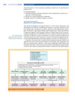
Figure 61-8
Neutrophil travel through the pulmonary capillaries is dependent on
neutrophil deformability. Neutrophil rigidity (e.g., caused by C5a) enhances
pulmonary trapping and response to pulmonary pathogens in a way that is not so
dependent on cell-surface receptors. Intraalveolar chemotactic factors, such as
those caused by certain bacteria (e.g., Streptococcus pneumoniae) lead to
diapedesis of neutrophils from the pulmonary capillaries into the alveolar space.
Neutrophil interaction with the endothelium of the systemic postcapillary venules
is dependent on molecules of attachment. The neutrophil "rolls" along the
endothelium using selectins: neutrophil CD15s (sialyl-Lewis
x
) binds to CD62E (E-
selectin) and CD62P (P-selectin) on endothelial cells; CD62L (L-selectin) on
neutrophils binds to CD34 and other molecules (e.g., GlyCAM-1) expressed on
endothelium. Chemokines or other activation factors stimulate integrin-mediated
"tight adhesion": CD11a/CD18 (LFA-1) and CD11b/CD18 (Mac-1, CR3) bind to
CD54 (ICAM-1) and CD102 (ICAM-2) on the endothelium. Diapedesis occurs
between endothelial cells: CD31 (PECAM-1) expressed by the emigrating
neutrophil interacts with CD31 expressed at the endothelial cell-cell junction
On cell stimulation, L-selectin is shed from neutrophils, and E-selectin
increases in the blood, presumably because it is shed from endothelial cells;
receptors for chemoattractants and opsonins are mobilized; and the phagocytes
orient toward the chemoattractant source in the extravascular space, increase their
motile activity (chemokinesis), and migrate directionally (chemotaxis) into tissues.
The process of migration into tissues is called diapedesis and involves the
crawling of neutrophils between postcapillary endothelial cells that open junctions
between adjacent cells to permit leukocyte passage. Diapedesis involves
platelet/endothelial cell adhesion molecule (PECAM) 1 (CD31), which is
expressed on both the emigrating leukocyte and the endothelial cells. The
endothelial responses (increased blood flow from increased vasodilation and
permeability) are mediated by anaphylatoxins (e.g., C3a and C5a) as well as
vasodilators such as histamine, bradykinin, serotonin, nitric oxide, vascular
endothelial growth factor (VEGF), and prostaglandins E and I. Cytokines regulate
some of these processes [e.g., TNF-α induction of VEGF, interferon (IFN)
γinhibition of prostaglandin E].
In the healthy adult, most neutrophils leave the body by migration through
the mucous membrane of the gastrointestinal tract. Normally, neutrophils spend a
short time in the circulation (half-life, 6–7 h). Senescent neutrophils are cleared
from the circulation by macrophages in the lung and spleen. Once in the tissues,
neutrophils release enzymes, such as collagenase and elastase, which help
establish abscess cavities. Neutrophils ingest pathogenic materials that have been
opsonized by IgG and C3b. Fibronectin and the tetrapeptide tuftsin also facilitate
phagocytosis.
With phagocytosis comes a burst of oxygen consumption and activation of
the hexose-monophosphate shunt. A membrane-associated NADPH oxidase,
consisting of membrane and cytosolic components, is assembled and catalyzes the
reduction of oxygen to superoxide anion, which is then converted to hydrogen
peroxide and other toxic oxygen products (e.g., hydroxyl radical). Hydrogen
peroxide + chloride + neutrophil myeloperoxidase generate hypochlorous acid
(bleach), hypochlorite, and chlorine. These products oxidize and halogenate
microorganisms and tumor cells and, when uncontrolled, can damage host tissue.
Strongly cationic proteins, defensins, and probably nitric oxide also participate in
microbial killing. Lactoferrin chelates iron, an important growth factor for
microorganisms, especially fungi. Other enzymes, such as lysozyme and acid
proteases, help digest microbial debris. After 1–4 days in tissues, neutrophils die.
The apoptosis of neutrophils is also cytokine-regulated; granulocyte colony-
stimulating factor (G-CSF) and IFN-γ prolong their life span. Under certain
conditions, such as in delayed-type hypersensitivity, monocyte accumulation
occurs within 6–12 h of initiation of inflammation. Neutrophils, monocytes,
microorganisms in various states of digestion, and altered local tissue cells make
up the inflammatory exudate, pus. Myeloperoxidase confers the characteristic
green color to pus and may participate in turning off the inflammatory process by
inactivating chemoattractants and immobilizing phagocytic cells.
Neutrophils respond to certain cytokines [IFN-γ, granulocyte-macrophage
colony-stimulating factor (GM-CSF), IL-8] and produce cytokines and
chemotactic signals [TNF-α, IL-8, macrophage inflammatory protein (MIP) 1] that
modulate the inflammatory response. In the presence of fibrinogen, f-met leu phe
or leukotriene B
4
induces IL-8 production by neutrophils, providing autocrine
amplification of inflammation. Chemokines (chemoattractant cytokines) are small
proteins produced by many different cell types, including endothelial cells,
fibroblasts, epithelial cells, neutrophils, and monocytes, that regulate neutrophil,
monocyte, eosinophil, and lymphocyte recruitment and activation. Chemokines
transduce their signals through heterotrimeric G protein–linked receptors that have
seven cell membrane–spanning domains, the same type of cell-surface receptor
that mediates the response to the classic chemoattractants f-metleuphe and C5a.
Four major groups of chemokines are recognized based on the cysteine structure
near the N terminus: C, CC, CXC, and CXXXC. The CXC cytokines such as IL-8
mainly attract neutrophils; CC chemokines such as MIP-1 attract lymphocytes,
monocytes, eosinophils, and basophils; the C chemokine lymphotactin is T cell
tropic; the CXXXC chemokine fractalkine attracts neutrophils, monocytes, and T
cells. These molecules and their receptors not only regulate the trafficking and
activation of inflammatory cells, but specific chemokine receptors serve as co-
receptors for HIV infection (Chap. 182) and have a role in atherogenesis.