Research ArticleGreen Synthesis of Silver Nanoparticles UsingPolyalthia longifoliaLeaf Extract along with D-Sorbitol:StudyofAntibacterialActivity
Bạn đang xem bản rút gọn của tài liệu. Xem và tải ngay bản đầy đủ của tài liệu tại đây (3.28 MB, 6 trang )
Hindawi Publishing Corporation
Journal of Nanotechnology
Volume 2011, Article ID 152970, 5 pages
doi:10.1155/2011/152970
Research Article
Green Synthesis of Silver Nanoparticles Using
Polyalthia longifolia
Leaf Extract along with D-Sorbitol:
StudyofAntibacterialActivity
S. Kaviya,
1
J. Santhanalakshmi,
1
and B. Viswanathan
2
1
Department of Physical Chemistry, University of Madras, Chennai 600 025, India
2
National Center for Catalysis Research, Indian Institute of Technology, Chennai 600 036, India
Correspondence should be addressed to S. Kaviya,
Received 23 March 2011; Accepted 16 June 2011
Academic Editor: Mallikarjuna Nadagouda
Copyright © 2011 S. Kaviya et al. This is an open access article distributed under the Creative Commons Attribution License,
which permits unrestricted use, distribution, and reproduction in any medium, provided the original work is properly cited.
Synthesis of silver nanoparticles (AgNPs) using Polyalthia longifolia leaf extract as reducing and capping agent along with D-
sorbitol used to increase the stability of the nanoparticles has been reported. The reaction is carried out at two different
concentrations (10
−3
Mand10
−4
M) of silver nitrate, and the effect of temperature on the synthesis of AgNPs is investigated by
stirring at room temperature (25
◦
C) and at 60
◦
C. The UV-visible spectra of NPs showed a blue shift with increasing temperature
at both concentrations. FT-IR analysis shows that the biomoites played an important role in the reduction of Ag
+
ions and the
growth of AgNPs. TEM results were utilized for the determination of the size and morphology of nanoparticles. The synthesized
silver nanoparticles are found to be highly toxic against Gram-positive bacteria than Gram-negative bacteria.
1. Introduction
An important area of research in nanotechnology is the syn-
thesis of nano silver particles. Silver has long been recognized
as having an inhibitory effect towards many bacterial strains
and microorganisms [1]. Antibacterial activity of the silver-
containing materials used in medicine to reduce infections
in burn treatment [2] and arthroplasty [3], as well as to
prevent bacteria colonization on prostheses [4], catheters [5],
vascular grafts, dental materials [6], stainless steel materials
[7], and human skin [8]. Silver nanoparticles also exhibit
a potent cytoprotective activity towards HIV-infected cells
[9]. Because of such wide range of applications, numerous
synthetic methods have been developed [10]. Biological routs
of nanoparticles synthesis using microorganism [11–13],
enzyme [14] and plant or plant extract [15–21]havebeen
suggested as possible ecofriendly alternatives to chemical and
physical methods. Using plant for nanoparticles synthesis
can be advantageous over other biological processes by
eliminating the elaborate process of maintaining cell cultures
[22]. It can also be suitably scaled up for large-scale synthesis
of nanoparticles. Specific surface area is relevant for catalytic
reactivity and other related properties such as antimicrobial
activity in silver nanoparticles.
Polyalthia longifolia is a lofty evergreen tree, native to
India, commonly planted due to its effectiveness in alleviat-
ing noise pollution. Methanolic extract of Polyalthia longifo-
lia have yielded 20 known and 2 new organic compounds,
some of which show cytotoxic properties [23]. Here in, we
report for the first time synthesis of silver nanoparticles using
aqueous extract derived from Polyalthia longifolia leafs with
D-sorbitol and their catalytic and antibacterial activity of the
synthesized NPs is described.
2. Experimental
The Polyalthia longifolia leaves were collected from University
of Madras Campus located at Chennai, India. All the
chemicals were obtained from Aldrich and experiments
done in triplicates. Double-distilled water was used for
the experiments. Fresh leaves of Polyalthia longifolia were
collected, washed thoroughly with double-distilled water,
and incised into small pieces. About 4 g of finely cut
Polyalthia longifolia leaves were weighed and transferred into
2 Journal of Nanotechnology
a 250 mL beaker containing 40 mL double-distilled water,
mixed well, and boiled for 2 min. The extract obtained was
filtered through Whatman number 1 filter paper, and the
filtrate was collected in 250 mL Erlenmeyer flask and stored
at 4
◦
C for further use.
Aqueous solution of 10
−3
Mand10
−4
M silver nitrate
(AgNO
3
)and10
−2
M of D-sorbitol was prepared and used
for the synthesis of silver nanoparticles. 3 mL of extract
and 1 mL of D-sorbitol were added to 40 mL of AgNO
3
solution. The effect of temperature on the synthesis of
silver nanoparticles was carried out at room temperature
(25
◦
C) and 60
◦
C. The silver nanoparticles synthesized using
Polyalthia longifolia leaf extract was tested for antimicrobial
activity by agar well diffusion method against pathogenic
bacteria Escherichia coli, Pseudomonas aeruginosa (Gram
negative), and Staphylococcus aureus (Gram positive). The
pure cultures of bacteria were subcultured on nutrient
agar medium. Each strain was swabbed uniformly onto
the individual plates using sterile cotton swabs. Wells of
10 mm diameter were made on nutrient agar plates using
gel puncture. Using a micropipette, 50 μL of nanoparticle
solutionwaspouredontoeachwellonallplates.After
incubation at 37
◦
C for 24 hours, the different levels of zone
of inhibition of bacteria were measured.
The bioreduction of Ag
+
ion in solution was monitored
using UV-visible spectrometer (Techomp 8500 spectrome-
ter). Further characterization was done using FTIR (Bruker
tensor 27) spectrometer. The extract was centrifuged at
5000 rpm for 30 min and the resulting suspension was
redispersed in 10 mL sterile distilled water. The centrifuging
and redispersing process was repeated three times. Finally,
the dried form of extract was palletized with KBr and
analyzed using FTIR. The morphology of the AgNPs was
examined using transmission electron microscopy (JEOL
3010 TEM). The films of the samples were prepared on a
carbon coated copper grid by dropping a small amount of
the sample and then allowing it to dry.
3. Results and Discussion
The time of addition of extract into the metal ion solution
was considered as the start of the reaction. It is well known
that silver nanoparticles exhibit yellowish brown color in
aqueous solution due to excitation of surface plasmon vibra-
tions in silver nanoparticles [15]. As the Polyalthia longifolia
leaf extract was mixed in the aqueous solution of the silver
ion complex and D-sorbitol, initially the color changed from
watery to yellowish brown due to the reduction of silver ion.
The reduction rate is found to increase with the reaction
temperature [24]. For 10
−3
M solution the addition of 3 mL
of extract to the reaction mixture, the reaction completed by
1.30 h, 1 h while 10
−4
M solution the reaction completed by
1h,40minat25
◦
Cand60
◦
C, respectively.
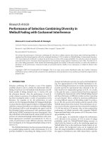
UV-vis spectroscopy could be used to examine size and
shape controlled nanoparticles in aqueous suspensions [25].
Figure 1 shows the UV-vis spectra which are recorded after
the completion of the reaction. For 10
−3
M solution, the
silver nanoparticles have absorbance peak at 451 nm and
435 nm, and 10
−4
M solution has peak at 425 nm and 422 nm
a
a
b
b
c
c
d
d
e
e
800700600500400
Wave lengt h (nm)
0
0
.5
1
1.5
Absorbance
Figure 1: UV-vis absorption spectrum of (a) Polyalthia longifolia
leaf extract, biosynthesized silver nanoparticles of different concen-
tration (10
−3
Mand10
−4
M) at (b and d) 25
◦
C, (c and e) 60
◦
C.
for reaction at 25
◦
Cand60
◦
C, respectively. The frequency
and width of the surface plasmon absorption depend on the
size and shape of the metal nanoparticles as well as on the
dielectric constant of the metal itself and the surrounding
medium [24]. Supposing the same particle shape, medium
dielectric constant and temperature, the mean diameter of
the nanoparticles strongly influence the SPR band in aqueous
solution [25]. The spectrum shows the blue shift with raising
temperature. This blue shift indicates the reduction of mean
diameter of the biogenic silver nanoparticles [24, 26, 27].
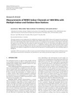
FT-IR measurements were carried out to identify the
possible biomolecules responsible for the reduction of the
Ag
+
ions and capping of the bioreduced silver nanoparticles
synthesized by Polyalthia longifolia leaf extract along with
D-sorbitol. Figure 2(b) represents the FTIR spectrum of D-
sorbitol and shows bands at 2938 cm
−1
(C–H stretching
in alkanes) and 1645 cm
−1
(C=O stretch of carbonyls).
Figure 2(a) represents the FTIR spectrum of the leaf extract
and shows peaks at 1637, 1418, and 1063 cm
−1
. These
peaks are known to be associated with the amide I arise
due to carbonyl stretch in proteins (1637 cm
−1
), –C–C–
stretch (in ring) aromatic (1418 cm
−1
)[28], and C–N
stretching vibration of amine (1063 cm
−1
)[29], respectively.
Proteins present in the extract can bind to AgNP through
either free amino or carboxyl groups in the proteins [30].
Experimentally, D-sorbitol does not have the potential to
reduce the silver ions in the solution, but it may cap the
formed silver nanoparticles through electrostatic attraction
or bind to the protein groups in the extract via hydrogen
bond and increase the stability of the silver nanoparticles.
It indicates that the functional groups in biomolecules are
mainly responsible for the reduction of silver ions.
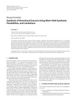
The silver nanoparticles are spherical in shape and
are not aggregated in solution with raising temperature
(Figure 3). This is due to the binding force between the
AgNPs and the capping molecules that may get decreased
with increasing temperature even though the size of the
Journal of Nanotechnology 3
100020003000
Wavenum ber (cm
−1
)
20
40
60
80
100
Transmittance (%)
(a)
500100015002000250030003500
Wavenum ber (cm
−1
)
20
30
40
50
60
70
80
90
100
Transmittance (%)
(b)
Figure 2: FTIR spectrum of (a) Polyalthia longifolia leaf extract and (b) D-sorbitol.
50 nm
(a)
35 nm
(b)
20 nm
(c)
15 nm
(d)
Figure 3: HRTEM image of the biosynthesized silver nanoparticles showing various particle sizes at (a and c) 25
◦
C, (b and d) 60
◦
C.
nanoparticles is reduced. In the 10
−3
M, the size of the
synthesized nanoparticle is 50 nm and 35 nm at 25
◦
Cand
60
◦
C, respectively. Similarly, in the case of 10
−4
M, the size
of the synthesized nanoparticle is 20 nm and 15 nm at 25
◦
C
and 60
◦
C, respectively.
The biologically synthesized silver nanoparticles exhib-
ited excellent antibacterial activity against the bacterial
pathogens Staphylococcus aureus (Gram positive), Escher ichia
coli, and Pseudomonas aeruginosa (Gram negative) [31]. It
has been reported that antibacterial effect was size and dose
dependant and was more pronounced against Gram-negative
bacteria than Gram-positive bacteria. But the present study
clearly indicates that the synthesized silver nanoparticles
have good antibacterial action against Gram-positive organ-
ism than Gram-negative organisms (Figure 4 and Ta bl e 1 ).
The antimicrobial activities of colloidal silver particles are
4 Journal of Nanotechnology
(a)
(a)
(b)
(b)
(c)
(c)
Extract
AgNPs at
25
◦
C
AgNPs at
60
◦
C
10
−3
M
Extract
AgNPs at
25
◦
C
AgNPs at
60
◦
C
10
−3
M
Extract
AgNPs at
25
◦
C
AgNPs at
60
◦
C
10
−3
M
Extract
AgNPs at
25
◦
C
AgNPs at
60
◦
C
10
−4
M
Extract
AgNPs at
25
◦
C
AgNPs at
60
◦
C
10
−4
M
Extract
AgNPs at
25
◦
C
AgNPs at
60
◦
C
10
−4
M
Figure 4: Zone of inhibition of silver nanoparticles against (a) Escherichia coli,(b)Pseudomonas aeruginosa, and (c) Staphylococcus aureus.
Table 1: Zone of inhibition (mm) of biologically synthesized silver
nanoparticles against bacterial pathogens.
S. NO Test organism
10
−3
MAgNPs 10
−4
MAgNPs
synthesized at synthesized at
25
◦
C60
◦
C25
◦
C60
◦
C
(1) Escherichia coli 7.3 7.7 7.3 8
(2)
Pseudomonas
aeruginosa
8.3 9 8.8 9.5
(3)
Staphylococcus
aureus
14 16 14.6 16.4
influenced by the dimensions of the particles. The smaller
particles lead to the greater antimicrobial effects [32]. The
effect of antibacterial activity is higher in the case of silver
nanoparticles synthesized at 60
◦
Ccomparedto25
◦
Cbecause
of being smaller in size [31, 33].
It is necessary to emphasize that the tested silver nanopar-
ticles have bactericidal effects resulting not only in inhibition
of bacterial growth but also in killing bacteria. Experiments
conducted using the scanning tunneling electron microscopy
(STEM) and X-ray energy dispersive spectrometer (EDS)
showed that silver nanoparticles not only at the surface of cell
membrane, but also inside the bacteria [34]. This suggests the
possibility that the silver nanoparticles may also penetrate
inside the bacteria and cause damage by interacting with
phosphorus and sulfur containing compounds such as DNA
[35]. The exact of inhibition of bacterial growth reported in
this study is dependent on the concentration and number of
nanoparticles in medium.
4. Conclusions
Silver nanoparticles were synthesized by Polyalthia longifolia
leaves extract along with D-sorbitol. The spectroscopic char-
acterization from UV-visible, FTIR, and TEM supports the
stability of the biosynthesized nanoparticles. The nanosilver
was found to have wider antimicrobial activity in Gram
positive than Gram negative organisms. We believe that the
silver nanoparticle has great potential for applications in
catalysis, biomedical, and pharmaceutical industries.
References
[1] H. Jiang, S. Manolache, A. C. L. Wong, and F. S. Denes,
“Plasma-enhanced deposition of silver nanoparticles onto
polymer and metal surfaces for the generation of antimicrobial
characteristics,” Journal of Applied Polymer Science, vol. 93, no.
3, pp. 1411–1422, 2004.
[2] D. V. Parikh, T. Fink, K. Rajasekharan et al., “Antimicro-
bial silver/sodium carboxymethyl cotton dressings for burn
wounds,” Textile Research Journal, vol. 75, no. 2, pp. 134–138,
2005.
[3] V.Alt,T.Bechert,P.Steinr
¨
ucke et al., “An in vitro assessment
of the antibacterial properties and cytotoxicity of nanopartic-
ulate silver bone cement,” Biomaterials, vol. 25, no. 18, pp.
4383–4391, 2004.
[4] G. Gosheger, J. Hardes, H. Ahrens et al., “Silver-coated
megaendoprostheses in a rabbit model—an analysis of the
Journal of Nanotechnology 5
infection rate and toxicological side effects,” Biomaterials, vol.
25, no. 24, pp. 5547–5556, 2004.
[5] M. E. Rupp, T. Fitzgerald, N. Marion et al., “Effect of
silver-coated urinary catheters: efficacy, cost-effectiveness,
and antimicrobial resistance,” American Journal of Infection
Control, vol. 32, no. 8, pp. 445–450, 2004.
[6] S. Ohashi, S. Saku, and K. Yamamoto, “Antibacterial activity
of silver inorganic agent YDA filler,” Journal of Oral Rehabili-
tation, vol. 31, no. 4, pp. 364–367, 2004.
[7] M. Bosetti, A. Masse, E. Tobin, and M. Cannas, “Silver coated
materials for external fixation devices: in vitro biocompatibil-
ity and genotoxicity,” Biomaterials, vol. 23, no. 3, pp. 887–892,
2002.
[8] H. J. Lee and S. H. Jeong, “Bacteriostasis and skin innox-
iousness of nanosize silver colloids on textile fabrics,” Textile
Research Journal, vol. 75, no. 7, pp. 551–556, 2005.
[9] R. W. Y. Sun, R. Chen, N. P. Y. Chung, C. M. Ho, C. L. S. Lin,
and C. M. Che, “Silver nanoparticles fabricated in hepes buffer
exhibit cytoprotective activities toward HIV-1 infected cells,”
Chemical Communications, no. 40, pp. 5059–5061, 2005.
[10] M. Gutierrez and A. Henglein, “Formation of colloidal
silver by “push-pull” reduction of Ag
+
,” JournalofPhysical
Chemistry, vol. 97, no. 44, pp. 11368–11370, 1993.
[11] T. Klaus, R. Joerger, E. Olsson, and C. G. Granqvist, “Silver-
based crystalline nanoparticles, microbially fabricated,” Pro-
ceedings of the National Academy of Sciences of the United States
of America, vol. 96, no. 24, pp. 13611–13614, 1999.
[12] Y. Konishi, K. Ohno, N. Saitoh et al., “Bioreductive deposition
of platinum nanoparticles on the bacterium Shewanella algae,”
Journal of Biotechnology, vol. 128, no. 3, pp. 648–653, 2007.
[13] B. Nair and T. Pradeep, “Coalescence of nanoclusters and
formation of submicron crystallites assisted by Lactobacillus ,”
Crystal Growth and Design, vol. 2, no. 4, pp. 293–298, 2002.
[14] I. Willner, R. Baron, and B. Willner, “Growing metal nanopar-
ticles by enzymes,” Advanced Materials,vol.18,no.9,pp.
1109–1120, 2006.
[15] S. S. Shankar, A. Rai, B. Ankamwar, A. Singh, A. Ahmad,
and M. Sastry, “Biological synthesis of triangular gold
nanoprisms,” Nature Materials, vol. 3, no. 7, pp. 482–488,
2004.
[16] D. Philip, C. Unni, S. Aromal, and V. K. Vidhu, “Murraya
Koenigii leaf-assisted rapid green synthesis of silver and gold
nanoparticles,” Spectrochimica Acta Part A,vol.78,no.2,pp.
899–904, 2011.
[17] R. Veerasamy, T. Z. Xin, S. Gunasagaran et al., “Biosynthesis
of silver nanoparticles using mangosteen leaf extract and
evaluation of their antimicrobial activities,” Journal of Saudi
Chemical Society, vol. 15, no. 2, pp. 113–120, 2011.
[18] D. Philip, “Mangifera Indica leaf-assisted biosynthesis of well-
dispersed silver nanoparticles,” Spectrochimica Acta Part A, vol.
78, no. 1, pp. 327–331, 2011.
[19] S. P. Dubey, M. Lahtinen, and M. Sillanpaa, “Tansy fruit
mediated greener synthesis of silver and gold nanoparticles,”
Process Biochemistry, vol. 45, no. 7, pp. 1065–1071, 2010.
[20] S. L. Smitha, D. Philip, and K. G. Gopchandran, “Green syn-
thesis of gold nanoparticles using Cinnamomum zeylanicum
leaf broth,” Spect rochimica Acta Part A, vol. 74, no. 3, pp. 735–
739, 2009.
[21] A. R. V. Nestor, V. S. Mendieta, M. A. C. Lopez, R. M. G.
Espinosa, M. A. C. Lopez, and J. A. A. Alatorre, “Solventless
synthesis and optical properties of Au and Ag nanoparticles
using Camellia sinensis ,” Materials Letters, vol. 62, no. 17-18,
pp. 3103–3105, 2008.
[22] S. S. Shankar, A. Rai, A. Ahmad, and M. Sastry, “Rapid synthe-
sis of Au, Ag, and bimetallic Au core—Ag shell nanoparticles
using neem (Azadirachta indica) leaf broth,” Journal of Colloid
and Interface Science, vol. 275, no. 2, pp. 496–502, 2004.
[23]C.Y.Chen,F.R.Chang,Y.C.Shihetal.,“Cytotoxic
constituents of Polyalthia longifolia var. pendula,” Journal of
Natural Products, vol. 63, no. 11, pp. 1475–1478, 2000.
[24] A. Rai, A. Singh, A. Ahmad, and M. Sastry, “Role of halide
ions and temperature on the morphology of biologically
synthesized gold nanotriangles,” Langmuir,vol.22,no.2,pp.
736–741, 2006.
[25] B. J. Wiley, S. H. Im, Z. Y. Li, J. McLellan, A. Siekkinen, and
Y. Xia, “Maneuvering the surface plasmon resonance of silver
nanostructures through shape-controlled synthesis,” Journal
of Physical Chemistry B, vol. 110, no. 32, pp. 15666–15675,
2006.
[26] J. Y. Song and B. S. Kim, “Rapid biological synthesis of
silver nanoparticles using plant leaf extracts,” Bioprocess and
Biosystems Engineering, vol. 32, no. 1, pp. 79–84, 2009.
[27]A.M.Fayaz,K.Balaji,P.T.Kalaichelvan,andR.Venkatesan,
“Fungal based synthesis of silver nanoparticles—an effect of
temperature on the size of particles,” Colloids and Surfaces B,
vol. 74, no. 1, pp. 123–126, 2009.
[28] H. Bar, D. K. Bhui, G. P. Sahoo, P. Sarkar, S. P. De, and A.
Misra, “Green synthesis of silver nanoparticles using latex of
Jatropha curcas,” Colloids and Surfaces A, vol. 339, no. 1–3, pp.
134–139, 2009.
[29] K. B. Narayanan and N. Sakthivel, “Coriander leaf mediated
biosynthesis of gold nanoparticles,” Materials Letters, vol. 62,
no. 30, pp. 4588–4590, 2008.
[30] A. Gole, C. Dash, V. Ramakrishnan et al., “Pepsin-gold col-
loid conjugates: preparation, characterization, and enzymatic
activity,” Langmuir, vol. 17, no. 5, pp. 1674–1679, 2001.
[31] M. Singh, S. Singh, S. Prasad, and I. S. Gambhir, “Nanotech-
nology in medicine and antibacterial effect of silver nanopar-
ticles,” Digest Journal of Nanomaterials and Biostructures, vol.
3, no. 3, pp. 115–122, 2007.
[32] C. Baker, A. Pradhan, L. Pakstis, J. Pochan Darrin, and S.
S. Ismat, “Synthesis and antibacterial properties of silver
nanoparticles,” Journal of Nanoscience and Nanotechnology,
vol. 5, no. 2, pp. 244–249, 2005.
[33] C. Carlson, S. M. Hussein, A. M. Schrand et al., “Unique
cellular interaction of silver nanoparticles: size-dependent
generation of reactive oxygen species,” Journal of Physical
Chemistry B, vol. 112, no. 43, pp. 13608–13619, 2008.
[34] J. R. Morones, J. L. Elechiguerra, A. Camacho et al., “The
bactericidal effect of silver nanoparticles,” Nanotechnology, vol.
16, no. 10, pp. 2346–2353, 2005.
[35] D. W. Hatchett and H. S. White, “Electrochemistry of sulfur
adlayers on the low-index faces of silver,” Journal of Physical
Chemistry, vol. 100, no. 23, pp. 9854–9859, 1996.
Submit your manuscripts at
Scientifica
Hindawi Publishing Corporation
Volume 2014
Corrosion
International Journal of
Hindawi Publishing Corporation
Volume 2014
Polymer Science
International Journal of
Hindawi Publishing Corporation
Volume 2014
Hindawi Publishing Corporation
Volume 2014
Ceramics
Journal of
Hindawi Publishing Corporation
Volume 2014
Composites
Journal of
Nanoparticles
Journal of
Hindawi Publishing Corporation
Volume 2014
Hindawi Publishing Corporation
Volume 2014
International Journal of
Biomaterials
Hindawi Publishing Corporation
Volume 2014
Nanoscience
Journal of
Textiles
Hindawi Publishing Corporation
Volume 2014
Journal of
Nanotechnology
Hindawi Publishing Corporation
Volume 2014
Journal of
Crystallography
Journal of
Hindawi Publishing Corporation
Volume 2014
The Scientic
World Journal
Hindawi Publishing Corporation
Volume 2014
Hindawi Publishing Corporation
Volume 2014
Coatings
Journal of
Advances in
Materials Science and Engineering
Hindawi Publishing Corporation
Volume 2014
Smart Materials
Research
Hindawi Publishing Corporation
Volume 2014
Hindawi Publishing Corporation
Volume 2014
Metallurgy
Journal of
Hindawi Publishing Corporation
Volume 2014
BioMed
Research International
Materials
Journal of
Hindawi Publishing Corporation
Volume 2014
Nanomaterials
Hindawi Publishing Corporation
Volume 2014
Journal of
Nanomaterials