Spinal Disorders: Fundamentals of Diagnosis and Treatment Part 80 pdf
Bạn đang xem bản rút gọn của tài liệu. Xem và tải ngay bản đầy đủ của tài liệu tại đây (278.59 KB, 10 trang )
Kyphosis correction by anterior instrumentation and fusion has been performed
in some centers very recently. The aims are to save spinal segments and to avoid
damage to the paraspinal muscles. There are, however, no reports yet on the out-
come of this procedure.
Posterior Approach
The basic steps of the classical posterior procedure for Scheuermann’s kyphosis
are:
posterior release
correction and internal fixation using posterior instrumentation
posteriorfusionwithbonegraft
Spinal cord monitoring and the possibility for a wake-up test are absolutely indis-
pensable for a safe surgical correction of the kyphotic deformity.
Posterior Release, Correction, and Fusion
The goal is shortening of the posterior column to allow for extension of the spine.
The posterior release encompasses the resection of:
spinous processes
ligamenta flava
upper and lower margins of the laminae
facet joints
in the area of the deformity (usually four to six segments) (
Fig. 9b, c).
Instrumentation includes
the upper kyphosis end
vertebra and the first
lordotic segment
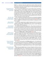
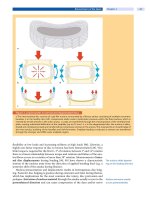
Instrumentation and correction of the deformity follow the cantilever and poste-
rior tension bend (compression) principle. The uppermost instrumented verte-
bra is the upper end vertebra of the deformity. Distally, the first lordotic segment
caudal to the apex should be included [39, 40, 41, 53, 56].
Claw constructs or pedicle screws areusedabovetheapexofthedeformity,
pedicle screws in the lower part of the instrumentation. A two-rod construct
(
Case Study 2) or a four-rod construct can be used for the correction maneuver
(
Fig. 10a, b). Stiff rods should be chosen to minimize the risk of loss of correction.
ab
Figure 10. Cantilever technique
Instrumentation/correction using cantilever and posterior tension band principle: a two-rod technique and b four-rod
technique.
Juvenile Kyphosis (Scheuermann’s Disease) Chapter 28 785
ab
c
de
f
g
Case Study 2
A 16-year-old male student was admitted for
assessment and treatment of thoracic hyperky-
phosis. The patient had no earlier treatment or
radiographs. The deformity had developed dur-
ing 3 years. He complained about mild thoraco-
lumbar pain after exercising and was dissatisfied
with the cosmetic appearance of his back. Other-
wise, he was healthy. Clinically, he exhibited the
typical features of Scheuermann’s kyphosis in
the lower thoracic spine (
a–c) . The deformity was pain free and corrected partially in extension. Bilateral hamstring tight-
ness of 50 degrees was present, and there were no pathologic neurological signs. On the standing lateral radiograph,
thoracic kyphosis measured 95 degrees (
d). It corrected to 54 degrees on the supine extension film (e). Around the apex
(T8) there were five wedge vertebrae. The standing posteroanterior radiograph was normal (
f). MRI showed typical
Scheuermann’s changes, and no cord compression or other pathology (
g).
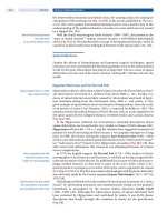
During the correction maneuver the area of the release should be watched very
carefully to detect and avoid cord compression due to translation of the vertebrae
or kinking of the laminae. The interlaminar gaps should not be fully closed at the
end of the correction maneuver to allow for drainage of possible hematoma.
After instrumentation the posterior elements of the area are decorticized with
786 Section Spinal Deformities and Malformations
hi jk
Case Study 2 (Cont.)
As the deformity was relatively mobile, brace treatment was considered. It was, however, discarded because of the mini-
mal remaining spinal growth left (Risser 4, skeletal age 18 years).Aposteriorrelease,UniversalSpineSystem(USS)instru-
mentation/correction using the two-rod cantilever tension band principle, and a posterior fusion from T2 to L2 were per-
formed. There were neither intraoperative nor postoperative complications. The cosmetic result looked very satisfactory
(
h, i). On radiographs 6 months after operation, thoracic kyphosis measured 48 degrees (j, k).
great care and packed with autogenous or allogenous bone graft to achieve a
thick solid fusion mass. Spinal cord monitoring and/or wake-up test are manda-
tory. Prophylactic antibiotics are recommended.
Combined Anterior/Posterior Approach
A combined anterior/poste-
rior approach is indicated in
very rigid kyphosis
In very rigid severe deformities, especially in adult patients, a combined
approach may be considered (
Fig. 9d). However, there are no scientifically based
numeric data available informing the surgeon which cases need additional ante-
rior release and which can be treated by posterior approach only. Halo-femoral
traction, used by some authors during the interval between staged anterior and
posterior surgery, does not seem to improve final results [12, 29].
Through an anterior approach the rib heads, the anterior longitudinal liga-
ment, the intervertebral discs down to the posterior longitudinal ligament, and
the cartilaginous vertebral endplates in the area of the deformity are resected.
The disc spaces are distracted and filled with bone graft (morcellized rib). Tradi-
tionally, this has been performed through a thoracotomy as an open procedure.
The literature has shown that thoracoscopic anterior release is effective in
Juvenile Kyphosis (Scheuermann’s Disease) Chapter 28 787
Scheuermann’s kyphosis [1]. Its definitive advantages over classic open thoracot-
omies are cosmesis and less morbidity. It does, however, have a considerable
learning curve [45].
Results of Operative Treatment
Surgery provides
a favorable outcome
in selected cases
Outcome data after operative treatment of Scheuermann’s kyphosis comprise
mainly retrospective short-term or mid-term follow-up reports. Results are ana-
lyzed usually according to the two major indications for which the surgery was
carried out: pain and deformity. As far as pain is concerned, all series report an
improvement in the amount of back pain of between 60% and 100% [12, 15, 29,
31, 60]. Hosman et al. showed a marked improvement concerning back pain in 31
out of 33 patients after a mean follow-up of 4.5 years. However, neck pain did not
seem to have improved after surgery. Interestingly, no relationship between the
amount of correction and the amount of residual back pain was found. As far as
patients’ satisfaction is concerned, most series report a very high satisfaction rate
of up to 96% [31].
As no cosmetic scale has been available for the assessment of juvenile kypho-
sis, one has to judge the cosmetic correction on plain radiographs, which repre-
sent an extrapolation of the cosmetic results. The rate of correction given in the
different surgical series is 21–51%. Loss of correction in the instrumented area
is minimal at present due to the rigidity of instrumentation systems used
(
Table 8). Ideally, the result of correction of juvenile kyphosis should be assessed
according to patient satisfaction and improvement of perceived self-image and
independent judgement of clinical photographs before and after the surgery by
non-medical observers. The literature definitively lacks such information. The
resultsofcorrectivesurgeryshouldnotbebasedonCobbanglecorrectionalone
but rather on outcome instruments such as the SRS 24, the sagittal balance of the
patient, and the assessment of spinal mobility and function. So far, only Poolman
et al. have used the SRS questionnaire instrument, which includes assessment of
the cosmetic situation [56].
Table 8. Surgical treatment of juvenile kyphosis
Author N Technique Follow-up
time
(months)
Kyphosis
(degrees)
Outcome/complications Conclusions
Bradford
et al.
(1974)
22 post Harrington
compression
35 (5 –92) pre 72 (50– 128) pain relief 100%, cosmesis
improved 100 %
complications frequent
cast for
9.8 months
follow-up 47
(29–88)
pseudarthrosis 3, infection 1,
thromboembolia 1, neurologi-
cal 1
indication restricted to
patients with severe pain
correction: 35% need for combined
approach to avoid loss
of correction
loss >10 in
15/22 patients
Taylor et
al. (1979)
27 post Harrington
compression
26.6 (6 –72) Pre 72 (55 –93) pain relief 100%, cosmesis
improved 100 %
instrument/fusion too
short leading to loss of
correction
cast for
5 months
follow-up 46
(23–63)
new neck/shoulder pain 9/27
patients
recommendation to
fuse whole curve
correction: 36%
intraoperative lamina fracture 1,
pneumothorax 1, donor-site
hematoma 3, transient paresthesia
1, gastrointestinal obstruction 1
loss of correction:
in fusion: 7
outside: 12
788 Section Spinal Deformities and Malformations
Table 8. (Cont.)
Author N Technique Follow-up
time
(months)
Kyphosis
(degrees)
Outcome/complications Conclusions
Bradford
et al.
(1980)
24 anterior release 24– 68 pre 77 (54– 110)
hook site pain 2, fusion extended
forpain1,pulmonaryembolus/
deep femoral thrombosis 2, deep
infection 1, vascular obstruction
of duodenum 1, hematothorax 1,
pericardial effusion 1, pseudar-
throsis 1, intercostal neuroma 1,
discomfort at lower hook 3 (2
removed)
correction after com-
bined approach supe-
rior to anterior only but
greater morbidity
Halo traction
2 weeks
follow-up 47
(30–67)
post Harrington
compression
correction: 39%
Risser cast
9–12 months
loss of correc-
tion: mean 6
outside fusion:
13–25 in 5
patients
Herndon
et al.
(1981)
13 anterior release 29 (12 –66) pre 78 (61–95) pain relief in 8/13 patients, cos-
mesis improved 100%
significant risk of
severe complications
Halo traction
2 weeks
follow-up 45
(30–73)
mortality 1, instrumentation
problems 2, transient neurology
1, pressure sore 1, urinary reten-
tion 1, deep thrombosis 1, psy-
chological problems in halo 1
no advantage from
preoperative halo;
deformities over 70°
need combined
approach
post Harrington
compression
correction: 51%
Risser cast
6 months
Lowe
(1987)
24 anterior release 32 (19 –48) pre 84 (72– 105) pain relief in 18/24 patients,
cosmesis improved 100%
longer follow-up neces-
sary
Halo gravity
1 week
follow-up 49
(30–65)
transient hyperesthesia of trunk
and lower extremity 4, rod
removal for bursa 4, fusion too
short distally 2, rod migration 1
hyperesthesia worri-
some
posterior Luque
double rod
correction: 43% good patient accep-
tance
no external sup-
port
loss of correc-
tion: mean 5
Lowe and
Kasten
(1994)
32 anterior release
+posterior
Cotrel-Dubous-
set instrumen-
tation in 28
patients
42 (24 –74) pre 85 (75– 105) preoperative back pain 27/28
patients, at follow-up 18/28
mild back discomfort with vig-
orous activities
indication for surgery
symptomatic kyphosis
>75°
4patientspost
C-D only
follow-up 47
(24–65)
cosmetically satisfied 26/28
patients
negative sagittal bal-
ance in Scheuermann’s
correction: 45% proximal junctional kyphosis
26° (12°–49°) in 10/28 patients
due to overcorrection (> 50%)
or short fusion
avoid overcorrection to
avoid junctional kypho-
sis
loss of correc-
tion: 4 (0– 19)
distal junctional kyphosis 17°
(10°–30°) in 9/28 patients due to
short fusion
include proximal end
vertebra and first lor-
dotic segment distally
sagittal balance:
pre –5.3 cm
follow-up
–6.6 cm
Otsuka et
al. (1990)
10 posterior heavy
Harrington
compression
27 (18 –33) pre 71 (63–90) pain relief 100 %, cosmesis
improved 100 %
good cosmesis
improvement and pain
relief
Brace
6–9months
follow-up 39
(28–57)
rod breakage after motor vehi-
cle accident 1, intraoperative
lamina fracture 1
in flexible kyphosis
(bending to <50°) pos-
terior surgery only is
sufficient
correction 45% lung problems in patient with
preoperative congenital
obstructive lung disease 1
loss of correc-
tion: 8
Fusion too short 3
in 3/10 patients
loss >10
Juvenile Kyphosis (Scheuermann’s Disease) Chapter 28 789
Table 8. (Cont.)
Author N Technique Follow-up
time
(months)
Kyphosis
(degrees)
Outcome/complications Conclusions
Reinhardt
and
Bassett
(1990)
14 post Harrington
compression
32 (12 –65) pre 71 (54 –101) clinical outcome and complica-
tions not mentioned
to avoid junctional
kyphosis, fusion
beyond the end verte-
bra to a non-wedged
(“square”) vertebra nec-
essary
anterior release
in 6/14 patients
follow-up 37
(15–54)
distal junctional kyphosis 23°
(15°–31°) in 5/14 patients
cast or brace for
6 months
correction: 48% proximal junctional kyphosis
34° in one patient
loss of correc-
tion: 8 (4– 14)
Poolman
et al.
(2002)
23 anterior release 75 (25 –126) pre 70 (62–78) SRS outcome instrument at fol-
low-up: total score 83 (55– 106),
7patients<72
outcome relatively fair
post Cotrel-
Dubousset
13/23
follow-up 55
(36–65)
back pain increased 4, back pain
improved 10, self-image
improved 10, self-image wors-
ened 3, would have the proce-
dure again 16, no correlation
SRS score vs. radiography
loss of correction after
implant removal
Moss-Miami
10/23
correction: 21% aorta + thoracic duct lesion 1,
proximal junctional kyphosis 3,
screw breakage 3, painful hard-
ware 6
indication for surgery
questioned
loss of correc-
tion: mean 15°
in 8 patients
after rod
removal
Hosman
et al.
(2002,
2003)
33 posterior H-
frame instru-
mentation
A. Post only
50 (25 –93)
A+BPre79
(70–103)
Oswestry Disability Index Pre
21.3 (0 –72), follow-up 6.6
(0–52)
good radiographic and
clinical results. No bene-
fit from anterior release.
Excessive correction
should be avoided to
minimize risk for postop-
erative sagittal malalign-
ment.
anterior release
in 17/33
patients,
B. Combined
55 (24 –98)
follow-up 52
(32–81)
no difference if compared pos-
terior only versus combined sur-
gery
orthosis
3 months
correction: 34% cosmesis improved 100%
loss of correc-
tion: mean 1.4°
infection 3, instrumentation
removal for prominence or irri-
tation 4, loss of distal fixation
(reop.) 1, rod breakage 1, proxi-
mal junctional kyphosis 1
patients with ham-
string tightness have
significantly higher risk
for postoperative sagit-
tal imbalance
no difference A
vs. B
Complications
Operative kyphosis
correction carries the risk
of major complications
Surgery on juvenile kyphosis is not benign and complications can occur. Neu-
rological complications due to spinal cord compression can arise during the
correction maneuver because of a rare but preoperatively undetected intraspi-
nal problem, or due to a surgical technique failure. The exact rate of neurologi-
cal complications is not known in surgery of juvenile kyphosis. Probably, it
is higher than for idiopathic scoliosis operations. Possible complications such
as death, dura lesion, vascular lesion, lamina fracture, Brown-S´equard
syndrome, pulmonary problems, venous thrombosis, gastrointestinal ob-
struction, infection, instrument failure, and pseudarthrosis have been
described as in any major corrective procedure for spinal deformities [2, 4, 12,
15, 29, 39, 53, 56].
Postoperative sagittal
imbalance must be avoided
Proximal junctional kyphosis due to overcorrection occurs in 20–30% of
cases according to Lowe and Kasten [41]. Distal junctional kyphosis due to short
fusion causing lossof correction (“adding on”)outside the instrumented area has
been reported by several authors [12, 26, 29, 41, 58, 67]. Reinhardt and Bassett
790 Section Spinal Deformities and Malformations
saw distal junctional kyphosis if fusion was carried out to a wedged caudal end
vertebra of the kyphosis. They recommend including the next “square” vertebra
to allow smooth transition into lumbar lordosis [58]. Lowe postulates three pos-
sible mechanisms: firstly, fusion that is too short, distally stopping above the first
lordotic disc, results in distal junctional kyphosis; secondly, fusion that is too
short proximally and does not include the whole kyphosis on the top may cause
proximal junctional kyphosis and a goose neck appearance. Finally, overcorrec-
tion seems to be a factor and one should not correct the kyphosis to more than
50% of its initial value [40]. In the case of overcorrection, possibly the remaining
mobile segments below the fusion are unable to adapt to the alignment changes
caused by excessive kyphosis correction. As a result this leads to permanent
increased flexion stress on the segment adjacent to the fusion, finally causing its
breakdown. This view is supported by Hosman et al. [30], who stressed the
importance of tight hamstrings for surgical correction.
According to Poolman et al., significant loss of correction occurs after removal
of the instrumentation even if the fusion is healed [56]. Therefore, the metal
should not be removed if it is not imperative to do so, e.g. in the case of infection.
The benign natural history
must be weighed against
therisksofthesurgery
Overall, surgery in Scheuermann’s kyphosis bears the risk of serious complica-
tions, a risk the surgeon should be aware of. The benign nature of the deformity
should be kept in mind, and the risks and benefits of an operation should be
weighed up carefully.
Recapitulation
The sagittal alignment of the human spine devel-
opsduringgrowthandshowsgreatindividualvari-
ability. The range of thoracic kyphosis in healthy
people ranges from 10 to 60 degrees. There are no
evidence-based “normal values”.
Definition and epidemiology. “Classic” juvenile ky-
phosis (Type I)isarigid thoracic or thoracolumbar
hyperkyphosis due to wedge vertebrae develop-
ing during adolescence. The incidence is 1–8% ac-
cording to the literature. Atypical juvenile kyphosis
(Type II, “lumbar” Scheuermann’s kyphosis) affects
mainly the lumbar spine, is characterized by end-
plate changes of the vertebral bodies without sig-
nificant wedging, and leads to loss of lumbar lordo-
sis (flat back).
Pathogenesis. The exact etiology is unknown.Ge-
netic, hormonal, and mechanical factors have been
discussed. A disturbance of the enchondral ossifica-
tion of the vertebral bodies leads to wedge verte-
bra formation, causing increased kyphosis. Type II is
frequently seen in athletes as a sequela of axial
overloading.
Clinical presentation. A rigid thoracic hyperkypho-
sis with or without pain is the reason for consulta-
tion. Hamstring tightness is common. Abnormal
neurological signs are rare. In Type II, the lumbar
spine is stiff and pain symptoms are more promi-
nent.
Diagnostic work-up. Diagnosis is based on typical
changes seen on lateral standing plain radiographs:
hyperkyphosis, irregularity of the endplates,
wedged vertebrae, increased sagittal length on the
vertebral bodies, and narrowed disc spaces.
Schmorl’s nodes may be present but they are not
pathognomonic. MRI is taken if abnormal neuro-
logical signs are observed or in connection with
preoperative work-up.
Non-operative treatment. The general objectives
of treatment are to prevent progression of the
kyphosis, to correct the deformity, and to relieve
pain. The choice of treatment must consider the
natural history, which is benign in the majority of
cases. In Type I, back pain is common but usu-
ally mild. Type II and kyphosis of greater than 70 de-
grees causes more clinical symptoms. Pulmonary
compromise occurs only in severe deformities
(>100 degrees). Bracing and casting are effective in
mobile deformities of between 45 and 60 degrees if
at least 1 year of growth is left.
Juvenile Kyphosis (Scheuermann’s Disease) Chapter 28 791
Operative treatment. Theonlyabsoluteindication
for surgery is a neurological compromise (spastic
paraparesis). Kyphosis greater than 75 degrees,
pain, and severe cosmetic impairment are relative
indications. The benign natural history should be
kept in mind and overtreatment must be avoided.
Posterior correction, instrumentation and fusion
are sufficient in the majority of cases. In very severe
rigiddeformitiesacombinedapproachwithaddi-
tional anterior release can be considered. The oper-
ative results are good in most cases concerning
pain relief and cosmesis. Severe intra- and postop-
erative complications have been described. The
risks and benefits of operative treatment must be
weighed carefully against the benign natural his-
tory.
Key Articles
Arlet V (2000) Anterior tho racoscopic spine release in deformity surgery: a meta-analy-
sis and review. Eur Spine J 9 Suppl 1:S17 – 23
This is a meta-analysis of all the literature available on thoracoscopic spine release done
for scoliosis or kyphosis. Thoracoscopic release has been effective in kyphosis for curves
with an average of 78 degrees that were corrected after video-assisted thoracoscopic
release and posterior surgery to 44 degrees. No report of the surgical outcome (balance,
rate of fusion, rib hump correction, cosmetic correction, pain, and patient satisfaction)
was available for any series.
Bernhardt M, Bridwell KH (1989) Segmental analysis of the sagittal plane alignment of
the normal thoracic and lumbar spines and thoracolumbar junction. Spine 14:717 –21
This is a review of the normal sagittal alignment of the spine segment by segment in 102
healthy individuals, indicating that there is a wide range of normal sagittal alignment of
the thoracic and lumbar spines. The thoracolumbar junction is for all practical purposes
straight; lumbar lordosis usually starts at L1–2 and gradually increases at each level cau-
dally to the sacrum.
Hosman AJ , de Kleuver M, Anderson PG, van Limbeek J, Langeloo DD, Veth RP, Slot GH
(2003) Scheuermann kyphosis: the importance of tight hamstrings in the surgical cor-
rection. Spine 19:2252 – 9
The author reviewed 33 patients with juvenile kyphosis who underwent surgical correc-
tion. Sixteen patients had tight hamstrings, and 17 patients had non-tight hamstrings.
Hamstrings were considered tight if the popliteal angle was >30 degrees. Patients with
tight hamstrings had a significantly greater risk of postoperative imbalance (p<0.05).
Tight hamstring patients can be classified as “lumbar compensators” and as such are
prone to overcorrection and imbalance.
HosmanAJ,LangelooDD,deKleuverM,AndersonPG,VethRP,SlotGH(2002)Analysis
of the sag ittal plane after surg ical management for Scheuermann’s disease: a view on
over correction and the use of an anterior release. Spine 2:167 –75
A cohort of 33 patients who had undergone surgery for their Scheuermann’s kypho-
sis were reviewed: Group A: posterior technique (n=16); Group B: anteroposterior
technique (n=17). At follow-up evaluation (4.5±2 years) there was no difference in
curve morphometry, correction, sagittal balance, average age, and follow-up period
between Groups A and B. In reducing postoperative sagittal malalignment, the
authors believe that surgical management should aim at a correction within the high
normal kyphosis range of 40–50 degrees, consequently providing good results and,
particularly in flexible adolescents and young adults, minimizing the necessity for an
anterior release.
MurrayPM,WeinsteinSL,SprattKF(1993) The natural history and long-term follow-up
of Scheuermann kyphosis. J Bone Joint Surg Am 75A:236 –48
Sixty-seven patients who had a diagnosis of Scheuermann kyphosis and a mean angle of
kyphosis of 71 degrees were evaluated after an average follow-up of 32 years. The results
were compared with those in a control group of 34 subjects who were matched for age and
sex: The patients who had juvenile kyphosis had more intense back pain, jobs that tended
to have lower requirements for activity, less range of motion of extension of the trunk and
792 Section Spinal Deformities and Malformations
less-strong extension of the trunk, and different localization of the pain. No significant
differences between the patients and the control subjects were demonstrated for level of
education, number of days absent from work because of low-back pain, extent that the
pain interfered with activities of daily living, presence of numbness in the lower extremi-
ties, self-consciousness, self-esteem, social limitations, use of medication for back pain,
or level of recreational activities.
Poolman RW, Been HD, Ubags LH (2002) Clinical outcome and radiographic results
after operative treatment of Scheuermann’s disease. Eur Spine J 11: 561 –9
This paper is a prospective study to evaluate radiographic findings, patient satisfaction
and clinical outcome, and to report complications and instrumentation failure after
operative treatment of Scheuermann’s kyphosis using a combined anterior and poste-
rior spondylodesis. Significant correction was maintained at 1 and 2 years follow-up but
recurrence of the deformity was observed at the final follow-up. The late deterioration of
correction in the sagittal plane was mainly caused by removal of the posterior instru-
mentation, and occurred despite radiographs, bone scans and thorough intraoperative
explorations demonstrating solid fusions. There was no significant correlation between
the radiographic outcome and the SRS score. Therefore, the indication for surgery in
patients with Scheuermann’s disease can be questioned and surgery should be limited to
patients with kyphosis greater than 75 degrees in whom conservative treatment has
failed.
Soo CL, Noble PC, Esses SI (2002) Scheuermann kyphosis: long-term follow-up. Spine J
2:49 – 56
Sixty-three patients were evaluated a mean of 14 years after treatment (10–28 years)
using a specially designed questionnaire. The patients had been treated using three dif-
ferent treatment modalities: exercise and observation, Milwaukee bracing, and surgical
fusion using the Harrington compression system. At the time of follow-up evaluation,
there were no differences in marital status, general health, education level, work status,
degree of pain and functional capacity between the various curve types, treatment
modality and degree of curve. Patients treated by bracing or surgery did have improved
self-image. Patients with kyphotic curves exceeding 70 degrees at follow-up had an infe-
rior functional result.
StagnaraP,DeMauroyJC,DranG,GononGP,CostanzoG,DimnetJ,PasquetA(1982)
Reciprocal angulation of vertebral bodies in a sagittal plane: Approach to references for
theevaluationofkyphosisandlordosis.Spine7:335 – 342
This report establishes a table of references for kyphosis and lordosis in a sample of
100 healthy adults (43 females, 57 males, age 20–29 years) from France. Segmental
measurements were carried out from standing lateral radiographs of the whole spine.
Mean thoracic kyphosis was 37 degrees (range 7–63); mean lumbar lordosis was
50 degrees (range 32–84). The majority of individuals had a thoracic kyphosis of
between 30 and 50 degrees. There was a correlation between sacral slope and lumbar
lordosis and thoracic kyphosis. The considerable variability is stressed. As the distri-
bution was found to be irregular, the authors consider it unreasonable to speak of nor-
mal kyphotic or lordotic curves. They state that average values are only indicative not
normative.
References
1. Arlet V (2000) Anterior thoracoscopic spine release in deformity surgery: a meta-analysis
and review. Eur Spine J 9 Suppl 1:S17–23
2. Ascani E, La Rosa G (1994) Scheuermann’s kyphosis. In: Weinstein SL (ed) The paediatric
spine: Principles and practice. Raven Press, New York, pp 557–584
3. Aufdermaur E (1981) Juvenile kyphosis (Scheuermann’s disease): Radiography, histology
and pathogenesis. Clin Orthop 154:166–174
4. Bauer R, Erschbaumer H (1983) Die operative Behandlung der Kyphose. Z Orthop 121:367
5. Bernhardt M, Bridwell KH (1989) Segmental analysis of the sagittal plane alignment of the
normal thoracic and lumbar spines and thoracolumbar junction. Spine 14:717–21
6. Bhojraj SY, Dandavate AV (1994) Progressive cord compression secondary to thoracic disc
lesions in Scheuermann’s kyphosis managed by posterolateral decompression, interbody
Juvenile Kyphosis (Scheuermann’s Disease) Chapter 28 793
fusion and pedicular fixation. A new approach to management of a rare clinical entity. Eur
Spine J 3:66–69
7. Blumenthal SL, Roach J, Herring JA (1987) Lumbar Scheuermann’s. A clinical series and
classification. Spine 9:929–32
8. Bosecker EH (1958) unpublished data cited in 10
9. Bouley C, Tardieu C, Hecquet J, Benaim C, Mouilleseaux B, Marty C, Prat-Pradal D, Legaye
J, Duval-Beaup´ere G, P´elissier J (2006) Sagittal alignment of spine and pelvis regulated by
pelvic incidence: standard values and prediction of lordosis. Eur Spine J 15:415–22
10. Bradford DS (1977) Editorial comment. Kyphosis. Clin Orthop Rel Res 128:2–4
11. Bradford DS (1977) Juvenile kyphosis. Clin Orthop Rel Res 128:45 –55
12. Bradford DS, Ahmed KB, Moe JH, Winter RB, Lonstein JE (1980) The surgical management
of patients with Scheuermann’s disease. J Bone Jt Surg [Am] 62A:705–12
13. Bradford DS, Garcia A (1969) Neurological complications in Scheuermann’s disease. J Bone
Jt Surg [Am] 51A:567–72
14. Bradford DS, Moe JH, Montalvo FJ, Winter RB (1974) Scheuermann’s kyphosis and round-
back deformity. Results of Milwaukee brace treatment. J Bone Joint Surg [Am] 56A:740–58
15. Bradford DS, Moe JH, Montalvo FJ, Winter RB (1975) Scheuermann’s kyphosis. Results of
surgical treatment by posterior spine arthrodesis in twenty-two patients. J Bone Joint Surg
[Am] 57A:439–48
16. Bruns I, Heise U (1994) Spastische Paraparese bei Morbus Scheuermann. Eine Kasuistik. Z
Orthop Ihre Grenzgeb 132:390– 393
17. Chiu KY, Luk KD (1995) Cord compression caused by multiple disc herniations and intra-
spinal cyst in Scheuermann’s disease. Spine 20:1075– 79
18. Edgar MA, Mehta MH (1988) Long-term follow-up of fused and unfused idiopathic scolio-
sis.JBoneJtSurg[Br]70B:712–16
19. Edgren W, Vainio S (1957) Osteochondrosis juvenilis lumbalis. Acta Chir Scand Suppl
227:3–47
20. Fallstrom K, Cochran T, Nachemson A (1986) Long-term effects on personality develop-
ment in patients with adolescent idiopathic scoliosis. Influence of type of treatment. Spine
11:756–58
21. Findlay A, Conner AN, Connor JM (1989) Dominant inheritance of Scheuermann’s juvenile
kyphosis. J Med Genet 26:400–403
22. Fowles JV, Drummond DS, L’Ecuyer S, Roy L, Kassab MT (1978) Untreated scoliosis in the
adult. Clin Orthop Rel Res 134:212–17
23. Greene TL, Hensinger RN, Hunter LY (1985) Back pain and vertebral changes simulating
Scheuermann’s disease. J Ped Orthop 5:1–7
24. Greulich WW, Pyle SI (1970) Radiographic atlas of skeletal development of the hand and
wrist. Stanford University Press, Stanford, CA
25. Griss P, Pfeil J (1983) Ergebnisse rein dorsaler und combiniert ventrodorsaler Aufrichtungs-
operationen bei der juvenilen Kyphose. Eine vergleichende Untersuchung am eigenen
Krankengut. Z Orthop 121:369
26. Gutowski WT, Renshaw TS (1988) Orthotic results in adolescent kyphosis. Spine 5:485–89
27. Haglund P (1923) Prinzipien der Orthopädie. Gustav Fischer Verlag, Jena, p 495
28. Halal F, Gladhill RB, Fraser C (1978) Dominant inheritance of Scheuermann’s juvenile
kyphosis. Am J Dis Child 132:1105–1107
29. Herndon WA, Emans BJ, Micheli LJ, Hall JE (1981) Combined anterior and posterior fusion
for Scheuermann’s kyphosis. Spine 6:125–130
30. Hosman AJ, de Kleuver M, Anderson PG, van Limbeek J, Langeloo DD, Veth RP, Slot GH
(2003) Scheuermann kyphosis: the importance of tight hamstrings in the surgical correc-
tion. Spine 19:2252–9
31. Hosman AJ, Langeloo DD, de Kleuver M, Anderson PG, Veth RP, Slot GH (2002) Analysis of
the sagittal plane after surgical management for Scheuermann’s disease: a view on overcor-
rection and the use of an anterior release. Spine 2:167 –75
32. Ippolito E, Bellocci M, Montanaro A, Ascani E, Ponseti IV (1985) Juvenile kyphosis: An
ultrastructural study. J Ped Orthop 5:315–322
33. Ippolito E, Ponsetti IV (1981) Juvenile kyphosis, histological and histochemical studies.
J Bone Jt Surg [Am] 63A:175–182
34. Lambrinudi C (1934) Adolescent and senile kyphosis. Br Med J 2:800–4
35. Lang G, Kehr P, Aebi J, Paternotte H (1983) Die Behandlung der regulären Kyphose beim
Jugendlichen. Z Orthop 121:368
36. Legaye J, Duval-Beaupere G, Hecquet J, Marty C (1998) Pelvic incidence: a fundamental pel-
vic parameter for three-dimensional regulation of spinal sagittal curves. Eur Spine J 7:
99–103
37. Lindemann K (1933) Die lumbale Kyphose im Adoleszentenalter. Z Orthop 58:54–65
38. Lonstein JE, Winter RB, Moe JH, Bradford DS, Chou SN, Pinto WC (1980) Neurologic deficit
secondary to spinal deformity. A review of the literature and report of 43 cases. Spine 5:
331–55
794 Section Spinal Deformities and Malformations


