OPTICAL BIOSENSORS PRESENT AND FUTURE - PART 1 doc
Bạn đang xem bản rút gọn của tài liệu. Xem và tải ngay bản đầy đủ của tài liệu tại đây (25.1 MB, 299 trang )
PREFACE
Since the birth of the field of optical biosensors, the pace of evolution of this
field has been swift. While myriad reports have appeared describing applications
and advancements in optical biosensor technology, few existing volumes are
dedicated to a synopsis of this field. Since the development of optical biosensors
mirrors the advances in the rapidly evolving telecommunications industry, we
deemed the time to be ripe for such an opus. In order to catch the wave of this
rapidly developing technology, we endeavored to focus both on the current state
of the art and on technologies that will influence tommorrow's state of the art.
We hope that this particular compendium of concepts will trigger new synapses
to foma in the brains of our readers and yield even more innovation in the years
to come. The history sections are included in order to recognize the contributions
of the giants upon whose shoulders we stand and we thank them for their
creativity and pioneering spirit. These sections are comparatively short, not so as
to minimize such contributions, but so that this book actually gets published in a
single volume.
According to the thematic focus on Present and Furore technology, the book is
divided into two parts. In the first part, we compiled a list of the most
outstanding optical biosensor technologies, while in the second part, the editors
used their crystal ball to select the science we deem exciting and promising in
terms of potential impact on biosensors. The optical biosensor technologies
include two very different fiber optic biosensors, planar waveguides, and the
displacement flow sensors, as well as sensors based on time-resolved
fluorescence, electrochemiluminescence, surface plasmon resonance, resonant
mirrors, and interferometry. The science for future technology development
includes four different methods for producing new recognition elements (genetic
engineering of proteins, chemical synthesis, combinatorial selection of
nucleotide-based receptors, and molecular imprinting), two methods for
immobilizing receptors on biosensors (sol gels and semi-synthetic membranes),
two methods for producing very bright signals (PEBBLES and quantum dots),
and soft lithography for surface patterning and microfluidics. We have asked
leaders in each field to provide our readers with as thorough and objective a
chapter as possible; they and their colleagues have been very patient with our
nagging and nit picking and, as will be obvious to you, have put inordinant
amounts of time into providing a conscientious review of their field.
We tasked the authors to describe the underlying principles behind each
technology, enumerate the types of applications for which it has been tested,
provide their opinions about the advantages and disadvantages of their favorite
vii
Preface
biosensor (and the objectivity each has provided is admirable!), and philosophize
on the future developments using that particular biosensor. The last section is
intended to be fun for the readers as well as the authors; however, it is available
for any clever venture capitalist to peruse as well.
Finally, the editors intend this book to be a gift of gratitude to our colleagues in
this rapidly expanding field. We appreciate the open sharing of ideas, the
encouragement, and the competition that motivates us to greater effort. To work
in the field of optical biosensors, one must be curious about biochemistry,
chemistry, physics, and engineering and the possibilities ever present in the
cracks between the disciplines. While information overload is a serious threat,
boredom never is. Since it is absolutely impossible to be expert in all these
fields, it behooves us to join forces with those who are. But even more than the
ideas and accomplishments of our fellows, we delight in their personalities and
camaraderie.
Sincerely,
Fran and Chris
viii
Optical Biosensors: Present and Future
F.S. Ligler and C.A. Rowe Taitt (editors)
9 2002 Elsevier Science B.V. All fights reserved
CHAPTER 1
OPTRODE-BASED FIBER OPTIC BIOSENSORS
(BIO-OPTRODE)
ISRAEL BIRAN, PH.D. AND DAVID R. WALT, PH.D.
The Max Tishler Laboratory for Organic Chemistry
Department Of Chemistry
Tufts University, Medford, MA 02155 USA
Optrode-based fiber optic biosensors (bio-optrodes) are analytical devices incorporating
optical fibers and biological recognition molecules. Optical fibers are small and flexible
"wires" made out of glass or plastic that can transmit light signals, with minimal loss,
over long distances. The light signals are generated by a sensing layer, which is usually
composed of biorecognition molecules and dyes, coupled to the fiber end. Light is
transmitted through the optical fibers to the sensing layer where different optical
phenomena such as absorption or luminescence are used to measure the interactions
between the analyte and the sensing layer. Bio-optrodes can be used for remote
analytical applications including clinical, environmental, and industrial process
monitoring. In the last decade, due to the rapidly growing use of fiber optics for
telecommunication applications, new fiber optic technologies have been developed
resulting in high-quality and inexpensive optical fibers that can be used for bio-optrode
applications. Recent advancements in bio-optrode technologies include the development
of nanoscale bio-optrodes, enabling measurements inside single living cells, and the
development of multi-analyte and reagentless bio-optrodes. Although currently no bio-
optrodes are commercially available, it is expected that the development of advanced
bio-optrode technologies will lead to commercially available devices for various
analytical applications.
Biran and Walt
Figure 1. Schematic diagram of optrode system.
I. Principle of Operation
The word "optrode" is a combination of the words "optical" and "electrode" and
refers to a fiber optic based analytical device that can measure the concentration
of a specific chemical or a group of chemicals in a sample of interest. The basic
design of an optrode system is shown in Figure 1. The main components of an
optrode are: (a) a light source; (b) an optical fiber to both transmit the light and
act as the substrate for (c) the sensing material, which is usually immobilized to
the surface of the end face of the fiber; and (d) a detector to measure the output
light signal. Computers or microprocessors are used to control the optrode
instrumentation and are employed to analyze the output signals.
The "heart" of the optrode is the sensing element. When the sensing element
interacts with the analyte, it undergoes physico-chemical transformations that
change its optical properties. This transduction mechanism generates optical
signals that can be correlated to the analyte concentration. The optical signals are
measured by launching light from the light source through the optical fiber to the
fiber end, where the sensing element is immobilized. The same fiber (Figure 1),
or a different fiber (Figure 6), is used to guide the output light to the detector
Optrode-based Fiber Optic Biosensors
Core (nl)
Cladding (n2) Jaclket
Figure 2. Schematic diagram of an optical fiber shows core and clad structure.
(e.g., spectrophotometer, fluorometer) where the reflected, emitted or absorbed
light is measured. Optrode biosensors or bio-optrodes are optrodes in which the
sensing elements are of biological origin. Biological sensing elements, such as
enzymes, nucleic acids, antibodies and cells, are immobilized on optical fibers
and used for specific recognition of many different analytes (Cunningham, 1998;
Kuswandi et al., 2001; Mehrvar et al., 2000; Wolfbeis, 2000). Since most
biological sensing elements and most analytes do not possess intrinsic spectral
properties, the biorecognition events are transduced to optical signals (e.g.
changes in fluorescence or absorbance) by coupling optically responsive reagents
to the sensing elements. For example, fluorescent dyes are used to label nucleic
acids and convert the biorecognition interaction between two complementary
DNA strands into a fluorescence signal. In another example, an indicator dye,
which is optically sensitive to changes in H + concentrations, is used to transduce
enzymatic activity that consumes or releases H § into an optical signal. The
signals are generated on the fiber optic face and transmitted by the optical fiber to
a remote measurement device. The small dimensions of bio-optrodes allow
measurement in very small sample volumes, which make them suitable for
various clinical applications (Meadows, 1996; Vo-Dinh and Cullum, 2000). Bio-
optrodes are also useful for different sensing applications in the industrial and
environmental fields (Rogers and Mascini, 1998; Rogers and Poziomek, 1996;
Marose et al., 1999; Mulchandani and Bassi, 1995; Scheper et al., 1996).
In this section, optical fibers, their basic characteristics, and the optical methods
used to transduce a biorecognition event to an optical signal are described. The
instrumentation employed in optrode biosensors, the biological sensing elements,
and the methods to immobilize them on the fiber optic surfaces are summarized.
Biran and Walt
Acceptance
cone
l n~ L: ' 7 [Cladding (a)
n 2 .
Cladding
(b)
Core
[ "- - n~
"
Core
(c)
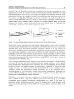
Figure 3. Propagation of light through the optical fiber occurs when the total internal
reflection condition exists at the interface between the core, (nl), and clad,
(n2)
such that
nl > n2. (a) Light entering the fiber is totally internally reflected (TIR), if the light angle is
greater than the critical angle (pc. (b) Light will be partially reflected and partially
refracted, if the light angle is less then the critical angle ~oc. (c) Light will propagate in
TIR only when the entering light angle is within the acceptance cone angle (~0m) range.
1.1. Optical fiber characteristics and use in bio-optrodes
Optical fibers are small and flexible "wires" made out of glass or plastic that can
transmit light signals, with minimal loss, for long distances. Optical fibers are
remarkably strong, flexible and durable and therefore can be used in harsh and
hazardous environments. Optical fibers are non-electrical, which make them
highly suitable for applications where the presence of electric current is
detrimental (e.g.,
in-vivo
monitoring inside a patient body). In the last decade,
due to the rapidly growing use of fiber optics for telecommunication applications,
new fiber optic technologies have been developed resulting in high-quality and
inexpensive optical fibers that can also be used for sensing applications. Optical
fibers can transmit multiple optical signals simultaneously, thereby offering
multiplexing capabilities for sensing.
Optrode-based Fiber Optic Biosensors
Optical fibers consist of a core with a refractive index, n~, surrounded by a
cladding with a lower refractive index, n2 (Figure 2). The difference in the
refractive indices between the core and the cladding enables the core-clad
interface to effectively act as a mirror such that a series of internal reflections
transmits the light from one end of the fiber to the other as shown in Figure 3 (a).
Light undergoes total internal reflection (TIR) at the core-clad interface if two
basic conditions are fulfilled: (a) The light strikes the cladding at an angle greater
than the
critical angle, (Pc,
(Figure 3 (a) and 3 (b)). The critical angle is defined by
the ratio between the clad and the core refractive indices, as shown in Equation
(1):
sin (p~
- n 2/n
1
(1)
(b) The angles of the light entering the fiber should be within the
acceptance
cone
as shown in Figure 3 (c). The acceptance cone angle, (am, depends on the
refractive indices of the core and the clad and also on the refractive index of the
medium from which the light enters the fiber,
no.
sin(Pm = (2)
l't o
Another important parameter that defines the fiber's light collection efficiency is
the
numerical aperture
(NA). This parameter is related to the acceptance cone's
angle and is given by:
NA = n0sinfo m (3)
A high NA indicates a wide acceptance cone and better light gathering
capabilities of the fiber. A typical NA value for a high quality glass fiber is 0.55,
but fiber NAs as high as 0.66 or as low as 0.22 have been used for sensing.
Optical fibers are usually made out of plastic and glass and have many different
configurations, formats, shapes, and sizes. Glass fibers are the most commonly
used fibers in optrode biosensors. Glass optical fibers can transmit light in the
visible and near-infrared regions of the optical spectrum (400 nm< ~, < 700 nm)
and are therefore suitable for measuring fluorescence signals generated by most
fluorescent dyes. For applications in which light in the UV region is required,
quartz (pure silica) is used as the fiber's core material and doped silica (with a
lower refractive index) is used as the cladding material. For most fiber optic-
based biosensors, optical fibers with diameters ranging from 50 to 500 ~tm are
employed.
Biran and Walt
Figure 4. Optical fiber bundle fabrication and its use for imaging. (a) Fiber bundles are
constructed from thousands of individual single fibers that are fused together. (b)
Coherent bundles can be used for imaging (Pantano and Walt, 1995). Reprinted with
permission from the American Chemical Society.
Recently, fiber optic bundles (Figure 4(a)) comprising thousands of identical
single fibers each with a diameter of a few micrometers, were employed for bio-
optrodes. The fibers can be bundled in a coherent or random fashion. In coherent
fiber bundles, the position of each fiber on one end is identical to its position on
the other end. These fibers were originally designed for imaging applications as
shown in Figure 4(b) and are also often called "optical imaging fibers". Imaging
fibers are suitable for multi-analyte optrode biosensor design (Healey and Walt,
1995; Healey et al., 1997a; Michael et al., 1998; Steemers and Walt, 1999; Walt,
2000) since each small individual fiber in the bundle can carry its own light
signal from one end of the bundle to the other. Moreover, optical imaging fiber-
based biosensors can be used for sensing and imaging simultaneously, providing
remote spatial sensing capabilities (Walt, 1998).
1.2. Optical phenomena employed for biosensing in bio-optrodes
In bio-optrodes, dyes are coupled to the biological sensing element and transduce
the biorecognition events to an optically detectable signal. Different optical
10
Optrode-based Fiber Optic Biosensors
phenomena, including fluorescence, luminescence and absorption, are employed
for monitoring these optical changes. In this section, the basic principles of these
phenomena and their use in bio-optrodes are described.
Fluorescence
is commonly used in bio-optrodes. Fluorescence occurs when
molecules are excited at a specific wavelength and re-emit radiation at a lower
energy, i.e., a longer wavelength. The absorption of the excitation light promotes
the molecule's energy from its ground state to a higher energy state. The
molecule emits fluorescent light when it returns to the ground state. Each
fluorescent molecule has a unique fluorescence spectrum since the excitation and
emission occur only at distinct energy levels corresponding to particular
wavelengths. The characteristic fluorescence spectrum of particular molecules
allows multiple fluorescent dyes to be used simultaneously in a single analytical
assay. In fluorescence-based bio-optrodes, the fluorescence signals are measured
by transmitting the excitation light through an optical fiber and measuring the
light emission using a detector. Usually the increase or decrease in fluorescence
intensity is measured and then correlated to the analyte concentration. For
example, when a fluorescent-labeled antibody is used as the sensing element, the
fluorescence intensity is proportional to the amount of antigen (analyte) bound to
the optical fiber. One method for measuring fluorescence lifetime is frequency-
domain. In this method, sinusoidally modulated light is used to excite the
fluorescent molecule. The resulting emission light also oscillates at the same
frequency. The emission light is phase shifted (delayed) and demodulated with
respect to the excitation light because of the finite lifetime of fluorescence. The
phase shift is expressed as a phase angle from which the lifetime can be
determined using simple relationships between the modulation frequency and the
degree of demodulation. The concentration of analyte that induces changes in the
molecule's fluorescence lifetime can be determined by measuring phase angle
values (Thompson et al., 1996).
A decrease in fluorescence intensity due to quenching can also used for sensing.
In this case, the biorecognition event causes a decrease in fluorescence
(quenching) of the fluorescent reporter molecule. The fluorescence decrease is
related to the analyte concentration. For example, a dye that undergoes
fluorescence quenching when the pH decreases can be coupled to an enzymatic
reaction that converts a substrate into an acidic product and results in a pH drop.
Thus, the decrease in fluorescence can be correlated to the analyte concentration
(see also Section 1.4.1). Fluorescence quenching is also one manifestation of
another fluorescence phenomenon used for sensing in bio-optrodes
-fluorescence
resonance energy transfer (FRET).
This phenomenon occurs when two distinct
fluorophores are present. If the emission spectrum of one fluorophore overlaps
with the excitation spectrum of a second fluorophore, and the two fluorophores
are in proximity (<100/~ ), then the excited fluorophore (donor) can transfer
energy non-radiatively to the second fluorophore (acceptor). There are two types
of acceptors. Quenchers are acceptors that are not fluorescent and therefore
11
Biran and Walt
cause the donor simply to decrease its fluorescence emission intensity.
Acceptors can also be fluorescent dyes that accept the energy non-radiatively
from the donor, and then re-emit the energy at specific emission wavelength.
This energy transfer results in an increase in light emission by the acceptor and a
decrease in light emission from the donor. When an energy transfer pair of
fluorophores is used to label two interacting molecules (e.g., antibody-antigen,
enzyme-substrate), they can be used for sensing. Recently, both the donor and the
acceptor molecules were incorporated into single biological molecules such as
proteins (Hellinga and Marvin, 1998) and nucleic acids (e.g., molecular beacons)
(Tyagi and Kramer, 1996; Tyagi et al., 2000). When these sensing molecules are
in their native conformation, the donor and the acceptor are in proximity and
therefore low fluorescence signals from the donor are obtained. When the
molecule interacts with the analyte, conformational changes occur that separate
the donor and the acceptor molecules and cause an increase in the fluorescence
from the donor (see Section 3.3).
The most commonly used fluorescent molecules in bio-optrodes are organic
dyes. Recently self-fluorescent proteins have also been used. The sources of
these proteins are marine organisms such as the jellyfish
Aequorea victoria
that
produce the green fluorescent protein (GFP) (Chalfie et al., 1994). When GFP is
excited, it emits light at a lower energy and therefore at a higher wavelength.
GFP is highly fluorescent, with a quantum efficiency of approximately 80% and
is very stable to heat and pH (5.5-12). The GFP has been expressed in different
cell types (bacteria, yeast, mammalian, plant) and used as reporter gene for
different cellular events (Naylor, 1999). In order to allow monitoring of several
cellular events simultaneously, several GFP mutants have been developed each
with unique excitation and emission wavelengths. Cells expressing fluorescent
proteins, and also the purified proteins have been used for constructing different
bio-optrodes (see Sections 1.4.1 and 3.3).
Time-resolved fluorescence spectroscopy
is another phenomenon used in bio-
optrodes. This method is based on the fluorescent molecule's excited state
lifetime. The light intensity emitted from a molecule excited by a short pulse of
light decays exponentially with time. The decay time pattern is unique for each
molecule and can be used for analytical purposes. Barker et al. (1999) used this
method to improve the performance of a bio-optrode for nitric oxide detection.
A different light emission phenomenon used in bio-optrodes is
chemiluminescence. In
contrast to fluorescence, chemiluminescence is produced
when a chemical reaction yields an excited species that emits light as it returns to
its ground state. The use of chemiluminescence in biosensors, including fiber
optic-based biosensors, was recently reviewed (Aboul-Enein et al., 2000; Gubitz
et al., 2001). In many bio-optrodes, the chemiluminescence of luminol is used to
generate the light signal. The reaction between luminol and H2Oz produces a
12
Optrode-based Fiber Optic Biosensors
Figure 5. Design of flow-cells incorporating bio-optrodes (Kuswandi et al., 2001).
Reproduced with permission of the Royal Society of Chemistry.
luminescence signal and is also catalyzed by certain ions or molecules (e.g.,
MnO42-, Iz, Cu2+). This reaction can be used, for example, in enzyme-based bio-
optrodes in which the enzymatic reaction generates H202 (see Section 3.3).
Enzymes such as horseradish peroxidase can also catalyze or induce a
chemiluminescence reaction by producing H2Oz. In addition, alkaline
phosphatase and 13-galactosidase can be used to label biological sensing elements
such as antibodies or nucleic acids. In the presence of a 1,2-dioxetane substrate
(Bronstein et al., 1996), these enzymes catalyze light formation proportional to
the analyte concentration. Chemiluminescence-based bio-optrodes are usually
used in conjunction with flow cells. An optical fiber with an immobilized sensing
element is placed inside the flow cell and transmits the light signals to the
detector (Figure 5).
Bioluminescence
is a biological chemiluminescent reaction. Many organisms
produce bioluminescence for signaling, self-protection, mating, attracting prey
and finding food (Campbell and Sala-Newby, 1993). The bioluminescence
reaction is catalyzed by the enzyme luciferase and requires the presence of
oxygen. The bioluminescent substrate used in this reaction is called luciferin.
Different luciferin molecules are used by different organisms. For example,
13
Biran and Walt
aldehydes and flavins are used by bacteria and imidazolopyrazines are employed
by some fish and squid. B ioluminescence can be applied for analytical
measurements in two ways: (1) One can detect cellular events inside living cells
by fusing the luciferase gene (e.g., the
luc
gene coding for firefly luciferase or the
lux
gene coding for the bacteria
Vibrio fischeri
luciferase) to the gene of interest.
The
in-vivo
activity of the selected gene can be detected by monitoring the
luminescence signal (LaRossa, 1998). (2) Alternatively, one can use purified
recombinant luciferase and synthetic luciferin substrates for
ex-vivo
detection
assays for analytes such as ATP, NADH and FMN (Blum et al., 1993). In bio-
optrodes, the cells or the purified enzymes are immobilized on the fiber tip and
the luminescence signals are transmitted through the fiber to the detector.
Absorption
is a simpler process than fluorescence and has also been used in bio-
optrodes. Absorption is a process in which light energy is absorbed by an atom or
a molecule, promoting the molecule from the ground energy state to a higher
energy excited state. The resulting energy is dissipated non-radiatively (i.e.,
thermally) to the medium when the excited state relaxes to the ground state. The
absorbance changes are related to the concentration [C] via the Beer-Lambert
relationship:
A = log(Io/I)= e.[C].l
(4)
where A is the optical absorbance, and
Io
and I are the intensities of transmitted
light in the absence and presence of the absorbing species respectively, 1 is the
effective path length, and e is the molar absorption coefficient. In practice,
optical fibers are used to measure absorbance by transmitting light through the
fiber to the-sensing layer and measuring changes in the scattered light.
Alternatively, light is transmitted through one arm of bifurcated optical fiber to
the sensing region and reflected light signals are measured through the other arm
of the fiber (Figure 6 (b)). In a different configuration, two fibers are placed with
one fiber facing the other creating an optical cell in which the distance between
excitation and collection fiber is the pathlength.
1.3. Optrode biosensor (bio-optrode) design and instrumentation
Different bio-optrode system designs have been used and recently reviewed
(Kuswandi et al., 2001; Mehrvar et al., 2000) The design of bio-optrodes is
similar to chemical optrode design and two basic configurations are used: (a) a
single fiber is used to transmit the light from the light source to the sample region
and back to the detector, as shown in Figure 1, or (b) multiple fibers are used in
which one fiber is employed to transmit the light to the sample region and the
other fiber or fibers are used to transmit light from the sample region to the
detector, as shown in Figure 6 (a). For the second configuration, the most
common format is a bifurcated fiber. Bifurcated fibers are fabricated by fusing
14
Optrode-based Fiber Optic Biosensors
(a)
()
) Lighi Source i
)' ~.etec;or ]
(b)
,,
, ight Source [
J ~ ' ~Detector. [
(c)
Sensing layer
Figure 6. Design principle of a bio-optrode. (a) Two fibers: one carries light to the
sensing layer and one carries the signal to the detector. (b) Bifurcated fiber: the
biosensing layer is placed on the fused end of the fiber (c) The biosensing layer is placed
on the central fiber and the surrounding fibers are used to collect the light signals.
two fibers on one end leaving the other ends free. The sensing elements are
immobilized on the fused side and the other ends of the bifurcated fiber are
connected to the light source and to the detector as shown in Figure 6 (b). In a
different configuration, multiple fibers comprising one central fiber surrounded
by several fibers are employed. The central fiber carries the immobilized sensing
elements and is connected to the light source; the surrounding fibers collect the
output light signals and transmit them to the detector (Figure 6 (c)).
The light sources used for bio-optrodes should provide sufficient light intensity
within the sensor wavelength operating range. In addition, the light output should
be stable over long time periods since light fluctuations may add noise to the
measurement and reduce the sensor sensitivity. The different light sources used
in bio-optrodes and their characteristics are summarized in Table 1.
In most fiber optic biosensor systems, the light transmitted from the sensing
element (output light) is measured by using photon detection devices, which
15
Biran and Walt
Type
Ill
Tungsten lamp
Deuterium lamp
LEDs
Laser (N2, Ar § He~
Ne)
Laser Diodes
Table 1. Li
Wavelength
(nm)
[]
IR/NIR, visible
200-300
200-1000
470-1300
377, ~188-568,
633
800-904
;ht sources
i ii
Characteristics
i i I Ill
High power output, bulky, expensive,
used together with wavelength
selection device.
LOW power Output, high stability,
long life, robust, compact size,
inexpensive.
Monochromatic, very high power
output, directional, bulky, expensiv e .
High power output, long life, narrow
spectral band, inexpensive, compact
.size.
I
Detector t~'l~e
Photomultipliers (PMT)
Photodiodes (PD)
Charge-coupled devices
Avalanche photodiodes
Table .2. Light detectors
Advantages
Sensitive, fast, low noise,
internal amplification,
compact.
Fast, robust, compact,
inexpensive
Very sensitive, can be
used for. imaging
Lower noise than PD, fast,
sensitive, can tolerate
intense illumination
Ill l I
Limitations
Need t~or high power
voltage supply,
destruction by over
exposure
High noise, no internal
amplifier .
Slow, expensive, need
for a cooling system.
More expensive than
PD
absorb photons and convert them into electrical signals. Several photon detectors
are available as shown in Table 2.
1.4. Biological sensing elements
Bi0-optrodes are constructed by immobilizing biological recognition
components, such as enzymes, antibodies, nucleic acids, or cells to optical fibers.
In nature, interactions between biological molecules, such as receptor-ligand,
antibody-antigen or two complementaryDNA strands, are highly specific. Some
of these recognition molecules can be purified and used in fiber optic biosensors.
Moreover, by using genetic engineering, the original recognition element's
structure can be modified and designed for a specific analytical application
(Hellinga and Marvin, 1998). Biological sensing compounds can be divided into
16
Optrode-based Fiber Optic Biosensors
two main categories based on their bioactivity: biocatalysts (enzymes and cells),
and bioaffinity molecules (antibodies, receptors, and nucleic acids).
1.4.1. Biocatalyst-based optrodes. Enzymes
are proteins that selectively bind
and catalyze the conversion of a substrate to product. Enzymes are used as
sensing elements in bio-optrodes based on their ability to bind specific substrates
(e.g., the analyte) and catalyze their conversion into an optically detectable
product (Kuswandi et al., 2001). The optical signal obtained, e.g., absorbance or
fluorescence, is proportional to the product concentration and consequently, to
the analyte concentration. Products that possess intrinsically optical properties
can be measured directly, but the most common enzymatic reactions products,
such as H § ammonia, oxygen, carbon dioxide and hydrogen peroxide, do not
possess optical properties and are therefore measured indirectly by using
indicators (Wolfbeis, 1997). The indicators change their optical properties when
interacting with these products. For example, fluorescein is a pH indicator and its
emission intensity can be correlated to changes in H + concentration. Other
indicators employed in enzyme bio-optrodes were recently reviewed (Kuswandi
et al., 2001).
An interesting example that demonstrates the simple fabrication and function of
enzyme-optrodes is the one used for glucose detection based on the enzyme
glucose oxidase. Glucose oxidase catalyzes the oxidation of glucose with oxygen
to produce gluconolactone and H202.
Glucose oxidase
Glucose + 0 2 . v Gluconic acid
+H202
Two approaches have been employed to determine the glucose concentration
with the enzyme: (1) measuring the amount of oxygen consumed in the
enzymatic reaction using a ruthenium complex as an indicator (Rosenzweig and
Kopelman, 1996a, 1996b) or (2) measuring the amount of H202 produced using a
chemiluminescence indicator (Marquette et al., 2000).
In many cases, a sequence of enzymatic reactions is required to detect a specific
analyte. In order to fabricate bio-optrodes for detection of such analytes, two or
three enzymes are immobilized together on the optical fiber in such a way that
sequential reactions can occur. The first enzyme catalyzes the conversion of the
analyte to a product that serves as a substrate for subsequent enzymatic reactions
that eventually convert the initial analyte to an optically detectable product
(Michel et al., 1998a, 1998b). Using this methodology, analytes that could not be
detected in a single reaction step can be detected. In addition, coimmobilizing
two enzymes can achieve signal amplification through enzyme recycling systems
as shown in Figure 7 (Zhang et al., 1997).
17
Biran and Walt
- " '. ~ iFiberfi~ . " i :" " ".!ii.: I i.i "ii[ i
.
.
NADH ~ S Pyruvate ~
S
NAD § Lactate S L
H202
02
Biocatalytic
Layer
~ ~ Bulk
NADH Pyruvate O~ Solution
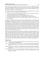
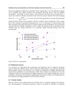
Figure 7. Schematic diagram of signal amplification using a dual-enzyme bio-optrode.
Pyruvate is detected using lactate dehydrogenase (LDH) and lactate oxidase (LDO),
which are co-immobilized on a fiber optic tip. Pyruvate concentration is determined by
measuring NADH fluorescence. Pyruvate and NADH diffuse from the bulk solution into
the enzyme layer, LDH catalyzes the formation of lactate and NAD § during the reduction
of pyruvate. LDO then catalyzes the regeneration of pyruvate causing additional
consumption of NADH by the LDH-catalyzed reaction. Thus, the signal obtained using a
dual-enzyme system is higher then when a single enzyme is used (Zhang et al., 1997).
Reprinted with permission from Elsevier Science.
Inhibition of enzymatic reactions can also be used as a sensing mechanism in
bio-optrodes (Freeman and Bachas, 1992). In this approach, the inhibitor is the
analyte and the measured signal is the decrease in enzymatic activity. One
example is detection of organophosphate and carbamate pesticides using an
enzyme inhibition-based optrode. The bio-optrode is based on the inhibition of
acetylcholinesterase (ACHE) by organophosphate pesticides. The enzyme is co-
immobilized together with a pH sensitive dye at the fiber's distal end. The
substrate acetylcholine is hydrolyzed by AChE causing a change in the local pH
and thereby the fluorescence signal. The inhibition of this reaction can be
correlated to the pesticide concentration in the sample (Doong and Tsai, 2001;
Hobel and Polster, 1992).
In
living cells,
cellular functions are carried out by enzymes that simultaneously
catalyze numerous biochemical reactions. Some enzymatic activities that occur in
cells have been applied for bio-optrode fabrication. Although enzymes can be
isolated and purified, their activity outside the cells is usually reduced compared
to their activity within the cells where they function in an optimum environment
containing all the necessary cofactors. Whole cell biocatalysts are particularly
advantageous when the detection is based on a sequence of multiple enzymatic
18
Optrode-based Fiber Optic Biosensors
reactions. These enzymatic cascade reactions are very difficult and complicated
to accomplish
ex-vivo
by coimmobilizing the enzymes but are relatively
straightforward when employing whole cells. In practice, whole cells that
produce Unique or enhanced enzymatic activity and can transform the analyte
(substrate) into detectable products or cells that produce cellular responses such
as changes in oxygen consumption are immobilized on optical fibers (Preininger
et al., 1994). The methods for detecting the products in cell-based fiber optic
biosensors are similar to those employed in enzyme optrodes. In a more recent
approach, cells are genetically engineered to over-express specific enzymes
involved in the analytical measurement. An example of this approach is the use
of
E. coli
cells that were engineered to over-express the enzyme
organophosphorus hydrolase (OPH) on their outer cell membrane (Mulchandani
et al., 1998). This enzyme catalyzes the hydrolysis of organophosphorus
pesticides to form a chromophoric product that can absorb light at a specific
wavelength. The cell optrode is fabricated by immobilizing the cells on a
bifurcated fiber optic tip and using a photomultiplier detection system to measure
the light signals. Although the specificity of whole cell optrodes is reduced
compared to enzyme optrodes, cells are very simple to use and obtain (e.g.,
growing the cells for a few hours), and there is no need for purification steps,
which makes cell bio-optrodes inexpensive to assemble.
A different approach for sensing with whole cells, which does not directly
involve biocatalysis, is based on utilizing genetic responses and signal
transduction mechanisms in living cells (Daunert et al., 2000; Kohler et al., 2000;
Naylor, 1999). Cells may express a specific gene or set of genes when a specific
molecule (e.g. analyte) is present in the cell's environment. By fusing reporter
genes, encoding for optically detectable enzymes or proteins (e.g., luciferase, [3-
galactosidase, GFP) to the responsive gene, the genetic response is measured and
correlated to the analyte concentration. For a more detailed description of this
approach, see Chapter 10 in this book.
1.4.2. Bioaffinity-based optrodes.
The natural high selectivity of antibodies,
receptors and nucleic acids make them very powerful sensing elements for
recognizing their binding partners. Such bioaffinity optrodes are used as probes
because the recognition reaction is essentially irreversible. The bio-optrode
sensing elements must be regenerated or recharged before the probe can be used
to make another measurement. In many cases, a probe-based bio-optrode
configuration involves the use of a permanent fiber optic and a disposable
sensing layer that can be placed on the fiber optic's distal end (Figure 8).
lmmuno/receptor optrodes
are a major group of bioaffinity fiber optic biosensors
based on transducing antibody-antigen (analyte) interactions into an optical
signal that is proportional to the antigen concentration. Monoclonal antibodies
that can recognize a specific antigenic epitope region (i.e., a specific spatial
19
B iran and Walt
Figure 8. Configuration of probe-based bio-optrodes with disposable biosensing
elements. (a) Biorecognition sensing molecules immobilized on membrane, which is
held by a screw cap on the optical fiber tip. (b) Disposable glass slide with gel entrapped
enzyme. (c) Nylon membrane, with immobilized sensing molecules, attached to the fiber
using an O-ring (Kuswandi et al., 2001). Reproduced with permission of the Royal
Society of Chemistry.
structure on the antigen molecule) or polyclonal antibodies that recognize
different antigenic epitopes are used in immuno-optrodes. Several detection
schemes are employed; the simplest scheme involves the detection of
intrinsically fluorescent analytes such as polynuclear aromatic hydrocarbons
(PAHs) (Vo-Dinh, et al., 2000). Antibodies are immobilized on the fiber surface
and a fluorescence signal is obtained when the analyte (antigen) binds to the
optrode's surface as shown in Figure 9(a).
A competition assay is a more generalized detection scheme that can be applied
to any antibody antigen pair. The detection is based on competition for the
antibody binding site between the antigen present in the sample (analyte) and an
externally added fluorescent-labeled antigen as shown in Figure 9 (b). A known
concentration of fluorescent-labeled antigen is added and captured by an
antibody, which is immobilized on the optical fiber surface. The fluorescence
signal obtained is measured and set as the initial signal. To perform an analysis,
the same fluorescent-labeled antigen concentration is mixed with a sample
containing an unknown antigen concentration. When this mixture is analyzed
using the bio-optrode, the resulting fluorescence signal obtained is lower than the
initial signal because of competition with the labeled antigen in the sample. The
relative decrease in the initial signal is proportional to the analyte concentration
in the sample. Using this detection scheme, bio-optrodes for the detection of
different analytes have been developed (Wittmann et al., 1996; Zhao et al.,
1995).
20
Optrode-based Fiber Optic Biosensors
(a)
(b)
Optical fiber ~-~ Optical
(c)
Optica
Optical
fiber
'~
Self-fluorescing antigen 0 Antigen
Antibody ~
Fluorescent dye
,.
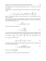
Figure 9. Schematic principle of immuno bio-optrodes. (a) Detection of intrinsically-
fluorescent molecules using immobilized antibodies. (b) Competition assay using a
fluorescent-labeled antigen. (c) Sandwich immunoassay using an immobilized antibody
and a fluorescent-labeled antibody.
The preferred detection scheme is the sandwich immunoassay, which involves
the use of two antibodies. The first antibody is immobilized to the fiber and used
to capture the antigen while a second antibody, which is labeled by a fluorescent
dye or enzyme, is used to generate the signal (Figure 9(c)).
The competition and sandwich assays require using labeled antigens or
antibodies. Fluorescent molecules and enzymes are employed for labeling using
different chemistries (Wortberg, 1997). Very low concentrations of enzymes can
be detected based on their enzymatic activity. The enzymes used for labeling,
such as alkaline phosphatase, catalyze the conversion of a non-fluorescent
substrate to a fluorescent product and can be detected by monitoring the
fluorescent signal generated (Michael et al., 1998). Other enzymes, such as
horseradish peroxidase, can catalyze chemiluminescence reactions and are
detected by monitoring the emitted light signals (Aboul-Enein et al., 2000; Diaz
et al., 1998; Gubitz et al., 2001; Spohn et al., 1995). Enzyme labeling is more
sensitive than fluorescent dye labeling since the signal is amplified by the
enzymatic reaction. Another new technology to increase the labeling efficiency
21
Biran and Walt
Figure 10. Principle of DNA fiber optic biosensors. (a) Single strand DNA probe
molecules, with a sequence complementary to the target DNA sequence, are immobilized
onto the fiber. (b) The fluorescent-labeled sample DNA molecules are first dehybridized
and the fiber is dipped into the sample solution. (c) After hybridization, the
complementary strands of the target DNA are attached to the probe DNA on the fiber and
a fluorescence signal is obtained.
of biological molecules was recently proposed and will be discussed in Chapter
17.
Nucleic acid-based optrodes
are the second major group of bioaffinity-based
optrodes. Nucleic acid base pairing is used as the sensing mechanism in bio-
optrodes for nucleic acid detection. The presence of a specific DNA sequence,
the "target", among millions of other different sequences is detected by
hybridization to its complementary DNA sequence, the "probe", which is
immobilized on the optical fiber, as shown in Figure 10. In a typical assay, the
target DNA is first amplified and fluorescently labeled using fluorescent primers
and the polymerase chain reaction (PCR). The resulting fluorescent double
stranded DNA molecules are dehybridized (usually by heating) (Figure 10 (b))
and then allowed to rehybridize (by cooling) to the single strand DNA probe
molecules immobilized on the fiber surface (Figure 10 (c)). The excess DNA
molecules are washed away, and if the complementary target DNA sequence is
present in the sample, a fluorescence signal is detected (Ferguson, et al., 1996).
For example, the target sequence can be a unique sequence found only in specific
22
Optrode-based Fiber Optic Biosensors
pathogenic bacteria (Iqbal et al., 2000; Pilevar et al., 1998). The target DNA can
be easily extracted from water, wastewater or clinical samples, and the presence
of pathogenic microorganisms can be determined by the bio-optrode. Recently,
new groups of nucleic acid molecules, such as aptamers (Lee and Walt, 2000)
and molecular beacons (Liu et al., 2000; Steemers et al., 2000), were
incorporated as sensing molecules into bio-optrodes. These different DNA
sensing schemes can be multiplexed by fabricating an array of hundreds to
thousands of probes as will described later in Section 3.2.
1.5. Sensing element immobilization
Immobilization of sensing biomolecules to the optical fiber is a key step in bio-
optrode development. A good immobilization method should be simple, fast and
durable but, more importantly, it should be gentle so the biological molecule
being immobilized can retain its biochemical activity. In addition, biological
recognition elements are often coimmobilized together with indicator dyes, so
that ideally the immobilization method should be suitable for both molecules. In
some cases, the recognition compounds are immobilized directly to the optical
fiber surface. Alternatively, the molecules are first immobilized on membranes,
such as cellulose acetate or polycarbonate that are later physically attached to the
optical fiber (Figure 8). There are three main methods for immobilizing a
biological sensing compound: adsorption/electrostatic, entrapment, and covalent
binding. A schematic representation of these methods is shown in Figure 11.
Adsorption immobilization methods involve adsorbing the sensing material onto
a solid surface or polymer matrix. Sensing materials can be adsorbed directly on
the fiber optic end. This immobilization method is very simple; however the
adsorbed molecules tend to gradually leach from the solid support, decreasing
sensing performance and/or lifetime. In order to overcome leaching problems, the
solid support surface may first be modified with complementary functional
groups. For example, a hydrophobic surface can be prepared to immobilize a
hydrophobic species. Electrostatic interaction can also be employed for
immobilization. This immobilization scheme is based on interaction between
oppositely charged molecules. For example, an optical-fiber surface can be
coated with a positively charged layer (i.e., using poly-L-lysine) that interacts
electrostatically with negatively charged recognition molecules (Figure 11 (a)).
The electrostatic immobilization method is very easy and highly reproducible but
may be affected by changes in the medium pH or by changes in other ion
concentrations.
Entrapment immobilization involves physical entrapment of sensing bio-
molecules within a porous matrix (Figure 11 (b)). The biomolecules are
suspended in a monomer solution, which is then polymerized to a gel causing the
molecules to be entrapped. Such polymers can be either thermally or
23
Biran and Walt
Figure 11. Schematic diagram of three different immobilization techniques employed in
biooptrodes. (a) Absorption/electrostatic. (b) Entrapment. (c) Covalent immobilization.
photochemically initiated and attached to the fiber surface by dip-coating
procedures (Healey et al., 1995). The immobilized molecules usually do not
leach out of the matrix and can retain their biochemical activity. Polyacrylamide
gels are most commonly used for entrapment immobilization, although agarose
and calcium alginate gels have also been used (Polyak et al., 2001). One
important limitation of this approach is the slow diffusion rates of the analytes
and products through the immobilization matrix, which increases the bio-optrode
response time.
Optically transparent sol-gel glasses are also used for biological sensing molecule
entrapment as described in Chapter 14 (Dunn et al., 1998, 2001; Jordan et al.,
1996). Sol-gel glasses are produced by hydrolysis and polycondensation of
organometallic compounds, such as tetraethyl orthosilicate (Si(OCH3)4). The
sensing biomolecules are added to the reaction mixture during the formation of
the sol or gel. Sol-gel glasses prepared by this method contain interconnected
pores formed by a three-dimensional SiO2 network. As a result, the biomolecules
and dyes are trapped but small analytes can readily diffuse in and out of the
pores. The main advantages of the sol-gel glass immobilization method are the
chemical, photochemical and mechanical stability of the immobilized layer.
24
Optrode-based Fiber Optic Biosensors
Disadvantages of sol-gel glass immobilization are the slow response times in
aqueous media and the fragility of thin sol-gel glass films compared with
polymer films.
Functional groups in the sensing biomolecules can be covalently bound to
reactive groups on the surface of optical fibers allowing robust immobilization
(Figure 11 (c)). The fiber surface can be chemically modified using silanization
reactions (Weetall, 1993). For example, the tiber surface can be aminosilanized
to form amine functional groups on the fiber surface followed by reaction with-
COOH groups on the enzyme or antibody. Amine-modified surfaces can also
covalently bind to the biomolecule's amine groups using bifunctional cross-
linkers such as glutaraldehyde. Covalent immobilization methods are usually
more complicated and time-consuming compared with the other immobilization
techniques, but are very reliable since the biomolecules and dyes are not likely to
leach out. It should be noted that covalent binding might change the biomolecule
activity. In some cases, if the binding occurs at crucial sites (e.g., an enzyme
active site or an antibody binding site), activity can be lost completely. To avoid
such inactivation, substrate, inhibitors and other effectors are often included in
the immobilization medium to protect the active or binding site of the
biomolecules. In recent years, new techniques have been developed which enable
the immobilized molecule's orientation on the sensing surface to be controlled
resulting in an increase in the immobilization efficiency (Sackmann, 1996).
A more generalized and widely used binding method involves the use of avidin-
biotin chemistry (Wilchek and Bayer, 1990). The fiber surface can be modified
with biotin groups and bind avidin-modified biomolecular conjugates or vice
versa. This method is very attractive since many biotin -~ or avidin-labeled
enzymes, antibodies and nucleic acids are commercially available.
2. History
Optical fiber-based biosensors evolved from chemical optrodes. The first optical
fiber-based chemical sensor was developed by Lubbers and Opitz (1975). Their
device was designed to measure CO2 and 02 and was used in biological fluids. A
few years later, biological molecules were coupled to the optical fiber-based
chemical sensors and bio-optrodes were formed. One of the first bio-optrodes
involved coupling the enzyme glucose oxidase to an Oz optrode to fabricate a
glucose biosensor (Arnold, 1985). In the following years, many bio-optrodes
with different recognition molecules were developed and reported in several
books (Blum et al., 1994; Wolfbeis, 1991), and reviews (Aboul-Enein et al.,
2000; Fraser, 1995; Mehrvar et al., 2000; Rabbany et al., 1994; Wolfbeis, 2000).
Although the bio-optrode basic configuration has not changed much from the one
proposed by Lubbers and Opitz (1975), new types of optical fibers, optical
instruments, biorecognition molecules and indicators have been integrated into
25
Brian and Walt
bio-optrodes. These materials, combined with new immobilization techniques
and advanced optical approaches, led to the development of more sophisticated,
selective and sensitive bio-optrodes. Advances in two fields influenced bio-
optrode development in the last decade. First, development of new fiber optic
technologies that were developed for telecommunication applications. Second,
advances in molecular biology techniques allow specific biorecognition
molecules to be designed. Integration of technologies from these two fields has
led to the development of advanced bio-optrode technologies such as multi-
analyte bio-optrodes, reagentless bio-optrodes and nano bio-optrodes.
3. Advanced Bio-Optrode Technologies and Applications
In this section, a few examples of new bio-optrode technologies and applications
will be described. Although many novel and interesting papers related to bio-
optrode developments have been published in recent years, we focus here on a
few examples that emphasize the diversity of existing bio-optrode technologies.
In addition, a few examples of bio-optrode applications in the industrial,
environmental and clinical fields will be described.
3.1. Nano bio-optrodes
One of the most exciting advances in bio-optrode development is the
miniaturization of sensors to submicron dimensions. Nanotechnology facilitates
research in this field and leads to development of new nano bio-optrodes (Cullum
and Vo-Dinh, 2000). The main importance of such biosensors is their ability to
monitor biomolecule concentrations inside a single living cell and thereby
expand our knowledge about complex intracellular process.
In order to prepare nano bio-optrodes, optical fibers a few nanometers in
diameter are fabricated. The fabrication process involves pulling optical fibers
with an initial diameter of a few microns using a modified micropipette puller
optimized for optical fiber pulling. After pulling, tapered fibers are formed with
typical distal end (tip) diameters of 20-80 nm. This technique was used by
Kopelman and coworkers to make a nano fiber optic chemical sensor for
monitoring intracellular pH inside living cells (Tan et al., 1992). Changes in pH
were measured by immobilizing a pH sensitive dye to the fiber tip. The same
design was used to prepare an enzyme-based nano bio-optrode for nitric oxide
detection (Barker et al., 1998). Fluorescently labeled cytochrome c', which
undergoes conformational changes in the presence of NO, was immobilized to
the fiber tip. Changes in NO concentrations were correlated to changes in the
energy transfer between cytochrome c' and the fluorescent dye.
26