Subcellular Proteomics : From Cell Deconstruction to System Reconstruction / Eric Bertrand
Bạn đang xem bản rút gọn của tài liệu. Xem và tải ngay bản đầy đủ của tài liệu tại đây (3.76 MB, 397 trang )
<span class="text_page_counter">Trang 2</span><div class="page_container" data-page="2">
BERTRAND: “BERTRAND_FM” — 2007/6/20 — 15:16 — PAGE 1 — #1
Subcellular Proteomics
Subcellular BiochemistryVolume 43
</div><span class="text_page_counter">Trang 3</span><div class="page_container" data-page="3">BERTRAND: “BERTRAND_FM” — 2007/6/20 — 15:16 — PAGE 2 — #2
<b>SUBCELLULAR BIOCHEMISTRY</b>
SERIES EDITOR
J. ROBIN HARRIS, University of Mainz, Mainz, Germany
ASSISTANT EDITORS
B.B. BISWAS, University of Calcutta, Calcutta, India P. QUINN, King’s College London, London, U.K.
<i>Recent Volumes in this Series</i>
Volume 31 <b>Intermediate Filaments</b>
Edited by Harald Herrmann and J. Robin Harris
Volume 32 <b>alpha-Gal and Anti-Gal: alpha-1,3-Galactosyltransferase, alpha-GalEpitopes and the Natural Anti-Gal Antibody</b>
Edited by Uri Galili and Jos-Luis Avila Volume 33 <b>Bacterial Invasion into Eukaryotic Cells</b>
Tobias A. Oelschlaeger and Jorg Hacker
Volume 34 <b>Fusion of Biological Membranes and Related Problems</b>
Edited by Herwig Hilderson and Stefan Fuller Volume 35 <b>Enzyme-Catalyzed Electron and Radical Transfer</b>
Andreas Holzenburg and Nigel S. Scrutton Volume 36 <b>Phospholipid Metabolism in Apoptosis</b>
Edited by Peter J. Quinn and Valerian E. Kagan Volume 37 <b>Membrane Dynamics and Domains</b>
Edited by P.J. Quinn
Volume 38 <b>Alzheimer’s Disease: Cellular and Molecular Aspects of Amyloid beta</b>
Edited by R. Harris and F. Fahrenholz Volume 39 <b>Biology of Inositols and Phosphoinositides</b>
Edited by Lahiri Majumder and B.B. Biswas Volume 40 <b>Reviews and Protocols in DT40 Research</b>
Edited by Jean-Marie Buerstedde and Shunichi Takeda Volume 41 <b>Chromatin and Disease</b>
Edited by Tapas K. Kundu and Dipak Dasgupta
Volume 42 <b>Inflammation in the Pathogenesis of Chronic Diseases</b>
Edited by Randall E. Harris
</div><span class="text_page_counter">Trang 4</span><div class="page_container" data-page="4">BERTRAND: “BERTRAND_FM” — 2007/6/20 — 15:16 — PAGE 3 — #3
</div><span class="text_page_counter">Trang 5</span><div class="page_container" data-page="5">BERTRAND: “BERTRAND_FM” — 2007/6/20 — 15:16 — PAGE 4 — #4 <small>A C.I.P. Catalogue record for this book is available from the Library of Congress.</small>
<i><small>Printed on acid-free paper</small></i>
<small>All Rights Reserved© 2007 Springer</small>
<small>No part of this work may be reproduced, stored in a retrieval system, or transmitted in any form or byany means, electronic, mechanical, photocopying, microfilming, recording or otherwise, without writtenpermission from the Publisher, with the exception of any material supplied specifically for the purposeof being entered and executed on a computer system, for exclusive use by the purchaser of the work.</small>
</div><span class="text_page_counter">Trang 6</span><div class="page_container" data-page="6">BERTRAND: “BERTRAND_FM” — 2007/6/20 — 15:16 — PAGE 5 — #5
<b>INTERNATIONAL ADVISORY EDITORIAL BOARD</b>
R. BITTMAN, Queens College, City University of New York, New York, USA D. DASGUPTA, Saha Institute of Nuclear Physics, Calcutta, India
H. ENGELHARDT, Max-Planck-Institute for Biochemistry, Munich, Germany L. FLOHE, MOLISA GmbH, Magdeburg, Germany
H. HERRMANN, German Cancer Research Center, Heidelberg, Germany A. HOLZENBURG, Texas A & M University, Texas, USA
H-P. NASHEUER, National University of Ireland, Galway, Ireland S. ROTTEM, The Hebrew University, Jerusalem, Israel
M. WYSS, DSM Nutritional Products Ltd., Basel, Switzerland P. ZWICKL, Max-Planck-Institute for Biochemistry, Munich, Germany
</div><span class="text_page_counter">Trang 7</span><div class="page_container" data-page="7">BERTRAND: “BERTRAND_FM” — 2007/6/20 — 15:16 — PAGE 7 — #7 To Michel and his speedy recovery. His experience and enthusiasm have been sorely missed in the final stages of preparing this volume.
To my son, Alexandre, far from me now, but still close to my heart.
To my mother, who has been a source of inspiration in many more ways than she could have imagined.
Eric
</div><span class="text_page_counter">Trang 8</span><div class="page_container" data-page="8">BERTRAND: “BERTRAND_FM” — 2007/6/20 — 15:16 — PAGE ix — #9
<b>TABLE OF CONTENTS</b>
<i>Thierry Rabilloud</i>
<i>René P. Zahedi, Jan Moebius and Albert Sickmann</i>
<b>3. Microparticles: A New Tool for Plasma Membrane</b>
<i>Laurent Miguet, Sarah Sanglier, Christine Schaeffer, Noelle Potier,Laurent Mauvieux and Alain Van Dorsselaer</i>
<b>4. Lipid Raft Proteomics: More than Just</b>
<i>Leonard J. Foster and Queenie W. T. Chan</i>
<b>5. Organelle Proteome Variation Among Different Cell Types:</b>
<i>Deirdre M. Kavanagh, William E. Powell, Poonam Malik,Vassiliki Lazou and Eric C. Schirmer</i>
<i>Fengju Bai and Frank A. Witzmann</i>
ix
</div><span class="text_page_counter">Trang 9</span><div class="page_container" data-page="9">BERTRAND: “BERTRAND_FM” — 2007/6/20 — 15:16 — PAGE x — #10
<i>Christine Olver and Michel Vidal</i>
<i>Vincent Collura and Guillaume Boissy</i>
<i>M.O. Collins and S.G.N. Grant</i>
<i>Ronald Roepman and Uwe Wolfrum</i>
<b>11. Systems Biology and the Reconstruction of the Cell:</b>
<i>Frank J. Bruggeman, Sergio Rossell, Karen van Eunen,Jildau Bouwman, Hans V. Westerhoff and Barbara Bakker</i>
<b>12. Automated, Systematic Determination of Protein</b>
<i>Elvira García Osuna and Robert F. Murphy</i>
<b>13. Systems Biology of the Endoplasmic Reticulum</b>
<i>Marie-Elaine Caruso and Eric Chevet</i>
<b>14. Systems Nanobiology: From Quantitative Single Molecule</b>
<i>Joerg Martini, Wibke Hellmich, Dominik Greif, Anke Becker,Thomas Merkle, Robert Ros, Alexandra Ros,</i>
<i>Katja Toensing and Dario Anselmetti</i>
<i>Michel Faupel, Débora Bonenfant, Patrick Schindler,</i>
<i>Eric Bertrand, Dieter Mueller, Markus Stoeckli, Francis Bitsch,Tatiana Rohner, Dieter Staab and Jan Van Oostrum</i>
</div><span class="text_page_counter">Trang 10</span><div class="page_container" data-page="10">BERTRAND: “BERTRAND_FM” — 2007/6/20 — 15:16 — PAGE xi — #11
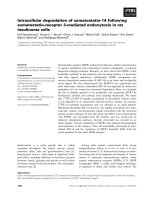
<b>16. Differential Epitope Identification of Antibodies AgainstIntracellular Domains of Alzheimer’s Amyloid Precursor</b>
<i>Xiaodan Tian, Madalina Maftei, Markus Kohlmann,Bernadette Allinquant and Michael Przybylski</i>
<b>17. LC-MALDI MS and MS/MS – An Efficient Tool in</b>
<i>Dieter R. Mueller, Hans Voshol, Annick Waldt, Brigitte Wiedmannand Jan van Oostrum</i>
</div><span class="text_page_counter">Trang 11</span><div class="page_container" data-page="11">BERTRAND: “BERTRAND_FM” — 2007/6/20 — 15:16 — PAGE xiii — #13
In light of the somewhat eventful circumstances in which the current volume came into being, we are sincerely grateful to the people who contributed to its preparation. Their hard work as well as their patience and kind understanding toward the editors were greatly appreciated. We are most grateful to Mary Johnson, who acted as the publishing editor for this volume. We wish to thank all of the contributors and their staff for submitting their respective chapters as needed, despite busy teaching and research schedules. We would also like to acknowledge several very good friends for their voluntary editorial assistance and even more importantly their unfailing moral support: Eileen Rojo, Hugues Ryckelynck and Claire Mc Donack. Last, but by no mean least, we are particularly grateful to Dr. Robin Harris, who gave us the opportunity to edit this volume for the “Subcellular Biochemistry” book series.
xiii
</div><span class="text_page_counter">Trang 12</span><div class="page_container" data-page="12">BERTRAND: “BERTRAND_FM” — 2007/6/20 — 15:16 — PAGE xv — #15
<b>LIST OF CONTRIBUTORS</b>
<b>Bernadette Allinquant</b>
INSERM U573, Centre Paul Broca, Paris, France.
<b>Dario Anselmetti</b>
Experimental Biophysics and Applied Nanoscience, Physics Faculty, Bielefeld University and Bielefeld Institute for Biophysics and Nanoscience (BINAS), Center for Biotechnology (CeBiTec), Bielefeld, Germany.
<b>Fengju Bai</b>
Charles River Laboratories Preclinical Services, Safety Sciences, Worcester, Massachusetts, United States.
<b>Barbara Bakker</b>
BioCentre Amsterdam, Free University Amsterdam, Faculty of Earth and Life Sciences, Department of Molecular Cell Physiology, Amsterdam, The Netherlands.
<b>Anke Becker</b>
Institute for Genome Research, Center for Biotechnology (CeBiTec) Bielefeld University, Bielefeld, Germany.
<b>Eric Bertrand</b>
Novartis Institutes for BioMedical Research, Genome and Proteome Sciences/ Systems Biology, Basel, Switzerland.
<b>Guillaume Boissy</b>
Hybrigenics S.A., Paris, France.
<b>Débora Bonenfant</b>
Novartis Institutes for BioMedical Research, Genome and Proteome Sciences/ Systems Biology, Basel, Switzerland.
<b>Jildau Bouwman</b>
BioCentre Amsterdam, Free University Amsterdam, Faculty of Earth and Life Sciences, Department of Molecular Cell Physiology, Amsterdam, The Netherlands.
xv
</div><span class="text_page_counter">Trang 13</span><div class="page_container" data-page="13">BERTRAND: “BERTRAND_FM” — 2007/6/20 — 15:16 — PAGE xvi — #16
<b>Frank J. Bruggeman</b>
BioCentre Amsterdam, Free University Amsterdam, Faculty of Earth and Life Sci-ences, Department of Molecular Cell Physiology, Amsterdam, The Netherlands and also Manchester Centre for Integrative Systems Biology, Manchester Inter-disciplinary BioCentre, University of Manchester, United Kingdom.
<b>Marie-Elaine Caruso</b>
Organelle signaling laboratory, Department of Surgery, McGill University, Montreal, Quebec, Canada.
<b>Queenie W. T. Chan</b>
UBC Centre for Proteomics, Department of Biochemistry and Molecular Biology, University of British Columbia, Vancouver, Canada.
<b>Eric Chevet</b>
Organelle signaling laboratory, Department of Surgery, McGill University, Montreal, Quebec, Canada.
<b>Mark O. Collins</b>
Proteomic Mass Spectrometry, The Wellcome Trust Sanger Institute, Hinxton, Cambridgeshire, United Kingdom.
<b>Vincent Collura</b>
Hybrigenics S.A., Paris, France.
<b>Michel Faupel</b>
Novartis Institutes for BioMedical Research, Genome and Proteome Sciences/ Systems Biology, Basel, Switzerland.
<b>Leonard J. Foster</b>
UBC Centre for Proteomics, Department of Biochemistry and Molecular Biology, University of British Columbia, Vancouver, Canada.
<b>Elvira García Osuna</b>
Center for Bioimage Informatics and Department of Biomedical Engineering, Carnegie Mellon University, Pittsburgh, Pennsylvania, United States.
<b>Seth G. N. Grant</b>
Genes to Cognition, The Wellcome Trust Sanger Institute, Hinxton, Cambridgeshire, United Kingdom.
<b>Dominik Greif</b>
Experimental Biophysics and Applied Nanoscience, Physics Faculty, Bielefeld Uni-versity and Bielefeld Institute for Biophysics and Nanoscience (BINAS), Center for Biotechnology (CeBiTec), Bielefeld, Germany.
<b>Wibke Hellmich</b>
Experimental Biophysics and Applied Nanoscience, Physics Faculty, Bielefeld Uni-versity and Bielefeld Institute for Biophysics and Nanoscience (BINAS), Center for Biotechnology (CeBiTec), Bielefeld, Germany.
</div><span class="text_page_counter">Trang 14</span><div class="page_container" data-page="14">BERTRAND: “BERTRAND_FM” — 2007/6/20 — 15:16 — PAGE xvii — #17
Department of Chemistry, Laboratory of Analytical Chemistry and Biopolymer Structure Analysis, University of Konstanz, Konstanz, Germany.
<b>Poonam Malik</b>
Wellcome Trust Centre for Cell Biology, University of Edinburgh, Edinburgh, United Kingdom.
<b>Joerg Martini</b>
Experimental Biophysics and Applied Nanoscience, Physics Faculty, Bielefeld Uni-versity and Bielefeld Institute for Biophysics and Nanoscience (BINAS), Center for Biotechnology (CeBiTec), Bielefeld, Germany.
<b>Laurent Mauvieux</b>
Institut d’Hématologie et d’Immunologie, Faculté de Médecine, Université Louis Pasteur and Laboratoire d’Hématologie, Hôpital Hautepierre, Strasbourg, France.
<b>Thomas Merkle</b>
Institute for Genome Research, Center for Biotechnology (CeBiTec) Bielefeld University, Bielefeld, Germany.
<b>Laurent Miguet</b>
Laboratoire de Spectrométrie de Masse Bio-Organique, ECPM, UMR/CNRS 7178, Institut Pluridisciplinaire Hubert CURIEN, Université Louis Pasteur, Strasbourg, France.
<b>Jan Moebius</b>
Protein Mass Spectrometry and Functional Proteomics Group, Rudolf-Virchow-Center for Experimental Biomedicine, University of Wuerzburg, Wuerzburg, Germany.
<b>Dieter R. Mueller</b>
Novartis Institutes for BioMedical Research, Genome and Proteome Sciences /Systems Biology, Basel, Switzerland.
<b>Robert F. Murphy</b>
Center for Bioimage Informatics and Departments of Biological Sciences, Biomed-ical Engineering and Machine Learning, Carnegie Mellon University, Pittsburgh, Pennsylvania, United States.
</div><span class="text_page_counter">Trang 15</span><div class="page_container" data-page="15">BERTRAND: “BERTRAND_FM” — 2007/6/20 — 15:16 — PAGE xviii — #18
<b>Christine Olver</b>
Clinical Pathology Section, Colorado State University, Ft Collins, United States.
<b>Noelle Potier</b>
Laboratoire de Spectrométrie de Masse Bio-Organique, ECPM, UMR/CNRS 7178, Institut Pluridisciplinaire Hubert CURIEN, Université Louis Pasteur, Strasbourg,
Department of Chemistry, Laboratory of Analytical Chemistry and Biopolymer Structure Analysis, University of Konstanz, Konstanz, Germany.
<b>Thierry Rabilloud</b>
CEA-DSV-iRTSV-LBBSI and UMR CNRS 5092, CEA-Grenoble, 17 rue des martyrs, 38054, Grenoble cedex 9.
<b>Ronald Roepman</b>
Department of Human Genetics, Radboud University Nijmegen Medical Centre and Nijmegen Centre for Molecular Life Sciences, Nijmegen, The Netherlands.
<b>Tatiana Rohner</b>
Novartis Institutes for BioMedical Research, Discovery Technologies, Basel, Switzerland.
<b>Robert Ros</b>
Experimental Biophysics and Applied Nanoscience, Physics Faculty, Bielefeld University and Bielefeld Institute for Biophysics and Nanoscience (BINAS), Center for Biotechnology (CeBiTec), Bielefeld, Germany.
<b>Alexandra Ros</b>
Experimental Biophysics and Applied Nanoscience, Physics Faculty, Bielefeld University and Bielefeld Institute for Biophysics and Nanoscience (BINAS), Center for Biotechnology (CeBiTec), Bielefeld, Germany.
<b>Sergio Rossell</b>
BioCentre Amsterdam, Free University Amsterdam, Faculty of Earth and Life Sciences, Department of Molecular Cell Physiology, Amsterdam, The Netherlands.
<b>Sarah Sanglier</b>
Laboratoire de Spectrométrie de Masse Bio-Organique, ECPM, UMR/CNRS 7178, Institut Pluridisciplinaire Hubert CURIEN, Université Louis Pasteur, Strasbourg, France.
<b>Christine Schaeffer</b>
Laboratoire de Spectrométrie de Masse Bio-Organique, ECPM, UMR/CNRS 7178, Institut Pluridisciplinaire Hubert CURIEN, Université Louis Pasteur, Strasbourg, France.
</div><span class="text_page_counter">Trang 16</span><div class="page_container" data-page="16">BERTRAND: “BERTRAND_FM” — 2007/6/20 — 15:16 — PAGE xix — #19
<b>Patrick Schindler</b>
Novartis Institutes for BioMedical Research, Genome and Proteome Sciences/Systems Biology, Basel, Switzerland.
<b>Eric C. Schirmer</b>
Wellcome Trust Centre for Cell Biology, University of Edinburgh, Edinburgh, United Kingdom.
<b>Albert Sickmann</b>
Protein Mass Spectrometry and Functional Proteomics Group, Rudolf-Virchow-Center for Experimental Biomedicine, University of Wuerzburg, Wuerzburg,
Department of Chemistry, Laboratory of Analytical Chemistry and Biopolymer Structure Analysis, University of Konstanz, Konstanz, Germany.
<b>Katja Toensing</b>
Experimental Biophysics and Applied Nanoscience, Physics Faculty, Bielefeld University and Bielefeld Institute for Biophysics and Nanoscience (BINAS), Center for Biotechnology (CeBiTec), Bielefeld, Germany.
<b>Alain Van Dorsselaer</b>
Laboratoire de Spectrométrie de Masse Bio-Organique, ECPM, UMR/CNRS 7178, Institut Pluridisciplinaire Hubert CURIEN, Université Louis Pasteur, Strasbourg, France.
<b>Karen van Eunen</b>
BioCentre Amsterdam, Free University Amsterdam, Faculty of Earth and Life Sciences, Department of Molecular Cell Physiology, Amsterdam, The Netherlands.
<b>Jan van Oostrum</b>
Novartis Institutes for BioMedical Research, Genome and Proteome Sciences/ Systems Biology, Basel, Switzerland.
<b>Michel Vidal</b>
UMR/CNRS 5235, University Montpellier II, Montpellier, France.
<b>Hans Voshol</b>
Novartis Institutes for BioMedical Research, Genome and Proteome Sciences/ Systems Biology, Basel, Switzerland.
</div><span class="text_page_counter">Trang 17</span><div class="page_container" data-page="17">BERTRAND: “BERTRAND_FM” — 2007/6/20 — 15:16 — PAGE xx — #20
<b>Annick Waldt</b>
Novartis Institutes for BioMedical Research, Genome and Proteome Sciences/ Systems Biology, Basel, Switzerland.
<b>Hans V. Westerhoff</b>
BioCentre Amsterdam, Free University Amsterdam, Faculty of Earth and Life Sciences, Department of Molecular Cell Physiology, Amsterdam, The Nether-lands and also Manchester Centre for Integrative Systems Biology, Manchester Interdisciplinary BioCentre, University of Manchester, United Kingdom.
<b>Brigitte Wiedmann</b>
Novartis Institutes for BioMedical Research, Infectious Disease Area, Cambridge, Massachusetts, United States.
<b>Frank A. Witzmann</b>
Department of Cellular & Integrative Physiology, Indiana University School of Medicine Biotechnology Research & Training Center, Indianapolis, United States.
<b>Uwe Wolfrum</b>
Department of Cell and Matrix Biology, Institute of Zoology, Johannes Gutenberg University of Mainz, Germany.
<b>René P. Zahedi</b>
Protein Mass Spectrometry and Functional Proteomics Group, Rudolf-Virchow-Center for Experimental Biomedicine, University of Wuerzburg, Wuerzburg, Germany.
</div><span class="text_page_counter">Trang 18</span><div class="page_container" data-page="18">BERTRAND: “BERTRAND_FM” — 2007/6/20 — 15:16 — PAGE xxi — #21
As proteomics technologies are reaching a plateau in the number of proteins that can be resolved and detected, pre-fractionation steps have become essential to increase the depth of proteomic analysis. So far, many pre-fractionation steps have been based on chromatography methods where the proteins are separated according to their individ-ual physicochemical properties. Subcellular fractionation methods proved to be very potent protein pre-fractionation steps: they allow the representation of low abundance proteins, and they can be combined with chromatography steps. Moreover, as the iso-lated subcellular components also represent functional units, subcellular fractionation allows the proteomic analysis of protein subsets that are functionally related in a bio-logically relevant manner. The first three sections of this volume deal with different levels of subcellular organization that also correspond to specific methodological approaches.
In his keynote chapter, Thierry Rabilloud superbly introduces the first section with a thorough definition of membrane proteomics where he pinpoints key theoretical and practical issues of this field, thereby setting the stage for the next contributions. Miguet et al. address the first key issue: the quality of the membrane preparation; they intro-duce and validate a microparticle strategy for plasma membrane purification. Zahedi et al. deal with the second major issue, the resolving of the hydrophobic proteins found in biological membrane samples, which they solve through two-dimensional BAC/SDS-PAGE gel electrophoresis. To close the first section, Foster and Chan review the proteomics of lipid rafts, membrane structures that are involved in intra-cellular trafficking and signal transduction. They describe a clever validation scheme based on the sensitivity of lipid rafts to cholesterol disruption.
A central theme in the second section on organelle subproteomes is the variability of their composition and how it can be exploited and interpreted. Kavanagh et al. describe a state-of-the-art substractive proteomics scheme that relies on an in sil-ico purification step based on the comparison of organelle subproteomes. With this approach, they could demonstrate variations in subproteome content across tissues. In the next chapter on synatosome proteomics, Bai and Witzmann review the current efforts to correlate synaptic plasticity and variations in synaptic subproteome content
xxi
</div><span class="text_page_counter">Trang 19</span><div class="page_container" data-page="19">BERTRAND: “BERTRAND_FM” — 2007/6/20 — 15:16 — PAGE xxii — #22
with a special emphasis on post-translational modifications. Finally, Olver and Vidal discuss how the proteomic analysis of exosomes would give clues to the molecular basis of their biogenesis and contribute to a better understanding of their function. Moreover, they propose that the variations observed in exosome protein content are useful for biomarker discovery.
The third section deals with protein complexes, which are considered as the molec-ular machinery that performs most cell functions. This area is certainly not a trivial one: there are several types of protein complexes and protein-protein interactions and it is not always clear which methodology is most suitable to use in either context. In their chapter, Boissy and Collura sort out for us the concepts and methods encountered in interactomics, guide us through data interpretation issues and share with us their insight on the very nature of interactions. They plead for a systematic integration of interaction maps with functional genomics and molecular genetics data: the potential of such an approach is strikingly demonstrated in the next two chapters. Collins and Grant examine the molecular architecture of membrane associated signalling com-plexes in the nervous system, they highlight the role scaffolding proteins within these complexes and point out to a few much needed construction rules. Aligning interac-tion and funcinterac-tional genomics data, they build a case for a modular organisainterac-tion of large complexes into functional sub-networks. To complete the picture, based on a thorough review on the complexes of the photoreceptor cilia, Roepman and Wolfrum sketch out an approach to organize complexes in functional modules and investigate their interactions.
Assuming that the protein content of an organelle has been inventoried and its protein complexes characterized, the next step is to translate this knowledge into functionally relevant interpretations. This is the purpose of systems biology: if we consider organelles as systems that function and communicate with each other through their protein machinery, it makes sense to apply such an approach at the subcellular level. In their chapter, Caruso and Chevet prove that this concept can actually be applied to reconstruct the stress signalling network of the endoplasmic reticulum. They build an integrative signalling map that qualitatively accounts for the interac-tions of the stress network with other endoplasmic reticulum machineries and also with other organelles. On the quantitative side, Bruggeman et al. introduce a theoret-ical framework for subcellular systems biology and thoroughly review the relevant mathematical approaches. Drawing on their extensive experience of metabolic net-works, they argue that a direct translation of subcellular units as modules within a mathematical model of the cell can be advantageous both for solving the problem and interpreting the results. Garcia Osuna and Murphy survey the current automated methods for high-throughput determination of protein subcellular location that are used to reconstruct subcellular anatomy at high resolution. These methods provide essential information on the dynamic aspect of subcellular events in individual cells: it would also be extremely interesting to combine them with the molecular switches described by Martini et al. in the next section.
This brings us to the fifth and last section of this volume, where the most recent technological developments in proteomics are reviewed. Martini et al. introduce
</div><span class="text_page_counter">Trang 20</span><div class="page_container" data-page="20">BERTRAND: “BERTRAND_FM” — 2007/6/20 — 15:16 — PAGE xxiii — #23
the emerging field of systems nanobiology that relies on ultrasensitive methods and instruments to investigate cellular processes at the single molecule level. An appli-cation, among others, is to monitor the dynamics of protein translocation from one subcellular compartment to another using two-photon laser scanning microscopy and a photoactivable GFP as a molecular switch. Faupel et al. review the current applica-tions of biophotonic technologies to proteomics with a focus on mass spectrometry based molecular imaging. Tian et al. describe a fast-track approach for the character-ization of antibody epitopes using Fourier transform ion cyclotron resonance mass spectrometry (FTICR-MS). The application of this method to the Amyloid Precursor Protein has important consequences for the study of intracellular processing path-ways relevant to Alzheimer’s disease. Finally, Mueller et al. describe the interfacing of LC and MALDI-MS and – MS/MS, discuss its performance, and present selected applications in the proteomics field including the analyses of membrane proteins and protein interactions.
</div><span class="text_page_counter">Trang 21</span><div class="page_container" data-page="21">BERTRAND: “BERTRAND_CH01” — 2007/6/8 — 20:07 — PAGE 1 — #1
<b>SECTION 1</b>
<b>MEMBRANE PROTEOMICS</b>
</div><span class="text_page_counter">Trang 22</span><div class="page_container" data-page="22">BERTRAND: “BERTRAND_CH01” — 2007/6/8 — 20:07 — PAGE 3 — #3 2. Sample Preparation Issues for Membrane Proteomics . . . . 5 3. Issues linked to the separation process . . . . 6 3.1. Constraints in Proteomics Based on Zone Electrophoresis. . . . 7 3.2. Constraints Proteomics Based on Two-dimensional Gel
Electrophoresis . . . . 8 3.3. Constraints in Peptide Separation-Based Approaches . . . . 9 3.4. Constraints in Protein Chromatography-Based Approaches . . . . 9 4. Concluding remarks . . . . 10 References . . . . 10
<b>1. INTRODUCTION</b>
Before addressing the key issues pertaining to proteomics of membrane proteins, which is an important subject in the wider topic of subcellular proteomics, the first question that must be asked is the definition of a membrane protein. While this might seem trivial at the first glance, the answer is far from being obvious. A commonly accepted definition for a membrane protein is a protein associated with a membrane, that is, a lipid bilayer. However, the meaning of the word “associated” in this case is the subject of almost infinite debate. In fact, two different situations arise. In the first, simple situation, the polypeptide chain spans the lipid bilayer a certain number of times. These proteins are defined as integral or intrinsic membrane proteins, as their association with the lipid bilayer is doubtless. The second, much more complicated case, is when the association with the lipid bilayer is not achieved by transmembrane segments or by transmembrane barrels (e.g. in porins), because many other types of association with membranes are encountered. In one particular type, the association is mediated by a post translational modification of the polypeptide, namely the grafting of an fatty acid or polyisoprenyl chain. Polyisoprenylation has been demonstrated to result in physical association with the membrane, as shown for small G proteins
<i><small>E. Bertrand and M. Faupel (eds.), Subcellular Proteomics, 3–11.© 2007 Springer.</small></i>
</div><span class="text_page_counter">Trang 23</span><div class="page_container" data-page="23">BERTRAND: “BERTRAND_CH01” — 2007/6/8 — 20:07 — PAGE 4 — #4
(Verma et al. 1994). Strong association of proteins with lipid membranes is also achieved by glycolipid anchors such as the glycosylphosphatidyl inositol common in
<i>eukaryotic species, or, as in bacterial lipoproteins, by an N -acyl diglyceride linkageto an N -terminal cysteine residue that becomes available after cleavage of a signal</i>
sequence (Cross 1991). Last but not least, the protein can also be acylated by a simple fatty acid chain. In this case, the reality of the anchoring to the lipid bilayer is much less clear.
Protein-membrane association via a post-translational modification introduces the notion of dynamic association and partitioning of proteins between the membrane phase of the cells and the aqueous phase (cytosol or inner phase of organelles). Con-sequently, such proteins can be found both as associated and membrane-free, which is not the case with intrinsic membrane proteins which are strictly membrane embedded. Another type of association to membrane is mediated by protein–protein interactions with other membrane proteins. A typical example of this situation is provided by the respiratory complexes. In the case of ubiquinol-cytochrome c oxidoreductase, core proteins 1 and 2 does not show any interaction with the lipid membrane, but only with the protein subunits spanning the membrane (e.g. cytochrome b) (Iwata et al. 1998).
However, the situation is often much less clear than this one, and drives quite fast into the infinite debate of what is a membrane protein apart from the integral ones. In addition to the dynamic view of association to membranes, this complicated situation arises mainly from the operational, biochemical, definition of membranes. When a cell is lysed in an aqueous, detergent-free, medium, the cell-limiting mem-brane and the network of inner memmem-branes (when applicable) will fragment into vesicles which can be separated from the bulk of cytosol by sedimentation or partition techniques. These techniques are also able to separate membrane-bound organelles (e.g. mitochondria and plasts) from other cellular components, providing good purity. However, not every protein present in such preparations can be con-sidered as a membrane protein. For example, when vesicles will form from larger structures upon lysis, simple physical entrapment will bring soluble proteins in the lumen of the vesicles. Furthermore, it appears more and more clearly that some classes of proteins (e.g. cytoskeletal proteins or ribosomal proteins) are directly or indirectly associated to bona fide transmembrane proteins or to the lipid bilayer. Should these proteins be classified as membrane proteins? Should the non-membrane spanning subunits of respiratory complexes be considered as membrane proteins? This gives an example of the debate that can take place on the notion of mem-brane proteins.
This could be seen as a very theoretical debate, but this has indeed very practical implications. Let us take the example of the analysis of a reticulum preparation. Endoplasmic reticulum is the place of synthesis of most secreted proteins and of most transmembrane proteins. As such, it contains many ribosomes (the Rough ER) and the reticulum vesicles are known to be associated with cytoskeletal filaments. The problem arises from the fact that the protein content of these cytoskeletal and ribosomal “contaminants” is concentrated in a few proteins. This means in turn that
</div><span class="text_page_counter">Trang 24</span><div class="page_container" data-page="24">BERTRAND: “BERTRAND_CH01” — 2007/6/8 — 20:07 — PAGE 5 — #5
even if these “contaminants” represent a few percent of the total protein mass of the preparation, the few proteins represented will be easily detected by any proteomics analysis method.
Conversely, the “real” reticulum proteins are scattered among almost all the secreted proteins and transmembrane proteins of the cells, plus the resident pro-teins of the ER. This means in turn that these propro-teins will appear as less prominent upon proteomics analysis, just because each protein species is more diluted than the few “contaminating” ones.
<b>2. SAMPLE PREPARATION ISSUES FORMEMBRANE PROTEOMICS</b>
A consequence of the above considerations is that sample preparation is very critical in membrane proteomics. As the volume occupied by the lipid bilayer in a membrane sample is very small in comparison to the volume of the aqueous phase, the transmem-brane proteins must be considered as rare components of the sample, although they often drive most of the interest of the researchers. In addition to that, these transmem-brane proteins often pose a very difficult solubilization problem. Transmemtransmem-brane proteins usually contain domains protruding in the extracellular environment or in the cytosol. These domains behave as classical protein domains and are therefore sta-ble in a water-based solvent. However, membrane proteins also contain domains that are embedded more or less deep in the lipid bilayer. Consequently, these domains are stable in a hydrocarbon-like environment. If a membrane protein is to be solubilized prior to its purification, this means that the solvent which is used to this purpose must show at the same time water-like and hydrocarbon-like properties. A water-based sol-vent will induce aggregation of the membrane domains via hydrophobic interactions, and thus protein precipitation. However, an organic solvent will induce in most cases precipitation of the membrane proteins via their water-soluble domains, which are denatured and coalesce in such a solvent. Some very hydrophobic proteins, however, are soluble in organic solvents (e.g. Molloy et al. 1999; Blonder et al. 2003). This is due to the reduced size of their non-membrane domains, and to the fact that all water-soluble protein domains contain a hydrophobic core, which can be soluble in organic solvents. In some cases, these positive solubilization forces can overcome the precipitation-deriving forces and make the protein soluble in organic solvents.
This is however not a general case, and the general rule for membrane proteins is that they require a solvent that has as the same time strong water-like and strong hydrocarbon-like properties. This is not the situation of mixed solvents, that is mixtures of water and water-miscible solvents, which only offer average proper-ties and not a combination of both properproper-ties. Such a combination is only offered by a stable dispersion of hydrocarbon chains in a water-based solvent. This is the definition of lipid membranes, but such assemblies are very difficult to handle along a purifi-cation process. Hopefully, this definition is also the one of detergent micelles, which are much easier to handle along a purification process. These constraints explain why
</div><span class="text_page_counter">Trang 25</span><div class="page_container" data-page="25">BERTRAND: “BERTRAND_CH01” — 2007/6/8 — 20:07 — PAGE 6 — #6
detergents are some kind of a universal tool for the disruption of cell membranes and for the solublization of membrane proteins.
The micelle-forming ability of detergents is driven by their chemical structure, which combine a hydrophobic part (the tail), promoting aggregation of the molecules into the micelles, which is linked to a hydrophilic one (the head) promoting water solubility of the individual molecules and of the micelles. By using different struc-tures or heads and tails and by combining various tails with various heads, an almost infinite range of detergents can be generated. Their protein and lipid solubilization properties are of course strongly dependent on the chemical properties of the heads and tails. As a rule of thumb, rigid tails (e.g. steroid-based) and weakly polar heads (e.g. glycosides, oligoethylene glycol) generally lead to “mild” detergents, that is chemicals that have good lipid solubilization properties, but that have weak protein dissociation properties. While these detergents do not denature proteins, they can prove unable to prevent protein–protein interactions promoting precipitation. Con-sequently, their protein solubilization properties are highly variable, and a lengthy optimization process in the detergent choice is usually needed for various membrane proteins.
Conversely, detergent having a flexible tail and a strongly polar (i.e. ionic) head are viewed as “strong” detergents. In addition of their lipid solubilizing properties, these detergents are able to disrupt the hydrophobic interactions maintaining the structure of the proteins. They are therefore denaturing. However, because of their ionic nature, the bound detergent imparts a net electrical charge to the denatured proteins and induces a strong electrostatic repulsion between protein molecules. Thus, even denatured proteins can no longer aggregate, and these ionic detergents are very powerful protein solubilizing agents.
These protein solubilization conditions have a key impact on the protein fraction-ation that can be carried out afterwards, in the sense that they will restrict the choice to techniques with which they are compatible. As an example, ionic detergents are not compatible with any technique using protein charge or pI as the fractionation parameter.
All of the above is mainly true when some fractionation is to be carried out at the protein level. In some approaches, the fractionation is carried out only on peptides arising from the digestion of the membrane preparation. In this case too, the choice of the solubilization media will be dictated by the constraints imposed by the subsequent peptide fractionation process. However, the use of chromatographic peptide fraction-ation tools, and especially those based on reverse-phase chromatography, will make the use of detergents more problematic, leading to specially designed, detergent-free protocols (Wu et al. 2003; Fischer et al. 2006).
<b>3. ISSUES LINKED TO THE SEPARATION PROCESS</b>
Once the membrane proteins have been solubilized, usually in a water-detergent medium, they must be fractionated. As each fractionation method brings its own constraints, this will further restrict the scope of possibilities that can be used for
</div><span class="text_page_counter">Trang 26</span><div class="page_container" data-page="26">BERTRAND: “BERTRAND_CH01” — 2007/6/8 — 20:07 — PAGE 7 — #7
sample solubilization. The following sections will exemplify some constraints linked to the most commonly used fractionation processes in proteomics.
<b>3.1. Constraints in Proteomics Based on Zone Electrophoresis</b>
In these proteomics methods, the separation process is split in two phases (e.g. in Bell et al. 2001). The first phase is a protein separation by denaturing zone electrophoresis, that is in the presence of denaturing detergents, most often sodium dodecyl sulfate (SDS). The second phase is carried out by chromatography on the peptides produced by digestion of the separated proteins. This has no impact on the sample preparation itself, which just needs to be compatible with the initial zone electrophoresis.
This is by far the simplest case. Sample preparation is achieved by mixing the initial sample with a buffered, concentrated solution of an ionic detergent, usually containing a reducer to break disulfide bridges and sometimes an additional nonionic chaotrope such as urea. Ionic detergents are among the most powerful protein dena-turing solubilizing agents. Their strong binding to proteins make all proteins to bear an electric charge of the same type, whatever their initial charge may be. This induces in turn a strong electrostatic repulsion between protein molecules, and thus maxi-mal solubility. The system of choice is based on SDS, as this detergent binds rather uniformly to proteins. However, SDS alone at room temperature, even at high con-centrations, may not be powerful enough to denature all proteins. This is why heating of the sample in the presence of SDS is usually recommended. The additional denat-uration brought by heat synergizes with SDS to produce maximal solubilization and denaturation. However, some hydrophobic transmembrane proteins do not withstand this heating step. In this case, their hydrophobic parts coagulate through the effect of heat much faster than binding of SDS can solubilize them. This leads to precipitation of these proteins.
The use of SDS is not always without drawbacks. One of the most important is encountered when the sample is rich in DNA. A terrible viscosity results, which can hamper the electrophoresis process. Moreover, some protein classes (e.g. glycopro-teins) bind SDS poorly and are thus poorly separated in the subsequent electrophoresis. In such cases, it is advisable to use cationic detergents. They are usually less potent than SDS, so that a urea-detergent mixture must be used for optimal solubilization (MacFarlane 1989). Moreover, electrophoresis in the presence of cationic detergents must be carried out at a very acidic pH, which is not technically simple but still feasible (MacFarlane 1989). This technique has however gained recent popularity as a double zone electrophoresis method able to separate even membrane proteins (Hartinger et al. 1996), and showing more separation power than SDS electrophoresis alone.
An important and often overlooked variegation of zone electrophoresis-based separations is the mixed native-denaturing two-dimensional (2D) electrophoresis developed mainly by Schägger and coworkers (Schägger and Von Jagow 1991). Because the first dimension is a native electrophoresis, this approach provides invalu-able information upon the assembly of membrane complexes. However, it poses
</div><span class="text_page_counter">Trang 27</span><div class="page_container" data-page="27">BERTRAND: “BERTRAND_CH01” — 2007/6/8 — 20:07 — PAGE 8 — #8
in turn tricky solubilization problems, as the powerful ionic detergents cannot be used because of their denaturing power. The answer to this difficult problem is the combined used of a salt (or a dielectric compound such as aminocaproic acid), a neutral, non-denaturing detergent and a reagent bringing additional electrical charges to the protein complexes to increase their solubility and prevent their aggregation during electrophoresis. This charge-transfer reagent is usually a protein dye such as Coomassie Blue.
Because of the complexity of this solubilization problem, the optimization of the system is usually difficult, and this system has been most often used for mitochondrial and chloroplastic membrane complexes (reviewed in Nijtmans et al. 2002), where it has shown the ability to analyze even very hydrophobic membrane proteins (Devreese et al. 2002). However, this separation approach has been recently applied to whole cell extracts (Camacho-Carjaval et al. 2004) and also to nuclear proteins (Novakova et al. 2006).
<b>3.2. Constraints Proteomics Based on Two-dimensionalGel Electrophoresis</b>
In this scheme, the proteins are first separated by isoelectric focusing (IEF) followed by SDS electrophoresis. The constraints made on sample preparation are thus those induced by the IEF step. One of these constraints is the impossibility to use ionic detergents at high concentrations, as they would mask the protein charge and thus dramatically alter its isoelectric point (pI). Ionic detergents can however be used at low doses to enhance initial solubilization (Wilson et al. 1977), but their amount is limited by the capacity of the IEF system (in terms of ions tolerated) and by the efficiency of the detergent exchange process with takes place during the IEF step. Another major constraint induced by IEF is the requirement for low ionic strength, induced by the high electric fields required for pushing the proteins to their isoelectric points. This means in turn that only uncharged compounds can be used to solubilize proteins, that is neutral chaotropes and detergents. The basic solubilization solutions for IEF thus contain high concentrations of a non-ionic chaotrope, historically urea but now more and more a mixture of urea and thiourea (Rabilloud et al. 1997), together with a reducer and a nonionic detergent. While CHAPS and Triton X-100 are the most popular detergents, it has been recently shown that other detergents can enhance the solubility of proteins and give better performances (Chevallet et al. 1998; Luche et al. 2003). In this solubilization process, detergents play a multiple role. They bind to proteins and help to keep them in solution, but they also break protein-lipids interactions and promote lipid solubilization.
Last but certainly not least, it must be recalled that proteins are at their pI at the end of the IEF process, and the pI is the solubility minimum. This means that solubility problems are very important in IEF. This is especially true with native proteins, and IEF is thus best performed with denatured proteins. However, even in this case, many proteins such as membrane proteins are poorly soluble under IEF conditions. Compared with detergent-based electrophoresis, this problem is further enhanced by
</div><span class="text_page_counter">Trang 28</span><div class="page_container" data-page="28">BERTRAND: “BERTRAND_CH01” — 2007/6/8 — 20:07 — PAGE 9 — #9
the fact that only electrically neutral detergents can be used. Ionic detergents would modify the pI of the proteins and thus prevent any correct separation through this parameter. This however means in turn that the beneficial electrostatic repulsion effect cannot be used in isoelectric focusing, thereby leaving proteins under conditions in which their solubility is minimal. While this is not so much a problem for proteins that are normally water-soluble, this turns to be a major problem for proteins that are poorly water-soluble, and especially transmembrane proteins. This explains why this class of proteins is strongly under-represented in this type of separation (for review see Santoni et al. 2000).
<b>3.3. Constraints in Peptide Separation-Based Approaches</b>
In these methods, the sample preparation process is designed to offer optimal digestion of the proteins into peptides. This means that proteins must be extracted from the sample and denatured to maximize exposure of the protease cleavage sites. This also means that the protease used for peptide production must be active in the separation method. In this case, the robustness of many proteases is a clear advantage. Classical extraction media usually contain either multimolar concentrations of chaotropes or detergents. In the latter case, the sample is usually solubilized and denatured in high concentrations of ionic detergents, and simple dilution is used to bring the detergent concentration down to a point compatible with other steps such as chemical labeling or proteolysis.
The choice between chaotropes and detergent is driven mainly by the constraints imposed by the peptide separation method. In the wide-scope approach based on online two-dimensional chromatography of complex peptide mixtures (Washburn et al. 2001), both the ion exchange and reverse phase steps are very sensitive to detergents. It must be mentioned that these online two-dimensional chromatographic methods are one of the rare cases where the interface between the two separation methods does not bring extra robustness, so that the sample preparation must be com-patible with both chromatographic methods. The modification approach (Gevaert et al. 2003), which uses intensively reverse phase chromatography, is also very sensi-tive to detergent interference. This rules out the use of detergent and favors the use of chatropes, generally nonionic ones because of the ion exchange step. Urea is used for these methods, with possible artefacts induced by urea-driven carbamylation of the sample during the lengthy digestion process. Inclusion of thiourea and lowering of the urea concentration could decrease the incidence of carbamylation in these methods. In methods where a detergent-resistant method is used, for example avidin selection of biotinylated peptides (Gygi et al. 1998), extraction by SDS is clearly the method of choice, as the above-mentioned drawbacks are absent.
<b>3.4. Constraints in Protein Chromatography-Based Approaches</b>
In these methods, the separation is carried out by a chromatographic setup. This setup may use various chromatographic principles for protein separation, and this
</div><span class="text_page_counter">Trang 29</span><div class="page_container" data-page="29"><small>BERTRAND:</small> “BERTRAND_CH01” — 2007/6/8 — 20:07 — PAGE 10 — #10
choice will of course alter the possibilities for protein solubilization. For example, the use of an ion-exchange step (Szponarski et al. 2004) will preclude the use of ionic detergents and thus produce a solubilization situation close to the one observed in 2D gels. However, an important advantage of ion exchange over chromatofocusing or IEF is that the proteins do not need to reach their pI in this separation scheme. This allows in turn to increase protein solubility, which is a major concern in transmembrane protein analysis.
Here again, not all chromatographic setups are usable for any proteomics question. The use of protein reverse phase chromatography, which has been advocated for plasma proteomics (Moritz et al. 2005), precludes in turn the use of any detergent of any type. This prevents the use of this chromatographic setup in most subcellular proteomics experiments, where detergents must be used to solubilize the membrane limiting the subcellular compartments.
<b>4. CONCLUDING REMARKS</b>
It should be obvious from the above that membrane proteomics strictly follows Murphy’s law. This is due to the fact that there is a mutual exclusion, for physico-chemical reasons, between on the one hand the conditions that must be used to solubilize in water all membrane proteins, including the most hydrophobic ones, and thus give a fair representation of the protein population in the sample, and on the other hand the conditions prevailing in the high resolution peptide separation methods. On top of this problem, there is a second problem linked to the membrane versus aque-ous phase volume in many subcellular preparations, which makes transmembrane proteins rare compared to water-soluble proteins.
Thus, everything concurs in making membrane proteomics probably one of the most difficult sub-field in proteomics. Up to now, the proteomics toolbox that we can use has proven quite imperfect to provide a thorough, quantitative and precise (i.e. including post-translational modifications analysis) analysis of membrane proteins.
<small>Bell, A.W., Ward, M.A., Blackstock, W.P., Freeman, H.N., Choudhary, J.S., Lewis, A.P., Chotai, D.,Fazel, A., Gushue, J.N., Paiement, J., Palcy, S., Chevet, E., Lafreniere-Roula, M., Solari, R.,Thomas, D.Y., Rowley, A. and Bergeron, J.J. (2001) Proteomics characterization of abundant Golgi</small>
<i><small>membrane proteins. J. Biol. Chem. 276, 5152–5165.</small></i>
<small>Blonder, J., Conrads, T.P., Yu, L.R., Terumuma, A., Janini, G.M., Issaq, H.J., Vogel, J. and Veenstra, T.D.(2004) A detergent- and cyanogen bromide-free method for integral membrane proteomics: application</small>
<i><small>to Halobacterium purple membranes and human epidermis. Proteomics 4, 31–45.</small></i>
<small>Camacho-Carvajal, M.M., Wollscheid, B., Aebersold, R., Steimle, V. and Schamel, W.W. (2004) Two-dimensional Blue native/SDS gel electrophoresis of multi-protein complexes from whole cellular</small>
<i><small>lysates: a proteomics approach. Mol. Cell. Proteomics 3, 176–182.</small></i>
<small>Chevallet, M., Santoni, V., Poinas, A., Rouquié, D., Fuchs, A., Kieffer, S., Rossignol, M., Lunardi, J.,Garin, J. and Rabilloud, T. (1998) New zwitterionic detergents improve the analysis of membrane</small>
<i><small>proteins by two-dimensional electrophoresis. Electrophoresis 19, 1901–1909.</small></i>
<i><small>Cross, G.A. (1990) Glycolipid anchoring of plasma membrane proteins. Ann. Rev. Cell. Biol. 6, 1–39.</small></i>
</div><span class="text_page_counter">Trang 30</span><div class="page_container" data-page="30">BERTRAND: “BERTRAND_CH01” — 2007/6/8 — 20:07 — PAGE 11 — #11
<small>Devreese, B., Vanrobaeys, F., Smet, J., Van Beeumen, J. and Van Coster, R. (2002) Mass spectromet-ric identification of mitochondrial oxidative phosphorylation subunits separated by two-dimensional</small>
<i><small>blue-native polyacrylamide gel electrophoresis. Electrophoresis 23, 2525–2533.</small></i>
<small>Fischer, F., Wolters, D., Rogner, M. and Poetsch, A. (2006) Toward the complete membrane proteome:high coverage of integral membrane proteins through transmembrane peptide detection. Mol. Cell.</small>
<i><small>Proteomics 5, 444–453.</small></i>
<small>Gevaert, K., Goethals, M., Martens, L., Van Damme, J., Staes, A., Thomas, G.R. and Vandekerckhove, J.(2003) Exploring proteomes and analyzing protein processing by mass spectrometric identification of</small>
<i><small>sorted N -terminal peptides. Nat. Biotechnol. 21, 566–569.</small></i>
<small>Gygi, S.P., Rist, B., Gerber, S.A., Turecek, F., Gelb, M.H. and Aebersold, R. (1999) Quantitative analysis</small>
<i><small>of complex protein mixtures using isotope-coded affinity tags. Nat. Biotechnol. 17, 994–999.</small></i>
<small>Hartinger, J., Stenius, K., Hogemann, D. and Jahn, R. (1996) 16-BAC/SDS-PAGE: a two-dimensional gel</small>
<i><small>electrophoresis system suitable for the separation of integral membrane proteins. Anal. Biochem. 124,</small></i>
<small>Iwata, S., Lee, J.W., Okada, K., Lee, J.K., Iwata, M., Rasmussen, B., Link, T.A., Ramaswamy, S. andJap, B.K. (1998) Complete structure of the 11-subunit bovine mitochondrial cytochrome bc1 complex.</small>
<i><small>Science 281, 64–71.</small></i>
<small>Luche, S., Santoni, V. and Rabilloud, T. (2003) Evaluation of nonionic and zwitterionic detergents as</small>
<i><small>membrane protein solubilizers in two-dimensional electrophoresis. Proteomics 3, 249–253.</small></i>
<small>Macfarlane, D.E. (1989) Two dimensional benzyldimethyl-n-hexadecylammonium chloride-sodiumdodecyl sulfate preparative polyacrylamide gel electrophoresis: a high capacity high resolution</small>
<i><small>technique for the purification of proteins from complex mixtures. Anal. Biochem. 176, 457–463.Molloy, M.P., Herbert, B.R., Williams, K.L. and Gooley, A.A. (1999) Extraction of Escherichia coli</small></i>
<i><small>proteins with organic solvents prior to two-dimensional electrophoresis. Electrophoresis 20, 701–704.</small></i>
<small>Moritz, R.L., Clippingdale, A.B., Kapp, E.A., Eddes, J.S., Ji, H., Gilbert, S., Connolly, L.M. andSimpson, R.J. (2005) Application of 2-D free-flow electrophoresis/RP-HPLC for proteomic analysis</small>
<i><small>of human plasma depleted of multi high-abundance proteins. Proteomics 5, 3402–3413.</small></i>
<small>Nijtmans, L.G., Henderson, N.S. and Holt, I.J. (2002) Blue Native electrophoresis to study mitochondrial</small>
<i><small>and other protein complexes. Methods 26, 327–334.</small></i>
<small>Novakova, Z., Man, P., Novak, P., Hozak, P. and Hodny, Z. (2006) Separation of nuclear protein complexes</small>
<i><small>by blue native polyacrylamide gel electrophoresis. Electrophoresis 27, 1277–1287.</small></i>
<small>Rabilloud, T., Adessi, C., Giraudel, A. and Lunardi, J. (1997) Improvement of the solubilization of proteins</small>
<i><small>in two-dimensional electrophoresis with immobilized pH gradients. Electrophoresis 18, 307–316.</small></i>
<small>Santoni, V., Molloy, M.P. and Rabilloud, T. (2000) Membrane proteins and proteomics: un amour</small>
<i><small>imposssible? Electrophoresis 21, 1054–1070.</small></i>
<small>Schagger, H. and von Jagow, G. (1991) Blue native electrophoresis for isolation of membrane protein</small>
<i><small>complexes in enzymatically active form. Anal. Biochem. 199, 223–231.</small></i>
<small>Szponarski, W., Sommerer, N., Boyer, J.C., Rossignol, M. and Gibrat, R. (2004) Large-scale char-acterization of integral proteins from Arabidopsis vacuolar membrane by two-dimensional liquid</small>
<i><small>chromatography. Proteomics 4, 397–406.</small></i>
<small>Verma, D.P.S., Cheon, C.I. and Hong, Z. (1994) Small GTP-binding proteins and membrane biogenesis in</small>
<i><small>plants. Plant Physiol. 106, 1–6.</small></i>
<small>Washburn, M.P., Wolters, D. and Yates, J.R. 3rd. (2001) Large-scale analysis of the yeast proteome by</small>
<i><small>multidimensional protein identification technology. Nat. Biotechnol. 19, 242–247.</small></i>
<small>Wilson, D., Hall, M.E., Stone, G.C. and Rubin, R.W. (1977) Some improvements in two-dimensional gel</small>
<i><small>electrophoresis of proteins. Protein mapping of eukaryotic tissue extracts. Anal. Biochem. 83, 33–44.</small></i>
<small>Wu, C.C., MacCoss, M.J., Howell, K.E. and Yates, J.R. 3rd. (2003) A method for the comprehensive</small>
<i><small>proteomic analysis of membrane proteins. Nat. Biotechnol. 21, 532–538.</small></i>
<small>Corresponding author: Dr. Thierry Rabilloud ()</small>
</div><span class="text_page_counter">Trang 31</span><div class="page_container" data-page="31"><small>BERTRAND:</small> “BERTRAND_CH02” — 2007/6/7 — 21:18 — PAGE 13 — #1
<b>CHAPTER 2</b>
<b>TWO-DIMENSIONAL BAC/SDS-PAGE FORMEMBRANE PROTEOMICS</b>
RENÉ P. ZAHEDI, JAN MOEBIUS, and ALBERT SICKMANN
<i><small>University of Wuerzburg, Germany</small></i>
<b><small>Abstract:</small></b> <small>Although often used in membrane proteome studies, conventional two-dimensional gel elec-trophoresis (2-DE) is not well suited for resolving hydrophobic proteins. Nevertheless, analternative technique, two-dimensional BAC/SDS-PAGE (2-DB) using the cationic </small>
<i><small>deter-gent benzyldimethyl-n-hexadecylammonium chloride (BAC) in the first and the anionic</small></i>
<small>detergent SDS in the second dimension can be utilized as a powerful tool for the separa-tion and analysis of membrane proteins. Systematic studies demonstrated the advantage of2-DB over one-dimensional SDS-PAGE and 2-DE with regard to membrane proteomics.While in 2-DE gels, in particular proteins with more than one transmembrane domain(TMD) are underrepresented, one-dimensional SDS-PAGE lacks sufficient resolution forlarge scale analyses. In contrast, 2-DB enabled the identification of extremely </small>
<i><small>hydropho-bic proteins like cytochrome-c oxidase subunit I from S. cerevisiae with a total of 12</small></i>
<small>known TMD. Especially the application of tube gels in the first dimension as well as therecent introduction of improved buffer systems hold a great potential for future 2-DB-basedmembrane studies.</small>
<i><small>E. Bertrand and M. Faupel (eds.), Subcellular Proteomics, 13–20.© 2007 Springer.</small></i>
</div><span class="text_page_counter">Trang 32</span><div class="page_container" data-page="32">BERTRAND: “BERTRAND_CH02” — 2007/6/7 — 21:18 — PAGE 14 — #2
<b>1. INTRODUCTION</b>
The separation of membrane proteins via conventional two-dimensional gel elec-trophoresis (2-DE) consisting of a first dimension isoelectric focussing step and a subsequent second dimension SDS-PAGE is, although still widely used, strongly biased against hydrophobic proteins. One the one hand, IEF is limited to the usage of zwitterionic or non-ionic detergents, providing weak solubilizing capa-bilities with regard to hydrophobic proteins in contrast to strong ionic detergents, for example SDS. On the other hand, proteins tend to precipitate upon reach-ing their isoelectric points (pI) or durreach-ing the transfer to the second dimension – this is particularly true for membrane proteins. Furthermore, membrane proteins often have pIs in the alkaline region, which in 2-DE is generally characterized by inferior resolution.
Although protocols have been constantly improved in recent years (Molloy 2000; Olsson et al. 2002; Luche et al. 2003), hydrophobic membrane proteins, especially those with multiple transmembrane domains (TMD) are generally underrepresented in 2-DE based proteome studies (Santoni et al. 2000). To account for these inherent limitations alternative electrophoretic techniques have to be applied.
While common one-dimensional SDS-PAGE is a powerful tool for separating hydrophobic membrane proteins (Reinders et al. 2006a), it only provides minor resolution of complex protein samples, and consequently is not suited for differ-ential analyses.
However, alternative 2-DE methodologies, for instance combining SDS-PAGE with PAGE systems based on the usage of cationic detergents like
<i>benzyldimethyl-n-hexadecylammonium chloride (BAC) (Macfarlane 1983, 1989; Hartinger et al.</i>
1996) or cetyltrimethylammonium bromide (CTAB) (Buxbaum 2003), enabling two-dimensional separation of hydrophobic membrane proteins, have gained more attention in recent years. Thereby, extremely hydrophobic membrane proteins with multiple TMD can be separated with relatively high resolution for subsequent identification by mass spectrometry.
<b>2. TWO-DIMENSIONAL BAC/SDS POLYACRYLAMIDEGEL ELECTROPHORESIS</b>
First introduced by Macfarlane et al. for the separation of base labile pro-tein methylation (Macfarlane, 1983), BAC-PAGE was then improved towards a two-dimensional technique by combination with a subsequent second dimension SDS-PAGE (Macfarlane, 1989). Although, both dimensions comprise a separation according to the molecular weight, slight differences in protein migration properties within the two systems lead to an enhanced resolution, resulting in an elliptical sepa-ration area. Already in 1996, Hartinger et al. demonstrated the potential of combined two-dimensional BAC/SDS-PAGE (2-DB) for the separation of membrane proteins (Hartinger et al. 1996). The second dimension SDS-PAGE provides full compati-bility with downstream methods like Western Blotting, a broad variety of staining
</div><span class="text_page_counter">Trang 33</span><div class="page_container" data-page="33">BERTRAND: “BERTRAND_CH02” — 2007/6/7 — 21:18 — PAGE 15 — #3
<i>Two-dimensional BAC/SDS-PAGE for membrane proteomics</i> 15
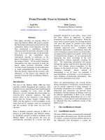
<i><small>Figure 1. Scheme of a two-dimensional BAC/SDS PAGE using slab gels. (A) Protein samples are </small></i>
<small>sep-arated on the first dimension BAC-PAGE. After visualization of proteins, an entire gel lane is excised.(B) The excised lane is re-buffered in 100 mM Tris, pH 6.8 for 30 min and afterwards incubated in 3×SDS sample buffer for another 5-10 min in order to exchange BAC for SDS. (C) The gel lane is transferredonto the second dimension SDS gel and fixed with a hot agarose solution. After separation staining revealsa characteristic spot pattern within an elliptical area.</small>
techniques and mass spectrometric analysis. Therefore, in recent years several studies have revealed the great potential of this alternative technique for membrane pro-teomics (Otto et al. 2001; Daub et al. 2002; Diao et al. 2003; Godl et al. 2003; Coughenour et al. 2004; Zahedi et al. 2006).
<b>3. GENERAL WORKFLOW</b>
The general scheme of 2-DB is depicted in Figure 1: Samples are first separated towards the cathode in an acidic PAGE system based on the cationic detergent BAC. Afterwards, protein lanes can be visualized by direct immersion into a col-loidal Coomassie staining solution. However, since Coomassie tends to precipitate in presence of cationic detergents which in turn leads to enhanced background staining, an intermediate washing step of at least 2 h is recommended in order to remove BAC from the gel surface.
After visualization whole protein lanes are excised and prepared for the second dimension. Since SDS-PAGE utilizes a more alkaline buffer system than BAC-PAGE, first of all gel lanes have to be re-buffered. Afterwards, BAC is exchanged for SDS by incubation in 3× SDS sample buffer. Finally, protein lanes are transferred onto a second dimension gel for separation and fixed by a hot agarose solution.
<b>4. IMPROVEMENTS</b>
While less complex samples can generally be separated using small gel sizes (approx-imately 7× 7 cm) in both dimensions, for samples of higher complexity switching
</div><span class="text_page_counter">Trang 34</span><div class="page_container" data-page="34">BERTRAND: “BERTRAND_CH02” — 2007/6/7 — 21:18 — PAGE 16 — #4
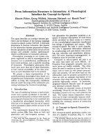
<i><small>Figure 2. Two-dimensional BAC/SDS-PAGE of platelet membrane proteins. While not only large gels are</small></i>
<small>recommended for complex samples, utilizing tube gels for the first dimension furthermore provides betterresolution and more efficient transfer of proteins to the second dimension (less vertical smearing). (A) Smallslab gel in the first dimension</small><i><small>(7 × 7 cm). (B) Large tube gel in the first dimension (1 mm × 15 cm).</small></i>
<small>(C) Summary of the human platelet membrane proteome study (Moebius et al. 2005). 158 proteins wereexclusively identified from 2-DB gels, 65 from 1D-PAGE. An overlap of 75 proteins was identified fromboth types of gels.</small>
to larger high resolution gels is recommended (Figure 2). Here, slab gels as well as tube gels can be utilized. However, the usage of slab gels is much more complicated in terms of handling (Figure 3).
In general, first dimension BAC-PAGE requires higher voltages and prolonged run-ning times than SDS-PAGE, resulting in an increased heat development – especially in case of large dimension slab gels. Therefore, cooling of running buffers during separation is mandatory.
Besides, an incomplete transfer of proteins from the first to the second dimension can be noticed for slab gels. The usage of tube gels with inner diameters of 1 mm and less totally abolishes this limitation and furthermore provides an improved resolution leading to a higher number of identifications in proteome studies. For this reason, the usage of tube gels is essential for differential studies. Moreover, after first dimension separation, time-consuming staining procedures which might be accompanied by loss of material can be omitted, as the entire gel can be transferred to the second dimension without the need for prior excision.
Resulting from our experience in the separation of a broad variety of samples with 2-DB, resolution strongly depends on the nature of the separated sample. Furthermore, upon long separation times, band broadening during first dimension separation can be observed.
</div><span class="text_page_counter">Trang 35</span><div class="page_container" data-page="35">BERTRAND: “BERTRAND_CH02” — 2007/6/7 — 21:18 — PAGE 17 — #5
<i>Two-dimensional BAC/SDS-PAGE for membrane proteomics</i> 17
<i><small>Figure 3. Processing of large slab (left) and tube (right) gels. Since transferring whole gel lanes of </small></i>
<small>approx-imately 15–20 cm length onto polymerized second dimension gels mostly results in extensive damage ofthe gel, a more complicated but also safer strategy is recommended here. First the excised gel lane is placedand fixed between two glass plates. Then the separation gel is poured beneath the lane. Finally, a stackinggel is poured on top, enclosing the first dimension gel lane. In case of tube gels, the gel is transferred ontothe already polymerized second dimension separation gel and fixed by hot agarose solution.</small>
In general, after membrane purification, it is highly recommended to introduce additional steps like carbonate- (Fujiki et al. 1982) and/or Triton X114-extraction prior to electrophoresis in order to reduce the amount of contaminating soluble proteins. Especially when separating plasma membrane enriched samples, resolution may be impaired in both dimensions. In that case protein precipitation prior to 2-DB may result in an improved separation, however it has to be kept in mind that precipitation procedures are generally not quantitative and might lead to unspecific loss of material. Recently, an improved BAC-PAGE protocol was introduced, particularly compen-sating for the inferior efficiency of the stacking gel when compared to common SDS-PAGE systems (Kramer 2006). By systematic studies the composition of gel buffers, running buffers as well as the sample buffer were optimized, result-ing in a higher resolution and shorter separation time, comparable to SDS-PAGE (Kramer 2006). Although the impact of these improvements was only investigated for one-dimensional BAC-PAGE, they nevertheless hold a great potential for 2-DB as well. However, this remains to be demonstrated in future studies.
<b>5. POTENTIAL FOR MEMBRANE PROTEOME STUDIES</b>
In systematic studies we demonstrated the potential of 2-DB compared to 2-DE (Zahedi et al. 2005) and SDS-PAGE (Moebius et al. 2005) regarding the separation of membrane proteins.
<i>While 2-DE separation of purified endoplasmatic reticulum membranes from Canis</i>
<i>familiaris yielded only a few spots after visualization by silver staining, an unequal</i>
</div><span class="text_page_counter">Trang 36</span><div class="page_container" data-page="36">BERTRAND: “BERTRAND_CH02” — 2007/6/7 — 21:18 — PAGE 18 — #6
higher amount of protein spots could be visualized after 2-DB. Among them, Sec61<i>α</i>
with a total of ten known TMD and a grand average hydrophobicity (GRAVY) index of 0.558 could be identified by Western blotting as well as mass spectrometry. Fur-thermore, 54 distinct ribosomal proteins were identified, which cannot be sufficiently resolved by 2-DE since their pIs range from 9 to 12. Furthermore, the separation of
<i>mitochondrial membranes from Saccharomyces cerevisiae yielded the subsequent</i>
identification of the extremely hydrophobic cytochrome-c oxidase subunit I with a total of 12 known TMD and a GRAVY index of 0.74 by mass spectrometry (MS).
In a comprehensive study of the human platelet membrane proteome we demon-strated the need for a combined application of 2-DB and one-dimensional SDS-PAGE for maximizing the amount of identified proteins. Since both techniques address different subproteomes (Figure 2C) they may be utilized in a complementary way. In 2-DB, a higher resolution increases the local protein concentration and facili-tates identification. However, in SDS-PAGE, whole lanes can be cut into equidistant slices, eliminating the need for protein visualization prior to excision. Thereby, even proteins, which cannot be visualized by staining procedures and therefore will escape detection after 2-DB, can be identified by subsequent MS.
<b>6. COMPARISON TO OTHER TECHNIQUES</b>
Another two-dimensional PAGE technique, doubled SDS (dSDS), introduced by Rais et al. (2004), is capable of resolving hydrophobic proteins with GRAVY indices of up to 0.86 – however it is has a lower resolution compared to 2-DB due to a smaller accessible separation area.
Since due to the lower resolution in 2-DB gels mostly several proteins co-localize within a single spot, in contrast to conventional 2-DE the usage of LC-MS/MS for protein identification is recommended instead of MALDI-MS.
Table 1 summarizes advantages and properties of the presented electrophoretic methods regarding (membrane) proteome studies.
<b>7. OUTLOOK</b>
Two-dimensional BAC/SDS polyacrylamide gel electrophoresis has been established as further tool in the field of proteome research, especially regarding the separation and analysis of membrane proteins. It is by far more efficient in resolving membrane proteins than common 2-DE and furthermore can be utilized in a complementary way to one-dimensional SDS-PAGE. Therefore, among other techniques, future proteome studies focussing on membrane proteins should include 2-DB as well.
Despite its lower resolution compared to 2-DE, 2-DB can also be applied in combination with the difference gel electrophoresis technique (DIGE) (Unlu et al. 1997; Reinders et al. 2006b), enabling the highly reproducible differential analysis of biological membrane samples.
However, for complex sample mixtures DIGE requires a very high resolution and distinct protein spots. Although the afore mentioned improvements by Kramer further
</div><span class="text_page_counter">Trang 37</span><div class="page_container" data-page="37"><small>BERTRAND:</small> “BERTRAND_CH02” — 2007/6/7 — 21:18 — PAGE 19 — #7
<i>Two-dimensional BAC/SDS-PAGE for membrane proteomics</i> 19
<i><small>Table 1. Comparison of different PAGE methods</small></i>
<small>Separation areaSmall laneEntire gelElliptical areaElliptical area</small>
enhance resolution during first dimension BAC-PAGE, additional prefractionation of complex mixtures might be necessary in order to account for the high demands of differential gel electrophoresis.
The authors thank the Deutsche Forschungsgemeinschaft (DFG) for continuous support within the SFB 688 and the FZT 82.
<small>Buxbaum, E. (2003) Cationic electrophoresis and electrotransfer of membrane glycoproteins. Anal.</small>
<i><small>Biochem. 314, 70–76.</small></i>
<small>Coughenour, H.D., Spaulding, R.S. and Thompson, C.M. (2004) The synaptic vesicle proteome: a</small>
<i><small>comparative study in membrane protein identification. Proteomics 4, 3141–3155.</small></i>
<small>Daub, H., Blencke, S., Habenberger, P., Kurtenbach, A., Dennenmoser, J., Wissing, J., Ullrich, A.and Cotten, M. (2002) Identification of SRPK1 and SRPK2 as the major cellular protein kinases</small>
<i><small>phosphorylating hepatitis B virus core protein. J. Virol. 76, 8124–8137.</small></i>
<small>Diao, A., Rahman, D., Pappin, D.J., Lucocq, J. and Lowe, M. (2003) The coiled-coil membrane protein</small>
<i><small>golgin-84 is a novel rab effector required for Golgi ribbon formation. J. Cell Biol. 160, 201–212.</small></i>
<small>Fujiki, Y., Hubbard, A.L., Fowler, S. and Lazarow, P.B. (1982) Isolation of intracellular membranes by</small>
<i><small>means of sodium carbonate treatment: application to endoplasmic reticulum. J. Cell Biol. 93, 97–102.</small></i>
<small>Godl, K., Wissing, J., Kurtenbach, A., Habenberger, P., Blencke, S., Gutbrod, H., Salassidis, K.,Stein-Gerlach, M., Missio, A., Cotten, M. and Daub, H. (2003) An efficient proteomics method</small>
<i><small>to identify the cellular targets of protein kinase inhibitors. Proc. Natl. Acad. Sci. U.S.A. 100,</small></i>
<small>Hartinger, J., Stenius, K., Hogemann, D. and Jahn, R. (1996) 16-BAC/SDS-PAGE: a two-dimensional gel</small>
<i><small>electrophoresis system suitable for the separation of integral membrane proteins. Anal. Biochem. 240,</small></i>
<small>126–133.</small>
</div><span class="text_page_counter">Trang 38</span><div class="page_container" data-page="38">BERTRAND: “BERTRAND_CH02” — 2007/6/7 — 21:18 — PAGE 20 — #8
<i><small>Kramer, M.L. (2006) A new multiphasic buffer system for benzyldimethyl-n-hexadecylammonium chlo-ride polyacrylamide gel electrophoresis of proteins providing efficient stacking. Electrophoresis 27,</small></i>
<small>Luche, S., Santoni, V. and Rabilloud, T. (2003) Evaluation of nonionic and zwitterionic detergents as</small>
<i><small>membrane protein solubilizers in two-dimensional electrophoresis. Proteomics 3, 249–253.Macfarlane, D.E. (1983) Use of benzyldimethyl-n-hexadecylammonium chloride (“16-BAC”), a cationic</small></i>
<small>detergent, in an acidic polyacrylamide gel electrophoresis system to detect base labile protein</small>
<i><small>methylation in intact cells. Anal. Biochem. 132, 231–235.</small></i>
<i><small>Macfarlane, D.E. (1989) Two dimensional benzyldimethyl-n-hexadecylammonium chloride – sodium</small></i>
<small>dodecyl sulfate preparative polyacrylamide gel electrophoresis: a high capacity high resolution</small>
<i><small>technique for the purification of proteins from complex mixtures. Anal. Biochem. 176, 457–463.</small></i>
<small>Moebius, J., Zahedi, R.P., Lewandrowski, U., Berger, C., Walter, U. and Sickmann, A. (2005) The humanplatelet membrane proteome reveals several new potential membrane proteins. Mol. Cell Proteomics</small>
<i><small>4, 1754–1761.</small></i>
<small>Molloy, M.P. (2000) Two-dimensional electrophoresis of membrane proteins using immobilized pH</small>
<i><small>gradients. Anal. Biochem. 280, 1–10.</small></i>
<small>Olsson, I., Larsson, K., Palmgren, R. and Bjellqvist, B. (2002) Organic disulfides as a means to generatestreak-free two-dimensional maps with narrow range basic immobilized pH gradient strips as first</small>
<i><small>dimension. Proteomics 2, 1630–1632.</small></i>
<small>Otto, H., Dreger, M., Bengtsson, L. and Hucho, F. (2001) Identification of tyrosine-phosphorylated proteins</small>
<i><small>associated with the nuclear envelope. Eur. J. Biochem. 268, 420–428.</small></i>
<small>Rais, I., Karas, M. and Schagger, H. (2004) Two-dimensional electrophoresis for the isolation of integral</small>
<i><small>membrane proteins and mass spectrometric identification. Proteomics 4, 2567–2571.</small></i>
<small>Reinders, J., Zahedi, R.P., Pfanner, N., Meisinger, C. and Sickmann, A. (2006a) Toward the Complete YeastMitochondrial Proteome: Multidimensional Separation Techniques for Mitochondrial Proteomics.</small>
<i><small>J. Proteome Res. 5, 1543–1554.</small></i>
<small>Reinders, Y., Schulz, I., Graf, R. and Sickmann, A. (2006b) Identification of novel centrosomal proteins</small>
<i><small>in Dictyostelium discoideum by comparative proteomic approaches. J. Proteome Res. 5, 589–598.</small></i>
<small>Santoni, V., Molloy, M. and Rabilloud, T. (2000) Membrane proteins and proteomics: un amour impossible?</small>
<i><small>Electrophoresis 21, 1054–1070.</small></i>
<small>Unlu, M., Morgan, M.E. and Minden, J.S. (1997) Difference gel electrophoresis: a single gel method for</small>
<i><small>detecting changes in protein extracts. Electrophoresis 18, 2071–2077.</small></i>
<small>Zahedi,R.P.,Meisinger,C. and Sickmann,</small> <i><small>A. (2005) Two-dimensional benzyldimethyl-n-hexadecylammonium chloride/SDS-PAGE for membrane proteomics. Proteomics 5, 3581–3588.</small></i>
<small>Zahedi, R.P., Sickmann, A., Boehm, A.M., Winkler, C., Zufall, N., Schonfisch, B., Guiard, B., Pfanner, N.and Meisinger, C. (2006) Proteomic analysis of the yeast mitochondrial outer membrane reveals</small>
<i><small>accumulation of a subclass of preproteins. Mol. Biol. Cell 17, 1436–1450.</small></i>
<small>Corresponding author: Dr. Albert Sickmann</small>
</div><span class="text_page_counter">Trang 39</span><div class="page_container" data-page="39">BERTRAND: “BERTRAND_CH03” — 2007/6/8 — 20:08 — PAGE 21 — #1
<b>CHAPTER 3</b>
<b>MICROPARTICLES: A NEW TOOL FOR PLASMAMEMBRANE SUB-CELLULAR PROTEOMIC</b>
LAURENT MIGUET<sup>1</sup>, SARAH SANGLIER<sup>1</sup>, CHRISTINE SCHAEFFER<sup>1</sup>, NOELLE POTIER<sup>1</sup>, LAURENT MAUVIEUX<sup>2</sup>, and
ALAIN VAN DORSSELAER<sup>1</sup>
<small>1</small><i><small>Laboratoire de Spectrométrie de Masse Bio-Organique, ECPM, UMR/CNRS 7178, InstitutPluridisciplinaire Hubert CURIEN, Université Louis Pasteur, Strasbourg, France</small></i>
<small>2</small><i><small>Institut d’Hématologie et d’Immunologie, Faculté de Médecine, Université Louis Pasteurand Laboratoire d’Hématologie, Hôpital Hautepierre, Strasbourg, France</small></i>
<b>Table of Contents</b>
1. Introduction . . . . 21 2. Microparticles . . . . 22 3. Strategy Used for Microparticles Proteomic Analysis . . . . 23 3.1. Presentation of the Strategy . . . . 23 3.2. Technical Details . . . . 26 4. Validation of Microparticles as a New Tool
for Plasma Membrane Preparation . . . . 30 5. Perspectives . . . . 31 References . . . . 33
<b>1. INTRODUCTION</b>
Membranes are critical components of cellular structure. It has been reported that plasma membrane proteins represent about 30% of all cellular proteins (Wallin and von Heijne 1998). Even if plasma membrane was considered for a long time as a simple biological barrier between the cytosol of the cell and the extra-cellular envi-ronment, these membrane proteins have been demonstrated to play a crucial role in the different fundamental biological processes as exchange of component or signal transduction. Also, more than half of all anticipated pharmacological drug targets are predicted to be localized to the plasma membrane (Jang and Hanash 2003). Plasma membrane can then clearly be considered as a sub-cellular compartment of first inter-est in regard to different diagnosis and/or therapeutic target proteins. Indeed, for each membrane protein there is potentially a specific antibody which can be used
<i><small>E. Bertrand and M. Faupel (eds.), Subcellular Proteomics, 21–34.© 2007 Springer.</small></i>
</div><span class="text_page_counter">Trang 40</span><div class="page_container" data-page="40">BERTRAND: “BERTRAND_CH03” — 2007/6/8 — 20:08 — PAGE 22 — #2
for the diagnosis of several pathologies and also for treatment using armed antibod-ies (Harris 2004). Therefore, proteomic analysis of plasma membrane proteins is of first importance. Despite the importance of plasma membrane proteins, there is less understanding in this class of proteins due to the difficulty to obtain enriched plasma membrane proteins preparation from eukaryotic cells. Until now many dif-ferent strategies have been applied but are still laborious and imperfect. In example of these different approaches, biotinylation (Peirce et al. 2004; Zhao et al. 2004) silica coated (Rahbar and Fenselau 2004), partition phase repartition (Qoronfleh et al. 2003) or partial tryptic surface digestion have been tested, but they still remain unsatisfactory. Although some studies of plasma membrane proteins using 2-DE have been reported (Galeva and Altermann 2002; Luche et al. 2003), but the separation of such hydrophobic proteins have been often poor (Santoni et al. 2000). We describe here a new strategy in order to increase the proportions of plasma membrane proteins identified with the highest possible coverage percentage in the different membrane proteins preparations analyzed by mass spectrometry. We will focus on microparticles as a source of plasma membrane proteins.
<b>2. MICROPARTICLES</b>
Each of the two leaflets of the plasma membrane bilayer has a specific lipid com-position. Aminophospholipids (phosphatidylserine and phosphatidylethanolamine) are specifically segregated in the inner layer of the membrane, whereas the phos-phatidylcholine and the sphingomyelin are enriched in the external leaflet (Bevers et al. 1998). When cells are submitted to various stress conditions as mitogenic activa-tions or apoptosis, the constitutive asymmetry between the inner and the outer leaflet of the plasma membrane is disrupted. The major changes in the plasma membrane constitution will be the delocalization of the phosphatidylserine to the outer leaflet and an augmentation of the Ca<sup>2</sup><small>+</small><sub>ion concentration in the cytoplasm. Such changes</sub> are going to disrupt the organisation of the cytoskeleton and drive to a blebbing of the plasma membrane and release microvesicles which are named microparticles (MPs) (Figure 1) (Hugel et al. 2005; Miguet et al. 2005).
Such microvesicles have size variable between 50 nm to 1µm and differ from other vesicles (like exosomes (30–100 nm)). In general, microparticles are phospholipids vesicles derived from eukaryotic cells as a result of different types of stimulation. Microparticles can also be defined as phospholipids microvesicles containing certain membrane proteins originating from the parental cell. Microparticles circulate in the blood and contribute to numerous physiological processes. MPs have been described in various haematopoietic cells as platelets (Heijnen et al. 1999), T-cells (Blanchard et al. 2002), polynuclear neutrophils (Mesri and Altieri 1999) or dendritic cells. After have been considered as cell dust, MPs are now considered to reflect cell activation. Platelet derived microparticles have been the most extensively studied until now. They are now accepted to play an important role in the procoagulant
</div>