Genomics : Applications in Human Biology / Sandy B. Primrose
Bạn đang xem bản rút gọn của tài liệu. Xem và tải ngay bản đầy đủ của tài liệu tại đây (2.65 MB, 229 trang )
<span class="text_page_counter">Trang 3</span><div class="page_container" data-page="3">
Sandy B. Primrose
<i>Senior Partner, Business & Technology Management,High Wycombe, UK</i>
Richard M. Twyman
<i>Department of Biology, University of York, York, UKManaging Director, Write Science, York, UK</i>
Applications inHuman Biology
</div><span class="text_page_counter">Trang 4</span><div class="page_container" data-page="4"><small>350 Main Street, Malden, MA 02148-5020, USA108 Cowley Road, Oxford OX4 1JF, UK</small>
<small>550 Swanston Street, Carlton, Victoria 3053, Australia</small>
<small>The right of Sandy B. Primrose and Richard M. Twyman to be identified as the Authors of thisWork has been asserted in accordance with the UK Copyright, Designs, and Patents Act 1988.All rights reserved. No part of this publication may be reproduced, stored in a retrieval system, ortransmitted, in any form or by any means, electronic, mechanical, photocopying, recording orotherwise, except as permitted by the UK Copyright, Designs, and Patents Act 1988, without theprior permission of the publisher.</small>
<i><small>Library of Congress Cataloging-in-Publication Data</small></i>
<small>Primrose, S. B.</small>
<small>Genomics : applications in human biology / Sandy B. Primroseand Richard Twyman.</small>
<small>p. ; cm.Includes index.</small>
<small>ISBN 1– 4051– 0819 –3 ( pbk.)</small>
<small>1. Medical genetics.2. Genomics.3. Pharmaceutical biotechnology.4. Molecular biology.I. Twyman, Richard M.II. Title.</small>
<small>[DNLM:1. Genomics.2. Biotechnology.3. Molecular Biology.</small>
<small>by Graphicraft Limited, Hong KongPrinted and bound in the United Kingdomby TJ International Ltd, Padstow, CornwallFor further information on</small>
<small>Blackwell Publishing, visit our website:</small>
</div><span class="text_page_counter">Trang 5</span><div class="page_container" data-page="5">Full Contents vii
C H A P T E R O N E Biotechnology and genomics in medicine 1
C H A P T E R T H R E E Genomics and the challenge of infectious disease 60 C H A P T E R F O U R Analyzing and treating genetic diseases 90 C H A P T E R F I V E Diagnosis and treatment of cancer 112 C H A P T E R S I X The large scale production of biopharmaceuticals 131 C H A P T E R S E V E N Genomics and the development of new chemical
</div><span class="text_page_counter">Trang 6</span><div class="page_container" data-page="6">CHAPTER ONE: Biotechnology and genomics
</div><span class="text_page_counter">Trang 7</span><div class="page_container" data-page="7">Applications of expression proteomics 51
CHAPTER THREE: Genomics and the challenge
Genomics and the development of new antibacterial agents 78
CHAPTER FOUR: Analyzing and treating
Finding genes for monogenic diseases and determining
CHAPTER FIVE: Diagnosis and treatment
</div><span class="text_page_counter">Trang 8</span><div class="page_container" data-page="8">New methods for the diagnosis of cancer 119
Using gene manipulation to facilitate downstream processing of
CHAPTER SEVEN: Genomics and the development
</div><span class="text_page_counter">Trang 9</span><div class="page_container" data-page="9">Nucleic acids as drugs 190
</div><span class="text_page_counter">Trang 10</span><div class="page_container" data-page="10">Fifty years ago, Watson and Crick detailed for us the structure of DNA and showed how it could be replicated faithfully from generation to generation. The impact of this discovery on medicine was barely considered. Rather, biologists wanted to know about the structure of genes and the genetic code. Twenty-five years ago the biotechnology revolution was underway following the development of
<i>recombin-ant DNA technology, which permitted the in vitro production of human proteins </i>
on a large scale. Then the vision for biotechnology was no more than factories producing recombinant molecules. Pharmaceutical biotechnology, as it then was known, was a very narrow subject.
Today we are in the midst of the genomics revolution, which was spearheaded by international projects aiming to sequence the complete genomes of organisms ranging from bacteria to mammals, including humans. Many of the genes in these organisms have been identified and good progress is being made towards under-standing the roles of these genes in health and disease. As a consequence, there is almost no aspect of medicine and drug development that has not been affected. For example, we now have a good understanding of the genes involved in microbial pathogenicity and this is facilitating the development of new diagnostics, new vac-cines, and new antibiotics. Similarly, we are rapidly dissecting the genetic basis of inherited diseases and cancer, which again is leading to new diagnostics and new treatments. The development of these new pharmaceuticals is being facilitated by the introduction of novel screening methodologies that are themselves based on recombinant DNA technology and genomics.
When Watson and Crick announced their momentous discovery almost all pharmaceuticals were small molecules, although insulin was a notable exception. Following the advent of recombinant DNA technology this drug repertoire was expanded to include a much wider range of natural human proteins including interferons, blood products, and further hormones. Today the diversity of drug molecules has expanded further, to include engineered proteins that are unlike any produced naturally, humanized antibodies, and even nucleic acids. Furthermore new medical procedures are being developed, such as gene therapy, cell therapy, and tissue therapy.
</div><span class="text_page_counter">Trang 11</span><div class="page_container" data-page="11">Given the pace at which the above developments are taking place it is not surpris-ing that students and their academic mentors have difficulty in seesurpris-ing the whole picture. This book has been written to provide them with the necessary overview, covering technologic developments, applications, and (where necessary) the eth-ical implications. The book is divided into three sections. The first section (Chapters 1 and 2) introduces the role of biotechnology and genomics in medicine and sets out some of the technologic advances that have been the basis of recent medical break-throughs. The second section (Chapters 3–5) takes a closer look at how biotech-nology and genomics are influencing the prevention and treatment of different categories of disease. Finally, in the third section (Chapters 6–8), we describe the contribution of biotechnology and genomics to the development of different types of therapy, including conventional drugs, recombinant proteins, and gene/cell therapies.
Throughout the book, the level of detail has been selected so that the reader can grasp what has been achieved without falling victim to “not seeing the wood for the trees.” A basic understanding of genetics and molecular biology has been assumed so we can avoid the obligatory chapters on DNA structure, gene expression, etc. that appear in most larger biology textbooks regardless of their actual focus. Readers requiring more detail of the recombinant DNA and genomics techniques
<i>should consult our more advanced textbooks on these subjects: Principles of GeneManipulation (POGM) and Principles of Genome Analysis and Genomics (POGA), also</i>
published by Blackwell Publishing. References to appropriate sections in these two books are included at the end of each chapter (with the relevant acronym indicating the book), plus a short bibliography mostly comprising review papers that have been selected for their clarity of presentation. The reader will also find the text con-tains several categories of boxed text, which include history boxes (describing the origins and development of particular technologies or treatments), molecular boxes (which describe the molecular basis of diseases or treatments in more detail), and ethics boxes (which discuss the ethical implications of technology development and new therapies).
Finally, we would like to thank the people who provided invaluable assistance in the preparation of the manuscript, particularly Sue Goddard and her team in the library at CAMR and Alistair Fitter at the Department of Biology, University of York. Richard Twyman would like to dedicate this book to his parents, Peter and Irene, his children, Emily and Lucy, and to Hannah, Joshua, and Dylan.
<i>Sandy B. Primrose and Richard M. Twyman</i>
<i>Primrose SB, Twyman RM (2003) Principles of Genome Analysis and Genomics, 3rd edn.</i>
Blackwell Publishing, Oxford.
<i>Primrose SB, Twyman RM, Old RW (2001) Principles of Gene Manipulation, 6th edn.</i>
Blackwell Science, Oxford.
</div><span class="text_page_counter">Trang 12</span><div class="page_container" data-page="12">Some figures and tables have been used from other sources. We thank the various authors and publishers for permission to use this material, which has come from the following sources:
Figures are extensively drawn from the following publications by the authors:
<i>Primrose SB (1991) Molecular Biotechnology, 2nd edn. Blackwell Science, Oxford.Primrose SB, Twyman RM (2003) Principles of Genome Analysis and Genomics, 3rd edn.</i>
Blackwell Publishing, Oxford.
<i>Primrose SB, Twyman RM, Old RW (2001) Principles of Gene Manipulation, 6th edn.</i>
Blackwell Science, Oxford.
Specific tables and figures have been taken from the following sources:
<i>Fig. 2.4: Coulson A, Sulston J, Brenner S et al. (1986) Toward a physical map of the</i>
<i><b>genome of the nematode Caenorhabditis elegans. Proc Natl Acad Sci USA 83,</b></i>
Fig. 2.8: EnsEMBL human genome browser www.ensembl.org
<i>Fig. 2.9: Veculescu VE et al. (1997) Characterization of the yeast transcriptome. Cell</i>
<b>88, 243–251.</b>
Fig. 2.12 inset: Görg A, Postel W, Baumer M, Weiss W (1992) Two-dimensional polyacrylamide gel electrophoresis, with immobilized pH gradients in the first dimension, of barley seed proteins: discrimination of cultivars with different mating
<i><b>grades. Electrophoresis 13, 192–203.</b></i>
Fig. 3.4: Courtesy of Catherine Arnold, UK Health Protection Agency.
<i><b>Fig. B3.3: Behr et al. (1999) Science 284, 1520–1523. [for Box 3.3]</b></i>
<i>Fig. 4.4: Nussbaum RL, McInnes RR, Willard HF (2001) Genetics in Medicine, WB</i>
Saunders, Philadelphia, figure 4.14. Original photograph courtesy of P. Wray, Hospital for Sick Children, Toronto.
<i>Fig. 4.6: Nussbaum RL, McInnes RR, Willard HF (2001) Genetics in Medicine, WB</i>
Saunders, Philadelphia.
</div><span class="text_page_counter">Trang 13</span><div class="page_container" data-page="13">Fig. 4.7: Thomson G (2001) Mapping of disease loci. In: Kalow W, Meyer UA,
<i>Tyndale R, eds. Pharmacogenomics, pp 337–361. Marcel Dekker, New York.</i>
Fig. 4.9: Judson R, Stephens JC, Windemuth A (2000) The predictive power of
<i><b>haplotypes in clinical response. Pharmacogenomics 1, 15–26.</b></i>
<i>Fig. 4.10: Nussbaum RL, McInnes RR, Willard HF (2001) Genetics in Medicine, WB</i>
Saunders, Philadelphia, figure 4.13.
Fig. 4.11: Johnson JA, Evans WE (2002) Molecular diagnostics as a predictive tool:
<i><b>genetics of drug efficacy and toxicity. Trends Mol Med 8, 300–305.</b></i>
<i>Fig. 5.6: Funaro A, Hovenstein AL, Santoro P et al. (2000) Monoclonal antibodies</i>
<i><b>and therapy of human cancers. Biotechnol Adv 18, 385 – 401, figure 2.</b></i>
Fig. B6.4b: Procognia Ltd.
Fig. 7.4: Croston GE (2002) Functional cell-based uHTS in chemical genomic drug
<i><b>discovery. Trends Biotechnol 20, 110–115, figure 2.</b></i>
<i><b>Fig. 7.5: Bandara, Kennedy (2002) Drug Discovery Today 7, 411– 418, figure 2.Fig. 7.7: Thompson, Ellman (1996) Chem Rev 96, 555, figure 10.29.</b></i>
<i>Fig. 7.8: Balkenhol F, von dem Bussche-Hunnefeld C, Lansky A et al. (1996) Angew</i>
<i><b>Chem Int Ed Engl 35, 2289, figure 10.30.</b></i>
Fig. 7.12: Castle AL, Carver MP, Mendrick DL (2002) Toxicogenomics: a new
<i><b>revolution in drug safety. Drug Discovery Today 7, 728–736, figure 4a.</b></i>
Table 7.1: Croston GE (2002) Functional cell-based uHTS in chemical genomic
<i><b>drug discovery. Trends Biotechnol 20, 110–115.</b></i>
<i>Table 7.2: DeVito JA et al. (2002) An array of target-specific screening strains for</i>
<i><b>antibacterial discovery. Nature Biotechnol 20, 478– 483.</b></i>
</div><span class="text_page_counter">Trang 14</span><div class="page_container" data-page="14">Over the last 300 years, there has been a growing understanding of how the human body functions in health and disease. However, our knowledge has not increased steadily. The history of medicine is punctuated by sudden breakthroughs and leaps of innovation. Very few of these key developments would have been possible
<b>with-out underlying advances in technology.</b>
As an example, consider the discovery of the first two antimicrobial substances by Alexander Fleming – lysozyme in 1922 and penicillin in 1928. Both discoveries were serendipitous, and neither would have been made if Fleming had been unable to culture bacteria on a solid growth medium. The use of agar for this purpose, initially proposed by Fanny Hesse, was put into practice by Robert Koch in 1882. Armed with such pure culture techniques, Robert Koch and Louis Pasteur were able to establish the principles of bacterial pathogenicity, thus founding the modern discipline of medical microbiology. In turn, the work of Fleming, Pasteur, and Koch stemmed from the discovery of bacteria by Anton van Leeuwenhoek in 1683, and this would have been impossible without the microscope. Van Leeuwenhoek made his own crude microscopes, but credit for the original invention goes to Hans and Zacharias Janssen in 1595. Similarly, the use of ether as an anesthetic, first demon-strated by Crawford Long in 1842,* would not have been possible without a method for ether synthesis. Such a method was first described by the German scientist Valerius Cordus in 1540. Thus, medical breakthroughs invariably have depended on technologic advances in physics, chemistry, and biology.
Since 1970, we have witnessed an unprecedented number of new medical
<b>innovations reflecting our increasing knowledge of the molecular basis of health</b>
and disease. While chemistry and physics have played their roles, much of this innovation is the direct result of two technologic revolutions in biology – the <small>*Crawford Long was the first to demonstrate the use of ether as an anesthetic, but prov-enance is often attributed to William Morton, who was the first to publish on the technique, in 1846.</small>
</div><span class="text_page_counter">Trang 15</span><div class="page_container" data-page="15"><b>recombinant DNA revolution and the genomics revolution, which are the</b>
subjects of this book. In this first chapter, we briefly summarize the impact of recom-binant DNA and genomics on the practice of medicine. In later chapters, we discuss the role of these technologies in the prevention, diagnosis and treatment of different types of disease, and examine the emerging technologies that may contribute to the medical breakthroughs of the future.
Recombinant DNA technology
<b>The recombinant DNA revolution began in about 1972 with the development of</b>
<i>tools and techniques for in vitro DNA manipulation. Until the 1970s, it was </i>
impos-sible to manipulate DNA precisely, which meant it was very difficult to study indi-vidual genes in a direct manner. In model organisms, genetic analysis could be used to find out about the structure and function of genes indirectly, but such methods could not be applied easily to humans. Recombinant DNA technology was enabled by the isolation and biochemical characterization of enzymes that bacteria use to manipulate DNA as part of their normal cellular processes (Box 1.1). It was soon realized that if such enzymes could be purified, they could be used to create novel
<i>combinations of different DNA fragments in vitro. Such novel fragments were</i>
<b>termed recombinant DNA molecules.</b>
<b>The central importance of cloning</b>
To study a particular DNA sequence experimentally it is necessary to generate enough copies for laboratory-scale handling. The first significant advance offered by recombinant DNA technology was the ability to prepare millions of copies of the
<b>same DNA sequence, a technique known as molecular cloning. Researchers had</b>
Box 1.1 Key enzymes used to manipulate DNA
<small>• Restriction endonucleases. These are bacterial</small><small>enzymes that cut DNA molecules internally at positionsdefined by specific target sequences, allowing largeDNA molecules to be cut into predictable fragments.Both DNA strands are cut and the cleavage sites may be opposite each other (generating bluntfragments) or staggered (generating overhangs).• DNA ligases. These are enzymes that join DNAfragments end to end. Some can join blunt fragments,while others require overhangs. The compatibility ofoverhanging ends depends on the restrictionendonuclease used.</small>
<small>• DNA polymerases. These are enzymes that synthesizeDNA on a complementary template. Different enzymesare used for DNA labeling, DNA sequencing, thepolymerase chain reaction, and reverse transcription of mRNA into cDNA.</small>
<small>• DNA modification enzymes. Examples includealkaline phosphatase (which removes phosphate groups from the ends of DNA fragments) andpolynucleotide kinase (which carries out the reverseprocess). These enzymes are used to control ligationreactions and for DNA labeling.</small>
</div><span class="text_page_counter">Trang 16</span><div class="page_container" data-page="16"><b>known for a long time that bacteria contained autonomous replicons, i.e. genetic</b>
elements such as plasmids and bacteriophage (phage) with the intrinsic ability to replicate to a high copy number. Recombinant DNA techniques were used to join such replicons to human DNA sequences, so that the human sequences were
<b>amplified. This principle led to the development of cloning vectors, i.e. DNA </b>
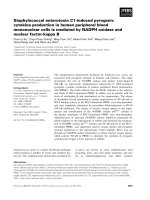
ele-ments based on plasmids, phage, or sometimes a combination of both, which are used specifically to clone fragments of donor or passenger DNA. The general tech-nique for cell-based molecular cloning is shown in Fig. 1.1.
<small>Fig. 1.1 The principle of cell-based molecular cloning with plasmid vectors. The vector is cut openwith a restriction enzyme that has only one recognition site in the vector sequence, thus cutting it at a predictable position. The insert, prepared with the same enzyme, is sealed into place with</small>
<i><small>DNA ligase. The recombinant vector is then introduced into the bacterium Escherichia coli by</small></i>
<small>transformation. The vector carries a selectable marker gene (see p. 184) which allows transformedbacteria, but not normal bacteria, to survive and proliferate. When the bacteria are spread on aplate of medium supplemented with antibiotic, transformed bacteria form colonies containingabout 1× 10⁶cells in which each cell carries several hundred copies of the plasmid. Individualcolonies are picked and grown in larger scale culture vessels under selection from which largeamounts of DNA can be isolated. The insert, now massively amplified, can be purified using thesame restriction enzyme used to insert it into the vector in the first place.</small>
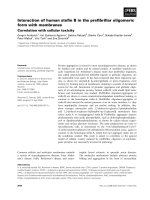
</div><span class="text_page_counter">Trang 17</span><div class="page_container" data-page="17"><small>Fig. 1.2 The basic polymerase chain reaction. A double-stranded DNA template is denatured (separated into single strands) and two primers are annealed. The primers face towards each other,anneal to opposite strands, and define the target fragment to be amplified. Primer extension copiesthe DNA in the region between the two primers and therefore doubles the amount of template. The process of template denaturation, primer annealing, and primer extension is repeated 25–30 times. In the presence of excess primers and other reaction components, 25 cycles can theoretically yield over 8 million copies of the same fragment.</small>
</div><span class="text_page_counter">Trang 18</span><div class="page_container" data-page="18">In the mid-1980s, a different technique for DNA amplification was developed
<i>that is carried out in vitro using purified DNA polymerase. This has become known</i>
<b>as the polymerase chain reaction (PCR). The basic PCR is shown in Fig. 1.2. Thetechnique requires primers, single-stranded DNA molecules that anneal at </b>
particu-lar sites on the template DNA. If two primers are designed to flank a target region of interest, face inwards, and anneal to opposite DNA strands, DNA synthesis across the region defined by the primers will double the amount of template available.
<b>Therefore, cyclical rounds of denaturation (separation of the template DNA intosingle strands), primer annealing, and primer extension by DNA synthesis can</b>
result in the exponential amplification of the target DNA sequence. Compared to traditional cell-based DNA cloning, the PCR is rapid, sensitive, and robust. It can be used to prepare large amounts of a specific fragment starting from a very small amounts of starting material, and that starting material does not have to be well preserved. For example, DNA can be extracted and amplified from fixed biologic specimens, blood and semen samples at crime scenes, and even Neanderthal bones! However, the PCR is generally less accurate than cell-based cloning because the DNA polymerases used in this procedure are error-prone. The standard technique is suitable for the amplification of fragments only up to about 5 kb in length, whereas large-capacity cloning vectors can easily amplify sequences that are several hun-dred kilobases long. Therefore cell-based cloning and the PCR have complementary although overlapping uses in human molecular biology.
Both of the cloning methods discussed above require a procedure that allows the progress of reactions to be followed and the products to be analyzed. The standard
<b>technique is gel electrophoresis, which separates DNA molecules on the basis of</b>
size (Box 1.2).
<b>Identification and cloning of specific genes</b>
Before a specific gene sequence can be cloned, it must be isolated from its natural source, and this is generally the bottleneck in any cloning procedure. The two
Box 1.2 Gel electrophoresis
<small>Gel electrophoresis is the standard method for the </small><small>size-separation of mixtures of DNA molecules. The basic principle is that DNA molecules in solution are negatively charged, and will therefore move towards the anode in an electric field. If the solution is dispersed within a matrix such as an agarose orpolyacrylamide gel, the pores of the gel have a sievingeffect, so that smaller molecules move towards theanode more rapidly than larger ones. The separatingrange of the gel depends on the pore size, whichdepends on the gel concentration. For example, a 5% agarose gel will separate DNA molecules within </small>
<small>the range 100–500 bp, while a 0.5% gel will separatemolecules in the range 5–20 kb. Polyacrylamide gels are used for smaller DNA fragments, and where it isnecessary to distinguish between molecules differing insize by a single nucleotide (e.g. in DNA sequencing). Inagarose gels, the fate of individual DNA molecules isfollowed using the intercalating fluorescent dyeethidium bromide, whereas in polyacrylamide gels theDNA is generally labeled prior to separation. Specialtechniques, such as pulsed-field gel electrophoresis, are required to separate molecules greater than 50 kb.</small>
</div><span class="text_page_counter">Trang 19</span><div class="page_container" data-page="19">major sources of DNA for cloning, genomic DNA and complementary DNA (cDNA), are both incredibly complex (Table 1.1). Individual genes are therefore diluted by millions of irrelevant DNA fragments.
In some rare cases, obtaining the desired sequence has been relatively straight-forward. For example, among the first human genes to be cloned were those encod-ingα-globin and β-globin because the mRNA is so highly enriched in reticulocytes (immature red blood cells) that cDNA clones could be obtained simply by random sequencing. However, few genes fall into this “superabundant” category and more sophisticated strategies are usually required.
<b>In cell-based molecular cloning, the general approach is to create a DNA library,</b>
in which a collection of cloned DNA fragments is assembled representing the entire
<b>source population (genomic DNA or cDNA). The library is then screened using</b>
one of the following procedures:
• Sequence-dependent screening. This is performed either by hybridization, using a labeled DNA or RNA probe (Box 1.3), or by PCR. In each case, the technique relies on the probe or PCR primer combination recognizing a particular clone in the library because it has the complementary sequence. Suitable probes or primer com-binations can be obtained from existing partial clones, from clones of similar genes in other species, from consensus sequences representing a particular gene family, or from the known amino acid sequences of proteins.
<b>• Immunologic screening. This requires an expression library, i.e. a cDNA</b>
library in which all the clones are expressed to produce proteins. If an antibody is available that recognizes the protein product of the target gene, the corresponding DNA clone can be isolated.
• Functional screening. This also requires an expression library. The screening pro-cedure is a test for protein function, e.g. a particular enzyme activity or a particular effect when introduced into cultured cells.
In contrast to cell based cloning, the PCR can be used to isolate DNA sequences directly from the source (i.e. without first creating a library), essentially following a sequence-dependent screening strategy. As stated above, the standard PCR can
<small>Table 1.1 Properties of genomic DNA and cDNA.Genomic DNA</small>
<small>With rare exceptions, genomic DNA is the same in all tissues from the same organismGenes in natural context (includes spacer DNA, regulatory elements, and introns)</small>
<small>All genes representedGenes represented equally</small>
<small>cDNA differs between tissues, and according todevelopmental stage and cell state</small>
<small>Only transcribed sequences represented. No spacerDNA, regulatory elements, or introns. Splice variantsrepresented by different cDNAs</small>
<small>Only genes expressed in the tissue from which mRNAwas obtained are represented</small>
<small>Different genes are not represented equally –strongly expressed genes will produce moretranscripts and give rise to more cDNA copies thanweakly expressed genes</small>
</div><span class="text_page_counter">Trang 20</span><div class="page_container" data-page="20"><b><small>1 kg</small></b>
<small>Hybridization, i.e. complementary base pairing between single-stranded nucleic acids, is one of the core techniques in molecular biology. It allows theidentification of specific DNA sequences in complexmixtures. One nucleic acid molecule is labeled in someway to facilitate detection and then used as a probe toidentify a specific target. For example, in Southern blothybridization, genomic DNA is fragmented, separatedby agarose gel electrophoresis, and then transferred to a membrane where it is immobilized as an imprint of the gel. The DNA is then denatured (to separate the strands) and a probe is added. The probe willhybridize to a specific target and will be revealed as a band when the label is detected (Fig. B1.3). Analogous procedures can be used to identify specificRNA molecules in mixtures separated by electrophoresis</small>
<i><small>(northern blot hybridization) or RNA molecules in situ</small></i>
<small>in tissue sections, embryos, or explants (</small><i><small>in situ</small></i>
<small>hybridization). Hybridization is also used to identifyclones in library screens (colony or plaquehybridization).</small>
<small>Traditionally, DNA and RNA probes have beenlabeled with radioactive substrates and detected byautoradiography (exposure to a radiation-sensitive film) or phosphorimaging (exposure to a radiation-sensitive screen). However, radioactive labels are beingprogressively replaced by nonradioactive alternatives,such as fluorophores, enzymes that can be detectedusing a colorimetric assay, chemiluminescent substrates, and haptens (which are detected withantibodies). Whatever label is used, incorporationinvolves either DNA/RNA synthesis with labelednucleotide analogs or end-labeling reactions using DNA modification enzymes (Box 1.1).</small>
<small>Fig. B1.3 The Southern blot demonstrates the value of hybridization in molecular biology. A complexpopulation of DNA molecules (e.g. cDNA, digested genomic DNA) containing a target sequence of interest(shown in bold) is separated by electrophoresis and transferred onto a membrane by capillary blotting. Thisinvolves placing the membrane on top of the gel and then stacking absorbent paper on top, so that the buffer isdrawn through and the DNA is transferred at the same time. The buffer is usually alkali so that the DNA isdenatured into single strands at the same time. The immobilized DNA is then hybridized with a labeled proberecognizing the target. When the signal is detected, a single band is revealed on the membrane.</small>
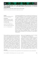
</div><span class="text_page_counter">Trang 21</span><div class="page_container" data-page="21"><small>Fig. 1.3 Chromosome walking. The top line shows a candidate region of the genome, 1 Mb in length, defined by two genetic markers (vertical lines). Underneath, the inserts of differentoverlapping BAC clones are arranged to form a clone contig map. To create this map, one of thegenetic markers (e.g. a restriction fragment length polymorphism (RFLP) or a microsatellite) isused as a probe to screen a BAC library, identifying clone 1. If the end of clone 1 is used as a probe,clone 2 is identified. Similarly, clone 2 will identify clones 3 and 4, either of which will find clone 5.Finally, clone 5 will hybridize to clones 6 and 7, either of which will identify clone 8. Clone 8 willalso hybridize to the second genetic marker, therefore generating a bridge of clones spanning thecandidate interval.</small>
</div><span class="text_page_counter">Trang 22</span><div class="page_container" data-page="22">amplify fragments up to about 5 kb in length. However, the more recent innovation
<b>of long PCR, which employs a mixture of DNA polymerases, can amplify muchlarger fragments (up to 50 kb). Reverse-transcriptase PCR (RT-PCR) is the</b>
standard procedure for amplifying cDNA directly from a source of mRNA. The RT-PCR is a single-tube reaction where mRNA is first reverse transcribed and the cDNA is then amplified.
The above methods can be applied only if a suitable probe/primer combina-tion can be designed or if some funccombina-tional informacombina-tion is available about the target gene. This is not the case for most human disease genes because generally the only information available is the overall disease phenotype. A widely used approach
<b>under these circumstances is positional cloning, where the disease gene is </b>
first mapped genetically to a particular genomic region. Known DNA sequences in the vicinity, generally the genetic markers used for the initial mapping study but sometimes other landmarks such as chromosome breakpoints, are then used
<b>to initiate a chromosome walk in which overlapping genomic clones are</b>
identified by library screening until the candidate interval is covered (Fig. 1.3). This interval is then searched for genes, with the ultimate aim of finding a gene that carries a mutation in individuals suffering from the disease but not in healthy individuals.
<b>Functional characterization of cloned genes</b>
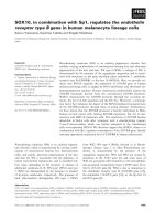
The cloning of a gene, e.g. a human disease gene, is only the first step in a long pro-cess. Once a clone is available, it is important to learn as much about the gene as possible, since this provides an insight into its normal function in the cell and its role in disease pathogenesis. A thorough understanding of the function of a gene in health and disease is valuable in the development of new therapies. There are many ways to learn about gene function (Fig. 1.4):
<small>Loss of functionGain of function</small>
<small>Fig. 1.4 A selection of approaches to study gene function on a global scale. Computers can be used to analyze protein sequences and structures, and predict their interactions from structuraldata, providing tentative functional annotations on the basis of information from relatedsequences and structures. Functions can be identified directly by mutation or interference to cause loss of function or by overexpression/ectopic expression to cause gain of function. Furtherevidence can be derived from mRNA/protein expression experiments, protein localization, directexperimental investigation of protein interactions, and assays for biochemical activity. Theseapproaches are described in more detail in Chapter 2.</small>
</div><span class="text_page_counter">Trang 23</span><div class="page_container" data-page="23">• Analysis of gene expression. Gene expression may be restricted to particular cells or tissues, to particular stages of development, or may be induced by external signals (e.g. hormones). Changes in gene expression patterns may be relevant in pathogenesis, and mutations in one gene may affect the expression patterns of others. Gene expression can be studied by methods such as northern blot
<i>hybridiza-tion and in situ hybridizahybridiza-tion (Box 1.3).</i>
• Analysis of protein localization. If the gene can be expressed to produce a re-combinant protein, antibodies can be raised and used as probes to study protein localization. Western blotting is analogous to northern blotting, and involves the separation of protein mixtures by electrophoresis followed by the use of antibody probes to detect specific proteins. Precise localization patterns in tissues and even
<i>within cells can be determined by in situ immunochemical analysis.</i>
• Analysis of protein interactions. A number of genetic and biochemical tech-niques can be used to investigate protein interactions with other proteins, with nucleic acids, and with small molecules. This can help to determine gene func-tion at the molecular and cellular levels and can link proteins into complexes or pathways.
• Altering gene expression or activity. Once a gene has been cloned, strategies can be developed to deliberately mutate that gene or to eliminate its function by interfering with its expression or the activity of its product. There are many
<b>different techniques that can be applied to study loss of gene function,</b>
including random mutagenesis, targeted gene mutation, interference with gene expression using antisense RNA, ribozymes or RNA interference, and interference with protein activity using antibodies (see Chapter 8). Conversely, the overexpres-sion of a gene, expresoverexpres-sion outside its normal spatial or temporal domain (ectopic expression), or the expression of a mutant version of the protein that is more active
<b>than normal can be used to determine the consequences of gain of gene function.</b>
Such techniques can help to elucidate gene function at the cellular and whole organism levels, and can be used to create models of human diseases in cells and animals.
• Analysis of protein structure. If the structure of the encoded protein is solved, interactions with other proteins and small molecules can be modeled.
From recombinant DNA to molecular medicine
The initial medical advances made possible by recombinant DNA technology reflected the isolation and characterization of individual genes with medical relev-ance, i.e. human disease genes, related genes from other animals, and genes from pathogenic organisms. As well as increasing our fundamental knowledge of the molecular basis of human diseases, this allowed the development of a new field of
<b>medicine, termed molecular medicine, which is the direct application of </b>
recom-binant DNA techniques to the prevention, diagnosis and treatment of human dis-ease. A whole new biotechnology industry has grown up around the potential of molecular medicine and several key areas are discussed below.
</div><span class="text_page_counter">Trang 24</span><div class="page_container" data-page="24"><b>The use of DNA sequences as diagnostic tools</b>
One of the first direct medical applications of recombinant DNA technology was the use of DNA sequences as diagnostic tools. In the same way that probes or PCR primers can be used to isolate genes from clone libraries, they can also be used to detect DNA sequences related to disease. Importantly, no disease symptoms need to be evident. For example, inherited disorders can be detected prenatally (e.g. by chorionic villus sampling) or before the onset of symptoms (in the case of a late-onset diseases like Huntington’s disease). Similarly, hybridization-based tests or PCR assays can be used to detect pathogens or malignant cells before conventional evidence of the infectious disease or cancer becomes apparent. This approach is particularly useful for screening blood products for latent pathogens, such as HIV. It is also of immense benefit for the rapid identification of pathogens in acute infections, as this allows the correct regimen of drug treatment to be implemented as soon as possible.
An early example of DNA-based diagnostics was the hybridization test used to detect hemoglobin disorders, which are known as hemoglobinopathies. As dis-cussed above, the globin genes were among the first human genes to be cloned because the cDNA sequences are so abundant. Labeled globin cDNA probes from healthy individuals were hybridized to Southern blots of genomic DNA from both healthy people and those suffering from different hemoglobinopathies. This allowed changes in DNA band patterns that were disease specific to be identified.
Some disease-causing mutations either create or destroy a restriction site, allow-ing the disease to be diagnosed directly by Southern blot analysis. This occurs in sickle-cell disease, which is caused by a point mutation in the β-globin gene. The
<i>mutation destroys the recognition site for the restriction endonuclease MstII,</i>
allowing sickle cell individuals (and carriers) to be detected because of the
<i>unusu-ally long MstII restriction fragments (Fig. 1.5). In other cases, one or more than one</i>
restriction fragments are absent and similar results occur with a number of different restriction endonucleases. This is suggestive of a larger deletion, as occurs in the thallasemias (Fig. 1.5b).
Very few diseases can be diagnosed on the basis of point mutations that change restriction sites, but restriction analysis is unnecessary for mutation detection. If a disease-causing point mutation can be identified, synthetic oligonucleotides can
<b>be made corresponding to both the normal and mutant sequences. These </b>
<b>allele-specific oligonucleotides (ASOs) can be used in two ways. Longer ASOs can be</b>
<b>used for allele-specific hybridization, a procedure in which the ASOs are labeled</b>
and hybridization conditions are adjusted to accept only perfect matches between such oligonucleotides and the target genomic DNA. Alternatively, shorter ASOs
<b>can be used as primers in an allele-specific PCR. In this case, the last nucleotide of</b>
the primer is chosen as the discriminant position because extension will not occur from a primer with a mismatched 3′ end (Fig. 1.6).
<b>The production of therapeutic proteins</b>
The modification of a cloning vector to include regulatory elements that control
<b>gene expression allows the cloned gene to be expressed as a recombinant protein.</b>
</div><span class="text_page_counter">Trang 25</span><div class="page_container" data-page="25"><small>Fig. 1.5 DNA sequences as diagnostic tools. (a) Disease diagnosis by testing for point mutationsthat alter the number of restriction sites using sickle cell anemia as an example. The top panelshows the human β-globin gene (the gray box represents the coding region and the first intron </small>
<i><small>is shown with darker shading). Vertical arrows represent MstII restriction sites. In normal</small></i>
<small>individuals, there are three sites and the probe will identify a fragment of genomic DNA 1.1 kb inlength. The mutation responsible for the disease (*) destroys the central restriction site so that theprobe detects a 1.3-kb fragment instead. The lower panel shows a Southern blot from normal(N/N), heterozygous (N/S), and sickle cell disease (S/S) individuals. The arrow shows the directionof electrophoresis. Note the similarity of this technique to the detection of RFLPs (see p. 25). (b)Disease diagnosis by testing for deletions that remove restriction fragments. The top panel showsthe</small><i><small>β-globin cluster with the genes and pseudogenes identified. The vertical arrows show EcoR1</small></i>
<small>restriction sites in the β-globin and δ-globin genes. The lower panel shows the result of a Southernblot experiment. In normal individuals (N), a β-globin cDNA probe (bar) would reveal severalfragments because cross-hybridization to the δ-globin gene would be possible under reducedstringency conditions. In individuals with βδ-thallasemia (BDT) these two genes are deleted, andhybridization to any residual fragments between the outer restriction sites would result in a single</small>
<i><small>hybridizing band. The same result would be expected for other restriction enzymes, e.g. HindIII.</small></i>
<small>Note the similarity of this technique to loss of heterozygosity mapping in cancer (see p. 118).</small>
<small>Fig. 1.6 Allele-specific PCR to detect sickle cell anemia. The top panels show the normal and mutant β-globinsequences, with * marking the position of the mutation. The lower panel shows amplification with a PCR primermatching the normal sequence. It will be extended on a normal template (left) but not on a mutant template becausethe final nucleotide does not anneal (right).</small>
</div><span class="text_page_counter">Trang 26</span><div class="page_container" data-page="26">There are many basic applications of this technology including, as discussed above, the use of expression libraries for gene isolation. In medicine, however, the primary application of expression technology is the production of recombinant therapeutic proteins.
<i>Human proteins as drugs</i>
Therapeutic protein synthesis was one of the first commercial applications of recombinant DNA technology and the initial products were simple proteins, like human growth hormone and insulin, for which there was a large demand and an unsatisfactory source. In many cases the authentic product had to be isolated from human cadavers or animals and there was a risk of contamination with pathogens. For example, some children treated with growth hormone extracted from human pituitary glands later developed Creutzfeld–Jakob disease, and many patients treated with human blood products have since developed hepatitis or HIV infections.
The first recombinant proteins were produced in bacteria in the late 1970s and large scale bacterial fermentation continues to be used today. However, while this approach is suitable for simple proteins, bacteria do not carry out many forms of protein post-translational modification, including glycosylation. Alternative systems are thus required for the production of complex glycoproteins. There have been some successes with yeast and insect cells, but the glycan chains added to recombinant proteins are radically different to those produced in mammals. Therefore, many complex recombinant human proteins are produced in large scale cultures of mammalian cells. Because this is very expensive, alternative production systems have been explored and the use of transgenic animals and plants is increas-ing in popularity. This topic is discussed in more detail in Chapter 6.
<i>Recombinant vaccines</i>
The prevention of infectious diseases by vaccination has a long and successful history beginning in 1796 when Edward Jenner injected a young boy with cowpox, thus conferring protection against a subsequent infection with the deadly smallpox virus. Most of the vaccines in use today are based on similar principles and are known as “Jennerian vaccines.” These include live but attenuated bacteria or viruses which cause the body to mount a protective immune response against the target pathogen (e.g. the measles, mumps, rubella, and tuberculosis vaccines) and “killed vaccines,” i.e. the pathogen itself is killed so it is no longer infectious but it can still stimulate the immune system.
Unfortunately, vaccines against all common diseases cannot be made using the above methods and other approaches are needed. An alternative strategy is the use
<b>of recombinant subunit vaccines, where the gene for one specific protein on the</b>
pathogen is expressed, and the protein used as the vaccine. The current hepatitis B and influenza vaccines are protein subunits produced in yeast. Since these inert subunits do not multiply inside the vaccinee, they do not generate an effective cellu-lar immune response. To address this, heterologous antigens have been expressed
</div><span class="text_page_counter">Trang 27</span><div class="page_container" data-page="27">in attenuated bacteria and viruses and used as surrogate live vaccines. For example, vaccinia virus has been used to express a wide range of proteins from different pathogens, including the rabies glycoprotein, leading to the eradication of rabies in some parts of Europe. More recently, genetically transformed plants have been used to produce oral vaccines which can be administered either by eating the plant material directly, or after minimal processing. Vaccines are discussed further in Chapter 3.
<i>The special status of recombinant antibodies</i>
Antibodies bind to target antigens with great specificity and are therefore used in molecular biology for the detection, quantification and purification of proteins. In medicine, antibodies can be used to prevent, detect and cure diseases. For example,
<i>antibodies against the surface adhesin of the oral pathogen Streptococcus mutans are</i>
being developed as a drug to prevent tooth decay, and antigens that recognize specific tumor antigens can be used to diagnose and treat cancer. The traditional way to produce monoclonal (single target specificity) antibodies is to fuse B lympho-cytes from immunized mice with immortalized myeloma cells, resulting in the
<b>recovery of hybridoma cell lines that produce the same antibody indefinitely. The</b>
disadvantage of murine antibodies is their immunogenicity in humans. Recom-binant DNA technology has been used to address this problem in a number of ways, including the production of humanized antibodies, recombinant antibody derivat-ives, and antibody fusion proteins. Furthermore, artificial immune diversity can be generated using libraries of antibody variable regions as in phage antibody display. Recombinant antibodies are discussed in Chapter 6.
Gene medicine
Traditionally, DNA sequences have been used to detect diseases while proteins and other “small molecule” drugs have been used to treat or prevent them. This distinc-tion is becoming blurred, however, with the development of novel forms of therapy
<b>known collectively as gene medicine (see Chapter 8). One form of gene medicine isknown as gene therapy and involves the introduction of DNA sequences into</b>
<i>human cells either in vitro or in vivo with the purpose of treating and hopefully </i>
cur-ing disease. In most cases, gene therapy is directed at diseases caused by mutations in human genes (inherited disorders, cancer) and ideally is meant to alter the genome and provide a permanent cure. In contrast to the use of drugs to alleviate disease symptoms, therapeutic DNA has the capability of correcting the actual cause of the disease by correcting or compensating for the mutation itself. Other forms of gene medicine are more similar to traditional drugs. They include the use of synthetic oligonucleotides, ribozymes, and most recently RNA interference to block the expression of particular mutant genes in the treatment of cancer or infectious diseases. For example, several gene therapy trials are underway which involve various strategies to combat HIV.
</div><span class="text_page_counter">Trang 28</span><div class="page_container" data-page="28"><b>A special category of gene medicine is the use of DNA vaccines. These are </b>
con-structs containing the gene corresponding to a pathogen antigen. When expressed in the human body, the antigen is made and induces an immune response pro-viding protection against subsequent infections. DNA vaccines are advantageous because the same strategy can be used to prepare vaccines against many different diseases, and because vaccines against new disease isolates can be developed rapidly. There are also logistic advantages in that DNA is easier to store and trans-port than proteins.
Disease models
Another major application of recombinant DNA technology is the introduction of
<i>predefined mutations into genes by in vitro mutagenesis followed by the transfer of</i>
such altered genes back into the source organism for functional testing. It is not
<b>pos-sible to do this with human genes for ethical reasons, but disease models can be</b>
created by mimicking human pathogenic mutations in other animals. Such models can be used to investigate the molecular basis of the disease and, importantly, to test novel drugs before clinical trials in humans.
Mammals have been used as human disease models for many years, but until comparatively recently this relied on the identification of spontaneous mutants or the screening of mutagenized populations to identify those with disease-like pheno-types. Recombinant DNA technology in combination with advances in
<b>mam-malian gene transfer techniques has made it possible to create exact replicas of</b>
human pathogenic mutations by integrating dominantly malfunctioning trans-genes or replacing the endogenous gene with a nonfunctional copy, a technique commonly described as “gene knockout.” More recently, it has been possible to model more complex diseases in mice by simultaneously introducing mutations into two or more genes.
The impact of genomics on medicine
The recombinant DNA revolution provided us with tools and techniques to isolate and characterize individual genes, but this approach has two major limitations. First, finding genes one at a time is extremely laborious and expensive work.
<b>Second, it encourages a reductionist approach to biomedical research, whereas it</b>
is well known that genes do not function in isolation. Thousands of genes must work together to coordinate the biologic activities that form a functioning human, or indeed any other organism. The second modern revolution in medicine, the
<b>genomics revolution, has addressed these drawbacks by encouraging a newholistic approach in which genes and their products are characterized in large</b>
numbers. Genomics is the study of entire genomes, incorporating mapping, sequencing, annotation (gene finding), and functional analysis. The tools and
</div><span class="text_page_counter">Trang 29</span><div class="page_container" data-page="29">techniques provided by the genomics revolution are high-throughput equivalents of those from the recombinant DNA era, allowing more data to be gathered and analyzed in a much shorter space of time.
The genomic revolution began in the early 1990s when the Human Genome Project began to gather pace. The initial aims of the project were to map and sequence the entire human genome, leading to the identification of all human genes. The first phase of the project involved the creation of a high-density genetic map that could be used as a framework or scaffold to assemble a physical map of DNA clones. These clones were then sequenced, systematically, and the sequences analyzed for genes. Technical innovations were required in all areas to achieve these aims but the most impressive advances came in the automation of DNA sequencing, which increased the rate of data production over 1000-fold compared to the 1980s. Technology improvements were stimulated by competition from the private sector, and during the progress of the Human Genome Project, the genomes of many bacteria and some eukaryotes were also sequenced. These included many human pathogens and a handful of important model experimental organisms, such
<i>as the fruit fly (Drosophila melanogaster), the nematode worm (Caenorhabditiselegans), and the humble baker’s yeast (Saccharomyces cerevisiae). We will not </i>
con-sider the methodology of genome mapping and sequencing here since this subject is explored in more detail in Chapter 2.
The output of the first phase of the Human Genome Project was a draft sequence
<b>extensively annotated with genes (a transcript map). The transcript map is the</b>
key to the potential medical benefits of the project because with further refinement it could provide access to all human genes. Therefore, while one of the first benefits of recombinant DNA was access to individual human genes, one of the first benefits
<b>of genomics was access to all of them. The transcript map is helping to accelerate</b>
the rate at which disease genes are discovered because it is now no longer necessary to devise elegant cloning strategies. Positional cloning is obsolete, because once a disease gene has been mapped to a particular genomic region, the transcript map can be inspected for candidate genes and these can be studied for evidence of disease association.
As well as large scale methods for gene isolation, the genomics revolution has also provided large scale methods for functional analysis. Indeed it seems impossible to read about genomics without the phrases “large scale” or “high-throughput” or “massively parallel” being used to describe the experimental methods. The emphasis of genomic technology is on maximizing the amount of data output while minimizing the amount of hands-on input through extensive automation, miniaturization, and parallelization. These techniques are described only very briefly below because they are discussed in more detail in the following chapter. However, compare the list below to the one on page 10:
• Analysis of gene expression. High-throughput expression analysis by large scale cDNA sequencing, sequence sampling techniques and the use of DNA microarrays allows the expression of thousands of genes to be analyzed simultaneously. This can show the global effect of different conditions on gene expression profiles, help to link
<b>genes into similar expression (synexpression) classes, and home in on </b>
differen-tially expressed genes.
</div><span class="text_page_counter">Trang 30</span><div class="page_container" data-page="30">• Analysis of protein expression. High-resolution separation techniques such as two-dimensional gel electrophoresis can be used to fractionate complex protein mixtures, and mass spectrometry can be used to identify individual proteins rapidly and accurately. The expression of thousands of proteins can be analyzed and com-pared across samples.
• Analysis of protein interactions. New high-throughput technologies such as phage display, the yeast two-hybrid system and mass spectrometric analysis of pro-tein complexes allow interacting propro-teins to be cataloged on a large scale. Propro-tein interaction maps of whole cells can be produced.
• Altering gene expression or activity. Large scale mutagenesis can be used to gen-erate populations with either random or targeted mutations in every single gene. Similarly, RNA interference can be applied on a large scale to inactivate all the genes in the genome systematically. Mutation techniques can be applied only to model organisms but RNA interference is used in human cells.
• Analysis of protein structure. Large scale “structural genomics” programs have been initiated to solve many protein structures. It is hoped that representatives of all protein families will be structurally solved to increase the rate at which functions are assigned to genes.
<b>Advances in bioinformatics (the use of computers to process biologic data) have</b>
gone hand in hand with advances in genomics because only computers have the power to analyze the large datasets produced by genomic-scale experiments. One of
<b>the most important contributions of bioinformatics is sequence analysis, which</b>
allows sequences of genes and whole genomes to be compared. There is extensive structural and functional conservation among genes and even whole molecular pathways between humans and model organisms such as the fruit fly, the nema-tode worm, and the baker’s yeast. Up to 20% of human disease genes have counter-parts in yeast and up to 60% have countercounter-parts in the worm and fly, allowing these organisms to be used for functional analysis and the screening of candidate drugs. Similarly, comparisons between bacterial sequences, especially those of harmless species and related pathogens, are helping to reveal virulence factors and patho-genesis-related proteins that could be used as new drug targets or candidates for new vaccines. Another important role of bioinformatics is the presentation of data in easily accessible and user-friendly databases, allowing the efficient dissemination of information. As we shall see later in the book, some databases are already having a real impact on our understanding of disease at the molecular level, and this will have a knock-on effect on the development of novel therapies. One example is the Cancer Genome Anatomy Project, which aims to assemble gene expression and functional data from all forms of cancer.
The new molecular medicine
The potential availability of all human disease genes, as well as genes in human pathogens that are responsible for infectious diseases, is likely to have a major impact on drug development. At the current time, most available drugs interact
</div><span class="text_page_counter">Trang 31</span><div class="page_container" data-page="31">with a small repertoire of 500 or so target proteins in the body. There are approxim-ately 30,000 genes in the human genome and many of these will represent novel drug targets. Therefore, the functional analysis of these genes and the structural analysis of their products could lead to an explosion in the number of drugs being developed in the next few decades. Furthermore, the growing recognition of the importance of conserved molecular pathways and the tendency of proteins to func-tion in large complexes will allow key regulatory molecules to be selected as drug targets. Pharmaceutical companies have not been slow to embrace the potential of genomics, and we discuss the process of drug development in Chapter 7.
Another aspect of genomics that is likely to have a large impact on medicine is
<b>the analysis of human variation. Earlier in this chapter, we discussed the use of</b>
DNA sequences as diagnostic tools to identify particular sequence variants associ-ated with disease. More recently, techniques based on the same principles have
<b>been streamlined and miniaturized for the high-throughput analysis of single</b>
<b>nucleotide polymorphisms (SNPs). Unlike disease-causing point mutations,</b>
SNPs are common variants that are widespread in the population. While they do
<b>not cause overt diseases, some are thought to contribute in a small and additive</b>
manner to disease susceptibility, and to other complex characteristics such as individual responses to drugs. Spin-offs from the Human Genome Project aim to catalog all the SNPs in the genome (there are thought to be 10 million in total, with any two individuals varying at about 3 million positions) as well as blocks
<b>of SNPs, known as haplotypes, that are tightly linked and tend to be inherited as </b>
a group. For the first time, it may be possible to pinpoint the genetic variants that predispose us to common diseases, such as asthma and diabetes (see Chapter 4). It may also be possible to identify genetic variants that influence our responses to drugs, raising the possibility of personalized medicines targeted to the genetic com-position of individual patients (see Chapter 7). We must be careful, however, to guard against the misuse of genetic information arising from the Human Genome Project and its subsidiaries. A large segment of the budget for this project has been set aside to address the social, legal and ethical issues involved, in order to protect the privacy of those contributing their DNA to the project and to prevent data from human genomic analysis being used to discriminate against individuals or ethnic groups.
Outline of this book
The aim of this book is to provide a broad and comprehensive account of how recombinant DNA technology and genomics are used in medicine. The next chap-ter explains the principles of genomics in enough detail for the reader to understand the material presented in later chapters. Chapters 3–5 discuss the role of recom-binant DNA and genomic analysis in the diagnosis, treatment and prevention of infectious diseases, inherited diseases, and cancer. The subsequent three chapters cover emerging types of therapy and modern approaches to drug development. A “roadmap” of the book is shown in Fig. 1.7.
</div><span class="text_page_counter">Trang 32</span><div class="page_container" data-page="32">Further reading
POGM: Chapter 1 provides an overview of recombinant DNA technology and describes the birth of the biotechnology industry. Chapter 2 introduces basic techniques while Chapters 3–6 discuss cloning vectors and strategies in more detail. Chapter 14 has sections on the applica-tions of recombinant DNA technology in medicine.
POGA: Chapter 1 introduces genomics and some of its applications. Chapter 12 has sections on the applications of genomics in medicine.
Williams SJ, Hayward NK (2001) The impact of the Human Genome Project on medical
<i><b>genetics. Trends Mol Med 7, 229–231.</b></i>
Yaspo M-L (2001) Taking a functional genomics approach in molecular medicine.
<i><b>Trends Mol Med 7, 494 –502.</b></i>
Two useful articles, one a summary and one an in-depth review, discussing the impact of genomics on molecular medicine.
Wren BW (2000) Microbial sequencing: insights into virulence, host adaptation and
<i><b>evolution. Nature Rev Genet 1, 30–38.</b></i>
A thorough article showing how microbial genomics is providing new leads in the fight against infectious disease.
<small>Fig. 1.7 A “roadmap” of the layout of this book.</small>
</div><span class="text_page_counter">Trang 33</span><div class="page_container" data-page="33">C H A P T E R T W O An overview of
In the previous chapter, we charted the history of molecular medicine from its origins in the aftermath of the recombinant DNA revolution to the present day, and briefly discussed some of the expected scientific and medical benefits of genomics. The position we are in now is one of enormous promise. At our fingertips, we have the complete sequence of the human genome and potential access to every single gene. This offers an unprecedented opportunity to study human biology, in health and disease, in a truly global and systematic way. Similar resources are available for a large number of other organisms of medical relevance, including some of our most important pathogens (Table 2.1). The focus of medical research is now turning to the systematic functional evaluation of genes and the elucidation of pathways and networks. A complete understanding of how genes function and interact to co-ordinate the biologic activities that make a healthy human provides enormous <small>Table 2.1 Some pathogen genomes (bacterial and protozoan) that have been sequenced.</small>
Copyright © 2004 by Blackwell Publishing Ltd
</div><span class="text_page_counter">Trang 34</span><div class="page_container" data-page="34">scope for the development of novel therapies. In this chapter, we review the sci-entific achievements that have led us to our current position and consider some of the emerging genomic technologies that may provide medical breakthroughs in the future.
A review of progress: the Human Genome Project
Genomics (Box 2.1) became a significant and independent field of research in 1990
<b>when the Human Genome Project (HGP) was officially launched. The stated </b>
aim of the project was to sequence the entire 3000-Mb human nuclear genome within 15 years. At the outset, however, it was acknowledged that a great deal of preliminary work was required before actual sequencing could begin, and that five model organism genomes should be sequenced in addition to the human genome to act as pilot projects for the validation of new technologies (Box 2.2). One of the first tasks was to construct a high-resolution genetic map of the human genome to act as a scaffold for the assembly of a physical map of DNA clones. Once the genetic and physical mapping phases were completed, then sequencing could begin. Technological advances were required in mapping, cloning, sequencing,
Box 2.1 What is genomics?
<small>The term genome was introduced in 1920 by the</small><small>German botanist Hans Winkler to describe the collectionof genes contained within a complete (haploid) set ofchromosomes. Nowadays, the term has expanded toinclude all the DNA in a haploid set of chromosomes,not just the genes, because in higher eukaryotes genesare in the minority. For example, only 2–3% of thehuman genome is represented by genes. Although the concept of the genome is longstanding, the termgenomics was not used for the first time until 1986. The mouse geneticist Thomas Roderick introduced this word to describe the mapping, sequencing andcharacterization of genomes. More recently, the </small>
<small>essence of genomics has become associated with anyform of large scale, high-throughput biologic analysisand has spawned a whole lexicon of derivative terms.Functional genomics encompasses any systematicapproach to the analysis of gene function, and many ofthe technologies of functional genomics are discussed in this chapter. Transcriptomics is the large scaleanalysis of mRNA expression. Proteomics is the largescale analysis of proteins, and can itself be divided intothe study of expression profiles, interactions, and proteinstructure. Proteomics is a very significant component ofthe new molecular medicine because most drug targetsare proteins.</small>
Box 2.2 Model organism genomes as initial targets of the Human Genome Project
<i><small>Escherichia coli (bacterium)Saccharomyces cerevisiae (yeast)Caenorhabditis elegans (nematode)</small></i>
<i><small>Drosophila melanogaster (fruit fly)Mus musculis (mouse)</small></i>
</div><span class="text_page_counter">Trang 35</span><div class="page_container" data-page="35">and bioinformatics, in order to achieve the goals of the HGP within the allotted time frame. A large part of the initial budget was also set aside to address the
<b>ethical, legal and social issues (ELSI) that arose from the project, such as </b>
preventing any data arising from the project being used to discriminate against individuals or populations (Box 2.3).
Box 2.3 The ethical, legal and social issues (ELSI) of the Human Genome Project
<small>Before the Human Genome Project was inaugurated, it was recognized that both the way in which the project was carried out and the data it produced wouldraise new and complex ethical issues. Particular areas of concern included matters relating to the collection of samples, the privacy of donors, and the availabilityand subsequent use of genetic information arising fromthe project. Therefore, both of the US organizationssponsoring the HGP – the US Department of Energy(DOE) and the National Institutes of Health (NIH) –devoted a significant proportion of their annual HGPbudgets (3% and 5% respectively) to fund a series ofprograms whose aim was to study the ethical, legal andsocial issues (ELSI) of the project. The function of theELSI programs was, and is, to promote education andguide policy decisions by consultation with a wide rangeof interested parties. A unique aspect of the HGP ELSIprograms is that they are integral to the project itselfrather than retrospective, and therefore help to foreseethe implications of new technology developments andaddress any important issues before problems arise.</small>
<small>The initial aims of the ELSI programs were stated as follows:</small>
<small>• To anticipate and address the implications forindividuals and society of mapping and sequencing the human genome</small>
<small>• To examine the ethical, legal and social</small>
<small>consequences of mapping and sequencing the humangenome</small>
<small>• To stimulate public discussion of the issues, and• To develop policy options that would assure that theinformation is used for the benefit of individuals andsociety.</small>
<small>In the 10 years since the ELSI programs were initiated, a large body of work has been produced to educatepolicymakers and the public. This has helped in thedevelopment of policies relating to the conduct ofgenetic research and the commercial exploitation of</small>
<small>genetic information and its associated technologies.Some of the more important challenges relate to the spin-off projects that focus on human geneticvariation, i.e. the SNP mapping project and thehaplotype mapping project. In these cases the privacy of individuals and communities contributing DNAsamples must be protected, but it is also necessary toobtain informed consent and to provide continuousliaison through advisory groups. A major concern is that information on genetic variation could be used to discriminate against individuals or populations interms of employment, insurance, or legislation. ELSIprograms have been established to anticipate how these data may affect concepts of race and ethnicity and to foresee the impact of technologic advances anddata availability on the entire concept of humanity. The educational resources not only help to keep thepublic and policymakers informed, but also helpscientists to present their results carefully to avoidmisinterpretation.</small>
<small>The aims of ELSI are updated every few years and the most recent are presented below:</small>
<small>• To examine issues surrounding the completion of the human DNA sequence and the study of humangenetic variation</small>
<small>• To examine issues raised by the integration of genetic technologies and information into health care and public health activities</small>
<small>• To examine issues raised by the integration ofknowledge about genomics and gene–environmentinteractions in nonclinical settings</small>
<small>• To explore how new genetic knowledge may interactwith a variety of philosophical, theological and ethicalperspectives</small>
<small>• To explore how racial, ethnic and socioeconomicfactors affect the use, understanding and interpretationof genetic information, the use of genetic services, andthe development of policy.</small>
</div><span class="text_page_counter">Trang 36</span><div class="page_container" data-page="36">To place the ambitious technical objectives of the HGP in context, consider that in the mid-1980s when the project was first conceived, it was possible to sequence about 1000 nucleotides of DNA per day. At that rate, armies of scientists doing nothing but sequencing would have been required to complete the whole genome. Sydney Brenner, one of the proponents of large scale biology, joked that sequenc-ing should be done by prisoners! It was envisaged that entirely new sequencsequenc-ing methods would be needed in order to increase data output to the required levels. However, although several new methods emerged during the HGP, the goal of increased output was met in the most part by the automation and multiplexing of existing technology. Using ultrarapid capillary sequencers that process 96 samples at once, it is now possible to produce upwards of half a million nucleot-ides of sequence per day with one machine. Further multiplexing, and the use of multiple machines, can increase this output even more.
<b>Breakthroughs in genetic mapping</b>
<b>Genetic maps are based on recombination frequencies, and in model </b>
organ-isms they are constructed by carrying out large scale crosses between different mutant strains. The principle of a genetic map is that the further apart two loci are on a chromosome, the more likely that a crossover will occur between them during meiosis. Recombination events resulting from crossovers can be scored in
<i>genetic-ally amenable organisms such as Drosophila and yeast by looking for new </i>
com-binations of the mutant phenotypes in the offspring of the cross. This approach cannot be used in human populations because it would involve setting up large scale matings between people with different inherited diseases. Instead, human
<b>genetic maps rely on the analysis of DNA sequence polymorphisms in existing</b>
family pedigrees (Box 2.4).
Prior to the HGP, low-resolution genetic maps had been constructed using
<b>restriction fragment length polymorphisms (RFLPs). These are naturally</b>
occurring variations that create or destroy sites for restriction enzymes and there-fore generate different sized bands on Southern blots (Fig. 2.1). The problem with RFLPs was that they were too few and too widely spaced to be of much use for constructing a framework for physical mapping – the first RFLP map had just over 400 markers and a resolution of 10 cM, equivalent to one marker for every 10 Mb of DNA. The necessary breakthrough came with the discovery of new
<b>polymorphic markers, known as microsatellites, which were abundant and</b>
widely dispersed in the genome (Fig. 2.2). By 1992, a genetic map based on microsatellites had been constructed with a resolution of 1 cM (equivalent to one marker for every 1 Mb of DNA) which was a suitable template for physical mapping. However, efforts in genetic mapping did not stop there. By 1996 a further map incorporating additional microsatellite markers was published, with a resolution of 0.5 cM. The most recent map, released in 2002 by the deCODE consortium in Iceland, has a resolution of 0.2 cM and incorporates over 5000 markers. The SNP and haplotype projects are also examples of high-resolution genetic maps (Box 2.4).
</div><span class="text_page_counter">Trang 37</span><div class="page_container" data-page="37">Box 2.4 Variation in the human genome
<small>The DNA used for the HGP came from 12 anonymous</small><small>volunteers. Since the genome sequences of any twounrelated humans are only 99.9% identical, there is no “correct” sequence. However, it is the 0.1%difference – amounting to 3 million base pairs of DNA – which is the most interesting, as this makeseach of us unique. Gene mutations that cause inheriteddiseases are very rare in the population as a whole andtherefore account for only a tiny proportion of thisvariation. The vast majority occurs in the form ofsequence polymorphisms, where several differentvariants (alleles) may be quite common. Thesevariations are used as markers to create genetic mapsbecause hybridization or PCR assays (see Chapter 1) can be used to detect and identify the alleles andtherefore establish whether recombination has occurred in a family pedigree.</small>
Types of variation <small>About 95% of polymorphic sequence variation isrepresented by single nucleotide polymorphisms(SNPs), i.e. single nucleotide positions that may be occupied by one base in some people but analternative base in others. Where these polymorphismsoccur in and around genes, they may occasionally haveovert phenotypic effects (e.g. polymorphisms affectinghair color). In most cases, however, the effects of SNPs are far more subtle, e.g. they may influence in a small but additive manner our disease susceptibility or response to certain drugs (see p. 108). The vastmajority of SNPs occur outside genes and probably have no effect. However, they are still useful as geneticmarkers. Some SNPs either create or destroy restrictionenzyme sites, so altering the pattern of bands seen on a Southern blot. These restriction fragment lengthpolymorphisms (RFLPs) were used to produce the first comprehensive genetic map of the human genome.</small>
<small>The remaining 5% of sequence polymorphism occurs mostly in the form of simple sequence repeat polymophisms (SSRPs) otherwise known as microsatellites. These are short sequences repeated a variable number of times. The most common form of microsatellite is CA(</small><i><sub>n</sub><small>), where n</small></i>
<small>represents the number of repeats (typically 5–50).Unlike SNPs, microsatellites have multiple alleles (i.e. there may be common variants with 12 repeats, 22 repeats, 31 repeats, etc.) whereas SNPs usually occur as one of two alternative forms. Microsatellitesrarely occur within genes, and often have pathogeniceffects when they do (e.g. Huntington’s disease), butthey are widely distributed and can be used to produce a much higher resolution map than RFLPs. The physicalmapping stage of the Human Genome Project used as a scaffold a genetic map based on microsatellitemarkers.</small>
Studying variation <small>Human variation has been used in forensic analysis for many years but interest in genome-wide variationbegan to grow only as the HGP gathered pace. A globaleffort to study human sequence diversity, the HumanGenome Diversity Project (HGDP), was initiated as a spin-off project from the HGP in 1991. However, itreceived little funding because the primary aim of theproject was to find markers corresponding to differentethnic groups for the study of population history andhuman origins. There has been much more support forSNP mapping projects, both public and private, sincethese provide concrete benefits to medical research. The ability to identify associations between SNPs anddisease susceptibility should greatly accelerate the rateat which disease genes are discovered, and associationsbetween SNPs and drug responses underlie the newmedical field of pharmacogenomics, where drugs canbe tailored to individuals based on their genotype (seeChapter 4). The International SNP Consortium Ltdstarted a systematic SNP mapping project in 1999 andhad produced a map containing nearly one and a halfmillion SNPs by 2001. More recently, it has been shownthat groups of SNPs tend to be inherited together ashaplotype blocks with little recombination within them. The estimated 10 million SNPs could therefore be represented by as few as 200,000 haplotypes which would make the process of establishing diseaseassociations much easier. An International HapMapProject, aiming to map haplotypes throughout thegenome, was inaugurated in October 2001.</small>
</div><span class="text_page_counter">Trang 38</span><div class="page_container" data-page="38"><b>Breakthroughs in physical mapping</b>
<b>Unlike genetic maps, physical maps are based on real units of DNA and </b>
there-fore provide a suitable basis for sequencing. The physical mapping phase of the HGP involved the creation of genomic DNA libraries (see Chapter 1) and the
<b>identification and assembly of overlapping clones to form contigs (unbroken series</b>
of clones representing contiguous segments of the genome). When the HGP was
<b>initiated, the highest-capacity vectors available for cloning were cosmids, with a</b>
maximum insert size of 40 kb. Because hundreds of thousands of cosmid clones would have to be screened to assemble a physical map, there was an immediate
<b>need for large-insert cloning vectors which would reduce the amount of work</b>
involved. New approaches were also required to find overlaps and assemble clone contigs on the genomic scaffold.
<small>Fig. 2.1 Restriction fragment length polymorphisms (RFLPs) are sequence variants that create or destroy a restriction site therefore altering the length of the restriction fragment detected by a given probe. The top panel shows two alternative alleles, in which the restriction fragmentdetected by a specific probe differs in length due to the presence or absence of the middle one of three restriction sites (represented by vertical arrows). Alleles a and b therefore producehybridizing bands of different sizes in Southern blots (lower panel). This allows the alleles to betraced through a family pedigree. For example, child II.2 has inherited two copies of allele a, onefrom each parent, while child II.4 has inherited one copy of allele a and one of allele b. Note the similarity of this method to the detection of disease alleles such as the sickle cell disease variantofβ-globin (Fig. 1.5). Essentially, the only difference is that RFLPs are more common in thepopulation than disease-related mutations because they do not have overt and striking effects onthe human phenotype.</small>
</div><span class="text_page_counter">Trang 39</span><div class="page_container" data-page="39">In the case of cloning vector technology, the necessary breakthrough came with
<b>the development of artificial chromosome vectors that could accept very largeinserts (Fig. 2.3). The first such vectors were yeast artificial chromosomes</b>
<b>(YACs), which could carry inserts of over 1 Mb reducing the number of clones</b>
required to cover the genome to just over 10,000. One problem with YACs,
<b>how-ever, was their tendency to incorporate chimeric inserts (i.e. inserts comprising</b>
segments of DNA from two or more nonadjacent locations in the genome). Therefore, higher-fidelity vectors were required to generate the final physical maps
<b>used for sequencing. BACs (bacterial artificial chromosomes) and PACs (P1</b>
<b>artificial chromosomes) were chosen because of their stability and relatively</b>
large insert size (200 –300 kb).
Various strategies have been devised to assemble physical clones into contigs, all of which involve the detection of overlaps between adjacent clones. These include:
<small>Fig. 2.2 Microsatellites are sequence variants that cause restriction fragments or PCR products todiffer in length due to the number of copies of a short tandem repeat sequence, 1–12 nt in length.The top panel shows four alternative alleles, in which the restriction fragment detected by aspecific probe differs in length due to a variable number of tandem repeats. All four alleles producebands of different sizes on Southern blots (lower panel) or different sized PCR products (not shown).Unlike RFLPs, multiple allelism is common for microsatellites so the precise inheritance patterncan be tracked. For example, the mother and father in the pedigree have alleles b/d and a/crespectively (the smaller DNA fragments move further during electrophoresis). The first child, II.1,has inherited allele b from his mother and allele a from his father.</small>
</div><span class="text_page_counter">Trang 40</span><div class="page_container" data-page="40"><b>• Chromosome walking. This technique has been widely used for positional</b>
cloning (see p. 9) and involves the stepwise use of clones as hybridization probes to identify overlapping ones (see Fig. 1.3). Alternatively, the end-sequences of each clone can be used to design primer pairs and overlapping clones can be detected by PCR.
<b>• Restriction enzyme fingerprinting. This technique involves the digestion of</b>
clones with panels of restriction enzymes. Two clones that overlap will share a significant number of identical restriction fragments. The patterns are complex and must be interpreted by computers (Fig. 2.4).
<b>• Repetitive DNA fingerprinting. As an extension of the above, Southern </b>
blots of the restriction fragments can be probed for genome-wide repeat sequences
<i>such as Alu. There are over a million copies of the Alu element dispersed in the</i>
genome (one every 4 kb), so a typical 100-kb BAC clone will contain 20–30 repeats. Overlapping clones will share a significant proportion of hybridizing bands. PCR-based fingerprinting tests based on repetitive DNA can also be used.
<b>• STS mapping. A STS (sequence tagged site) is a unique sequence in the</b>
genome, 100–200 bp long, which can be detected easily by PCR. If two clones share the same STS, then by definition they overlap and can be united in a contig. STS mapping was the most valuable strategy for contig assembly in the HGP
<b>because a physical reference map containing 15,000 STS markers with an </b>
average spacing of 200 kb was published in 1995 (Box 2.5). Therefore, clones containing particular STS markers could be anchored to the reference map to show their precise chromosomal location, not just their relationship to other clones. Importantly, some of the STSs contained polymorphic microsatellite sequences,
<small>Fig. 2.3 Two artificial chromosome vectors that were invaluable in the human genome project.(a) Yeast artificial chromosome, maximum insert size up to 2 Mb. TEL, telomere; TRP, tryptophansynthesis selectable marker; ARS, yeast origin of replication (autonomous replication sequence);CEN, centromere; LEU, leucine synthesis selectable marker. (b) Bacterial artifical chromosome,maximum insert size up to 200 kb. Cm</small><sub>R</sub><small>, antibiotic resistance marker; oriS/repE, sequencesrequired for replication; parA/parB, sequences required for copy number regulation. Arrowsindicate promoters for T3 and T7 RNA polymerases, which are used to prepare labeled probescorresponding to the end-sequences of the insert.</small>
</div>