SARS-COV-2 RECEPTOR ACE2 IS AN INTERFERON- STIMULATED GENE IN HUMAN AIRWAY EPITHELIAL CELLS AND IS DETECTED IN SPECIFIC CELL SUBSETS ACROSS TISSUES
Bạn đang xem bản rút gọn của tài liệu. Xem và tải ngay bản đầy đủ của tài liệu tại đây (29.56 MB, 40 trang )
Article
SARS-CoV-2 Receptor ACE2 Is an Interferon-
Stimulated Gene in Human Airway Epithelial Cells
and Is Detected in Specific Cell Subsets across
Tissues
Graphical Abstract Authors
Carly G.K. Ziegler, Samuel J. Allon,
Sarah K. Nyquist, ..., Alex K. Shalek,
Jose Ordovas-Montanes, HCA Lung
Biological Network
Correspondence
(A.K.S.),
jose.ordovas-montanes@childrens.
harvard.edu (J.O.-M.),
(HCA
Lung Biological Network)
Highlights In Brief
d Meta-analysis of human, non-human primate, and mouse Analysis of single-cell RNA-seq datasets
single-cell RNA-seq datasets for putative SARS-CoV-2 targets from human, non-human primate, and
mouse barrier tissues identifies putative
d Type II pneumocytes, nasal secretory cells, and absorptive cellular targets of SARS-CoV-2 on the
enterocytes are ACE2+TMPRSS2+ basis of ACE2 and TMPRSS2 expression.
ACE2 represents a previously
d Interferon and influenza increase ACE2 in human nasal unappreciated interferon-stimulated
epithelia and lung tissue gene in human, but not mouse, epithelial
tissues, identifying anti-viral induction of
d Mouse Ace2 is not upregulated by interferon, raising a host tissue-protective mechanism, but
implications for disease modeling also a potential means for viral
exploitation of the host response.
Ziegler et al., 2020, Cell 181, 1016–1035
May 28, 2020 ª 2020 The Authors. Published by Elsevier Inc. ll
ll
Article
SARS-CoV-2 Receptor ACE2 Is an Interferon-
Stimulated Gene in Human Airway Epithelial Cells
and Is Detected in Specific Cell Subsets across Tissues
Carly G.K. Ziegler,1,2,3,4,5,6,50 Samuel J. Allon,2,4,5,7,50 Sarah K. Nyquist,2,4,5,8,9,50 Ian M. Mbano,10,11,50
Vincent N. Miao,1,2,4,5 Constantine N. Tzouanas,1,2,4,5 Yuming Cao,12 Ashraf S. Yousif,4 Julia Bals,4 Blake M. Hauser,4,13
Jared Feldman,4,13,14 Christoph Muus,5,15 Marc H. Wadsworth II,2,3,4,5,7 Samuel W. Kazer,2,4,5,7 Travis K. Hughes,1,4,5,16
Benjamin Doran,2,4,5,7,17,18 G. James Gatter,2,4,5 Marko Vukovic,2,3,4,5,7 Faith Taliaferro,5,18 Benjamin E. Mead,2,3,4,5,7
Zhiru Guo,12 Jennifer P. Wang,12 Delphine Gras,19 Magali Plaisant,20 Meshal Ansari,21,22,23 Ilias Angelidis,21,22
Heiko Adler,22,24 Jennifer M.S. Sucre,25 Chase J. Taylor,26 Brian Lin,27 Avinash Waghray,27 Vanessa Mitsialis,18,28
Daniel F. Dwyer,29 Kathleen M. Buchheit,29 Joshua A. Boyce,29 Nora A. Barrett,29 Tanya M. Laidlaw,29 Shaina L. Carroll,30
(Author list continued on next page)
1Program in Health Sciences & Technology, Harvard Medical School & Massachusetts Institute of Technology, Boston, MA 02115, USA
2Institute for Medical Engineering & Science, Massachusetts Institute of Technology, Cambridge, MA 02139, USA
3Koch Institute for Integrative Cancer Research, Massachusetts Institute of Technology, Cambridge, MA 02139, USA
4Ragon Institute of MGH, MIT, and Harvard, Cambridge, MA 02139, USA
5Broad Institute of MIT and Harvard, Cambridge, MA 02142, USA
6Harvard Graduate Program in Biophysics, Harvard University, Cambridge, MA 02138, USA
7Department of Chemistry, Massachusetts Institute of Technology, Cambridge, MA 02139, USA
8Program in Computational & Systems Biology, Massachusetts Institute of Technology, Cambridge, MA 02139, USA
9Computer Science & Artificial Intelligence Lab, Massachusetts Institute of Technology, Cambridge, MA 02139, USA
10Africa Health Research Institute, Durban, South Africa
11School of Laboratory Medicine and Medical Sciences, College of Health Sciences, University of KwaZulu-Natal, Durban, South Africa
12University of Massachusetts Medical School, Worcester, MA 01655, USA
13Department of Microbiology, Harvard Medical School, Boston, MA 02115, USA
(Affiliations continued on next page)
SUMMARY
There is pressing urgency to understand the pathogenesis of the severe acute respiratory syndrome corona-
virus clade 2 (SARS-CoV-2), which causes the disease COVID-19. SARS-CoV-2 spike (S) protein binds angio-
tensin-converting enzyme 2 (ACE2), and in concert with host proteases, principally transmembrane serine
protease 2 (TMPRSS2), promotes cellular entry. The cell subsets targeted by SARS-CoV-2 in host tissues
and the factors that regulate ACE2 expression remain unknown. Here, we leverage human, non-human pri-
mate, and mouse single-cell RNA-sequencing (scRNA-seq) datasets across health and disease to uncover
putative targets of SARS-CoV-2 among tissue-resident cell subsets. We identify ACE2 and TMPRSS2 co-ex-
pressing cells within lung type II pneumocytes, ileal absorptive enterocytes, and nasal goblet secretory cells.
Strikingly, we discovered that ACE2 is a human interferon-stimulated gene (ISG) in vitro using airway epithe-
lial cells and extend our findings to in vivo viral infections. Our data suggest that SARS-CoV-2 could exploit
species-specific interferon-driven upregulation of ACE2, a tissue-protective mediator during lung injury, to
enhance infection.
INTRODUCTION genic CoVs: severe acute respiratory syndrome (SARS)-CoV
and Middle East respiratory syndrome (MERS)-CoV. SARS-
Human coronaviruses (CoVs) are single-stranded positive-sense CoV-2, which causes the disease known as COVID-19, was first
RNA viruses that can cause mild to severe respiratory disease reported in late 2019 (Coronaviridae Study Group of the Interna-
(Fung and Liu, 2019). Over the past two decades, zoonotic trans- tional Committee on Taxonomy of, 2020; Lu et al., 2020; Paules
mission events have led to the emergence of two highly patho- et al., 2020). COVID-19 is characterized by pneumonia, fever,
1016 Cell 181, 1016–1035, May 28, 2020 ª 2020 The Authors. Published by Elsevier Inc.
This is an open access article under the CC BY license ( />
ll
Article
Lucrezia Colonna,31 Victor Tkachev,17,32,33 Christopher W. Peterson,34,35 Alison Yu,17,36 Hengqi Betty Zheng,31,36
Hannah P. Gideon,37,38 Caylin G. Winchell,37,38,39 Philana Ling Lin,38,40,41 Colin D. Bingle,42 Scott B. Snapper,18,28
Jonathan A. Kropski,43,44,45 Fabian J. Theis,23 Herbert B. Schiller,21,22 Laure-Emmanuelle Zaragosi,20 Pascal Barbry,20
Alasdair Leslie,10,11,46 Hans-Peter Kiem,34,35 JoAnne L. Flynn,37,38 Sarah M. Fortune,4,5,47 Bonnie Berger,9,48
Robert W. Finberg,12 Leslie S. Kean,17,32,33 Manuel Garber,12 Aaron G. Schmidt,4,13 Daniel Lingwood,4
Alex K. Shalek,1,2,3,4,5,6,7,8,16,33,49,51,52,* and Jose Ordovas-Montanes5,16,18,49,51,52,53,* HCA Lung Biological Network*
14Program in Virology, Harvard Medical School, Boston, MA 02115, USA
15John A. Paulson School of Engineering & Applied Sciences, Harvard University, Cambridge, MA 02138, USA
16Program in Immunology, Harvard Medical School, Boston, MA 02115, USA
17Division of Pediatric Hematology/Oncology, Boston Children’s Hospital, Boston, MA 02115, USA
18Division of Gastroenterology, Hepatology, and Nutrition, Boston Children’s Hospital, Boston, MA 02115, USA
19Aix-Marseille University, INSERM, INRA, C2VN, Marseille, France
20Universite´ Coˆ te d’Azur, CNRS, IPMC, Sophia-Antipolis, France
21Comprehensive Pneumology Center & Institute of Lung Biology and Disease, Helmholtz Zentrum Muă nchen, Munich, Germany
22German Center for Lung Research, Munich, Germany
23Institute of Computational Biology, Helmholtz Zentrum Muă nchen, Munich, Germany
24Research Unit Lung Repair and Regeneration, Helmholtz Zentrum Muă nchen, Munich, Germany
25Division of Neonatology, Department of Pediatrics, Vanderbilt University Medical Center, Nashville, TN 37232, USA
26Divison of Allergy, Pulmonary, and Critical Care Medicine, Department of Medicine, Vanderbilt University Medical Center, Nashville, TN
37232, USA
27Center for Regenerative Medicine, Massachusetts General Hospital, Boston, MA 02114, USA
28Division of Gastroenterology, Brigham and Women’s Hospital, Boston, MA 02115, USA
29Division of Allergy and Clinical Immunology, Department of Medicine, Brigham and Women’s Hospital, Boston, MA 02115, USA
30University of California, Berkeley, CA 94720, USA
31University of Washington, Seattle, WA 98195, USA
32Dana Farber Cancer Institute, Boston, MA 02115, USA
33Harvard Medical School, Boston, MA 02115, USA
34Stem Cell & Gene Therapy Program, Fred Hutchinson Cancer Research Center, Seattle, WA 98109, USA
35Department of Medicine, University of Washington, Seattle, WA 98195, USA
36Division of Gastroenterology and Hepatology, Seattle Children’s Hospital, Seattle, WA 98145, USA
37Department of Microbiology & Molecular Genetics, University of Pittsburgh School of Medicine, Pittsburgh, PA 15219, USA
38Center for Vaccine Research, University of Pittsburgh School of Medicine, Pittsburgh, PA 15261, USA
39Division of Pulmonary, Allergy, and Critical Care Medicine, University of Pittsburgh School of Medicine, Pittsburgh, PA 15213, USA
40UPMC Children’s Hospital of Pittsburgh, Pittsburgh, PA 15224, USA
41Department of Pediatrics, University of Pittsburgh School of Medicine, Pittsburgh, PA 15224, USA
42Department of Infection, Immunity & Cardiovascular Disease, The Medical School and The Florey Institute for Host Pathogen Interactions,
University of Sheffield, Sheffield, S10 2TN, UK
43Department of Medicine, Vanderbilt University Medical Center, Nashville, TN 37232, USA
44Department of Cell and Developmental Biology, Vanderbilt University Medical Center, Nashville, TN 37240, USA
45Department of Veterans Affairs Medical Center, Nashville, TN 37212, USA
46Department of Infection & Immunity, University College London, London, UK
47Harvard T.H. Chan School of Public Health, Boston, MA 02115, USA
48Department of Mathematics, Massachusetts Institute of Technology, Cambridge, MA 02139, USA
49Harvard Stem Cell Institute, Cambridge, MA 02138, USA
50These authors contributed equally
51These authors contributed equally
52Senior author
53Lead Contact
*Correspondence: (A.K.S.), (J.O.-M.),
(HCA Lung Biological Network)
/>
cough, and occasional diarrhea (Guan et al., 2020; Holshue et al., dividual, although these figures might evolve as more accurate
2020; Huang et al., 2020), and SARS-CoV-2 RNA has been reli- epidemiological data become available (Kucharski et al., 2020).
ably detected in nasopharyngeal swabs, sputum, and stool sam-
ples (Wang et al., 2020; Woă lfel et al., 2020; Zou et al., 2020). As of Work during the first SARS-CoV epidemic identified the hu-
April 19, 2020, SARS-CoV-2 continues to spread worldwide, and man host factor angiotensin-converting enzyme 2 (ACE2) as
there are over 2,401,379 confirmed cases, 165,044 deaths, and the receptor for SARS-CoV (Li et al., 2003). SARS-CoV-2 spike
623,903 recovered individuals in 185 countries and regions (S) protein has been experimentally shown to bind ACE2 on
(Dong et al., 2020a). Early models of COVID-19 transmission dy- host cells with significantly higher affinity than SARS-CoV-S
namics estimate one infectious individual infects slightly over (Hoffmann et al., 2020; Wrapp et al., 2020). The main host prote-
two individuals; travel restrictions reduce that spread to one in- ase that mediates S protein activation on primary target cells and
initial viral entry is the type II transmembrane serine protease
Cell 181, 1016–1035, May 28, 2020 1017
ll
Article
TMPRSS2 (Glowacka et al., 2011; Hoffmann et al., 2020; Iwata- et al., 2018; Smillie et al., 2019). This has been particularly evident
Yoshikawa et al., 2019; Matsuyama et al., 2010; Shulla et al., in the elucidation of novel human epithelial and stromal cell sub-
2011; Walls et al., 2020). Other host proteases, such as furin, sets and states (Ordovas-Montanes et al., 2018; Regev et al.,
have also been suggested to promote the pathogenesis of this 2017; Ruiz Garc´ıa et al., 2019; Schiller et al., 2019; Smillie et al.,
pandemic SARS-CoV-2 clade, but when and where they process 2019; Vieira Braga et al., 2019). Recently, scRNA-seq has been
S protein remains to be determined (Boă ttcher-Friebertshaă user applied to better understand the cellular variation present during
et al., 2013; Bugge et al., 2009; Coutard et al., 2020; Walls viral infection in vitro and in vivo (Russell et al., 2018; Steuerman
et al., 2020). Binding of SARS-CoV-S to ACE2 results in recep- et al., 2018). Global single-cell profiling efforts such as the Human
tor-mediated internalization (Grove and Marsh, 2011; Kuba Cell Atlas (HCA) initiative are ideally poised to rapidly share critical
et al., 2005). Importantly, ACE2 functions as a key tissue-protec- data and enhance our understanding of disease during emergent
tive component during severe acute lung injury (Imai et al., 2005; public health challenges (Sungnak et al., 2020).
Kuba et al., 2005).
Here, using published and unpublished datasets (all from non-
A tissue-level basis for understanding SARS-CoV tropism was SARS-CoV-2-infected samples), we analyze human, NHP, and
proposed based on ACE2 histological staining and expression in mouse tissues that have been clinically identified to harbor virus
human epithelia of the lung and small intestine (Hamming et al., in patients exhibiting COVID-19 symptoms. We provide a
2004; Harmer et al., 2002; Jonsdottir and Dijkman, 2016). How- cautionary note on the interpretation of the scRNA-seq data pre-
ever, unlike the specific expression of CDHR3 (the rhinovirus-C sented below, given that many factors such as dissociation,
receptor), which is resolved to ciliated epithelial cells of the upper profiling method, and sequencing depth can influence results
airway (Griggs et al., 2017), the specific cell subsets within each (STAR Methods). Here, we focus our analysis and discussion
tissue that express ACE2 remain unknown. Identifying the cell on the specific subsets where ACE2 and TMPRSS2 are enriched
subsets targeted by SARS-CoV-2 (ACE2+) and those at greatest and on relative comparisons within each dataset, rather than be-
risk of direct infection (ACE2+TMPRSS2+) is critical for under- tween datasets or equivalence to absolute numbers of total cells.
standing and modulating host defense mechanisms and viral Across several studies of human and NHP tissues, we found
pathogenesis. ISGs upregulated in ACE2-expressing cells.
After cellular detection of viral entry into a host cell, interferon Strikingly, by treating primary human upper airway basal cells
(IFN) induction of interferon-stimulated genes (ISGs) is essential with distinct types of inflammatory cytokines, we demonstrate
for host antiviral defense in mice, non-human primates (NHPs), that IFN-a drives ACE2 expression. Human influenza infection
and humans (Bailey et al., 2014; Deeks et al., 2017; Dupuis also induces broader expression of ACE2 in upper airway epithe-
et al., 2003; Everitt et al., 2012; Schneider et al., 2014; Utay lial cells and is corroborated by publicly available databases.
and Douek, 2016). There are three distinct types of IFNs: type I Overall, our data provide motivation to better understand the
IFNs (IFN-a and IFN-b), type II IFNs (IFN-g), and type III IFNs trade-offs of antiviral and/or IFN therapy in humans infected
(IFN-l) (Broggi et al., 2020; Muă ller et al., 1994; Stetson and with SARS-CoV-2 in order to balance host restriction, tissue
Medzhitov, 2006). Each appears to converge on almost indistin- tolerance, and viral enhancement mechanisms (Davidson
guishable responses, mediated through the binding of STAT1 et al., 2015; Fung and Liu, 2019; Imai et al., 2005; Iwasaki
homodimers or STAT1/STAT2 heterodimers to ISGs. However, et al., 2017; Kuba et al., 2005; Lei et al., 2020; Medzhitov et al.,
mounting evidence suggests that each type of IFN might have 2012; Zou et al., 2014). Importantly, although our findings identify
a non-redundant role in host defense or immunopathology, similar cell subsets enriched for Ace2 in mice, neither in vitro nor
particularly at epithelial barriers (Broggi et al., 2020; Iwasaki in vivo IFN-stimulation nor in vivo viral challenge substantially
et al., 2017; Iwasaki and Pillai, 2014; Jewell et al., 2010). alter Ace2 expression levels. The dynamic, species-specific
and multifaceted role of IFN raises implications for pre-clinical
Although the host response to SARS-CoV highlighted a role for COVID-19 disease modeling.
IFNs, most studies assessed the effect of IFN restriction in cell
lines that might not fully recapitulate the repertoire of ISGs pre- RESULTS
sent in primary human target cells (Bailey et al., 2014; de Lang
et al., 2006; Sainz et al., 2004; Zheng et al., 2004). One study Lung Epithelial Cell Expression of Host Factors Used by
of SARS-CoV suggested the timing of the type I IFN response SARS-CoV-2 in Non-Human Primates and Humans
was critical in vivo (Channappanavar et al., 2016). Clinical ther- To investigate which cells within human and NHP tissues repre-
apy using approved IFNs has been attempted for SARS-CoV, sent likely SARS-CoV-2 targets, we analyzed new and existing
MERS-CoV, and SARS-CoV-2 in the absence of a controlled trial scRNA-seq datasets to assess which cell types express ACE2,
to mixed effect, resulting in anecdotal evidence suggesting alone or with TMPRSS2. In a previously unpublished dataset
either rapid improvement or worsening of symptoms (Dong consisting of NHP (Macaca mulatta) lung tissue collected after
et al., 2020b; Lei et al., 2020; Li and De Clercq, 2020). Elucidating necropsy of healthy adult animals and analyzed by using Seq-
tissue- and cell-type-specific ISGs and their activity is essential Well v1 (Gierahn et al., 2017), we recovered at least 17 distinct
for understanding the role of IFNs in host defense during human major cell types, including various lymphoid, myeloid, and stro-
SARS-CoV-2 infection. mal populations (Figures 1A–1C; Table S1; STAR Methods).
ACE2 and TMPRSS2 were primarily expressed in epithelial cells,
Massively parallel single-cell RNA-sequencing (scRNA-seq) is with 6.7% of type II pneumocytes expressing ACE2 and 3.8%
transforming our ability to comprehensively map the cell types, co-expressing ACE2 and TMPRSS2 (Figures 1B and 1C).
subsets, and states present during health and disease in barrier
tissues (Ordovas-Montanes et al., 2020; Ordovas-Montanes
1018 Cell 181, 1016–1035, May 28, 2020
Article ll
A B
C D
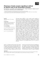
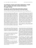
Figure 1. Expression of ACE2 in Type II Pneumocytes in Healthy Lungs of Non-human Primates
(A) Schematic of protocol for isolation of lung tissue at necropsy from healthy non-human primates (M. mulatta, n = 3), creation of scRNA-seq libraries by using
Seq-Well v1, and computational analysis to identify cell types by using unbiased methods. UMAP projection of 3,793 single cells, points colored by cell identity
(see STAR Methods).
(B) Uniform manifold approximation and projection (UMAP) as in (A), points colored by detection of ACE2 (coronavirus receptor, top) or TMPRSS2 (coronavirus S
protein priming for entry, bottom). Color coding is as follows: black, RNA positive; blue, RNA negative.
(C) Dot plot of 2 defining genes for each cell type (Table S1) (Bonferroni-adjusted p < 0.001) and ACE2 and TMPRSS2. Dot size represents fraction of cells within
that type expressing a given gene, and color intensity represents binned count-based expression amount (log(scaled UMI+1)) among expressing cells. ACE2 is
enriched in type II pneumocytes (6.7% expressing, Bonferroni-adjusted p = 8.62EÀ33), as is TMPRSS2 (29.5% expressing, Bonferroni-adjusted p = 8.73EÀ153).
Of all type II pneumocytes, 3.8% co-express ACE2 and TMPRSS2 (Table S9). Red arrow indicates cell type with largest proportion of ACE2+TMPRSS2+ cells.
(D) Genes differentially expressed among ACE2+ and ACE2À type II pneumocytes. (SCDE package, FDR-adjusted p < 0.05 for IFNGR2, NT5DC1, ARL6IP1, and
TRIM27; full results can be found in Table S1).
See also Table S1.
Notably, the only double-positive cells observed were classified cells, and type I pneumocytes, albeit at diminished abundance
within the type II pneumocyte population; however, we also iden- and frequency compared with type II pneumocytes (Figure 1C;
tified TMPRSS2 expression within club cells, ciliated epithelial Table S1).
Cell 181, 1016–1035, May 28, 2020 1019
ll
Article
A B
C D
E
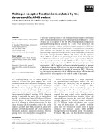
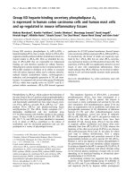
Figure 2. Select Lung Epithelial Cells from Control, HIV-1-Infected, and Mycobacterium-tuberculosis-Infected Human Donors Co-Express
ACE2 and TMPRSS2
(A) Schematic of protocol for isolation of human lung tissue from surgical excess, creation of scRNA-seq libraries by using Seq-Well S3, and computational
analysis to identify cell types by using unbiased methods. Shown on the right is a UMAP projection of 18,915 cells across 8 donors (n = 3 TB+HIV+; n = 3 TB+; n = 2
non-infected patients). Cells represented by points, colored according to cell type (see STAR Methods).
(B) UMAP projection as in (A), points colored by detection of ACE2 (top) or TMPRSS2 (bottom). Color coding is as follows: black, RNA positive; blue, RNA
negative.
(C) Dot plot of 2 defining genes for each cell type (FDR-adjusted p < 0.001), and ACE2 and TMPRSS2; dot size represents fraction of cells within cell type ex-
pressing a given gene, and color intensity represents binned count-based expression amount (log(scaled UMI+1)) among expressing cells. All cluster-defining
genes are provided in Table S2. Red arrow indicates cell types with largest proportion of ACE2+TMPRSS2+ cells.
(D) Volcano plot identifying significantly upregulated genes in ACE2+TMPRSS2+ pneumocytes compared with all remaining pneumocytes. Red points represent
genes with a FDR-adjusted p < 0.05, and log2(fold change) >1.5. Text highlighting specific genes; the full list is available in Table S2.
(E) Expression of ACE2 across human donors by HIV and TB status (p = 0.009 by likelihood-ratio test).
See also Table S2.
Next, we compared ACE2+ with ACE2À type II pneumocytes ACE2+TMPRSS2+ co-expressing type II pneumocytes, but its
to explore broader gene programs that differentiate putative level of upregulation compared with all remaining pneumocytes
SARS-CoV-2 target cells from cells of a similar phenotype and did not meet statistical significance (FDR-adjusted p = 0.11). This
ontogeny (Figure 1D; Table S1). Among genes significantly upre- analysis finds ACE2+ cells enriched within a rare fraction of
gulated in ACE2+ type II pneumocytes, we observed IFNGR2 secretory cells in NHPs and that ACE2 expression is co-regu-
(false discovery rate [FDR]-adjusted p = 0.022), a receptor for lated with genes involved in IFN responses.
type II IFNs. Notably, previous work has demonstrated limited
anti-viral potency of IFN-g for SARS-associated coronaviruses, To assess whether the findings from NHP lung cells were simi-
compared with that of type I IFNs, at least in vitro (Sainz et al., larly present in humans, we analyzed a previously unpublished
2004; Zheng et al., 2004). Other co-regulated genes of potential scRNA-seq dataset derived from surgical resections of fibrotic
interest include TRIM27 (FDR-adjusted p = 0.025), as well as lung tissue collected with Seq-Well S3 (Hughes et al., 2019). Un-
NT5DC1 (FDR-adjusted p = 0.003) and ARL6IP1 (FDR-adjusted supervised analysis identified multiple cell types and subtypes of
p = 0.047), which were upregulated in the A549 adenocarcinoma immune cells (Figures 2A–2C; STAR Methods), as defined by the
alveolar basal epithelial cell line after exposure to IFN-a and genes displayed in Figure 2C (full lists available in Table S2).
IFN-g for 6 h (Sanda et al., 2006). We found IFNAR1 consistently Here, we found that ACE2 and TMPRSS2 were primarily ex-
expressed among both ACE2+ type II pneumocytes and pressed within type II pneumocytes and ciliated cells, in line
with our analysis of the NHP-derived cells (Figures 1 and 2A,
1020 Cell 181, 1016–1035, May 28, 2020
ll
Article
A
B C
D E
F
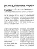
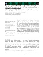
Figure 3. NHP and Human Ileal Absorptive Enterocytes Co-Express ACE2 and TMPRSS2
(A) Expression ACE2 across diverse tissues in healthy NHPs (n = 3 animals; 52,858 cells).
(B) Schematic of protocol for isolation of NHP ileum (n = 5) at necropsy for scRNA-seq using Seq-Well v1, and computational pipeline to identify cell types by using
unbiased methods. Shown on the right is a UMAP projection of 4,515 cells colored by cell type.
(C) Dot plot of 2 defining genes for each cell type, with ACE2 and TMPRSS2. Dot size represents fraction of cells within cell type expressing a given gene, and color
intensity represents binned count-based expression amounts (log(scaled UMI+1)) among expressing cells. All cluster defining genes are provided in Table S4.
Red arrow indicates cell type with largest proportion of ACE2+TMPRSS2+ cells.
(D) Schematic of protocol for isolation of human ileal cells from endoscopic pinch biopsies in non-inflamed regions (n = 13). Shown on the right is a tSNE plot of
13,689 epithelial cells selected from original dataset generated by 10x 30 v2 (see Figure S2), colored by cellular subsets.
(legend continued on next page)
Cell 181, 1016–1035, May 28, 2020 1021
ll
Article
2B). In type II pneumocytes (identified by unique expression of these cells and other cells within those clusters. Altogether, we
surfactant proteins SFTPC, SFTPB, and SFTPA1), we found identify ACE2+TMPRSS2+ cells in lower airways of humans
1.4% of cells expressing ACE2 (FDR-adjusted p = 1.35EÀ21), and NHPs with consistent cellular phenotypes and evidence
34.2% expressing TMPRSS2 (FDR-adjusted p < 1EÀ300), and supporting a potential role for IFN-associated inflammation in
0.8% co-expressing both. In ciliated cells, we found 7% were upregulation of ACE2.
ACE2+ (FDR-adjusted p = 5EÀ64), 24.6% were TMPRSS2+
(FDR-adjusted p = 3.8EÀ30), and 5.3% co-expressed both. Ileal Absorptive Enterocytes Express Host Factors Used
by SARS-CoV-2
As above, to assess for cellular pathways significantly co-ex- Next, we examined several other tissues for ACE2-expressing
pressed within putative target cells for SARS-CoV-2, we com- cells on the basis of the location of hallmark symptoms of
puted differentially expressed genes between ACE2+TMPRSS2+ COVID-19, focusing on the gastrointestinal tract due to reports
type II pneumocytes and all other type II pneumocytes (Figures of clinical symptoms and viral shedding (Xiao et al., 2020).
2C and 2D; Table S2). We found significant enrichment of Leveraging a previously unpublished scRNA-seq atlas of NHP
BATF among ACE2+TMPRSS2+ cells (FDR-adjusted p = (M. mulatta) tissues collected with Seq-Well v1, we observed
3.25EÀ7), which has been demonstrated previously to be upre- that the majority of ACE2+ cells reside in the small intestine, prin-
gulated by type I and type II IFNs (Murphy et al., 2013). Of note, cipally within the ileum, jejunum, and, to a lesser extent, the liver
we also observed TRIM28 co-expressed with ACE2 and and colon (Figure 3A; STAR Methods). Critically, we note that, in
TMPRSS2 among type II pneumocytes in this dataset (FDR- this experiment, the dissociation method used on each tissue
adjusted p = 2.34EÀ9), which might play a role in potentiating was optimized to preserve immune cell recovery, and therefore
an IFN response in lung epithelial cells (Krischuns et al., 2018). under-sampled stromal and epithelial populations, as well as
Within this cohort of donors, 3 individuals were human immuno- neurons from the brain. Within the ileum, we identified ACE2+
deficiency virus (HIV)+ and diagnosed with active tuberculosis, 3 cells as absorptive enterocytes on the basis of specific expres-
donors had active tuberculosis and were HIVÀ, and 2 were nega- sion of ACE2 within cells defined by APOA1, SI, FABP6, and EN-
tive for both pathogens. Surprisingly, we found that all of the PEP, among others, by a likelihood-ratio test (Figures 3B and 3C)
ACE2+ cells across all cell types were derived from HIV+ Myco- (p < 1EÀ300, 62% of all absorptive enterocytes; see Table S4).
bacterium tuberculosis (Mtb)+ donors despite approximately All other epithelial subtypes expressed ACE2 to a lesser extent,
equivalent recovery of epithelial cell types from all donors (likeli- and variably co-expressed ACE2 with TMPRSS2 (see Table S4
hood-ratio test, p = 0.009) (Figure 2E). Given limited cell and pa- for full statistics).
tient numbers combined with potential sampling biases, we
caution that this observation requires much broader cohorts to Persistent viral RNA in rectal swabs has been detected in pe-
validate a potential role for co-infections; still, we note our obser- diatric infection, even after negative nasopharyngeal tests (Xu
vation is suggestive of a role for chronic IFNs in the induction of et al., 2020). In an additional dataset consisting of endoscopic bi-
ACE2, given that HIV infection is associated with persistent up- opsies from the terminal ileum of a human pediatric cohort (n =
regulation of ISGs, and we observed elevated amounts of IF- 13 donors, ranging in age from 10 to 18 years old), collected
NAR2, IFI30, and IKBKB (Utay and Douek, 2016) (FDR-adjusted with 10X 30 v2, we confirmed a large abundance of ACE2+ cells
p = 1.1EÀ6, 8.8EÀ9, 1.57EÀ7, respectively; HIV+ versus HIVÀ with selective expression within absorptive enterocytes (29.7%
epithelial cells). ACE2+, FDR-adjusted p = 2.46EÀ100) (Figures 3D and 3E; Table
S5; STAR Methods). Furthermore, we identified a subset (888
Next, using a previously unpublished scRNA-seq dataset con- cells, $6.5% of all epithelial cells) that co-express both genes
sisting of granuloma and adjacent, uninvolved lung samples (Figures S2A–S2C). We performed differential expression testing
from Mtb-infected NHPs (Macaca fascicularis) collected with and GO-term enrichment using these cells relative to matched
Seq-Well S3, we identified subsets of epithelial cells expressing non-expressers to highlight putative biological functions en-
ACE2 and TMPRSS2 (Figure S1; Table S3; STAR Methods). The riched within them, such as metabolic processes and catalytic
majority of ACE2+TMPRSS2+ cells were, once again, type II activity, and to identify shared phenotypes of ACE2+TMPRSS2+
pneumocytes (22%) and type I pneumocytes (9.7%) and were ileal cells across both human and NHP cohorts (Table S5). We
largely enriched within granulomatous regions compared with speculate that viral targeting of these cells, taken from patients
those in adjacent uninvolved lung (Figures S1B and S1C) (p = without overt clinical viral infection, might help explain intestinal
0.006, Fisher Exact Test). ACE2+TMPRSS2+ type II pneumo- symptoms. Finally, we compared ileal absorptive enterocytes
cytes expressed significantly higher amounts of antimicrobial ef- from healthy NHPs and NHPs infected with simian-human immu-
fectors such as LCN2 compared with remaining type II pneumo- nodeficiency virus (SHIV) and then treated for 6 months with anti-
cytes (Figure S1D). Cells with club cell/secretory, type I retroviral therapy (animal and infection characteristics published
pneumocyte, and ciliated cell types also contained some in Colonna et al., 2018) (STAR Methods). We found significant
ACE2+TMPRSS2+ cells, but we did not have sufficient power upregulation of ACE2, STAT1, and IFI6 within the absorptive
to detect significantly differentially expressed genes between
(E). Dot plot of 2 defining genes for each cell type, with ACE2 and TMPRSS2. Dot size represents fraction of cells within cell type expressing a given gene, and
color intensity represents binned count-based expression amounts (log(scaled UMI+1)) among expressing cells. All cluster defining genes are provided in Table
S5. Red arrow indicates cell type with largest proportion of ACE2+TMPRSS2+ cells.
(F). Expression of ACE2 (left) and TMPRSS2 (right) among all epithelial subsets from human donors.
See also Figure S2 and Tables S4 and S5.
1022 Cell 181, 1016–1035, May 28, 2020
Article ll
A B C
D E F
G I
H
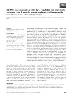
Figure 4. Healthy and Allergic Inflamed Human Nasal Mucosa Co-Express ACE2 and TMPRSS2 in a Subset of Goblet Secretory Cells
(A) Schematic for sampling of n = 12 ethmoid sinus surgical samples and n = 9 inferior turbinate nasal scrapings to generate scRNA-seq libraries by using Seq-
Well v1. See Ordovas-Montanes et al., (2018).
(B) Dot plot of all cell types from ethmoid-sinus-derived cells (n = 6 non-polyp CRS samples, n = 6 polyp CRS samples). Two defining genes for each cell type, in
addition to CDHR3 (rhinovirus receptor), ACE2, TMPRSS2, and JAK1. Dot size represents fraction of cells within that type expressing a given gene, and color
(legend continued on next page)
Cell 181, 1016–1035, May 28, 2020 1023
ll
Article
enterocytes of SHIV-infected animals (which maintain chroni- FDR-adjusted p = 7.32EÀ28; 28% express TMPRSS2, FDR-
cally elevated amounts of IFNs and ISGs) compared with those adjusted p = 2.15EÀ132; Table S6). We next explicitly gated cells
of uninfected controls (FDR-adjusted p < 2E-7) (Figure S2D) by their TMPRSS2 and ACE2 expression, identifying a rare sub-
(Deeks et al., 2017; Utay and Douek, 2016). set that co-expresses both, the majority of which fall within the
‘‘Secretory Cluster 7’’ cell type (Figures 4E and 4F) (30 cells,
Upper Airway Expression of Host Factors Used by SARS- $0.3% of all upper airway secretory cells, 1.6% of goblet
CoV-2 ‘‘Secretory Cluster 7’’). These findings are aligned with concur-
To identify potential viral target cells in nasal and sinus tissue, rent work by the HCA Lung Biological Network on human nasal
two regions that are frequently primary sites of exposure for co- scRNA-seq data, which identified nasal secretory cells to be en-
ronaviruses, we analyzed existing scRNA-seq datasets from the riched for ACE2 and TMPRSS2 expression (Sungnak
human upper airway (inferior turbinate and ethmoid sinus mu- et al., 2020).
cosa) across a spectrum of healthy donors and individuals with
allergic inflammation due to chronic rhinosinusitis (CRS) Although we identified co-expression of ACE2 and TMPRSS2
collected with Seq-Well v1 (Figure 4A; STAR Methods) (Ordo- in few airway cells overall, we detected ACE2 and TMPRSS2 sin-
vas-Montanes et al., 2018). We had previously noted a signifi- gle- and double-positive cells in over 20 donors and thus posit
cantly enriched IFN-dominated gene signature in inferior turbi- that these genes are enriched in secretory cells and are not a
nate secretory epithelial cells from both healthy and CRS product of individual-patient-driven variability (Figure S3A). Infe-
donors compared with CRS samples from the ethmoid sinus, rior turbinate scrapings collected on Seq-Well S3, which in-
which were significantly enriched for interleukin-4 (IL-4)/IL-13 creases the resolution of lower-abundance transcripts
gene signatures (Giovannini-Chami et al., 2012; Ordovas-Mon- compared with Seq-Well v1, revealed consistent and specific
tanes et al., 2018). We speculate that these cells, taken from clin- expression restricted to goblet secretory cells, but at a greater
ically non-virally infected patients, yet constantly exposed to detection frequency in samples from the same donors (Fig-
environmental viruses, might provide one of the earliest locations ure S3B) (ACE2+ from 4.7% v1 to 9.8% S3; ACE2+TMPRSS2+
for coronaviruses to infect before spreading to other tissues. We from 1.9% v1 to 4% S3) (Hughes et al., 2019). Using the gated
observed significant enrichment of ACE2 expression in apical ACE2+TMPRSS2+ cells, we tested for differentially expressed
epithelial cells and, to a lesser extent, ciliated cells compared genes compared to the remaining secretory epithelial cells (full
with all cell types recovered from surgically resected mucosa results provided in Table S6). Notably, we observed significant
(1% of apical epithelial cells, FDR-adjusted p = 4.55EÀ6, n.s. upregulation of ADAR, GBP2, OAS1, JAK1, and DUOX2 (FDR
in ciliated cells) (Figure 4B; Table S6). adjusted, all p < 0.02) within ACE2+TMPRSS2+ cells, potentially
indicative of IFN signaling (Figure 4G). Almost all ‘‘Secretory
To better map putative SARS-CoV-2 targets among epithelial Cluster 7’’ cells were from inferior turbinate scrapings of healthy
subsets, we employed a finer-grained clustering method applied and allergically inflamed individuals, few cells were from the
to both ethmoid sinus surgical specimens and scrapings from ethmoid sinus tissue of patients with chronic rhinosinusitis
the inferior turbinate and ethmoid sinus (Figures 4C–4F). Once without nasal polyps, and no cells were detected in polyp tissue
again, we observed selective expression of ACE2 within a minor- (Figure 4H). Gene Ontology (GO) analysis of enriched genes in
ity of cell types, with 1.3% of all secretory cells expressing ACE2 double-positive cells include processes related to intracellular
(Figure 4C) (FDR-adjusted p = 0.00023), specifically sub-clusters cytoskeleton and macromolecular localization and catabolism,
7 and 13, which represent two varieties of secretory epithelial cell potentially involved in viral particle entry, packaging, and exocy-
(Figures 4C, 4F, and 4G). Cluster 7 secretory cells are marked by tosis (Fung and Liu, 2019).
S100P, LYPD2, PSCA, CEACAM5, and STEAP4; encompass
some MUC5AC goblet cells; and contain the most significantly We next utilized IFN-inducible gene sets of relevance to hu-
enriched ACE2 and TMPRSS2 expression (4% express ACE2, man airway epithelial cells, which we derived from a prior study
by performing differential expression on a published dataset
intensity represents binned count-based expression amounts (log(scaled UMI+1)) among expressing cells (see Table S6 for statistics by subset). Red arrow
indicates cell types with largest proportion of ACE2+TMPRSS2+ cells.
(C) Dot plot for 2 defining genes for each cell type identified from granular clustering of epithelial cells (18,325 single cells) derived from both ethmoid sinus and
inferior turbinate sampling (healthy inferior turbinate [3,681 cells; n = 3 samples], polyp-bearing patient inferior turbinate [1,370 cells; n = 4 samples], non-polyp
ethmoid sinus surgical samples [5,928 cells; n = 6 samples], and polyp surgical and scraping samples directly from polyp in ethmoid sinus [7,346 cells; n = 8
samples]). Red arrow indicates cell type with largest proportion of ACE2+TMPRSS2+ cells.
(D) tSNE of 18,325 single epithelial cells from inferior turbinate and ethmoid sinus (omitting immune cells). Colored by cell types 3,152 basal, 3,089 differentiating,
8,840 secretory, 1,105 ciliated, and 2,139 glandular cells.
(E) tSNE as in (D), identifying epithelial cells co-expressing ACE2 and TMPRSS2 (30 cells, black points).
(F) tSNE as in (D), colored by detailed cell types with higher granularity, as in (C).
(G) Individual differentially expressed genes between ACE2+TMPRSS2+ cells and all other secretory epithelial cells (see Table S6 for full gene list with statistics).
Bonferroni-adjusted likelihood-ratio test p < 0.02 for all genes displayed.
(H) Stacked bar plot of each subset of epithelial cells among all epithelial cells by donor (each bar) and sampling location (noted below graph) (unpaired t test p <
0.00035 for Secretory Goblet 7 inferior turbinate versus ethmoid sinus; see Table S6 for raw values).
(I) Violin plot of cell clusters in respiratory epithelial cells (from Figures 4C and 4F) ordered by average expression of IFN-a-induced gene signatures, presented as
a gene module score; non-normal distribution by Lilliefors test, Mann-Whitney U-test p = 2.2EÀ16, 1.21 effect size, IFN-a signature for Secretory Goblet Cluster 7
versus all epithelial cells. Arrow indicates cluster containing majority ACE2+TMPRSS2+ cells.
See also Figure S3 and Table S6.
1024 Cell 181, 1016–1035, May 28, 2020
ll
Article
where air-liquid interface cultures from primary human nasal cells from two human donors (Figure 5A-D). We confirmed
epithelial cells were treated with IFN-aA/D, IFN-b1a, IFN-g, appropriate induction of an IFN response in each cell type by
IL-4, or IL-13 (Giovannini-Chami et al., 2012; Ordovas-Montanes performing differential expression testing between untreated
et al., 2018). Using these gene lists, we scored the human nasal cells and IFN-treated cells for each condition (Table S7). Within
epithelial cells analyzed by scRNA-seq described in Figures 4C each cell type, stimulation with IFN-a2, IFN-g, or IFN-b resulted
and 4F and found significant concomitant upregulation of the in dose-dependent upregulation of canonical ISGs, including
IFN-a-stimulated gene set within ACE2+TMPRSS2+ secretory STAT1/Stat1, BST2/Bst2, XAF1/Xaf1, IFI35/Ifi35, MX1/Mx1,
goblet cluster 7 (Figure 4I). and GBP2/Gbp2. Notably, Ace2 expression was not robustly
induced in basal cells derived from healthy mouse trachea under
Type I Interferon IFN-a Drives ACE2 Expression in any interferon stimulation condition (Figure 5A). The magnitude
Primary Human Nasal Epithelial Cells of ACE2 upregulation was diminished in BEAS-2B cells
The meta-analysis described above consistently identified an compared to that in our original findings in primary human upper
association between ACE2 expression and canonical ISGs or airway epithelial cells, but reached statistical significance
components of the IFN-signaling pathway. This prompted us compared with that of the untreated condition after IFN-g expo-
to investigate whether IFNs might play an active role in regulating sure (Figure 5B). In primary basal cells derived from healthy nasal
ACE2 expression levels in specific target cell subsets, thus mucosa, we confirmed significant induction of ACE2 after IFN-
potentially allowing for a tissue-protective host response or a2 stimulation and, to a lesser extent, after stimulation with
increased viral binding of SARS-CoV-2 through ACE2. Our initial IFN-g (IFN-a2-stimulated: both Bonferroni-adjusted p < 0.001;
literature search indicated that IFN-g and IL-4 downregulate the IFN-g-stimulated: both Bonferroni-adjusted p < 0.05) (Figures
SARS-CoV receptor ACE2 in Vero E6 cells (African green mon- 5C and 5D). Expression of ACE2 was significantly correlated
key kidney epithelial cells [de Lang et al., 2006]), appearing to with expression of STAT1 in all human cell types, with a larger ef-
invalidate this hypothesis. Relatedly, in vitro stimulation of fect size and correlation coefficient in primary human basal cells
A549 cells, a commonly used cell line model for lung epithelia, (Figure 5E-H). These experiments support a relationship be-
with IFN-a, IFN-g, and IFN-a+IFN-g for 24 h did not identify tween induction of the canonical IFN response, including key
ACE2 as an ISG (Russell et al., 2018). This is potentially explained transcription factors and transcriptional regulation of the ACE2
by recent work that aimed to understand SARS-CoV-2 receptor locus. Finally, among primary human samples, we confirmed
usage by performing screening studies within cell line models the dose-dependence of ACE2 upregulation after IFN-a2 or
and found that A549 cells did not express ACE2 and therefore IFN-g treatment and significant induction of ACE2 after IFN-a2
represents a poor model to understand regulation of this gene stimulation at concentrations as low as 0.1–0.5 ng/mL (Fig-
(Letko et al., 2020). While conducting experiments to directly ure 5I-L).
test the hypothesis that ACE2 is an ISG, we noted in our own
gene lists used for scoring from Ordovas-Montanes et al., 2018 Next, using a publicly available resource (interferome.org) that
and in a supplementary extended table available from Giovan- hosts genomic and transcriptomic data from cells or tissues
nini-Chami et al., 2012 that ACE2 was in upregulated gene lists treated with IFN, we queried ACE2 expression within human
after exposure to Type I IFN. and mouse cells, searching for datasets with a log2-fold-change
of >1 or < À1 compared with untreated samples, including all IFN
We directly tested whether IFN-a induces ACE2 in primary hu- types (Rusinova et al., 2013). We recovered 21 datasets span-
man upper airway epithelial cells in greater detail. We cultured ning 8 distinct primary tissues or cell lines with non-trivial
human primary basal (stem and progenitors) epithelial cells to changes in ACE2 expression after both type I and type II IFN
confluence and treated them with increasing doses (0.1–10 ng/ treatment (Figure S4A). We observed substantial upregulation
mL) of IFN-a2, IFN-g, IL-4, IL-13, IL-17A, or IL-1b for 12 h and of ACE2 in primary skin and primary bronchial cells treated
then performed bulk RNA-seq (Figure S3C). Only IFN-a2 and with either type I or type II IFN (> 5-fold upregulation compared
IFN-g led to upregulation of ACE2 over the time period tested, with that in untreated cells), in strong support of our in vitro
and compared with all other cytokines, IFN-a2 lead to greater data (Figures 5C, 5D, 5G–5L, and S3D–S3F). Immune cell types,
and more significant upregulation over all doses tested (Fig- such as CD4 T cells and macrophages, were noticeably absent
ure S3D,Wilcoxon test: IFN-a2 FDR-adjusted p = 4.1EÀ07; from datasets with a significant change in ACE2 expression after
IFN-g p = 9.3E-03,Figures S3E and S3F, all statistical tests IFN stimulation or were even found to downregulate ACE2 (e.g.,
compared with 0 ng/mL dose). We confirmed substantial and primary CD4 T cells + type I IFN) (Figure S4A, and in our analysis
dose-dependent induction of canonical members of the inter- of scRNA-seq peripheral blood mononuclear cell data from But-
feron response after IFN-a2 and IFN-g (Figures S3G and S3H). ler et al., (2018); data not shown).
Conversely, we found that IFN-g, relative to IFN-a2, induced
potent upregulation of GBP5, a GTPase-like protein thought to Given that the majority of cells robustly upregulating ACE2
act as a viral restriction factor through inhibiting furin-mediated were epithelial, this observation potentially explains why previ-
protease activity, which could limit viral processing from infected ous analyses to define canonical ISGs within immune popula-
cells, whereas IFN-a2 more robustly induced IFITM1 (Fig- tions did not identify ACE2 as an induced gene. Furthermore, us-
ure S3G–S3K) (Braun and Sauter, 2019). ing both Transcription Factor database (TRANSFAC) data
hosted by the interferome database, as well as chromatin immu-
To further extend and substantiate these findings, as above, noprecipitation sequencing (ChIP-seq) data (provided by the
we stimulated primary mouse tracheal basal cells, the commonly ENCODE Factorbook repository), we found evidence for
used human bronchial cell line BEAS-2B, and upper airway basal STAT1, STAT3, IRF8, and IRF1 binding sites within À1500–
Cell 181, 1016–1035, May 28, 2020 1025
ll
Article
A B C D
E F G H
I J K L
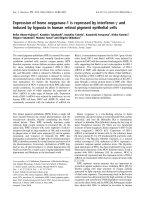
Figure 5. ACE2 is an Interferon-Stimulated Gene in Primary Human Barrier Tissue Epithelial Cells
(A–D) Basal epithelial cells from distinct sources were cultured to confluence and treated with increasing doses (0.1–10 ng/mL) of IFN-a2, IFN-g, IL-4, IL-17A, and/
or IFN-b for 12 h and bulk RNA-seq analysis was performed. Expression of ACE2 (human) or Ace2 (mouse) by cell type and stimulation condition. (A) Primary
mouse basal cells from tracheal epithelium are shown. (B) BEAS-2B human bronchial cell line is shown. (C) Primary human basal cells from nasal scraping, Donor
1, is shown. (D) Primary human basal cells from nasal scraping, Donor 2. Abbreviation is as follows: TP10K, transcripts per 10,000 reads. ***p < 0.001, **p < 0.01,
*p < 0.05, Bonferroni-corrected t test compared with untreated condition.
(E–H) Co-expression of STAT1/Stat1 and ACE2/Ace2 by cell type. (E) Primary mouse basal cells from tracheal epithelium are shown. (F) BEAS-2B human
bronchial cell line is shown. (G) Primary human basal cells from nasal scraping, Donor 1, are shown. (H) Primary human basal cells from nasal scraping, Donor 2
are shown. Abbreviation is as follows: TP10K, transcripts per 10,000 reads. Statistical significance assessed by Spearman’s rank correlation.
(I–L) Expression of ACE2 in primary human basal cells from nasal scrapings across a range of concentrations of IFN-g or IFN-a2. (I) IFN-a2 dose response in Donor
1 (p < 0.001 by one-way ANOVA) is shown. (J) IFN-g dose response in Donor 1 (p < 0.01 by one-way ANOVA) is shown. (K) IFN-a2 dose response in Donor 2 (p <
0.001 by one-way ANOVA) is shown. (L) IFN-g dose response in Donor 2 (p < 0.001 by one-way ANOVA). Abbreviation is as follows: TP10K, transcripts per 10,000
reads. ***p < 0.001, **p < 0.01, *p < 0.05, Bonferroni-corrected post hoc testing compared with 0 ng/mL condition.
See also Figures S3 and S4 and Table S7.
500 bp of the transcription start site of ACE2 (all in human with saline and two mice intranasally with 10,000 units of IFN-a
studies, Figure S4B) (Gerstein et al., 2012; Matys et al., 2003; (Guerrero-Plata et al., 2005). After 12 h, we isolated the nasal mu-
Wang et al., 2012; Wang et al., 2013). This finding is supportive cosa, consisting of both respiratory and olfactory epithelium, with
of our current hypothesis that ACE2 represents a previously un- underlying lamina propria, and performed scRNA-seq using Seq-
appreciated ISG in epithelial cells within barrier tissues. Well S3 (Figure S5A). We collected from both tissue sites because
of early reports of anosmia in COVID-19 (Lechien et al., 2020). We
Given minimal upregulation of Ace2 among primary mouse recovered 11,358 single cells, including epithelial, stromal,
basal cells in vitro, we were curious as to whether Ace2 repre- neuronal, and immune cell types, generating the largest single-
sented a murine ISG in vivo. We treated two mice intranasally
1026 Cell 181, 1016–1035, May 28, 2020
Article ll
A C
B
D
E F G
Figure 6. In Vivo Administration of Interferons in Mice Does Not Induce Ace2, and ACE2 Is Induced in Goblet Secretory Cells during Human
Influenza Infection
(A) UMAP of 11,358 single cells from mouse nasal epithelium (n = 4).
(B) UMAP projection as in (A), points colored by detection of Ace2 (SARS-CoV-2 receptor homolog). Color coding is as follows: black, RNA positive; blue, RNA
negative.
(C) Percent of Ace2+ cells by treatment condition (n = 4 arrays per condition; n = 2 arrays per mouse). Black bars indicate Ace2+ cells; white bars indicate Ace2À
cells. p = 0.4 by Student’s t test.
(D) Heatmap of cell-type-defining genes (Trp63 and Krt17), interferon-induced genes (Irf7, Stat1, Irf9, and Oasl2), and Ace2 among basal epithelial cells,
separated by cells derived from saline-treated mice (left) and IFN-a-treated mice (right). Statistical significance by likelihood-ratio test with Bonferroni correction
is shown. A full list of differentially expressed genes can be found in Table S8.
(E) Schematic for sampling cells derived from nasal washes of n = 18 human donors with and without current influenza A or B infection for Seq-Well v1 (35,840
single cells). See Cao et al., (2020).
(F and G) ACE2 expression among goblet cells (F) and squamous cells (G) by infection status. Shown are Healthy Donor cells from influenza-negative donors
(white); Bystander Cells from influenza A (IAV)- or influenza B (IBV)-infected donors, no intracellular viral RNA detected (black); Flu Viral RNA+ Cells with detectable
intracellular influenza A or B viral RNA (red). Statistical significance by Wilcoxon test with Bonferroni correction, n.s. for Bystander versus Flu Viral RNA+.
See also Figure S5 and Tables S6 and S8.
cell atlas of mouse respiratory and olfactory mucosa to date (Fig- and S5B) (Brann et al., 2020; Dear et al., 1991; Montoro et al.,
ures 6A and S5B). We annotated all 36 clusters, focusing our 2018; Tepe et al., 2018). Notably, Furin was enriched within olfac-
attention on epithelial cell clusters, given that we noted enrich- tory epithelial gland cells (Table S8). Next, we asked whether a
ment for Ace2 and Tmprss2 within epithelial cell subsets, consis- 12 h stimulation with IFN-a would upregulate Ace2 in vivo.
tent with our human and NHP results (Table S8). Specifically, we Focusing on basal epithelial cells, which contain the highest abun-
found Ace2 enriched within olfactory epithelial gland cells, dance of Ace2+ cells, we found that despite robust upregulation of
Muc5b+Scgb1c1+ goblet cells, basal epithelial cells, and myofi- canonical murine ISGs, Ace2 expression was only slightly
broblasts/pericytes (Bonferroni-corrected p < 0.01) (Figures 6B elevated after IFN-a treatment (Figures 6C, 6D, S5C, and S5D).
Cell 181, 1016–1035, May 28, 2020 1027
ll
Article
This observation was supported by analysis of scRNA-seq in the human upper airway by inducing ACE2, we attempted to
data from 5,558 epithelial cells from the lungs of mice 3–6 days extend our transcriptomic data on IFN-driven expression of
after intranasal infection with murine gamma herpesvirus-68 ACE2 to protein-level induction of ACE2. As testing of various
(MHV68) (Figure S5E). Here, we found significant enrichment of commercially available polyclonal antibody preparations found
Ace2+ cells within type II pneumocytes, in line with our data broad evidence for non-specific or inconclusive staining in histo-
from NHP and human lungs (Figures S5F). We did not observe logical immunofluorescent based readouts (data not shown), we
changes in Ace2 expression among viral-transcript-positive cells assessed whether IFN-g-stimulated human bronchial air-liquid
or ‘‘bystander’’ type II pneumocytes (those without detectable interface cultures induced ACE2 within 24 h. Our results show
cell-associated viral RNA in MHV68-infected animals), nor did that cells from one patient robustly induced ACE2 (+2.02x), cells
we see significant alterations in Ace2+ cell abundance among from another mildly induced ACE2 (+1.21x) and two patient’s
MHV68-infected mice lacking IFN-gR (Figure S5G and S5H). cells showed minor changes (+/À1.12x) (Figure S5M). We pro-
These observations were in agreement with our in vitro murine vide a note of caution as these cells were derived from asthmatic
basal cell assay (Figure 5A and 5E). patients, and the overall changes did not reach significance.
Furthermore, we could not determine cell surface localization
Finally, we sought to validate our hypothesis that ACE2 is upre- of ACE2 but do note that these results align with our transcrip-
gulated in human epithelial cells during upper airway viral infec- tomic data.
tions, which are known to induce a robust IFN response (Bailey
et al., 2014; Everitt et al., 2012; Iwasaki and Pillai, 2014; Jewell DISCUSSION
et al., 2010; Russell et al., 2018; Steuerman et al., 2018). We re-
analyzed a publicly available dataset of RNA-seq from human Here, we utilize scRNA-seq across various barrier tissues and
lung explants isolated after surgical resections that were infected model organisms to identify the potential initial cellular targets
with influenza A virus ex vivo for 24 h. Here, we found that ACE2 of SARS-CoV-2 infection. To review the data presented: (1) we
expression was significantly correlated with that of SFTPC, sup- found that expression of the cellular entry receptor for SARS-
porting our hypothesis that ACE2 is expressed within type II CoV-2, ACE2, is primarily restricted to type II pneumocytes in
pneumocytes (Figures 1C, 2C, S5I, and S5J) (Matos et al., the lung, absorptive enterocytes within the gut, and goblet
2019). Furthermore, although the abundance of SFTPC was not secretory cells of the nasal mucosa; (2) ACE2 and TMPRSS2
significantly altered by influenza A virus infection, ACE2 expres- co-expression in respiratory tissues is consistently found only
sion was significantly upregulated after viral exposure (p = among a rare subset of epithelial cells; (3) we observed similar-
0.0054, ratio paired t test) (Figures S5K and S5L). This suggests ities in the cellular identities and frequencies of putative SARS-
that influenza A virus infection increases ACE2 expression. CoV-2 target cells across human and NHP cohorts; (4) we
Nevertheless, these population-level analyses are not able to observe increased expression of ACE2 during SHIV and TB
definitively resolve specific cell subsets of relevance, nor whether infection of NHPs, and HIV/TB co-infection and influenza infec-
they are directly infected cells or bystanders of infection. tion of humans compared with that in matched controls but
caution that none of the datasets presented here were designed
In order to address these questions, we leveraged an ongoing to answer this specific query. Specific targeting of these cell sub-
scRNA-seq study of nasal washes from 18 individuals with sets has only been described for a handful of viruses, including
confirmed influenza A virus or influenza B virus infection or the following: goblet cells by human adenovirus-5p and entero-
healthy controls collected with Seq-Well v1, which yielded virus 71, type II pneumocytes by H5N1 avian influenza, and
35,840 cells resolved into 17 distinct cell types (Figure 6E; absorptive enterocytes by rotavirus (Fleming et al., 2014; Good
STAR Methods) (Cao et al., 2020). We investigated the cell types et al., 2019; Holly and Smith, 2018; Weinheimer et al., 2012).
with greatest enrichment for ACE2 and TMPRSS2 in non-in-
fected controls and individuals with influenza A and B. Strikingly, Additionally, we provide an overall note of caution when inter-
ACE2 was most upregulated in samples from influenza-virus-in- preting scRNA-seq data for low abundance transcripts like ACE2
fected individuals within bystander goblet or squamous cells not and TMPRSS2 because detection inefficiencies might result in
directly infected by virus (Figures 6F and 6G). ACE2+TMPRSS2+ an underestimation of the actual frequencies of ACE2+ or
goblet cells during influenza infection exhibited enrichment for ACE2+TMPRSS2+ cells in a tissue. Moreover, the protein
canonical ISGs such as the CXCL9/CXCL10/CXCL11 gene clus- amounts of each might differ from their mRNA abundances
ter; correspondence with ACE2+TMPRSS2+ goblet cells in (Genshaft et al., 2016; Jovanovic et al., 2015; Rabani et al.,
healthy and allergic nasal scrapings; and a shared overlap in 2011; Shalek et al., 2013). We also present datasets separately,
ISGs including GBP2, ZNFX1, ADAR, and ACE2 (significantly given that each study differed in its methods of tissue processing
differentially expressed gene lists) (Table S6). Together, our and collection, which can influence the frequency of recovered
data suggest that ACE2 is an ISG in vitro and in vivo in human pri- cell subsets (STAR Methods). We provide Table S9 as a sum-
mary upper airway epithelial basal cells, but that the murine ho- mary of ACE2+ and ACE2+TMPRSS2+ cells across various data-
molog Ace2 is not in airway epithelial basal cells or pulmonary sets. Moreover, we present Figure S6, which describes statisti-
epithelial cells in vitro or in vivo. Collectively, our findings suggest cal modeling and power calculations underlying detection and
that careful considerations of animal and cellular models will be dropout of ACE2, to help guide interpretation of these data.
needed for assessing therapeutic interventions targeting the IFN This includes an examination of the probability to detect a lowly
system when studying ACE2/Ace2-associated biology. expressed transcript like ACE2 within a cell, as well as upper
bound estimates on the percentage of positive cells within a
Finally, because our in vivo and in vitro work indicate that IFN
might promote human cellular targets for SARS-CoV-2 infection
1028 Cell 181, 1016–1035, May 28, 2020
ll
Article
cluster, considering the effects of transcript counts, sequencing adds to the growing evidence that coronaviruses, as well as
depth, and cell numbers in these calculations (STAR Methods). other viruses, have evolved to leverage features of the human
IFN pathway (Fung and Liu, 2019; Mar et al., 2018; Zhao et al.,
Whether ACE2 and TMPRSS2 are needed on the same cell or 2014). Whether type I IFNs are net protective or detrimental to
soluble proteases can activate SARS-CoV-2 S protein to invade the host might depend on the stage of infection; cell subsets in
ACE2 single-positive cells is an area of active inquiry (Coutard question; the SARS viral clade (Channappanavar et al., 2016;
et al., 2020; Letko et al., 2020). Importantly, rapidly evolving liter- Channappanavar et al., 2019; Channappanavar and Perlman,
ature has identified that SARS-CoV-2-S might have a furin cleav- 2017; Davidson et al., 2015); and other factors such as co-infec-
age site, leading to a broader set of host proteases that could tion, age, gender, and co-morbidities, among others. Under-
mediate S protein activation (Bugge et al., 2009; Coutard et al., standing the specific host restriction factors targeting SARS-
2020; Walls et al., 2020). However, because an active S protein CoV-2 and identifying specific drivers of these genes in the
has a finite lifetime to find a target cell membrane, the timing absence of ACE2 upregulation might provide strategies to disso-
and cellular location of S protein activation is key to consider. ciate the dual roles of IFN in certain coronavirus infections.
Activation events proximal to the plasma membrane have been Whether IFNs upregulate ACE2 in putative target cell subsets
shown to be most effective for SARS-CoV entry (Shulla in vivo will be of significant interest to define in future work
et al., 2011). once current COVID-19-related restrictions on basic scientific in-
quiry are lifted (Qian et al., 2013).
Our study finds that type I IFNs, and to a lesser extent type II
IFNs, upregulate ACE2. This is based on several lines of evi- ACE2 is a central component of the renin-angiotensin system,
dence: (1) we identified a human goblet secretory cell subset in which has emerged as a key regulator of sterile- or microbially
upper airway nasal epithelium enriched for ACE2 expression to induced lung pathology (Imai et al., 2005). In brief, ACE cleaves
have the highest IFN-a-induced gene signature; (2) we found angiotensin I to generate angiotensin II (Skeggs et al., 1980).
that IFN-a, and to a lesser extent IFN-b or IFN-g, induced Angiotensin II then acts to drive acute lung injury through various
ACE2 expression in a published dataset of air-liquid interface mechanisms, including increased vascular permeability (Imai
cultures derived from human nasal epithelial cells (Giovannini- et al., 2005). Amounts of angiotensin II in humans and mice are
Chami et al., 2012; Ordovas-Montanes et al., 2018); (3) we elevated during influenza infection, and ACE2 exerts tissue-pro-
extended our search through the Interferome database (Rusi- tective functions by reducing amounts of angiotensin II (Zou
nova et al., 2013) and found that, in epithelial barrier tissues, et al., 2014). Binding of SARS-CoV-S to mouse ACE2 in vivo
type I IFNs upregulate ACE2 in multiple studies, especially in pri- reduced ACE2 expression leading to acute acid-aspiration-
mary bronchial cells and keratinocytes (Rusinova et al., 2013); (4) induced lung failure (Kuba et al., 2005). Depending on the ques-
we found two STAT1 binding sites in the promoter of ACE2; (5) in tions asked in future work, there are mouse models available on
our unpublished atlas of SHIV-infected macaques, known to the basis of transgenic expression of human ACE2 (required for
have elevated amounts of chronic IFN signaling, we found overt infectious pathology of SARS-CoV in mice), there are es-
ACE2 upregulation in absorptive enterocytes; (6) we directly pro- tablished NHP models available of SARS-CoV infection in
vided evidence for IFN-a, and to some extent IFN-g, inducing M. fascicularis and C. aethiops, and early reports suggest symp-
ACE2 expression in primary human upper airway basal cells; tomatic infection in M. mulatta and M. fascicularis models for
and (7) influenza infection in humans, a known inducer of the SARS-CoV-2 (Bao et al., 2020; McCray et al., 2007; Munster
IFN pathway, leads to increased ACE2 expression in goblet et al., 2020; Rockx et al., 2020; Smits et al., 2011). For example,
secretory cells of the nasal epithelium (Cao et al., 2020). examining the efficacy of recombinant human ACE2 to act as a
decoy receptor or the effect of ‘‘ACE inhibitors’’ in patients
Altogether, our own and publicly available data highlight that with, or at risk for, COVID-19 will require careful experimentation
ACE2 might have been missed as a canonical ISG because of in appropriate models together with well-controlled clinical trials
its notable absence in peripheral blood mononuclear cell data- (Hofmann et al., 2004; Monteil et al., 2020; Vaduganathan
sets and in lung-derived transformed cell lines such as the et al., 2020).
A549 cell line (Butler et al., 2018; Letko et al., 2020; Rusinova
et al., 2013). Importantly, other groups have independently IFN responses that induce ISGs are essential for host antiviral
analyzed publicly available datasets, some referenced in our defense in mice, NHPs, and humans (Bailey et al., 2014; Dupuis
work, and observed ACE2’s behavior as an ISG (Wang and et al., 2003; Everitt et al., 2012). Canonical ISGs function by
Cheng, 2020). Furthermore, we found weak IFN- or virally driven directly restricting viruses and reducing burden (Schneider
induction of Ace2 in murine cells and tissues. This highlights the et al., 2014). More recently, disease tolerance to equivalent path-
importance of studying primary human epithelial cells and the ogen burden by factors that increase the ability of the host to
careful consideration of appropriately selected gene lists and tolerate tissue damage has been identified as part of a combined
in vitro models of in vivo cellular systems for understanding hu- host defense strategy (Iwasaki et al., 2017; Iwasaki and Pillai,
man biology (Jonsdottir and Dijkman, 2016; Mead and Karp, 2014; Medzhitov et al., 2012; Schneider and Ayres, 2008). Dis-
2019; Regev et al., 2017). ease tolerance factors in the lung include IL-22 and amphiregulin
(Iwasaki et al., 2017). During acute infection in the respiratory sys-
As SARS-CoV-S leads to ACE2-receptor-mediated internali- tem, ACE2 is critical for early tissue tolerance responses to respi-
zation, the host IFN response could thus promote the ability for ratory infection, including H5N1 influenza (Huang et al., 2014; Zou
SARS-CoV and SARS-CoV-2 to maintain cellular targets in et al., 2014). However, our discovery that ACE2 is an ISG in hu-
neighboring human upper airway epithelial cells. Altogether man epithelial cells, along with SARS-CoV-2 utilizing host ACE2
along with a study of HCoV-OC43, which co-opts IFN-inducible
transmembrane 2 (IFITM2) and IFITM3 to promote viral entry, this
Cell 181, 1016–1035, May 28, 2020 1029
ll
Article
to gain entry to cells, suggests that SARS-CoV and SARS-CoV-2 et al., 2020). Given the unappreciated complexities of host-path-
might exploit the ACE2-mediated tissue-protective response to ogen interactions between humans and SARS-CoV-2, the best
provide further cellular targets for entry. This potential strategy measures to combat this pandemic continue to be surveillance
employed by SARS-CoV-2 could present a unique challenge and avoidance—especially given that a deep understanding of
for the human host and is distinct from HCoV-OC43, which tar- the full spectrum of resistance and tolerance mechanisms will
gets the two restriction factors IFITM2 and IFITM3 (Zhao et al., require the concerted efforts of scientists around the globe
2014). Our study provides motivation to understand the specific (Amanat et al., 2020; Chu et al., 2020; Hadfield et al., 2018).
role and balance of type I and type II IFNs, as well as type III Here, we seek to share our initial findings and data so that other
IFNs, in tissue protection during, and host restriction of, groups might build on this discovery of ACE2 as an ISG and
SARS-CoV-2 infection. Key experiments to understand ACE2 further consider the careful balance between tissue tolerance
as an ISG in tissue protection or genuine tolerance will require and viral infection needed at the human airway epithelium.
the appropriate mouse, NHP, or other model in BSL3 or BSL4
facilities to execute SARS-CoV-2 viral infections and measure STAR+METHODS
host tissue health along with viral loads. Further work will
also be needed to understand how co-infections, as well as Detailed methods are provided in the online version of this paper
other host factors, might affect both the susceptibility to, and and include the following:
dynamics of, host SARS-CoV-2 infection. Moreover, carefully
controlled clinical trials will be essential to determine the overall d KEY RESOURCES TABLE
effects of different IFNs (Prokunina-Olsson et al., 2020). d RESOURCE AVAILABILITY
Altogether, we anticipate that comprehensive characterization B Lead Contact
of the putative cellular targets of SARS-CoV-2 will be critical to B Materials Availability
understand basic mechanisms of viral tropism and disease path- B Data and Code Availability
ophysiology, inform differential susceptibility among vulnerable d EXPERIMENTAL MODEL AND SUBJECT DETAILS
populations, and potentially suggest unanticipated targets for B Human Intestinal Biopsies
drug inhibitors of viral infection. The cellular targets we nominate B Human Lungs, Surgical Excess
will need to be confirmed by specific reagents for SARS-CoV-2, B Human Nasal Polyps and Scrapings
as done for SARS-CoV (Ding et al., 2004). Furthermore, the tran- B Human Nasal Washes, Healthy and Influenza Infected
scriptional response to the virus will need to be rigorously char- B Cell Culture of Primary Basal Cells and Cell Lines
acterized in appropriate in vitro and in vivo model systems B Non-Human Primates (M. mulatta)
(Blanco-Melo et al., 2020). We provide gene lists associated B Non-Human Primates (M. fascicularis)
with target cells in specific tissues and diseases to aid the com- B Mouse Nasal and Olfactory Epithelium and
munity in understanding this emergent disease. A concurrent
HCA Lung Biological Network study assessing ACE2 and Tracheal Cells
TMPRSS2 across more tissues also identified enrichment in B Mouse Lungs, MHV68 Infection
nasal goblet and ciliated cells (Sungnak et al., 2020). Other d METHOD DETAILS
studies are considering additional tissues; co-variates such as B Methods of Sample Collection and Tissue Preparation
age, sex, and co-infection state; and represent a large coordi-
nated international effort to the ongoing crisis (Pinto et al., for Single-Cell RNA-Seq
2020). One study in particular identified upregulation of ACE2 B Methods to Generate Single-Cell and Bulk RNA-seq
by respiratory viruses and TMPRSS2 by IL-13 in a pediatric
cohort, suggesting further links to how underlying allergic condi- Libraries
tions or co-infections might modulate these two SARS-CoV-2- B Human and Mouse Basal Cell Cytokine Stimulation
related host factors (Sajuthi et al., 2020). B Western blot for human ACE2
d QUANTIFICATION AND STATISTICAL ANALYSIS
During the preparation of this manuscript, several papers have B Non-Human Primate Lung and Ileum
been posted to bioRxiv assessing patterns of ACE2+ and B Human Lung Tissue
TMPRSS2+ cells in barrier tissues (Brann et al., 2020; Lukassen B Human Ileum
et al., 2020; Qi et al., 2020; Wu et al., 2020; Zhang et al., 2020). At B Human Adult Nasal Mucosa
a high level, these studies are largely in agreement with B Granulomatous Tissue from Mycobacterium Tubercu-
our report. Furthermore, another study appeared on medRxiv
profiling bronchoalveolar lavage fluid from 3 severe and 3 mild losis Infected NHPs
COVID-19 patients, though they were unable to profile sufficient B Basal Cell Cytokine Stimulation
numbers of epithelial cells (Liao et al., 2020). B Interferon Treatment of Mouse Nasal Mucosa
B Lung from MHV68-Infected WT and IFNgR KO Mice
Our study highlights the power of scRNA-seq datasets, both B Nasal Washes during Influenza Infection
existing and novel, to derive hypotheses relevant to human dis- B Power Calculations for Detection of Rare Transcripts
ease that might differ from paradigms established by using cell B Statistical Testing
lines. Further work will be critical to determine how SARS-
CoV-2 influences temporal dynamics of host responses at sin- SUPPLEMENTAL INFORMATION
gle-cell resolution and which host factors might affect this (Kazer
Supplemental Information can be found online at /> cell.2020.04.035.
1030 Cell 181, 1016–1035, May 28, 2020
ll
Article
CONSORTIA Merck, Honeycomb Biotechnologies, Cellarity, Cogen Therapeutics, Orche
Bio, and Dahlia Biosciences. L.S.K. is on the SAB for HiFiBio; she reports
The members of HCA Lung Biological Network are Nicholas E. Banovich, research funding from Kymab Limited, Bristol Meyers Squibb, Magenta Ther-
Pascal Barbry, Alvis Brazma, Tushar Desai, Thu Elizabeth Duong, Oliver Eick- apeutics, BlueBird Bio, and Regeneron Pharmaceuticals and consulting fees
elberg, Christine Falk, Michael Farzan, Ian Glass, Muzlifah Haniffa, Peter Hor- from Equillium, FortySeven, Inc, Novartis, Inc, EMD Serono, Gilead Sciences,
vath, Deborah Hung, Naftali Kaminski, Mark Krasnow, Jonathan A. Kropski, and Takeda Pharmaceuticals. A.S. is an employee of Johnson and Johnson.
Malte Kuhnemund, Robert Lafyatis, Haeock Lee, Sylvie Leroy, Sten Linnarson, N.K. is an inventor on a patent using thyroid hormone mimetics in acute lung
Joakim Lundeberg, Kerstin B. Meyer, Alexander Misharin, Martijn Nawijn, injury that is now being considered for intervention in COVID-19 patients.
Marko Z. Nikolic, Jose Ordovas-Montanes, Dana Pe’er, Joseph Powell, Ste- J.L. is a scientific consultant for 10X Genomics, Inc. O.R.R, is a co-inventor
phen Quake, Jay Rajagopal, Purushothama Rao Tata, Emma L. Rawlins, on patent applications filed by the Broad Institute to inventions relating to sin-
Aviv Regev, Paul A. Reyfman, Mauricio Rojas, Orit Rosen, Kourosh Saeb- gle-cell genomics applications, such as in PCT/US2018/060860 and US Pro-
Parsy, Christos Samakovlis, Herbert Schiller, Joachim L. Schultze, Max A. Sei- visional Application No. 62/745,259. S.T. in the last three years was a consul-
bold, Alex K. Shalek, Douglas Shepherd, Jason Spence, Avrum Spira, Xin Sun, tant at Genentech, Biogen, and Roche and is a member of the SAB of Foresite
Sarah Teichmann, Fabian Theis, Alexander Tsankov, Maarten van den Berge, Labs. M.H.W. is now an employee of Pfizer. F.J.T. reports receiving consulting
Michael von Papen, Jeffrey Whitsett, Ramnik Xavier, Yan Xu, Laure-Emma- fees from Roche Diagnostics GmbH and ownership interest in Cellarity, Inc.
nuelle Zaragosi, and Kun Zhang. Pascal Barbry, Alexander Misharin, Martijn P.H. is a co-inventor on a patent using artificial intelligence and high-resolution
Nawijn, and Jay Rajagopal serve as the coordinators. microscopy for COVID-19 infection testing based on serology.
ACKNOWLEDGMENTS Received: March 13, 2020
Revised: April 3, 2020
We are grateful to the study participants who made this work possible. We Accepted: April 20, 2020
would like to thank Bruce Horwitz, Ivan Zanoni, Matt Sampson, Michael Published: April 27, 2020
Retchin, Peter Winter, Andrew Navia, Jamie Cohen, and Audrey Sporrij for dis-
cussions. Mengyang (Vicky) Li Horst, Timothy Tickle, Jonathan Bistline, Jean REFERENCES
Chang, Eric Weitz, Eno-Abasi Augustine-Akpan, and Devon Bush for develop-
ment and support of the Broad Institute Single Cell Portal. This work was sup- Adler, H., Messerle, M., Wagner, M., and Koszinowski, U.H. (2000). Cloning
ported in part by the Searle Scholars Program, the Beckman Young Investi- and mutagenesis of the murine gammaherpesvirus 68 genome as an infectious
gator Program, the Pew-Stewart Scholars Program for Cancer Research, a bacterial artificial chromosome. J. Virol. 74, 6964–6974.
Sloan Fellowship in Chemistry, the MIT Stem Cell Initiative through Fondation
MIT, the NIH (5U24AI118672 and BAA-NIAID-NIHAI201700104), and the Bill Amanat, F., Nguyen, T., Chromikova, V., Strohmeier, S., Stadlbauer, D., Javier,
and Melinda Gates Foundation to A.K.S., as well as NIH R56 AI139053 to A., Jiang, K., Asthagiri-Arunkumar, G., Polanco, J., Bermudez-Gonzalez, M.,
J.L.F and P.L.L., and the Aeras Foundation to J.L.F. B.B. and S.K.N. are et al. (2020). A serological assay to detect SARS-CoV-2 seroconversion in hu-
partially supported by NIH 5R01GM081871. We acknowledge support from mans. medRxiv. />the Damon Runyon Cancer Research Foundation (DRG-2274-16) and Richard
and Susan Smith Family Foundation to J.O.-M; from a National Science Foun- Ardain, A., Domingo-Gonzalez, R., Das, S., Kazer, S.W., Howard, N.C., Singh,
dation Graduate Research Fellowship (1122374) to S.K.N., S.J.A., and C.N.T.; A., Ahmed, M., Nhamoyebonde, S., Rangel-Moreno, J., Ogongo, P., et al.
from a Fannie and John Hertz Foundation Fellowship to C.N.T.; by (2019). Group 3 innate lymphoid cells mediate early protective immunity
T32GM007753 from the National Institute of General Medical Sciences to against tuberculosis. Nature 570, 528–532.
C.G.K.Z. This work was further supported by the UMass Center for Clinical
and Translational Science Project Pilot Program; and the Office of the Assis- Bailey, C.C., Zhong, G., Huang, I.C., and Farzan, M. (2014). IFITM-Family Pro-
tant Secretary of Defense for Health Affairs, through the Peer Reviewed Med- teins: The Cell’s First Line of Antiviral Defense. Annu. Rev. Virol. 1, 261–283.
ical Research Program (W81XWH-15-1-0317) to R.W.F. We also acknowledge
support from NIH grants AI078908, HL111113, HL117945, R37AI052353, Bao, L., Deng, W., Gao, H., Xiao, C., Liu, J., Xue, J., Lv, Q., Liu, J., Yu, P., Xu, Y.,
R01AI136041, R01HL136209, and U19AI095219 to J.A.B.; by grants from et al. (2020). Reinfection could not occur in SARS-CoV-2 infected rhesus ma-
the NIH and National Heart, Lung, and Blood Institute (U19 HL129902) to caques. bioRxiv. />H.P.K and L.S.K; from National Institute of Allergy and Infectious Diseases
(UM1 AI126623) to H.P.K.; and to P.B. from the Fondation pour la Recherche Blanco-Melo, D., Nilsson-Payant, B.E., Liu, W.-C., Møller, R., Panis, M.,
Me´ dicale (DEQ20180339158), and the Agence Nationale pour la Recherche Sachs, D., Albrecht, R.A., and tenOever, B.R. (2020). SARS-CoV-2 launches
(ANR-19-CE14-0027); and by the following grants to L.S.K: NIH/NIAID U19 a unique transcriptional signature from in vitro, ex vivo, and in vivo systems.
AI051731, NIH/NHLBI R01 HL095791 NIH/NIAID R33-AI116184, NIH/NIAID bioRxiv. />U19 AI117945, and DHHS/NIH 1UM1AI126617. B.E.M. was supported by
the Massachusetts Institute of Technology - GlaxoSmithKline (MIT-GSK) Ger- Boă ttcher-Friebertshaă user, E., Klenk, H.D., and Garten, W. (2013). Activation of
trude B. Elion Postdoctoral Fellowship; T.M.L. by the NIH/NHLBI influenza viruses by proteases from host cells and bacteria in the human
1R01HL128241-01, K.M.B. by NIH/NIAID K23AI139352; and D.L. by NIH airway epithelium. Pathog. Dis. 69, 87–100.
R01AI137057, DP2DA042422, and R01AI124378. This publication is part of
the Human Cell Atlas (www.humancellatlas.org/publications). Brann, D., Tsukahara, T., Weinreb, C., Logan, D.W., and Datta, S.R. (2020).
Non-neural expression of SARS-CoV-2 entry genes in the olfactory epithelium
AUTHOR CONTRIBUTIONS suggests mechanisms underlying anosmia in COVID-19 patients. bioRxiv.
/>Document S1 details contributions of all authors.
Braun, E., and Sauter, D. (2019). Furin-mediated protein processing in infec-
DECLARATION OF INTERESTS tious diseases and cancer. Clin. Transl. Immunology 8, e1073.
A.R. is an SAB member of ThermoFisher Scientific, Neogene Therapeutics, Broggi, A., Granucci, F., and Zanoni, I. (2020). Type III interferons: Balancing
Asimov, and Syros Pharmaceuticals; a co-founder of and equity holder in tissue tolerance and resistance to pathogen invasion. J. Exp. Med. 217,
Celsius Therapeutics; and an equity holder in Immunitas Therapeutics. e20190295.
A.K.S. reports compensation for consulting and/or SAB membership from
Bugge, T.H., Antalis, T.M., and Wu, Q. (2009). Type II transmembrane serine
proteases. J. Biol. Chem. 284, 23177–23181.
Butler, A., Hoffman, P., Smibert, P., Papalexi, E., and Satija, R. (2018). Inte-
grating single-cell transcriptomic data across different conditions, technolo-
gies, and species. Nat. Biotechnol. 36, 411–420.
Cao, Y., Guo, Z., Vangala, P., Donnard, E., Liu, P., McDonel, P., Ordovas-Mon-
tanes, J., Shalek, A.K., Finberg, R.W., Wang, J.P., et al. (2020). Single-cell
Cell 181, 1016–1035, May 28, 2020 1031
ll
Article
analysis of upper airway cells reveals host-viral dynamics in influenza infected Dupuis, S., Jouanguy, E., Al-Hajjar, S., Fieschi, C., Al-Mohsen, I.Z., Al-Jumaah,
adults. bioRxiv. S., Yang, K., Chapgier, A., Eidenschenk, C., Eid, P., et al. (2003). Impaired
response to interferon-alpha/beta and lethal viral disease in human STAT1
Channappanavar, R., and Perlman, S. (2017). Pathogenic human coronavirus deficiency. Nat. Genet. 33, 388–391.
infections: causes and consequences of cytokine storm and immunopa-
thology. Semin. Immunopathol. 39, 529–539. Everitt, A.R., Clare, S., Pertel, T., John, S.P., Wash, R.S., Smith, S.E., Chin,
C.R., Feeley, E.M., Sims, J.S., Adams, D.J., et al.; GenISIS Investigators;
Channappanavar, R., Fehr, A.R., Vijay, R., Mack, M., Zhao, J., Meyerholz, D.K., MOSAIC Investigators (2012). IFITM3 restricts the morbidity and mortality
and Perlman, S. (2016). Dysregulated Type I Interferon and Inflammatory associated with influenza. Nature 484, 519–523.
Monocyte-Macrophage Responses Cause Lethal Pneumonia in SARS-CoV-
Infected Mice. Cell Host Microbe 19, 181–193. Fleming, F.E., Boă hm, R., Dang, V.T., Holloway, G., Haselhorst, T., Madge, P.D.,
Deveryshetty, J., Yu, X., Blanchard, H., von Itzstein, M., and Coulson, B.S.
Channappanavar, R., Fehr, A.R., Zheng, J., Wohlford-Lenane, C., Abrahante, (2014). Relative roles of GM1 ganglioside, N-acylneuraminic acids, and a2b1
J.E., Mack, M., Sompallae, R., McCray, P.B., Jr., Meyerholz, D.K., and Perl- integrin in mediating rotavirus infection. J. Virol. 88, 4558–4571.
man, S. (2019). IFN-I response timing relative to virus replication determines
MERS coronavirus infection outcomes. J. Clin. Invest. 130, 3625–3639. Fung, T.S., and Liu, D.X. (2019). Human Coronavirus: Host-Pathogen Interac-
tion. Annu. Rev. Microbiol. 73, 529–557.
Chu, H.Y., Boeckh, M., Englund, J.A., Famulare, M., Lutz, B.R., Nickerson,
D.A., Rieder, M.J., Starita, L.M., Adler, A., Brandstetter, E., et al. (2020). The Genshaft, A.S., Li, S., Gallant, C.J., Darmanis, S., Prakadan, S.M., Ziegler,
Seattle Flu Study: a multi-arm community-based prospective study protocol C.G., Lundberg, M., Fredriksson, S., Hong, J., Regev, A., et al. (2016). Multi-
for assessing influenza prevalence, transmission, and genomic epidemiology. plexed, targeted profiling of single-cell proteomes and transcriptomes in a sin-
medRxiv. gle reaction. Genome Biol. 17, 188.
Colonna, L., Peterson, C.W., Schell, J.B., Carlson, J.M., Tkachev, V., Brown, Gerstein, M.B., Kundaje, A., Hariharan, M., Landt, S.G., Yan, K.K., Cheng, C.,
M., Yu, A., Reddy, S., Obenza, W.M., Nelson, V., et al. (2018). Evidence for Mu, X.J., Khurana, E., Rozowsky, J., Alexander, R., et al. (2012). Architecture of
persistence of the SHIV reservoir early after MHC haploidentical hematopoiet- the human regulatory network derived from ENCODE data. Nature
ic stem cell transplantation. Nat. Commun. 9, 4438. 489, 91–100.
Coronaviridae Study Group of the International Committee on Taxonomy of V. Gierahn, T.M., Wadsworth, M.H., 2nd, Hughes, T.K., Bryson, B.D., Butler, A.,
(2020). The species Severe acute respiratory syndrome-related coronavirus: Satija, R., Fortune, S., Love, J.C., and Shalek, A.K. (2017). Seq-Well: portable,
classifying 2019-nCoV and naming it SARS-CoV-2. Nat. Microbiol. https:// low-cost RNA sequencing of single cells at high throughput. Nat. Methods 14,
doi.org/10.1038/s41564-020-0695-z. 395–398.
Coutard, B., Valle, C., de Lamballerie, X., Canard, B., Seidah, N.G., and Decr- Giovannini-Chami, L., Marcet, B., Moreilhon, C., Chevalier, B., Illie, M.I., Leb-
oly, E. (2020). The spike glycoprotein of the new coronavirus 2019-nCoV con- rigand, K., Robbe-Sermesant, K., Bourrier, T., Michiels, J.F., Mari, B., et al.
tains a furin-like cleavage site absent in CoV of the same clade. Antiviral Res. (2012). Distinct epithelial gene expression phenotypes in childhood respiratory
176, 104742. allergy. Eur. Respir. J. 39, 1197–1205.
Davidson, D.J., Gray, M.A., Kilanowski, F.M., Tarran, R., Randell, S.H., Shep- Glowacka, I., Bertram, S., Muă ller, M.A., Allen, P., Soilleux, E., Pfefferle, S., Stef-
pard, D.N., Argent, B.E., and Dorin, J.R. (2004). Murine epithelial cells: isolation fen, I., Tsegaye, T.S., He, Y., Gnirss, K., et al. (2011). Evidence that TMPRSS2
and culture. J. Cyst. Fibros. 3 (Suppl 2 ), 59–62. activates the severe acute respiratory syndrome coronavirus spike protein for
membrane fusion and reduces viral control by the humoral immune response.
Davidson, S., Maini, M.K., and Wack, A. (2015). Disease-promoting effects of J. Virol. 85, 4122–4134.
type I interferons in viral, bacterial, and coinfections. J. Interferon Cytokine
Res. 35, 252–264. Good, C., Wells, A.I., and Coyne, C.B. (2019). Type III interferon signaling re-
stricts enterovirus 71 infection of goblet cells. Sci Adv. />de Lang, A., Osterhaus, A.D., and Haagmans, B.L. (2006). Interferon-gamma 1126/sciadv.aau4255.
and interleukin-4 downregulate expression of the SARS coronavirus receptor
ACE2 in Vero E6 cells. Virology 353, 474–481. Griggs, T.F., Bochkov, Y.A., Basnet, S., Pasic, T.R., Brockman-Schneider,
R.A., Palmenberg, A.C., and Gern, J.E. (2017). Rhinovirus C targets ciliated
Dear, T.N., Boehm, T., Keverne, E.B., and Rabbitts, T.H. (1991). Novel genes airway epithelial cells. Respir. Res. 18, 84.
for potential ligand-binding proteins in subregions of the olfactory mucosa.
EMBO J. 10, 2813–2819. Grove, J., and Marsh, M. (2011). The cell biology of receptor-mediated virus
entry. J. Cell Biol. 195, 1071–1082.
Deeks, S.G., Odorizzi, P.M., and Sekaly, R.P. (2017). The interferon paradox:
can inhibiting an antiviral mechanism advance an HIV cure? J. Clin. Invest. Guan, W.J., Ni, Z.Y., Hu, Y., Liang, W.H., Ou, C.Q., He, J.X., Liu, L., Shan, H.,
127, 103–105. Lei, C.L., Hui, D.S.C., et al.; China Medical Treatment Expert Group for Covid-
19 (2020). Clinical Characteristics of Coronavirus Disease 2019 in China.
Derr, A., Yang, C., Zilionis, R., Sergushichev, A., Blodgett, D.M., Redick, S., N. Engl. J. Med. />Bortell, R., Luban, J., Harlan, D.M., Kadener, S., et al. (2016). End Sequence
Analysis Toolkit (ESAT) expands the extractable information from single-cell Guerrero-Plata, A., Baron, S., Poast, J.S., Adegboyega, P.A., Casola, A., and
RNA-seq data. Genome Res. 26, 1397–1410. Garofalo, R.P. (2005). Activity and regulation of alpha interferon in respiratory
syncytial virus and human metapneumovirus experimental infections. J. Virol.
Ding, Y., He, L., Zhang, Q., Huang, Z., Che, X., Hou, J., Wang, H., Shen, H., Qiu, 79, 10190–10199.
L., Li, Z., et al. (2004). Organ distribution of severe acute respiratory syndrome
(SARS) associated coronavirus (SARS-CoV) in SARS patients: implications for Hadfield, J., Megill, C., Bell, S.M., Huddleston, J., Potter, B., Callender, C., Sa-
pathogenesis and virus transmission pathways. J. Pathol. 203, 622–630. gulenko, P., Bedford, T., and Neher, R.A. (2018). Nextstrain: real-time tracking
of pathogen evolution. Bioinformatics 34, 4121–4123.
Dong, E., Du, H., and Gardner, L. (2020a). An interactive web-based dash-
board to track COVID-19 in real time. Lancet Infect. Dis., S1473-3099(20) Hamming, I., Timens, W., Bulthuis, M.L., Lely, A.T., Navis, G., and van Goor, H.
30120-1. (2004). Tissue distribution of ACE2 protein, the functional receptor for SARS
coronavirus. A first step in understanding SARS pathogenesis. J. Pathol.
Dong, L., Hu, S., and Gao, J. (2020b). Discovering drugs to treat coronavirus 203, 631–637.
disease 2019 (COVID-19). Drug Discov. Ther. 14, 58–60.
Harmer, D., Gilbert, M., Borman, R., and Clark, K.L. (2002). Quantitative mRNA
Dunston, D., Ashby, S., Krosnowski, K., Ogura, T., and Lin, W. (2013). An effec- expression profiling of ACE 2, a novel homologue of angiotensin converting
tive manual deboning method to prepare intact mouse nasal tissue with pre- enzyme. FEBS Lett. 532, 107–110.
served anatomical organization. J. Vis. Exp. (78) />3791/50538. Hoffmann, M., Kleine-Weber, H., Schroeder, S., Kruă ger, N., Herrler, T., Erich-
sen, S., Schiergens, T.S., Herrler, G., Wu, N.-H., Nitsche, A., et al. (2020).
1032 Cell 181, 1016–1035, May 28, 2020
ll
Article
SARS-CoV-2 Cell Entry Depends on ACE2 and TMPRSS2 and Is Blocked by a Kucharski, A.J., Russell, T.W., Diamond, C., Liu, Y., Edmunds, J., Funk, S., and
Clinically Proven Protease Inhibitor. Cell 181, 271–280.e8. Eggo, R.M.; Centre for Mathematical Modelling of Infectious Diseases COVID-
19 working group (2020). Early dynamics of transmission and control of
Hofmann, H., Geier, M., Marzi, A., Krumbiegel, M., Peipp, M., Fey, G.H., Gram- COVID-19: a mathematical modelling study. Lancet Infect. Dis. https://doi.
berg, T., and Poă hlmann, S. (2004). Susceptibility to SARS coronavirus S pro- org/10.1016/S1473-3099(20)30144-4.
tein-driven infection correlates with expression of angiotensin converting
enzyme 2 and infection can be blocked by soluble receptor. Biochem. Bio- Lechien, J.R., Chiesa-Estomba, C.M., De Siati, D.R., Horoi, M., Le Bon, S.D.,
phys. Res. Commun. 319, 1216–1221. Rodriguez, A., Dequanter, D., Blecic, S., El Afia, F., Distinguin, L., et al. (2020).
Olfactory and gustatory dysfunctions as a clinical presentation of mild-to-
Holly, M.K., and Smith, J.G. (2018). Adenovirus Infection of Human Enteroids moderate forms of the coronavirus disease (COVID-19): a multicenter Euro-
Reveals Interferon Sensitivity and Preferential Infection of Goblet Cells. J. Virol. pean study. Eur. Arch. Otorhinolaryngol. />92, e00250-18. 020-05965-1.
Holshue, M.L., DeBolt, C., Lindquist, S., Lofy, K.H., Wiesman, J., Bruce, H., Lei, J., Li, J., Li, X., and Qi, X. (2020). CT Imaging of the 2019 Novel Coronavirus
Spitters, C., Ericson, K., Wilkerson, S., Tural, A., et al.; Washington State (2019-nCoV) Pneumonia. Radiology 295, 18.
2019-nCoV Case Investigation Team (2020). First Case of 2019 Novel Corona-
virus in the United States. N. Engl. J. Med. 382, 929–936. Letko, M., Marzi, A., and Munster, V. (2020). Functional assessment of cell en-
try and receptor usage for SARS-CoV-2 and other lineage B betacoronavi-
Huang, F., Guo, J., Zou, Z., Liu, J., Cao, B., Zhang, S., Li, H., Wang, W., Sheng, ruses. Nat. Microbiol. 5, 562–569.
M., Liu, S., et al. (2014). Angiotensin II plasma levels are linked to disease
severity and predict fatal outcomes in H7N9-infected patients. Nat. Commun. Li, G., and De Clercq, E. (2020). Therapeutic options for the 2019 novel coro-
5, 3595. navirus (2019-nCoV). Nat. Rev. Drug Discov. 19, 149–150.
Huang, C., Wang, Y., Li, X., Ren, L., Zhao, J., Hu, Y., Zhang, L., Fan, G., Xu, J., Li, W., Moore, M.J., Vasilieva, N., Sui, J., Wong, S.K., Berne, M.A., Somasun-
Gu, X., et al. (2020). Clinical features of patients infected with 2019 novel coro- daran, M., Sullivan, J.L., Luzuriaga, K., Greenough, T.C., et al. (2003). Angio-
navirus in Wuhan, China. Lancet 395, 497–506. tensin-converting enzyme 2 is a functional receptor for the SARS coronavirus.
Nature 426, 450–454.
Hughes, T.K., Wadsworth, M.H., Gierahn, T.M., Do, T., Weiss, D., Andrade,
P.R., Ma, F., de Andrade Silva, B.J., Shao, S., Tsoi, L.C., et al. (2019). Highly Liao, M., Liu, Y., Yuan, J., Wen, Y., Xu, G., Zhao, J., Chen, L., Li, J., Wang, X.,
Efficient, Massively-Parallel Single-Cell RNA-Seq Reveals Cellular States Wang, F., et al. (2020). The landscape of lung bronchoalveolar immune cells in
and Molecular Features of Human Skin Pathology. bioRxiv. COVID-19 revealed by single-cell RNA sequencing. medRxiv. />10.1101/689273. 10.1101/2020.02.23.20026690.
Imai, Y., Kuba, K., Rao, S., Huan, Y., Guo, F., Guan, B., Yang, P., Sarao, R., Lu, R., Zhao, X., Li, J., Niu, P., Yang, B., Wu, H., Wang, W., Song, H., Huang, B.,
Wada, T., Leong-Poi, H., et al. (2005). Angiotensin-converting enzyme 2 pro- Zhu, N., et al. (2020). Genomic characterisation and epidemiology of 2019
tects from severe acute lung failure. Nature 436, 112–116. novel coronavirus: implications for virus origins and receptor binding. Lancet
395, 565–574.
Iwasaki, A., and Pillai, P.S. (2014). Innate immunity to influenza virus infection.
Nat. Rev. Immunol. 14, 315–328. Lukassen, S., Chua, R.L., Trefzer, T., Kahn, N.C., Schneider, M.A., Muley, T.,
Winter, H., Meister, M., Veith, C., Boots, A.W., et al. (2020). SARS-CoV-2 re-
Iwasaki, A., Foxman, E.F., and Molony, R.D. (2017). Early local immune de- ceptor ACE2 and TMPRSS2 are predominantly expressed in a transient secre-
fences in the respiratory tract. Nat. Rev. Immunol. 17, 7–20. tory cell type in subsegmental bronchial branches. bioRxiv. /> 1101/2020.03.13.991455.
Iwata-Yoshikawa, N., Okamura, T., Shimizu, Y., Hasegawa, H., Takeda, M.,
and Nagata, N. (2019). TMPRSS2 Contributes to Virus Spread and Immunopa- Lun, A.T., McCarthy, D.J., and Marioni, J.C. (2016). A step-by-step workflow
thology in the Airways of Murine Models after Coronavirus Infection. J. Virol. for low-level analysis of single-cell RNA-seq data with Bioconductor.
93, e01815-18. F1000Res. 5, 2122. />
Jewell, N.A., Cline, T., Mertz, S.E., Smirnov, S.V., Flan˜ o, E., Schindler, C., Macosko, E.Z., Basu, A., Satija, R., Nemesh, J., Shekhar, K., Goldman, M., Tir-
Grieves, J.L., Durbin, R.K., Kotenko, S.V., and Durbin, J.E. (2010). Lambda osh, I., Bialas, A.R., Kamitaki, N., Martersteck, E.M., et al. (2015). Highly Par-
interferon is the predominant interferon induced by influenza A virus infection allel Genome-wide Expression Profiling of Individual Cells Using Nanoliter
in vivo. J. Virol. 84, 11515–11522. Droplets. Cell 161, 1202–1214.
Jonsdottir, H.R., and Dijkman, R. (2016). Coronaviruses and the human airway: Mar, K.B., Rinkenberger, N.R., Boys, I.N., Eitson, J.L., McDougal, M.B., Ri-
a universal system for virus-host interaction studies. Virol. J. 13, 24. chardson, R.B., and Schoggins, J.W. (2018). LY6E mediates an evolutionarily
conserved enhancement of virus infection by targeting a late entry step. Nat.
Jovanovic, M., Rooney, M.S., Mertins, P., Przybylski, D., Chevrier, N., Satija, Commun. 9, 3603. />R., Rodriguez, E.H., Fields, A.P., Schwartz, S., Raychowdhury, R., et al.
(2015). Immunogenetics. Dynamic profiling of the protein life cycle in response Martin, C.J., Cadena, A.M., Leung, V.W., Lin, P.L., Maiello, P., Hicks, N.,
to pathogens. Science 347, 1259038. Chase, M.R., Flynn, J.L., and Fortune, S.M. (2017). Digitally Barcoding Myco-
bacterium tuberculosis Reveals In Vivo Infection Dynamics in the Macaque
Kazer, S.W., Aicher, T.P., Muema, D.M., Carroll, S.L., Ordovas-Montanes, J., Model of Tuberculosis. MBio 8, e00312-17.
Miao, V.N., Tu, A.A., Ziegler, C.G.K., Nyquist, S.K., Wong, E.B., et al. (2020).
Integrated single-cell analysis of multicellular immune dynamics during hyper- Matos, A.D.R., Wunderlich, K., Schloer, S., Schughart, K., Geffers, R., Seders,
acute HIV-1 infection. Nat. Med. 26, 511–518. M., Witt, M., Christersson, A., Wiewrodt, R., Wiebe, K., et al. (2019). Antiviral
potential of human IFN-a subtypes against influenza A H3N2 infection in hu-
Kharchenko, P.V., Silberstein, L., and Scadden, D.T. (2014). Bayesian man lung explants reveals subtype-specific activities. Emerg. Microbes Infect.
approach to single-cell differential expression analysis. Nat. Methods 11, 8, 1763–1776.
740–742.
Matsuyama, S., Nagata, N., Shirato, K., Kawase, M., Takeda, M., and Taguchi,
Krischuns, T., Guă nl, F., Henschel, L., Binder, M., Willemsen, J., Schloer, S., Re- F. (2010). Efficient activation of the severe acute respiratory syndrome corona-
scher, U., Gerlt, V., Zimmer, G., Nordhoff, C., et al. (2018). Phosphorylation of virus spike protein by the transmembrane protease TMPRSS2. J. Virol. 84,
TRIM28 Enhances the Expression of IFN-b and Proinflammatory Cytokines 12658–12664.
During HPAIV Infection of Human Lung Epithelial Cells. Front. Immunol. 9,
2229. Matys, V., Fricke, E., Geffers, R., Goă ssling, E., Haubrock, M., Hehl, R., Horn-
ischer, K., Karas, D., Kel, A.E., Kel-Margoulis, O.V., et al. (2003). TRANSFAC:
Kuba, K., Imai, Y., Rao, S., Gao, H., Guo, F., Guan, B., Huan, Y., Yang, P., transcriptional regulation, from patterns to profiles. Nucleic Acids Res. 31,
Zhang, Y., Deng, W., et al. (2005). A crucial role of angiotensin converting 374–378.
enzyme 2 (ACE2) in SARS coronavirus-induced lung injury. Nat. Med. 11,
875–879.
Cell 181, 1016–1035, May 28, 2020 1033
ll
Article
McCray, P.B., Jr., Pewe, L., Wohlford-Lenane, C., Hickey, M., Manzel, L., Shi, Regev, A., Teichmann, S.A., Lander, E.S., Amit, I., Benoist, C., Birney, E., Bod-
L., Netland, J., Jia, H.P., Halabi, C., Sigmund, C.D., et al. (2007). Lethal infec- enmiller, B., Campbell, P., Carninci, P., Clatworthy, M., et al.; Human Cell Atlas
tion of K18-hACE2 mice infected with severe acute respiratory syndrome co- Meeting Participants (2017). The Human Cell Atlas. eLife 6, e27041.
ronavirus. J. Virol. 81, 813–821.
Robinson, M.D., McCarthy, D.J., and Smyth, G.K. (2010). edgeR: a Bio-
Mead, B.E., and Karp, J.M. (2019). All models are wrong, but some organoids conductor package for differential expression analysis of digital gene expres-
may be useful. Genome Biol. 20, 66. sion data. Bioinformatics 26, 139–140.
1677-4.
Rockx, B., Kuiken, T., Herfst, S., Bestebroer, T., Lamers, M.M., Oude Munnink,
Mead, B.E., Ordovas-Montanes, J., Braun, A.P., Levy, L.E., Bhargava, P., B.B., de Meulder, D., van Amerongen, G., van den Brand, J., Okba, N.M.A.,
Szucs, M.J., Ammendolia, D.A., MacMullan, M.A., Yin, X., Hughes, T.K., et al. (2020). Comparative pathogenesis of COVID-19, MERS, and SARS in a
et al. (2018). Harnessing single-cell genomics to improve the physiological fi- nonhuman primate model. Science. />delity of organoid-derived cell types. BMC Biol. 16, 62. abb7314.
1186/s12915-018-0527-2.
Rodriguez, A., and Laio, A. (2014). Machine learning. Clustering by fast search
Medzhitov, R., Schneider, D.S., and Soares, M.P. (2012). Disease tolerance as and find of density peaks. Science 344, 1492–1496.
a defense strategy. Science 335, 936–941.
Ruiz Garcı´a, S., Deprez, M., Lebrigand, K., Cavard, A., Paquet, A., Arguel, M.J.,
Monteil, V., Kwon, H., Prado, P., Hagelkryus, A., Wimmer, R.A., and al, e. Magnone, V., Truchi, M., Caballero, I., Leroy, S., et al. (2019). Novel dynamics
(2020). Inhibition of SARS-CoV-2 infections in engineered human tissues using of human mucociliary differentiation revealed by single-cell RNA sequencing of
clinical-grade soluble human ACE2. Cell. nasal epithelial cultures. Development 146, dev177428. />04.004. 1242/dev.177428.
Montoro, D.T., Haber, A.L., Biton, M., Vinarsky, V., Lin, B., Birket, S.E., Yuan, Rusinova, I., Forster, S., Yu, S., Kannan, A., Masse, M., Cumming, H.,
F., Chen, S., Leung, H.M., Villoria, J., et al. (2018). A revised airway epithelial Chapman, R., and Hertzog, P.J. (2013). Interferome v2.0: an updated database
hierarchy includes CFTR-expressing ionocytes. Nature 560, 319–324. of annotated interferon-regulated genes. Nucleic Acids Res. 41,
D1040D1046.
Muă ller, U., Steinhoff, U., Reis, L.F., Hemmi, S., Pavlovic, J., Zinkernagel, R.M.,
and Aguet, M. (1994). Functional role of type I and type II interferons in antiviral Russell, A.B., Trapnell, C., and Bloom, J.D. (2018). Extreme heterogeneity of
defense. Science 264, 1918–1921. influenza virus infection in single cells. eLife 7, e32303. /> 7554/eLife.32303.
Munster, V.J., Feldmann, F., Williamson, B.N., van Doremalen, N., Pe´ rez-
Pe´ rez, L., Schulz, J., Meade-White, K., Okumura, A., Callison, J., Brumbaugh, Sainz, B., Jr., Mossel, E.C., Peters, C.J., and Garry, R.F. (2004). Interferon-beta
B., et al. (2020). Respiratory disease and virus shedding in rhesus macaques and interferon-gamma synergistically inhibit the replication of severe acute
inoculated with SARS-CoV-2. bioRxiv. respiratory syndrome-associated coronavirus (SARS-CoV). Virology
001628. 329, 11–17.
Murphy, T.L., Tussiwand, R., and Murphy, K.M. (2013). Specificity through Sajuthi, S.P., DeFord, P., Jackson, N.D., Montgomery, M.T., Everman, J.L.,
cooperation: BATF-IRF interactions control immune-regulatory networks. Rios, C.L., Pruesse, E., Nolin, J.D., Plender, E.G., Wechsler, M.E., et al.
Nat. Rev. Immunol. 13, 499–509. (2020). Type 2 and interferon inflammation strongly regulate SARS-CoV-2
related gene expression in the airway epithelium. bioRxiv. />Ordovas-Montanes, J., Dwyer, D.F., Nyquist, S.K., Buchheit, K.M., Vukovic, 1101/2020.04.09.034454.
M., Deb, C., Wadsworth, M.H., 2nd, Hughes, T.K., Kazer, S.W., Yoshimoto,
E., et al. (2018). Allergic inflammatory memory in human respiratory epithelial Sanda, C., Weitzel, P., Tsukahara, T., Schaley, J., Edenberg, H.J., Stephens,
progenitor cells. Nature 560, 649–654. M.A., McClintick, J.N., Blatt, L.M., Li, L., Brodsky, L., and Taylor, M.W.
(2006). Differential gene induction by type I and type II interferons and their
Ordovas-Montanes, J., Beyaz, S., Rakoff-Nahoum, S., and Shalek, A.K. combination. J. Interferon Cytokine Res. 26, 462–472.
(2020). Distribution and storage of inflammatory memory in barrier tissues.
Nat. Rev. Immunol. Satija, R., Farrell, J.A., Gennert, D., Schier, A.F., and Regev, A. (2015). Spatial
reconstruction of single-cell gene expression data. Nat. Biotechnol. 33,
Paules, C.I., Marston, H.D., and Fauci, A.S. (2020). Coronavirus Infections- 495–502.
More Than Just the Common Cold. JAMA. />2020.0757. Schiller, H.B., Montoro, D.T., Simon, L.M., Rawlins, E.L., Meyer, K.B., Strunz,
M., Vieira Braga, F.A., Timens, W., Koppelman, G.H., Budinger, G.R.S., et al.
Pinto, B.G., Oliveira, A.E., Singh, Y., Jimenez, L., Goncalves, A.N., Ogava, R.L., (2019). The Human Lung Cell Atlas: A High-Resolution Reference Map of the
Creighton, R., Peron, J.P., and Nakaya, H.I. (2020). ACE2 Expression is Human Lung in Health and Disease. Am. J. Respir. Cell Mol. Biol. 61, 31–41.
Increased in the Lungs of Patients with Comorbidities Associated with Severe
COVID-19. medRxiv. Schneider, D.S., and Ayres, J.S. (2008). Two ways to survive infection: what
resistance and tolerance can teach us about treating infectious diseases.
Prokunina-Olsson, L., Alphonse, N., Dickenson, R.E., Durbin, J.E., Glenn, J.S., Nat. Rev. Immunol. 8, 889–895.
Hartmann, R., Kotenko, S.V., Lazear, H.M., O’Brien, T.R., Odendall, C., et al.
(2020). COVID-19 and emerging viral infections: The case for interferon Schneider, W.M., Chevillotte, M.D., and Rice, C.M. (2014). Interferon-stimu-
lambda. J. Exp. Med. 217, e20200653. lated genes: a complex web of host defenses. Annu. Rev. Immunol. 32,
513–545.
Qi, F., Qian, S., Zhang, S., and Zhang, Z. (2020). Single cell RNA sequencing of
13 human tissues identify cell types and receptors of human coronaviruses. Shalek, A.K., Satija, R., Adiconis, X., Gertner, R.S., Gaublomme, J.T., Ray-
bioRxiv. chowdhury, R., Schwartz, S., Yosef, N., Malboeuf, C., Lu, D., et al. (2013). Sin-
gle-cell transcriptomics reveals bimodality in expression and splicing in im-
Qian, Z., Travanty, E.A., Oko, L., Edeen, K., Berglund, A., Wang, J., Ito, Y., mune cells. Nature 498, 236–240.
Holmes, K.V., and Mason, R.J. (2013). Innate immune response of human alve-
olar type II cells infected with severe acute respiratory syndrome-coronavirus. Shulla, A., Heald-Sargent, T., Subramanya, G., Zhao, J., Perlman, S., and Gal-
Am. J. Respir. Cell Mol. Biol. 48, 742–748. lagher, T. (2011). A transmembrane serine protease is linked to the severe
acute respiratory syndrome coronavirus receptor and activates virus entry.
Rabani, M., Levin, J.Z., Fan, L., Adiconis, X., Raychowdhury, R., Garber, M., J. Virol. 85, 873–882.
Gnirke, A., Nusbaum, C., Hacohen, N., Friedman, N., et al. (2011). Metabolic
labeling of RNA uncovers principles of RNA production and degradation dy- Skeggs, L.T., Dorer, F.E., Levine, M., Lentz, K.E., and Kahn, J.R. (1980). The
namics in mammalian cells. Nat. Biotechnol. 29, 436–442. biochemistry of the renin-angiotensin system. Adv. Exp. Med. Biol. 130, 1–27.
Smillie, C.S., Biton, M., Ordovas-Montanes, J., Sullivan, K.M., Burgin, G., Gra-
ham, D.B., Herbst, R.H., Rogel, N., Slyper, M., Waldman, J., et al. (2019). Intra-
1034 Cell 181, 1016–1035, May 28, 2020