Salmonella A Diversified Superbug Part 8 potx
Bạn đang xem bản rút gọn của tài liệu. Xem và tải ngay bản đầy đủ của tài liệu tại đây (2.09 MB, 30 trang )
Antibiotic Resistance in Salmonella: A Risk for Tropical Aquaculture
197
Salmonella strains with phenotypical profile of antibacterial resistance may be submitted to
plasmid curing in Luria-Bertani broth supplemented with acridine orange dye at 100 μg·mL
-1
.
The method makes it possible to determine whether resistance stems from chromosomal or
plasmidial elements (Molina-Aja et al., 2002).
2.3 Determination of resistance genes and plasmid profile
Polymerase chain reaction (PCR) has been used to detect genes encoding resistance to
tetracycline in Salmonella strains from fish farms. Restriction enzymes used in PCR include
SmaI (for detecting the gene tetA), SalI (for tetC), SphI (for tetB, tetD and tetY), EcoRI (for G)
and NdeII (for tetE, tetH and tetI) (Furushita et al., 2003).
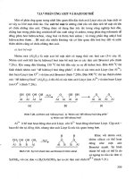
The extraction of plasmidial DNA from salmonelas is usually done by alkaline lysis, as
proposed by Birnboim and Doly (1979), with or without modification, or with acidic phenol,
as described by Wang and Rossman (1994). For small plasmids, the extraction product may
be submitted to electrophoresis in 1% agarose gel following the protocol of Akiyama et al.
(2011). The protocol for electrophoresis of mega-plasmid DNA molecules in 1% agarose gel
is described in Ponce et al. (2008).
3. Results
3.1 Salmonella in tropical aquaculture
Salmonelas are recognized worldwide as one of the main etiological agents of gastroenteritis
in humans. Despite variations in the regulation of microbiological quality of foods around
the world, the largest importers of seafoods only buy products completely free from
Salmonella, based on the claim that salmonelas are not part of the indigenous microbiota of
aquatic environments and that, therefore, the presence of salmonelas in aquatic organisms is
associated with poor sanitation and inadequate hygiene practices (Dalsgaard, 1998).
Several studies published in the 1990s reported Salmonella in shrimp farming environments
in tropical countries. Reilly and Twiddy (1992) found Salmonella in 16% of their shrimp
samples and 22.1% of their pond water and sediment samples collected on farms in
Southeast Asia. Weltevreden was the most abundant Salmonella serovar identified, followed
by Anatum, Wandsworth and Potsdam. According to the authors, the incidence of
Salmonella was higher in ponds located near urban areas and, not surprisingly, the bacterial
load increased during the rainy season. Bhaskar et al. (1995) detected Salmonella in 37.5%,
28.6% and 37.4% of shrimp, sediment and water samples, respectively, collected from semi-
intensive grow-out ponds in India.
In contrast, despite detecting high indices of thermotolerant and total coliforms, Dalsgaard
et al. (1995) found no Salmonella in water, sediment and shrimp samples from sixteen
different penaeid shrimp farms in Thailand.
Hatha and Rao (1998) reported only one Salmonella-positive sample out of 1,264 raw shrimp.
They believed the presence of the bacteria was due to pond contamination from different
sources, including the use of untreated fertilizer of animal origin. Likewise, Hatha et al. (2003)
found the incidence of Salmonella to be low in shrimp farm products exported by India.
Koonse et al. (2005) investigated the prevalence of Salmonella in six major shrimp-producing
countries in Southeast Asia (n=2), Central Asia (n=1), Central America (n=1), North America
Salmonella – A Diversified Superbug
198
(n=1) and the Pacific (n=1). In four of these countries, Salmonella was detected in 1.6% of
shrimp samples, and two serovars were identified (Paratyphi B var. Java and Weltevreden
Z6). The authors highlighted the need to control or eliminate potential sources of fecal
matter polluting the water bodies adjacent to the grow-out ponds.
In Brazil, the microbiological quality of shrimp (Litopenaeus vannamei) farmed in Ceará was
evaluated by Parente et al. (2011) and Carvalho et al. (2009), both of whom detected
Salmonella in shrimp and water samples (Table 1). The authors associated the presence of
salmonelas with discharge of fecal matter into the respective estuaries where the farms are
located. The detection of Salmonella in estuaries in Ceará is not an isolated finding. Farias et
al. (2010) found salmonelas in samples of the bivalve Tagelus plebeius collected in the estuary
of the Ceará river and identified the serovars Bredeny, London and Muechen. Similar
findings were reported by Silva et al. (2003) in a study on Salmonella in the oyster Crassostrea
rhizophorae obtained from natural oyster grounds in the estuary of the Cocó river, on the
outskirts of Fortaleza, Ceará.
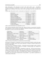
Country Sample N° Sorovars Source
Brazil
Water and
Shrimp
3 S. ser. Saintpaul e S. ser. Newport
Parente et al.
(2011)
Brazil Fish 30
S. ser. Agona, S. ser. Albany, S. ser.
Anatum, S. ser. Brandenburg, S. ser.
Bredeney, S. ser. Cerro, S. ser.
Enteretidis, S. ser. Havana, S. ser.
Infantis, S. ser. Livingstone, S.
ser.London, S. ser. Mbandaka, S. ser.
Muenchen, S. ser. Newport, S. ser.
Saintpaul, S. ser. Thompson, S. ser.
O4,5:i:-, S. ser. O4,5:-:1,7, S. O:17
Ribeiro et al.,
2010
Brazil
Water,
Sediment and
Shrimp
23
S. ser. Anatum,
S. ser. Newport, S. ser. Soahanina e S.
ser. Albany
Carvalho et al.
(2009)
Vietnam Shrimp 29
S. ser. Bovismorbificans, S. ser.
Derby, S. ser. Dessau, S. ser.
Lexington, S. ser. Schleissheim, S. ser.
Tennessee, S. ser. Thompson, S. ser.
Virchow, S. ser. Weltevreden, S. ser.
II heilbron
Ogasawara
et al. (2008)
India Shrimp 54
S. ser. Bareilly, S. ser. Braenderup, S.
ser. Brancaster, S. ser. Derby, S. ser.
Kottbus, S. ser. Lindenburg, S. ser.
Mbandaka, S. ser. Oslo, S. ser. Rissen,
S. ser. Takoradi, S. ser. Typhi, S. ser.
Typhimurium, S. ser. Weltevreden,
Salmonella VI
Kumar et al.
(2009)
*Nº: number of positive samples.
Table 1. Salmonella in tropical seafood.
Antibiotic Resistance in Salmonella: A Risk for Tropical Aquaculture
199
Thus, Shabarinath et al.
(2007), who also detected Salmonella in shrimp, concluded that since
salmonelas inhabit the intestinal tract of warm-blooded animals, their presence in rivers and
in marine/estuarine sediments exposed to fecal contamination is not surprising.
Tropical fish species may also be infected with salmonelas (Ponce et al., 2008; Heinitz et al.,
2000; Ogbondeminu, 1993); in fact, microorganisms of this genus have recently been
associated with farmed catfish (McCoy et al., 2011).
3.2 Antimicrobial susceptibility profile of Salmonella
The use of antibiotics for prophylaxis in aquaculture not only favors the selection of
resistant bacteria in the pond environment, thereby changing the natural microbiota of pond
water and sediments, but also increases the risk of transferring resistance genes to
pathogens infecting humans and terrestrial animals
(Cabello, 2006). Thus, Le and Munekage
(2005) reported high levels of drug residues (sulfamethoxazole, trimetoprim, norfloxacin
and oxolinic acid) in pond water and sediments from tiger prawn farms in Northern and
Southern Vietnam due to indiscriminate use of antibiotics.
According to Seyfried et al. (2010), autochthonous communities in aquatic environments
may serve as a reservoir for elements of antibacterial resistance. However, the contribution
of anthropic activities to the development of such reserves has not been fully clarified.
Holmström et al. (2003) reported the use, often indiscriminate, of large amounts of
antibiotics on shrimp farms in Thailand, and concluded that at a regional scale human
health and the environmental balance may be influenced by such practices. Adding to the
impact, many of the antibiotics used for prophylaxis in shrimp farming are very persistent
and toxic.
Heuer et al. (2009) presented a list of the major antibacterials used in aquaculture and
their respective routes of administration: amoxicillin (oral), ampicillin (oral),
chloramphenicol (oral, bath, injection), florfenicol (oral), erythromycin (oral, bath,
injection), streptomycin (bath), neomycin (bath), furazolidone (oral, bath), nitrofurantoin
(oral), oxolinic acid (oral), enrofloxacin (oral, bath), flumequine (oral), oxytetracycline
(oral, bath, injection), chlortetracycline (oral, bath, injection), tetracycline (oral, bath,
injection) and sulfonamides (oral).
Current aquaculture practices can potentially impact human health in variable, far-
reaching and geographically specific ways. On the other hand, the increasing flow of
aquaculture products traded on the global market exposes consumers to contaminants,
some of which from production areas (Sapkota et al., 2008).
Antibacterial susceptibility in microorganisms associated with aquaculture livestock is an
increasingly frequent topic in the specialized literature (Molina-Aja et al., 2002; Peirano et
al., 2006; Akinbowale et al., 2006; Costa et al., 2008; Newaja-Fyzul et al., 2008; Dang et al.,
2009; Del Cerro et al., 2010; Fernández-Alarcón et al., 2010; Patra et al., 2010; Vieira et al.,
2010; Tamminem et al., 2011; Laganà et al., 2011; Millanao et al., 2011; Rebouças et al., 2011;
Dang et al., 2011).
In this respect, salmonelas are one of the most extensively investigated groups of intestinal
bacteria. Thus, in China salmonelas isolated from fish ponds were resistant to ampicillin
Salmonella – A Diversified Superbug
200
(20%), erythromycin (100%), cotrimoxazole (20%), gentamicin (20%), nalidixic acid (40%),
penicillin (100%), streptomycin (20%), sulfanomides (40%), tetracycline (40%) and
trimethoprim (20%) (Broughton and Walker, 2009).
Ubeyratne et al. (2008) detected Salmonella resistant to erythromycin, amoxicillin and
sulfonamides in shrimp (Penaeus monodon) farmed in Sri Lanka. Likewise, Ogasawara et
al. (2008) found salmonelas resistant to oxytetracycline and chloramphenicol in
Vietnamese shrimp samples but concluded ARI values were not as high as in neighboring
or developing countries.
Low ARI values were also reported by Boinapally and Jiang (2007) who in a single sample of
shrimp imported to the US detected Salmonella resistant to ampicillin, ceftriaxone,
gentamicin, streptomycin and trimethoprim. This is in accordance with published findings
for shrimp in tropical regions, where the major exporters of farmed shrimp are located.
Zhao et al. (2003) evaluated the profile of antibacterial resistance in salmonelas isolated from
seafood from different countries and found that most of the resistant bacteria came from
Southeast Asia. The authors believe the use of antibiotics in aquaculture, especially in
Southeast Asia, favors the selection of resistant Salmonella strains which may find their way
into the US market of imported foods.
In Brazil, Ribeiro et al. (2010) reported an antibacterial resistance index of 15.1% among
salmonelas isolated from an aquaculture system. The Salmonella serovars Mbandaka (n=1)
and Agona (n=2) were resistant to tetracycline, Albany (n=1) was resistant to
sulfamethoxazole-trimethoprim, and London (n=2) was resistant to chloramphenicol. In
addition, Carvalho et al. (2009) collected samples from three penaeid shrimp farms in Ceará
(Northeastern Brazil) and found Salmonella serovars Newport and Anatum to be resistant to
tetracycline and nalidixic acid. Water and sediment samples collected in the vicinity of the
three farms contained the Salmonella serovars Newport, Soahanina, Albany and Anatum,
which were likewise resistant to tetracycline and nalidixic acid, suggesting the ponds were
contaminated by water drawn from the estuaries.
Bacterial resistance in Salmonella may be of either chromosomal or plasmidial nature (Frech
e Schwarz, 1999; Mirza et al., 2000; Govender et al., 2009; Tamang et al., 2011; Glenn et al.,
2011). In bacteria, the acquisition and diffusion of resistance genes may be influenced by
exchanges of DNA mediated by conjugative plasmids and by the integration of resistance
genes into specialized genetic elements (Carattoli et al., 2003).
Evidence of plasmidial mediation of antibacterial resistance in Salmonella has been
available since the 1970s and 1980s (Anderson e Threlfall, 1974; Frost et al., 1982). Thus,
Anderson et al. (1977) detected three types of resistance plasmids in Salmonella strains
from different countries. According to the authors, plasmids of the F
Ime
type confer
resistance to penicillin, ampicillin and streptomycin, whereas, for example, resistance to
furazolidone in all Salmonella isolates from Israel was considered to be chromosomal.
Mohan et al. (1995) determined the plasmid profile of Salmonella strains isolated from
different regions in India and found a large diversity of small plasmids (2.7 to 8.3 kb) in
strains resistant to ampicillin, chloramphenicol, kanamycin, streptomycin,
sulphamethoxazole, tetracycline and trimethoprim.
Antibiotic Resistance in Salmonella: A Risk for Tropical Aquaculture
201
In one study, salmonelas isolated from food animals were found to carry CMY-2, a plasmid-
mediated AmpC-like β-lactamase (Winokur et al., 2001). Doublet et al. (2004) found florR (a
florfenicol resistance gene) and bla
CMY-2
plasmids to be responsible for resistance to wide-
spectrum cephalosporines in salmonelas isolated from clinical samples, animals and foods
in the US. The authors added that the use of phenicols in animal farming environments may
place a selective pressure on organisms and favor the dissemination of bla
CMY-2
plasmids. In
addition, florR is known to confer cross-resistance to chloramphenicol.
Kumar et al. (2010) found evidence that tropical seafood can serve as vehicle for resistant
salmonela strains, some of which resistant to as many as four antibiotics (sulfamethizole,
carbenicillin, oxytetracycline and nalidixic acid). The authors also identified low-molecular-
weight plasmids in the Salmonella serovars Braenderup, Lindenburg and Mbandaka.
Six isolates of Salmonella serovar Saintpaul from samples of shrimp and fish from India,
Vietnam and Saudi Arabia presented one or more resistance plasmids of varying size (2.9 to
86 kb). One of these carried a Incl1 plasmid (Akiyama et al., 2011).
As discussed above, the indiscriminate use of antibiotics in aquaculture is one of the major
causes of the emergence of resistant bacteria in the environment. Several of the mechanisms
of resistance in Salmonella have been investigated, especially with regard to beta-lactams
(Alcaine et al., 2007) and quinolones (Piddock et al., 1998; Piddock, 2002)―two families of
antibiotics widely used in aquaculture.
4. Conclusion
The growing incidence of Salmonella in tropical aquaculture environments is a worldwide
concern which may have local impacts (in the culture area) or global impacts (considering
the dynamics of the international seafood market). Human health and environmental
balance are further threatened by the emergence of salmonelas resistant to antibiotics
employed in farming, in some cases mediated by mobile genetic elements. The elimination
of sources of fecal pollution from tropical areas used for aquaculture seems to be the main
strategy for minimizing the risk of transference of salmonelas to foods destined for human
consumption. As a final consideration, studies should be encouraged on the presence,
antibacterial susceptibility and mechanisms of resistance in salmonelas occurring in tropical
areas destined for culture of fish, crustaceans and mollusks.
5. References
Akinbowale OL, Peng H, Barton MD. Antimicrobial resistance in bacteria isolated from
aquaculture sources in Australia. Journal of Applied Microbiology, v. 100, p. 1103-
1113, 2006.
Akiyama T, Khan AA, Cheng CM, Stefanova R. Molecular characterization of Salmonella
enteric serovar Saintpaul isolated from imported seafood, pepper, environmental
and clinical samples. Food Microbiology, v. 28, p. 1124-1128, 2011.
Alcaine SD, Warnick LD, Martin W. Antimicrobial Resistance in Nontyphoidal Salmonella.
Journal of Food Protection, v. 70, n. 3, p. 780-790, 2007.
Anderson ES, Threlfall EJ, Carr JM, Mcconnell MM, Smith HR. Clonal distribution of
resistance plasmid-carrying Salmonella typhimurium, mainly in the Middle East.
Journal of Hygiene, v. 79, p. 425-448, 1977.
Salmonella – A Diversified Superbug
202
Anderson ES, Threlfall EJ. The characterization of plasmids in the enterobacteria. Journal of
Hygiene, v. 72, p. 471-87, 1974.
Andrews WH, Hammack T. Salmonella. In: Bacteriological Analytical Manual. U.S. Food and
Drug Administration. 2011. Available in:
Research/LaboratoryMethods/BacteriologicalAnalyticalManualBAM/default.htm.
Angulo FJ, Griffin PM. Changes in antimicrobial resistance in Salmonella enterica serovar
Typhimurium. Emerging Infectious Diseases, v. 6, n. 4, p. 436-437, 2000.
Asai Y, Kaneko M, Ohtsuka K, Morita Y, Kaneko S, Noda H, Furukawa I, Takatori K, Hara-
kudo Y. Samonella prevalence in seafood imported into Japan. Journal of Food
Protection, v. 71, n. 7, p. 1460-1464, 2008.
Bhaskar N, Setty TMR, Reddy GVS, Manoj YB, Anantha CS, Raghunath BS, Antony JM.
Incidence of Salmonella in cultured shrimp Penaeus monodon. Aquaculture, v. 138, p.
257-266, 1995.
Bhaskar N, Setty TMR, Mondal S, Joseph MA, Raju CV, Raghunath BS, Anantha CS.
Prevalence of bacteria of public health significance in the cultured shrimp Penaeus
monodon. Food Microbiology, v. 15, p. 511-519, 1998.
Birnboim HC, Doly J. A rapid alkaline extraction procedure for screening recombinant
plasmid DNA. Nucleic Acids Research, v. 7, n. 6, p. 1513-1523, 1979.
Boinapally K, Jiang X. Comparing antibiotic resistance in commensal and pathogenic
bacteria isolated from wild-caught South Carolina shrimps vs. farm-raised
imported shrimps. Canadian Journal of Microbiology, v. 53, n. 7, p. 919-924, 2007.
Bremer PJ, Fletcher GC, Osborne C. Salmonella in seafood. New Zealand Institute for Crop &
Food Research Limited, 2003.
Broughton EI, Walker DG. Prevalence of antibiotic-resistant Salmonella in fish in
Guangdong, China. Foodborne pathogens and disease, v. 6, n. 4, p. 519-521, 2009.
Cabello FC. Heavy use of prophylactic antibiotics in aquaculture: a growing problem for
human and animal health and for the environment. Environmental Microbiology,
v. 8, p. 1137-1144, 2006.
Carattoli A. Plasmid-mediated antimicrobial resistance in Salmonella enterica. Current Issues
in Molecular Biology, v. 5, p. 113-122, 2003.
Carvalho FCT, Barreto NSE, Reis CMF, Hofer E, Vieira RHSF. Susceptibilidade
antimicrobiana de Salmonella spp. Isoladas de fazendas de carciniculturas no
Estado do Ceará. Revista Ciência Agronômica, v. 40, n. 4, p. 549-556, 2009.
CLSI. Clinical and Laboratory Standards Institute. Performance Standards for Antimicrobial
Susceptibility Testing; Twentieth Informational Supplement: Supplement M100-
S19, Wayne, PA, USA, 2009.
Costa RA, Vieira GHF, Silva GC, Vieira RHSF, Sampaio SS. Susceptibilidade "in vitro" a
antimicrobianos de estirpes de Vibrio spp isoladas de camarões (Litopenaeus
vannamei) e de água de criação destes animais provenientes de uma fazenda de
camarões no Ceará - Nota prévia. Brazilian Journal of Veterinary Research and
Animal Science, v. 45, n. 6, p. 458-462, 2008.
Dang H, Zhao J, Song L, Chen M, Chang Y. Molecular characterizations of chloramphenicol-
and oxytetracycline-resistant bacteria and resistance genes in mariculture waters of
China. Marine Pollution Bulletin, v. 58, n. 7, p. 987-994, 2009.
Dang ST, Petersen A, Van Truong D, Chu HT, Dalsgaard A. Impact of medicated feed on the
development of antimicrobial resistance in bacteria at integrated pig-fish farms in
Vietnam. Applied and Environmental Microbiology, v. 77, n. 13, p. 4494-4498, 2011.
Dalsgaard A. The occurrence of human pathogenic Vibrio s
pp. and Salmonella in aquaculture.
International Journal of Food Science and Technology, v. 33, p. 127-138, 1998.
Antibiotic Resistance in Salmonella: A Risk for Tropical Aquaculture
203
Dalsgaard A, Huss HH, H-Kittikun A, Larsen JL. Prevalence of Vibrio cholerae and Salmonella
in a major shrimp production area in Thailand. International Journal of Food
Protection, v. 28, p. 101-113, 1995.
Del Cerro A, Márquez I, Prieto JM. Genetic diversity and antimicrobial resistance of
Flavobacterium psychrophilum isolated from cultured rainbow trout, Onchorynchus
mykiss (Walbaum), in Spain. Journal of Fish Diseases, v. 33, n. 4, p. 285-291, 2010.
Doublet B, Carattoli A, Whichard JM, White DG, Baucheron S, Chaslus Dancla E, Cloeckaert
A. Plasmid-mediated florfenicol and ceftriaxone resistance encoded by the floR and
bla
CMY-2
genes in Salmonella enterica serovars Typhimurium and Newport isolated
in the United States. FEMS Microbiology Letters, v. 233, n. 2, p. 301-305, 2004.
Fauconneau B. Health value and safety quality of aquaculture products. Revue de Médecine
Vétérinaire, v. 153, n. 5, p. 331-336, 2002.
Farias MF, Rocha-Barreira CA, Carvalho FCT, Silva CM, Reis EMF, Costa RA, Vieira RHSF.
Condições microbiológicas de Tagelus plebeius (Lightfoot 1786) (Mollusca: Bivalvia:
Solecurtidae) e da água no estuário do rio Ceará, em Fortaleza-CE. Boletim do
Instituto de Pesca, v. 36, n. 2, p. 135-142, 2010.
Fernández-Alarcón C, Miranda CD, Singer RS, López Y, Rojas R, Bello H, Domínguez M,
González-Rocha G. Detection of the floR gene in a diversity of florfenicol resistant
Gram-negative bacilli from freshwater salmon farms in Chile. Zoonoses and Public
Health, v. 57, n. 3, p.181-188, 2010.
Frech G, Schwarz S. Plasmid-encoded tetracycline resistance in Salmonella enteric subsp.
enterica serovars choleraesuis and typhimurium: identification of complete and
truncated Tn1721 elements. FEMS Microbiology Letters, v. 176, p. 97-103, 1999.
Frost JA, Rowe B, Ward LR, Threlfall EJ. Characterization of resistance plasmids and carried
phages in an epidemic clone of multi-resistant Salmonella typhimurium in India.
Journal of Hygiene, v. 88, p. 193-204, 1982.
Furushita M, Shiba T, Maeda T, Yahata M, Kaneoka A, Takahashi Y, Torii K, Hasegawa T,
Ohta M. Similarity of tetracycline resistance genes isolated from fish farm bacteria
to those from clinical isolates. Applied and Environmental Microbiology, v. 69, n. 9,
p. 5336–5342, 2003.
Glenn LM, Lindsey RL, Frank JF, Meinersmann RJ, Englen MD, Fedorka-Cray PJ, Frye JG.
Analysis of antimicrobial resistance genes detected in multidrug-resistant
Salmonella enterica serovar Typhimurium isolated from food animals. Microbial
Drug Resistance, 2011. DOI:10.1089/mdr.2010.0189.
Govender N, Smith AM, Karstaedt AS, Keddy KH. Plasmid-mediated quinolone resistance
in Salmonella from South Africa. Journal of Medical Microbiology, v. 58, p. 1393-
1394, 2009.
Hatha AAM, Rao NPB. Bacteriological quality of individually quick-frozen (IQF) raw and
cooked ready-to-eat shrimp produced from farm raised black tiger shrimp (Penaeus
monodon). Food Microbiology, v. 15, p. 177-183, 1998.
Hatha AAM, Maqbool TK, Kumar SS. Microbial quality of shrimp products of export trade
produced from aquacultured shrimp. International Journal of Food Microbiology,
v. 82, p. 213-221, 2003.
Heinitz ML, Ruble RD, Wagner DE, Tatini SR. Incidence of Salmonella in fish and seafood.
Journal of Food Protection, v. 63, n. 5, p. 579-592, 2000.
Heuer OE, Kruse H, Grave K, Collignon P, Karunasagar I, Angulo FJ. Human health
consequences of use of antimicrobial agents in aquaculture. Clinical Infectious
Diseases, v. 49, p. 1248-1253, 2009.
Salmonella – A Diversified Superbug
204
Holmström K, Gräslund S, Wahlström A, Poungshompoo S, Bengtsson BE, Kautsky N.
Antibiotic use in shrimp farming and implications for environmental impacts and
human health. International Journal of Food Science and Technology, v. 38, p. 255-
266, 2003.
Jones JG, Gardener S, Simon BM, Pickup RW. Factors affecting the measurement of
antibiotic resistance in bacteria isolated from lake water. Journal of Applied
Microbiology, v. 60, n. 5, p. 455-462, 1986.
Khan AA, Cheng CM, Khanh TV, Summage-West C, Nawaz MS, Khan SA. Characterization
of class 1 integron resistance gene cassettes in Salmonella enteric serovars Oslo and
Bareily from imported seafood. Journal of Antimicrobial and Chemotherapy, v. 58,
p. 1308-1310, 2006.
Kimura B, Kawasaki S, Fujii T, Kusunoki J, Ithoh T, Flood SJ. Evaluation of TaqMan PCR
assay for detecting Salmonella in raw meat and shrimp. Journal of food protection,
v. 62, n. 4, p. 329-335, 1999.
Koonse B, Burkhardt III W, Chirtel S, Hoskin GP. Salmonella and the sanitary quality of
aquacultured shrimp. Journal of Food Protection, v. 68, n. 12, p. 2527-2532, 2005.
Krumperman PH. Multiple antibiotic resistance indexing of Escherichia coli to indentify high-
risk sources of fecal contamination of foods. Applied and Environmental
Microbiology, v. 46, p. 165-170, 1983.
Kumar HS, Sunil R, Venugopal MN, Karunasagar I, Karunasagar I. Detection of Salmonella
spp. in tropical seafood by polymerase chain reaction. International Journal of
Food Microbiology, v. 88, p. 91-95, 2003.
Kumar R, Surendran PK, Thampuran N. Analysis of antimicrobial resistance and plasmid
profiles in Salmonella serovars associated with tropical seafood of India. Foodborne
Pathogens and Disease, v. 6, n. 5, p. 621-625, 2009.
Kumar R, Surendran PK, Thampuran N. Distribuition and genotypic characterization of
Salmonella serovars isolated from tropical seafood of Cochin, India. Journal of
Applied Microbiology, v. 106, p. 515-524, 2009.
Kumar R, Surendran PK, Thampuran N. Rapid quantification of Salmonella in seafood using
real-time PCR assay. Journal of Microbiology and Biotechnology, v. 20, n. 3, p. 569-
573, 2010.
Le TX, Munekage Y, Kato S. Antibiotic resistance in bacteria from shrimp farming in
mangrove areas. The Science of the Total Environment, v. 349, p. 96-105, 2005.
Laganà P, Caruso G, Minutoli E, Zaccone R, Santi D. Susceptibility to antibiotics of Vibrio
spp. and Photobacterium damsela ssp. piscicida strains isolated from Italian
aquaculture farms. The New Microbiologica, v. 34, n. 1, p. 53-63, 2011.
Ling ML, Goh KT, Wang GCY, Neo KS, Chua T. An outbreak of multidrug-resistant
Salmonella enterica subsp. enterica serotype Typhimurium, DT104L linked to dried
anchovy in Singapore. Epidemiology and Infection, v. 128, p. 1-5, 2002.
Malorny B, Paccassoni E, Fach P, Bunge C, Martin A, Helmuth R. Diagnostic Real-Time PCR
for Detection of Salmonella in Food. Applied and Environmental Microbiology, v.
70, n. 12, p. 7046-7052, 2004.
McCoy E, Morrison J, Cook V, Johnston J, Eblen D, Guo C. Foodborne agents associated
with the consumption of aquaculture catfish. Journal of Food Protection, v. 74, n. 3,
p. 500-516, 2011.
Millanao AB, Barrientos MH, Gómez GC, Tomova A, Buschmann A, Dölz H, Cabello FC.
Uso inadecuado y excesivo de antibióticos: Salud pública y salmonicultura em
Chile. Revista Médica de Chile, v. 139, p. 107-118, 2011.
Antibiotic Resistance in Salmonella: A Risk for Tropical Aquaculture
205
Mirza S, Kariuki S, Mamun KZ, Beeching NJ, Hart CA. Analysis of plasmid and
chromosomal DNA of multidrug-resistant Salmonella enterica Serovar Typhi from
Asia. Journal of Clinical Microbiology, v. 38, n. 4, p. 1449-1452 2000.
Molina-Aja A, García-Gasca A, Abreu-Grobois A, Bolán-Mejía C, Roque A, Gomez-Gil B.
Plasmid profiling and antibiotic resistance of Vibrio strains isolated from cultured
penaeid shrimp. FEMS Microbiology Letters, v. 213, p. 7-12, 2002.
Mohan VP, Sharma KB, Agarwal DS, Purnima G, Pillai PR. Plasmid profile and phage type of
Salmonella typhimurium strains encountered in different regions of India. Comparative
Immunology, Microbiology and Infectious Diseases, v. 18, n. 4, p. 283-290, 1995.
Newaj-Fyzul A, Mutani A, Ramsubhag A, Adesiyun A. Prevalence of bacterial pathogens
and their anti-microbial resistance in Tilapia and their pond water in Trinidad.
Zoonoses and Public Health, v. 55, n. 4, p. 206-213.
Ogasawara N, Tran TP, Ly TLK, Nguyen TT, Iwata T, Okatani AT, Watanabi M, Taniguchi
T, Hirota Y, Hayashidani H. Antimicrobial susceptibilities of Salmonella from
domestic animals, food and human in the Mekong delta, Vietnam. The Journal of
veterinary medical science, v. 70, n. 11, p. 1159-1164, 2008.
Ogbondeminu FS. The occurrence and distribution of enteric bacteria in fish and water of
tropical aquaculture ponds in Nigeria. Journal of Aquaculture in the Tropics, v. 8,
n. 1, p. 61-66. 1993.
Parente LS, Costa RA, Vieira GHF, Reis EMF, Hofer E, Fonteles AA, Vieira RHSF. Bactérias
entéricas presentes em amostras de água e camarão marinho Litopenaeus vannamei
oriundos de fazendas de cultivo no Estado do Ceará, Brasil. Brazilian Journal of
Veterinary Research and Animal Science, v. 48, n. 1, p. 46-53, 2011.
Patra S, Das TK, Gosh SCh, Sarkar D, Jana BB. Cadmium tolerance and antibiotic resistance
of Pseudomonas sp. isolated from water, sludge and fish raised in wastewater-fed
tropical ponds. Indian Journal of Experimental Biology, v. 48, n. 4, p. 383-393, 2010.
Peirano G, Agerso Y, Aarestrup FM, Reis EMF, Rodrigues DP. Occurrence of integrons and
antimicrobial resistance genes among Salmonella enterica from Brazil. Journal of
Antimicrobial Chemotherapy, v. 58, p. 305-309, 2006.
Piddock LJV, Ricci V, McLaren I, Griggs DJ. Role of mutations in the gyrA and parC genes of
nalidixic-acid-resistant Salmonella serotypes isolated from animals in the United
Kingdom. The Journal of antimicrobial chemotherapy, v. 41, n. 6, p. 635-642, 1998.
Piddock LJV. Fluoroquinolone resistance in Salmonella serovars isolated from humans and
food animals. FEMS Microbiology Reviews, v. 26, n. 1, p. 3-16, 2002.
Ponce E, Khan AA, Cheng C-M, Summage-West C, Cerniglia CE. Prevalence and
characterization of Salmonella enteric serovar Weltevreden from imported seafood.
Food Microbiology, v. 25, p. 29-35, 2008.
Raj KT, Jeyasekaran G, Shakila RJ, Thangarani AJ, Sukumar D. Multiplex polymerase chain
reaction assay for the detection of Salmonella enteric serovars in shrimp in 4 h.
Journal of Bacteriology Research, v. 3, n. 3, p. 56-62, 2011.
Rebouças RH, Sousa OV, Lima AS, Vasconcelos FR, Carvalho PB, Vieira RHSF.
Antimicrobial resistance profile of Vibrio species isolated from marine shrimp
farming environments (Litopenaeus vannamei) at Ceará, Brazil. Environmental
Research, v. 111, p. 21-24, 2011.
Reilly PJ, Twiddy DR. Salmonella and Vibrio cholerae in brackishwater cultured tropical
prawns. International Journal of Food Microbiology, v. 16, n. 4, p. 293-301, 1992.
Ribeiro RV, Reis EMF, Reis CMF, Freitas-Almeida AC, Rodrigues DP. Incidence and
antimicrobial resistance of enteropathogens isolated from an integrated
aquaculture system. Letters in Applied Microbiology, v. 51, p. 611-618, 2010.
Salmonella – A Diversified Superbug
206
Sapkota A, Sapkota AR, Kucharski M, Burke J, McKenzie S, Walker P, Lawrence R.
Aquaculture practices and potential human health risks: Current knowledge and
future priorities, v. 34, p. 1215-1226, 2008.
Seyfried EE, Newton RJ, Rubert IV KF, Pedersen JA, McMahon KD. Occurrence of
tetracycline resistance genes in aquaculture facilities with varying use of
oxytetracycline. Microbial Ecology, v. 59, p. 799-807, 2010.
Shabarinath S, Sanath Kumar H, Khushiramani R, Karunasagar I, Karunasagar I. Detection
and characterization of Salmonella associated with tropical seafood. International
Journal of Food Microbiology, v. 114, n. 2, p. 227-33, 2007.
Silva A.I.M., Vieira R.H.S.F., Menezes F.G.R., Lima L.N.G.C., Nascimento S.M.M., Carvalho
F.C.T. Bactérias fecais em ostras, Crassostrea rhizophorae. Arquivos de Ciências do
Mar, v. 36, p. 63-66, 2003.
Tamang M.D., Nam H.M., Kim T.S., Jang G.C., Jung S.C., Lim S.K. Emergence of extended-
spectrum {beta}-lactamase (CTX-M-15 and CTX-M-14) - producing nontyphoid
Salmonella with reduced susceptibility to ciprofloxacin among food animals and
humans in Korea. Journal of Clinical Microbiology, v. 49, n. 7, p. 2671-2675, 2011.
Tamminen M., Karkman A., Lõhmus A., Muziasari W.I., Takasu H., Wada S., Suzuki S., Virta
M. Tetracycline resistance genes persist at aquaculture farms in the absence of
selection pressure. Environmental Science & Technology, v. 45, n. 2, p. 386-391, 2010.
Upadhyay B.P., Utrarachkij F., Thongshoob J., Mahakunkijcharoen Y., Wongchinda N.,
Suthienkul O., Khusmith S. Detection of Salmonella invA gene in shrimp
enrichment culture by polymerase chain reaction. The Southeast Asian Journal of
Tropical Medicine and Public Health, v. 41, n. 2, p. 426-435, 2010.
Ubeyratne K.H., Hildebrandt G., Kleer J., Khattiya R., Padungtod P. Microbiological quality
of marketed Penaeus Monodon shrimps in north western province, Sri Lanka.
Proceedings, The 15
th
Congress of FAVA. OIE Joint Symposium on Emerging
Diseases Bangkok, Thailand, p. P63-P65, 2008.
Vieira R.H.S.F., Carvalho E.M.R., Carvalho F.C.T, Silva C.M., Sousa O.V., Rodrigues D.P.
Antimicrobial susceptibility of Escherichia coli isolated from shrimp (Litopenaeus
vannamei) and pond environment in northeastern Brazil. Journal of Environmental
Science and Health. Part. B, Pesticides, Food Contaminants, and Agricultural
Wastes, v. 45, n. 3, p. 198-203, 2010.
Velge P., Cloeckaert A., Barrow P. Emergence of Salmonella enterica serotype Enteritidis and
multiple antibiotic resistance in other major serotypes. Veterinary Research, v. 36,
p. 267-288, 2005.
Wang Z., Rossman T.G Large-scale supercoiled plasmid preparation by acidic phenol
extraction. Biotechniques, v. 16, n. 3, p. 460-463, 1994.
Winokur P.L., Vonstein DL, Hoffman L.J., Uhlenhopp E.K., Doern G.V. Evidence for
Transfer of CMY-2 AmpC β-Lactamase Plasmids between Escherichia coli and
Salmonella isolates from food animals and humans. Antimicrobial Agents and
Chemotherapy, v. 45, n. 10, p. 2716-2722, 2001.
Zhao S., Datta A.R., Ayers S., Friedman S., Walker R.D., White D.G. Antimicrobial-resistant
Salmonella serovars isolated from imported foods. The International Journal of
Food Microbiology, v. 84, p. 87–92, 2003.
Part 3
Genetics
12
Reticulate Evolution Among the Group I
Salmonellae: An Ongoing Role for
Horizontal Gene Transfer
Eric W. Brown, Rebecca L. Bell, Marc W. Allard, Narjol Gonzalez-Escalona,
Andrei Perlloni, Joseph E. LeClerc and Thomas A. Cebula
Center for Food Safety and Applied Nutrition
Food and Drug Administration, College Park, MD
USA
1. Introduction
Salmonella enterica is responsible for 1.4 million cases of foodborne salmonellosis in the
United States annually making it the number one causative agent of bacterial foodborne
illnesses (CDC, 2007). Infection can occur after eating undercooked meat, poultry and eggs
that have been contaminated with Salmonella (CDC, 2007). In recent years several outbreaks
have occurred in the United States that were associated with Salmonella contamination of
produce, the most recent being a S. enterica Saintpaul outbreak associated with tomatoes,
jalapeño and serrano peppers that sickened over 1400 individuals (CDC, 2008). The
movement of several serovars of Salmonella into previously naïve niches (i.e., produce-
growing environs) suggests that the pathogen is readily adapting to new environments. An
understanding of the reticulate evolutionary mechanisms that underpin the acquisition and
composition of the requisite genetic and phenotypic features of Salmonella is essential to
more accurate risk assessment of this pathogen (Hohmann, 2001).
It is now widely accepted that horizontal gene transfer (HGT) has driven the emergence of
more aggressive and virulent strains of Salmonella in the environment, on the farm, and in
the food supply. Such assault by various salmonellae has fueled the in-depth examination of
specific genotypes and conditions that permit reticulate evolutionary change and the rise of
deleterious phenotypes (LeClerc et el., 1996; 1998; 1999; Cebula and LeClerc, 1997). The
hypermutable phenotype represents one scheme by which reticulate evolution of the
bacterial chromosome may occur (Trobner and Piechoki, 1984; Haber et al., 1988; Haber and
Walker, 1991; LeClerc et al., 1996; Matic et al., 1997; Radman et al., 1999; Cebula and LeClerc;
2000; Funchain et al., 2000). Methyl-directed mismatch repair (MMR) defects, leading to a
mutator or hypermutable phenotype, are found in more than 1% of the isolates within
naturally-occurring populations of Salmonella enterica (LeClerc et al., 1996) and at even
greater frequencies in the food supply where oxidative and other anti-microbial stressors are
applied (Cebula et al., 2001). Up to 73% of the MMR defects found in feral settings are due to
lesions within the mutS gene, resulting in increased nucleotide substitution rates, enhanced
DNA transposition, and, perhaps most importantly, a relaxation of the internal barriers that
Salmonella – A Diversified Superbug
210
normally restrict homeologous recombination following HGT of foreign DNA (Cebula and
LeClerc, 1997; Radman et al., 1999).
This latter role, as a major sentinel for recombination, led to a substantial focus on the
genetics and evolution of the mutS gene and its adjacent sequences located at 63 min on the
Salmonella chromosome (Brown et al, 2002; 2003; Kotewicz et al., 2003; 2003). Phylogenetic
analyses of mutS alleles from strains of the SAR (Salmonella reference) collections (i.e., SARA,
SARB, and SARC)―largely taken to represent the extent of genetic variability within the
species (Boyd et al., 1993; 1996; Beltran et al., 1991)―have revealed striking levels of
phylogenetic discordance between trees derived from mutS alleles and whole-
chromosome trees of the same strains based on MLEE (multilocus enzyme
electrophoresis) analysis (Brown et al., 2002, 2003). These differences were interpreted as
numerous examples of HGT among mutS alleles in Salmonella. Similar observations have
been made among sequences abutting the mutS gene in Salmonella, E. coli, and Shigella spp
(Kotewicz et al., 2002; 2003; Brown et al., 2001b). Our laboratory showed previously that
the 61.5 min mutS-rpoS region retains a novel and highly polymorphic 2.9 kb sequence in
the genome of all E. coli O157:H7 strains, Shigella dysenteriae type 1, and several other E.
coli strains (LeClerc et al., 1999) but not in Salmonella enterica (Kotewicz et al., 2003). This
highly polymorphic stretch of DNA (previously coined the mutS-rpoS “unusual region”) is
varied in its distribution among enteric bacterial lineages and is absent in others entirely
(Kotewicz et al., 2003). Sequence analysis of the region revealed an IS1 insertion element
in place of the prpB gene in S. dysenteriae type 1 suggesting the existence of a
recombinational crossover in the mutS-rpoS region for this strain (LeClerc et al., 1999).
Evidence for additional crossovers in the same region were also obtained for other E. coli
strains (Brown et al., 2001b). These findings support the notion that HGT helped forge
current relationships among Salmonella and other enteric pathogens in this region and
throughout numerous other locales in the Salmonella chromosome.
Indeed, as evidenced from global efforts involving whole-genome sequencing, microarray,
and multi-locus sequence typing, the substantial impact that HGT has played in structuring
the chromosome of Salmonella enterica is now indisputable (Porwollik and McClelland, 2003;
Fricke et al., 2011; Kelly et al., 2009; Hall, 2010). Previous estimates indicate that at least one-
quarter of the Salmonella genome may have been forged through HGT and reticulate
evolutionary events (Porwollik and McClelland, 2003), although this number seems
conservative from current views. In addition to the 61.5 min region surrounding mutS, HGT
has played a key role in structuring many other regions of the Salmonella chromosome.
Notably, SPI el
ements (Salmonella pathogenicity islands) have likely been acquired through
HGT (Groisman and Ochman, 2000; Ochman et al., 2000; Hacker and Kaper, 2000; Baumler
et al., 1997). For example, the SPI-1 pathogenicity island, comprising the genes encoding a
type III secretion system, was probably acquired early in Salmonella evolution (Kingsley and
Baumler, 2000; Li et al., 1995), yet several inv–spa alleles seem to have converged
horizontally more recently between S. enterica groups IV and VII (Boyd et al., 1997; Brown et
al., 2002). Additionally, type 1 pilin genes that encode fimbrial adhesins retain unusually
low GC contents and aberrant DNA sequence phylogenies relative to other fim genes (Boyd
and Hartl, 1999). Other studies focusing on numerous housekeeping gene loci have reported
evolutionary histories for these genes that are strikingly decoupled from S. enterica strain
history (Nelson and Selander, 1994; Thampapillae et al., 1994; Brown et al., 2002; Boyd et al.,
Reticulate Evolution Among the Group I
Salmonellae: An Ongoing Role for Horizontal Gene Transfer
211
Christensen and Olsen, 1998; Groisman et al., 1992; Li et al., 1994; Liu and Sanderson, 1996;
Nelson and Selander, 1994; Nelson et al., 1992; 1997).
The now incontrovertible connection between horizontal transfer and MMR gene evolution
has led to the thesis that genetic exchange of mutS alleles could simultaneously quiet the
mutator phenotype while rescuing adaptive changes from the population (LeClerc et al.,
1996; Denamur et al., 2000). Consistent with this hypothesis, the mutS gene is evolutionarily
scrambled by HGT in subspecies I Salmonella enterica. Our laboratories documented the
prevalence of horizontal gene transfer (HGT) among strains of Salmonella enterica (Brown et
al., 2002; 2003). In comparing across and within subspecies of Salmonella, a recombination
gradient was noted wherein the incidence of HGT was inversely correlated with the genetic
diversity separating individual strains. It appears that a genetic threshold exists that
tolerates free exchange of sequences within a framework delimited by sequence variation
and niche diversity of individual strains. We demonstrated this through identification of
intragenic (patch-like) recombination as the primary outcome across disparate Salmonella
subspecies and assortative (whole-allele) recombination which caused extensive
reassortment of alleles among more genetically homogeneous populations of group I
Salmonella pathogens, all sharing a common niche restricted to warm-blooded mammals.
A torrent of scientific information has accrued over the past decade to support the important
role of HGT in the genetic and evolutionary diversification of S. enterica subspecies,
serovars, and individual pathogenic clones (McQuiston et al., 2008; Octavia and Lan, 2006;
Lan et al., 2009; Fricke et al., 2011). Our understanding in reconstructing the horizontal
acquisitions of important features including those involved in virulence, drug resistance,
and other adaptations that foster an enhanced fitness for Salmonella persistence in foods,
animals, and people is expanding at a pace which we could not have foreseen even a decade
ago (Sukhnanand et al., 2005). It is important to recall however that reticulate evolutionary
pressures do not subside once selectively advantageous traits are gained. Rather, horizontal
exchange likely continues to dapple the evolutionary landscape between even the most
closely related salmonellae (Brown et al., 2003). Here, we provide results of several
previously unreported phylogenetic studies that evidence (i) the continued role of HGT in
the intra-operon shuffling of SPI-1 alleles among subspecies I S. enterica strains; (ii) the often
under-appreciated role for HGT and recombination in the homogenization of allele
structure in a closely related population of S. enterica; and (iii) the panmictic and reticulate
nature of restriction-modification (R-M) genes among group I salmonellae. This last finding,
noting free exchange of R-M (i.e., hsd) alleles, provides phylogenetic evidence of the
compatibility of S. enterica subspecies I R-M complexes, likely accounting for the
documented successful HGT of entire gene sequences among closely (e.g., intra-subspecies)
related strains as DNA exchange between strains that shared or recently shared common R-
M alleles would not be subject to substantial restriction (Sharp et al., 1992).
2. Reticulate evolution in SPI-1 of Salmonella enterica subspecies I
Salmonella pathogenicity island 1 (SPI-1) specifies a type III secretion system essential for
host cell invasion and macrophage apoptosis (Galan and Curtiss, 1989; Galan and
Collmer, 1999). SPI-1 comprises a cluster of virulence genes (e.g., the inv/spa gene cluster)
that encode, in part, the “needle complex”, a key delivery component for transporting
virulence associated effector molecules into the host cell (Galan and Collmer, 1999). The
Salmonella – A Diversified Superbug
212
disparate phylogenetic distribution, lack of chromosomal synteny, and diverse base
compositions of SPI-1 and its homologues indicate that these sequences were obtained
independently across enteric species of bacteria. It is presumed that SPI-1 was present in
the last common ancestor of all Salmonella lineages. Horizontal acquisition of the inv/spa
gene cluster, however, is thought to have been a pivotal event for the emergence of
Salmonella as a pathogenic species (Boyd et al., 1997; Groisman and Ochman, 2000). The
gene complex lies adjacent to the polymorphic mutS–rpoS region of the chromosome. We
and others previously presented phylogenetic evidence for intragenic recombination of
sequences within several SPI-1 invasion loci (Boyd et al., 1997; Brown et al., 2002),
primarily among S. enterica subspecies IV and VII. However, in order to determine the
extent to which HGT may have disrupted SPI-1 evolution across the more ecologically
and genetically homologous group I salmonellae, we examined nine SPI-1 invasion loci
from nearly half of the SARB reference collection of strains (Boyd et al., 1993), composed
exclusively of subspecies I Salmonella serovars.
2.1 SPI-1 gene evolution is decoupled from Salmonella chromosome evolution
Using a cladistic approach (Forey et al., 1992; Allard et al., 1999; Bell et al., 2011), the
nucleotide sequences from nine invasion gene sequences were subjected to phylogenetic
analysis. The resultant invasion gene phylogenies were then compared to phylogenetic
groupings from the mdh gene, a chromosomal anchor locus that is taken largely to reiterate
chromosome evolution within subspecies I (Boyd et al., 1994) and MLEE (multi-locus
enzyme electrophoresis), also applied here as a metric of strain/chromosome evolution for
the group I salmonellae (Boyd et al., 1993). As shown in Fig. 1, strains composing single
SARB mdh and MLEE lineages were, for the most part, distributed across disparate inv/spa
gene clades for all nine invasion genes tested indicating that many of these strains, although
linked tightly in chromosome evolution, retain invasion gene alleles with unrelated
evolutionary histories, presumably as a result of HGT.
Evolutionary incongruence between inv/spa genes and the Salmonella chromosome was
affirmed using the ILD (incongruence length difference) test, which evaluates the likelihood
of a common evolutionary history between genes (Farris et al., 1995; LeCointre et al., 1998;
Brown et al., 2001a). Seven of the nine invasion genes yielded significant ILD scores (p <
0.05), indicating that a hypothesis of congruence could be rejected for these strains and
further reinforcing the discordance evident in the clade comparisons. The only exceptions
were invB (p = 0.08) and spaP (p = 0.59), albeit both still retained cladistic signatures of HGT
from broken clade structures in the tree analysis.
2.2 SPI-1 gene evolution is decoupled from mutS gene evolution
The mutS gene, downstream and adjacent to SPI-1 in S. enterica, has been shuffled extensively
by HGT (Brown et al., 2003). In order to determine whether mutS may have been linked in the
recombination now evident among SPI-1 genes, cladistic comparisons were made between
mutS phylogeny and inv/spa gene phylogeny revealing substantial incongruence between
inv/spa trees and mutS
trees. Six of these comparisons are shown in the form of tanglegrams
(F
ig. 2). Again, strains composing SARB mutS clades were distributed across disparate inv/spa
gene clades for all nine invasion genes tested, and seven of nine inv/spa genes were further
Reticulate Evolution Among the Group I
Salmonellae: An Ongoing Role for Horizontal Gene Transfer
213
confirmed as discordant with mutS based on ILD testing. Taken together, these findings
indicate that inv/spa gene sequences and mutS sequences from the same strains are decoupled
in their evolution. These data suggest that reticulate evolution has repeatedly forged this
contiguous region of the Salmonella chromosome such that different strains appear to have
been affected by assortative (allelic) HGT between the two loci.
Fig. 1. Phylogenetic discordance between SPI-1invasion genes and the Salmonella
chromosome. mdh and MLEE comparisons are shown to each of nine different inv/spa genes
indicated. Identical letters denote strains from the same mdh or MLEE lineage. It is
important to note that letters are only relevant to their respective data column and do not
cross-over columns. The column to the left of the dividing line designates mdh clade
assignments for the respective S. enterica strain while the column of letters to the right of the
divider corresponds to MLEE clade assignments. The number at the base of each tree
denotes the ILD score (p-value) relative to a comparison for congruence between the
respective inv/spa gene and the mdh gene sequence alignment for the same strains. Trees
shown were rooted using S. bongori as an outgroup. Nucleotide sequence alignments were
performed using CLUSTAL X (Thompson et al., 1998). Most parsimonious trees were
generated in PAUP* v.10 (Swofford et al., 2002).
Salmonella – A Diversified Superbug
214
Fig. 2. Tanglegrams of several invasion gene and mutS revealing the phylogenetic
incongruence between inv/spa genes and the mutS, which lies adjacent to SPI-1 on the
Salmonella chromosome. Lines connect the discordant, potentially recombinagenic
(incongruent) strains. inv/spa to mutS comparisons with an ILD score of p < 0.01 were
displayed. Trees shown were again rooted using S. bongori as an outgroup taxa.
Reticulate Evolution Among the Group I
Salmonellae: An Ongoing Role for Horizontal Gene Transfer
215
2.3 Intra-island HGT within the SPI-1 region of subspecies I Salmonella strains
In order to determine the presence and extent to which HGT has shuffled individual alleles
within SPI-1 among more closely related subspecies I strains, a pairwise ILD approach was
adopted wherein congruence was scored for individual comparisons of all nine of the
inv/spa genes included in this study (Fig. 3). Several findings were noteworthy. Although no
individual invasion gene showed unanimous evolutionary discordance with its neighbors,
three inv/spa loci (invA, invB, and spaP) were incongruent (p < 0.10) with a significant
majority of other genes. invA and invB showed discordance with all other loci except spaN
and spaQ, while spaP showed discordance to all but spaM and spaQ. Conversely, with the
exception of spaQ, no inv/spa gene was congruent with every other. Thus, a hypothesis of
extensive intra-island shuffling begins to emerge with an evolutionary decoupling of
individual invasion loci one from another. Additional tree comparisons buttressed this
conclusion. Akin to the selfish operon theory (Lawrence and Roth, 1996), these data suggest
that the SPI-1 region is a chromosomal mosaic, composed of inv/spa gene sequences that
have converged within this island but with each retaining unique evolutionary paths.
Fig. 3. ILD test results for intragenic comparisons among inv/spa invasion genes. ILD tests
(Farris et al., 1995) were performed with 1000 partitions using the Partition Heterogeneity
command in PAUP* v.10 (Swofford et al., 2002). A p-value of 0.05 or less allows for a
rejection of the null hypothesis of congruence (vertical evolution) and accepts the alternative
hypothesis of incongruence which is interpreted among bacterial phylogeny as evidence for
HGT (LeCointre et al., 1998).
Salmonella – A Diversified Superbug
216
2.4 Key observations
i. The inv/spa complex of S. enterica subspecies I appears to have undergone extensive
intra-island allelic shuffling due to HGT. This suggests that the SPI-1 region is a mosaic
composed of SPI-1 gene sequences with distinct evolutionary origins.
ii. Invasion genes within this Salmonella population are not only decoupled
phylogenetically from mutS and other flanking sequences but also from the
chromosomes of group I S. enterica strains, suggesting that these genes have been re-
assorted by HGT.
iii. Much of the recombination observed here appears to be assortative transfer, a finding
that contrasts to the inv genes in S. enterica as a whole, where tree structure was
largely intact with HGT limited mostly to subspecies IV and VII (Boyd et al., 1997;
Brown et al., 2002).
iv. Allele shuffling appears to be most prominent within the subspecies I taxonomic
boundary and not across other subspecies of S. enterica. This finding is consistent with a
relaxed and compatible restriction-modification system among more closely related
Salmonella strains (Brown et al., 2003).
3. HGT homogenizes the mutS gene among ‘Typhimurium’ complex strains
Here, we present phylogenetic and genetic analyses of Salmonella reference collection A
(SARA), also known as the Typhimurium strain complex—the most homogeneous S. enteric
reference collection, consisting solely of five closely related subspecies I serovars
(Typhimurium, Paratyphi B, Muenchen, Saintpaul, and Heidelberg) (Beltran et al., 1991).
Given the evolutionary similarity shared among these pathogens and trend noted
previously that highlight the inverse relationship between Salmonella diversity and
recombination, one would expect to observe an even greater role for HGT in the population
structure of the S. enterica SARA collection of pathogens.
3.1 Cladistic evidence for horizontal exchange of mutS alleles among ‘Typhimurium’
complex strains
As was done for SPI-1 gene sequences, a phylogenetic tree was derived from 72 SARA mutS
sequences and was compared to phylogenetic trees derived from multi-locus enzyme
electrophoresis (MLEE)
and mdh (malate dehydrogenase) gene sequences for the same strains.
Phylogenies derived from horizontally exchanged sequences display evolutionary discordance
(incongruence) when compared to mdh and MLEE trees. In the tree shown, six clades of mutS
alleles were observed and compared to the distribution of four mdh and six MLEE multi-strain
containing clades (Fig. 4). Two of the four SARA mdh clades were found to be displaced into
multiple clades on the mutS tree. Two additional mdh clades were found to have converged
into a single mutS clade, suggesting that HGT may have homogenized mutS diversity of these
particular mutS lineages. Similarly, strains from five of the six MLEE lineages were displaced
into separate clades on the mutS tree. The only exception was a single clade of MLEE SARA
strains (A57, A58, A59, and A60), which was also found intact in the mutS tree except for the
inclusion of SARA strain A56. Nonetheless, numerous examples of evolutionary discordance
between the 1.1 kb mutS segment and the chromosome of the ‘Typhimurium’ complex strains
indicate that horizontal exchanges of mutS alleles have accumulated during the rather shallow
radiation of even these highly homogeneous group I pathogens. As an aside, it was
Reticulate Evolution Among the Group I
Salmonellae: An Ongoing Role for Horizontal Gene Transfer
217
noteworthy that full-length mutS alleles were horizontally transferred among SARA S. enterica
strains, lending further credence to a model for R-M compatibility among closely related S.
enterica serovars and strains.
Fig. 4. Most-parsimonious relationships of SARA mutS alleles. mutS clades are bracketed
and numbered to the right of the tree. Distributions of mutS, mdh, and MLEE clades are
presented in column form. Note that strains originating from the same clade retain a
common shape and common internal shading. Bootstrap nodal support values (Felsenstein
et al., 1985) are presented on the mutS tree as follows: ^, 76-100%; *, 51-75%; +, 26-50%; o, 1-
25%. In this case, mdh and MLEE are taken to represent the evolution of the strain in general
(Boyd et al., 1994; Beltran et al., 1991). The tree shown is rooted with two E. coli outgroups.
Salmonella – A Diversified Superbug
218
3.2 Homogenization of mutS sequence diversity among S. Typhimurium and S.
Heidelberg strains
Curiously, a single clade in the SARA mutS tree was found to comprise three distinct
Salmonella serovars. In this clade, every strain representing S. Typhimurium (n=21) and S.
Heidelberg (n=11), along with a single strain of S. Saintpaul, converged into a single
evolutionary lineage of mutS alleles. In the SARA mdh tree (Fig. 5), mdh alleles for these same
SARA serovars formed three disparate clades in the tree such that S. Typhimurium strains
clustered only with other S. Typhimurium and S. Heidelberg strains only with other S.
Heidelberg. S. Saintpaul strains formed a single lineage at the tip of the tree with strains of
S. Muenchen and a single S. Paratyphi B. It should be noted that these distinct clades
retained substantial statistical support with bootstrap values around 90% (Felsenstein, 1985).
Thus, phylogenetic comparison of mutS and mdh sequences supported the notion that these
serovars have converged into a single mutS clade, possibly as a result of the repeated HGT
of only one or a few preferred mutS alleles.
Fig. 5. Phylogenetic tree revealing the most-parsimonious relationships of SARA mdh alleles.
mdh clades are bracketed and lettered while SARA serovars are labeled to the right of the tree.
For sample sizes greater than one, multiple strains of the same serovar are depicted as a cone
on the tree terminal nodes. Note that strains originating from the same clade are designated by
a common bracket and letter. Bootstrap nodal support values are presented on the mdh tree as
follows: ^, 76-100%; *, 51-75%; +, 26-50%; o, 1-25%. Note the bifurcations between specific
clusters in the tree, signaling sequence diversity among distinct serovars using the mdh gene.
Reticulate Evolution Among the Group I
Salmonellae: An Ongoing Role for Horizontal Gene Transfer
219
In order to further investigate the genetic structure of this converged clade, we examined mutS
sequence homogeneity across the strains composing this lineage as well as the remaining mutS
alleles of the SARA collection (Fig. 6). Evaluation of polymorphic positions in the mutS
alignment revealed several findings consistent with homogeneous clade structure surrounding
these serovars. First, five substitutions were observed across the entire 1,115 bp sequence for
all 33 strains that define this mutS clade (#2). Second, with the exception of the polymorphism
at position 913 in SARA strains 12 and 13, no clade #2 substitution was retained by more than
one strain. Thus, none of the substitutions present within this clade partitioned any member
serovar from another. The near structural uniformity of this clade at the nucleotide level
further suggests that HGT has homogenized mutS alleles among these particular serovars.
This is consistent with the thesis of Dykhuizen and Green (1991) who reminded that
recombination can not only diversify the genome but can also homogenize it as well.
Fig. 6. mutS nucleotide sequence homogeneity among S. enterica serovars Typhimurium,
Heidelberg, and a strain of Saintpaul. Periods indicate exact nucleotide identity to the
reference sequence at the top of the alignment while listed nucleotides represent actual
substitutions. The synonymous/nonsynonymous status (blackened ovals indicate
synonymous change) of each substitution is noted below the alignment. Nucleotide sequences
were generated using a PCR-based approach and automated CE-sequencing technology.
Salmonella – A Diversified Superbug
220
3.3 Distinct roles for HGT across various taxonomic tiers of S. enterica
With the inclusion of the SARA analysis reported here, we have been able to define varying
roles for HGT across three taxonomically distinct populations of S. enterica (SARA, B, and C)
(Fig. 7). Within S. enterica as a whole, a model for HGT begins to emerge that tolerates near-
free HGT among closely-related subspecies I strains. As genetic divergence increases across
serovars, however, the extent of HGT appears to decrease. The analysis reported here
suggested two unique findings for SARA, the most genetically monomorphic population.
Fig. 7. Model for the frequency and effects of HGT among various taxonomic tiers of
Salmonella enterica. Graphic representation of the various effects of HGT on the
taxonomically distinct SARA, SARB, and SARC strain collections as well as an interspecies
comparison. The S. enterica collections are plotted relative to genetic divergence versus the
extent of HGT observed. Specific effects and trends associated with the HGT occurring at
each taxonomic level are noted below each of the Salmonella populations shown.
First, SARA revealed evidence for a substantial convergence of mutS alleles between distinct
serovars suggesting, that, recombination can have a homogenizing effect on sequence
diversity in this population. Second, despite yielding numerous examples of assortative
(allelic) exchange, SARA appears to be―at least from a phylogenetic perspective―refractory
to intragenic (mosaic) HGT within the mutS gene. Thus, the SARA and SARB groups seem
Reticulate Evolution Among the Group I
Salmonellae: An Ongoing Role for Horizontal Gene Transfer
221
to have been influenced more extensively by HGT than SARC possibly because they are not
so diverged that exchange is inhibited due to extreme niche or R-M (restriction-
modification) system variability. Moreover, it is also possible that much of the HGT among
SARA strains have gone undetected here since identical alleles would leave no phylogenetic
footprint following an exchange event.
3.4 Key observations
i. Horizontal gene transfer of mutS alleles in Salmonella appears to play a prominent role
in the evolutionary structure of the five closely-related serovars representing the SARA
(‘Typhimurium’ complex) collection, a finding consistent with extensive HGT that has
been documented among subspecies I serovars in general (Brown et al., 2003).
ii. Cladistic analysis of SARA strains revealed the first example of a substantial
convergence of mutS alleles from disparate serovars into a single clade. This suggests
that HGT is homogenizing allele diversity among certain Salmonella strains and
serovars―an observation reminiscent of allele homogenization observed for the E. coli
polA gene (Patel and Loeb, 2000).
iii. Among closely related ‘Typhimurium’ complex strains, mutS alleles appear to have
shuffled largely as single units rather than in intragenic segments. One explanation for
this might be a more recent evolutionary divergence of the five serovars composing the
highly homogeneous ‘Typhimurium’ strain complex. Alternatively, recombination of
highly homologous mosaic segments of the mutS gene would do little to obscure
phylogeny and likely go undetected in these analyses.
iv. Retrospective comparison of SARA HGT patterns with that of SARB and SARC strains
yields a gradated model for HGT whereby different taxonomic tiers of Salmonella are
subject to different HGT effects. The differences appear coupled to the extent of genetic
diversity that defines these three different “tiers” of Salmonella population structure.
4. HGT among restriction-modification (R-M) genes of subspecies I
salmonellae
The restriction and modification (R-M) system is a defense mechanism developed by
bacteria to protect the bacterial genome from invasion by foreign DNA (Bullas et al., 1980).
Foreign sequences entering the cell are cleaved by restriction enzyme(s), while the bacterial
DNA itself is modified by methylase(s), thus providing protection from its own restriction
enzyme (Murray, 2000). R-M systems are composed of genes that encode a specific
restriction endonuclease and modification methylase. There are several types of R-M
systems, namely type I (e.g., EcoKI), type II (e.g., EcoRI), and type III (e.g., Sty LTI) (Barcus et
al., 1995). Types of R-M systems are classified on the basis of their composition and cofactor
requirements, the nature of the target sequence, and the site of DNA cleavage with respect
to the target sequence (Murray, 2000; Naderer et al., 2002).
Compatibility of R-M systems among strains was proposed as one explanation to account
for contrasting recombination rates (Brown et al., 2003). In this model, compatible R-M
complexes would permit the successful transfer of larger gene segments among closely
related Salmonella pathogens; crosses between strains with identical R-M systems would not
be subject to restriction (Sharp et al., 1992). A gradation in the size limits of DNA segments
exchanged would depend on the polymorphic character of R-M systems in natural strains.