Salmonella A Diversified Superbug Part 6 pot
Bạn đang xem bản rút gọn của tài liệu. Xem và tải ngay bản đầy đủ của tài liệu tại đây (1.47 MB, 30 trang )
Salmonella – A Diversified Superbug
138
Weckesser S, Engel K, Simon-Haarhaus B, Wittmer A, Pelz K, Schempp CM (2007).
Screening of plant extracts for antimicrobial activity against bacteria and yeasts
with dermatological relevance. Phytomedicine, 14: 508-516.
Wedel, S.D., Bender, J.B., Leano, F.T., Boxrud, D.J., Hedberg, C. & Smith, K. (2005)
Antimicrobial-drug susceptibility of human and animal Salmonella
typhimurium, Minnesota, 1997–2003. Emerging Infection Diseases, Vol. 11, pp.
1899–1906, ISSN 1080-0059.
Yu, H.; Zhang, L.; Li, L.; Zheng, C.; Guo, L.; Li, W.; Sun, P. & Qin, L. (2010), Recent
developments and future prospects of antimicrobial metabolites produced by
endophytes. Microbiological Research, Vol. 165, No. 6 (August 2010), pp. 437-449,
ISSN 0944-5013.
Zhang, Y.; Mu, J.; Feng, Y.; Kang, Y.; Zhang, J.; Gu, P.; Wang, Y.; Ma, L.; Zhu, Y. (2009),
Broad-spectrum antimicrobial epiphytic and endophytic fungi from marine
organisms: isolation, bioassay and taxonomy. Marine drugs, Vol. 7 (April 2009),
pp.97-112, ISSN 1660-3397.
8
Nanotechnology Tools for Efficient
Antibacterial Delivery to Salmonella
Ali Nokhodchi
1,2
, Taravat Ghafourian
1
and Ghobad Mohammadi
3
1
Medway School of Pharmacy, Universities of Kent and Greenwich, Chatham
2
Drug Applied Research Center and Faculty of Pharmacy
Tabriz University of Medical Sciences, Tabriz
3
Faculty of Pharmacy, Kermanshah University of Medical Sciences, Kermanshah
1
UK
2
Iran
1. Introduction
In recent years, an increasing number of salmonellosis outbreaks have been recorded
around the world, and probably there should be more cases that were not detected or
reported (1). Many different types of Salmonella exist, some of which cause illness in both
animals and people, and some types cause illness in animals but not in people. The various
forms of Salmonella that can infect people are referred to as serotypes, which are very closely
related microorganisms that share certain structural features. Some serotypes are only
present in certain parts of the world (1). Salmonella spp are gram negative anaerobic and
intracellular bacteria. Salmonellosis, mainly due to Salmonella typhimurium, occurs more
frequently in HIV-infected patients than in healthy individuals and the frequency of
bacteraemia is much higher in such patients (2).
Despite the discovery of new antibiotics, treatment of intracellular infections often fails to
eradicate the pathogens completely. One major reason is that many antimicrobials are
difficult to transport through cell membranes and have low activity inside the cells, thereby
imposing negligible inhibitory or bactericidal effects on the intracellular bacteria (3). In
addition, antimicrobial toxicity to healthy tissues poses a significant limitation to their use
(3). Therefore, the delivery of the drug to the bacterial cells is currently a big challenge to the
clinicians. This is on top of the problems posed by the emerging Multi-Drug Resistant
species. Moreover, the reduced membrane permeability of microorganisms has been cited as
a key mechanism of resistance to antibiotics (4).
Indeed, the challenge is to design the means of carrying an antibiotic into bacterial cells.
The pioneer concept of targeted drugs was developed by Ehrlich in 1906 and defined as
the ‘magic bullet’. Since then targeted drug delivery has involved design and
development of small molecule drugs that can specifically interact with the intended
receptors in intended tissues. For example prodrugs can be designed for brain delivery of
the active drug (5). Another common example is colon delivery of prodrugs designed to
release the drug by taking advantage of the bacterial reductase enzymes in colon (6).
Salmonella – A Diversified Superbug
140
However, the drug development process is inevitably lengthy and breakthroughs are
quite scarce which has led to the ever increasing cost of discovery and development of
new drugs (7). On the other hand, nanotechnology offers a more convenient method for
targeted therapy.
Logistic targeting strategies can be employed to enable the drug to be endocytosed by
phagocytic cells and then released into the bacteria. To reach the above goal, a drug carrier is
generally needed for a drug to arrive at the target site (8). The first study employing a drug
carrier for targeted drug delivery was published approximately 40 years ago, using antibodies
as carriers of radioactivity for the specific recognition of tumor cells (9). The ideal drug carrier
ensures the timely release of the drug within the therapeutic window at the appropriate site, is
neither toxic nor immunogenic, is biodegradable or easily excreted after action, and is
preferably cheap and stable upon storage (10). Out of different types of drug carriers that have
been investigated, many are soluble macromolecular carriers or liposomes (11-15).
By searching all published work on drug carriers it can be concluded that ''the ideal drug
carrier" does not exist. The suitability of a drug carrier is determined by the disease that will
be targeted, its access to the pathological site, and the carriers’ ability to achieve appropriate
drug retention and timely drug release (16). When these types of formulations are
administered by the intravenous route, phospholipidic, polymeric or metal particles are
localized preferentially in organs with high phagocytic activity and in circulating
monocytes, ensuring their clearance (8). The ability of circulating carriers to target these cells
is highly dependent on tissue characteristics and on the carrier’s properties. The liver rather
than the spleen or bone marrow captures the submicronic particles (8). Immediately after
injection, the foreign particles are subjected to opsonization by plasma proteins. This is the
process by which bacteria are altered by opsonins so as to become more readily and more
efficiently engulfed by phagocytes. In this way, ‘classical’ or ‘conventional’ carriers are
recognized by the mononuclear phagocytic system (8).
The approaches for drug carrier to improve the drug’s antibacterial efficacy are shown in
Figure 1. In most cases, i.v. administration of the formulation is needed particularly for
passive and active targeting.
The local administration of drug/carriers will increase the residence time of antibiotics at the
site of infection (17-19). These carriers are generally investigated with the intention to treat
local infections in body parts with limited blood flow as in bone, joint, skin, and cornea.
In passive targeting after i.v. administration of carriers which tend to be taken by phagocytic
cells, drug-carrier complex will target intracellular infections. These infections are often
difficult to treat as a result of limited ability of the antimicrobial agent to penetrate into cells.
This approach makes use of the recognition of drug carriers (nanoparticles) as foreign
material in the bloodstream by the phagocytic cells of the mononuclear phagocyte system,
the cell type often infected with microorganisms (20, 21).
Regarding the other two approaches (passive targeting with long-circulation time, and
active targeting) the targeting of infectious foci is not restricted to mononuclear phagocyte
system tissues. In passive targeting a drug carrier with long duration of circulation is used
and this is an area which has extensively been investigated, whereas in active targeting
carriers specifically bind to the infectious organism or host cells involved in the
inflammatory response.
Nanotechnology Tools for Efficient Antibacterial Delivery to Salmonella
141
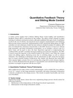
Fig. 1. Drug carrier approaches targeting bacterial infections to improve antibacterial
efficacy of drugs.
This chapter focuses mainly on the current research for increasing anti-salmonella
performance of antibiotics by means of liposomes and nanoparticle systems. Structure,
properties, advantages and disadvantages of these drug delivery systems have been
discussed. It is clear that such systems may improve the antibiotic efficacy by increasing the
drug concentration at the surrounding of the bacteria.
2. Liposomes for antisalmonellosis drug delivery
2.1 Introduction
Liposomes are composed of small vesicles of a bilayer of phospholipid, encapsulating an
aqueous space ranging from about 30 to 10000 nm in diameter (Figure 2). They are
composed of one or several lipid membranes enclosing discrete aqueous compartments.
The enclosed vesicles can encapsulate water-soluble drugs in the aqueous spaces, and
lipid soluble drugs can be incorporated into the membranes. They are used as drug
carriers in the cosmetic and pharmaceutical industry. The main routes of liposome
administration are parenteral, topical and inhalation, and, in a few occasions, possibly
other routes of administration can be used. Majority of current products are administered
parenterally (22).
Salmonella – A Diversified Superbug
142
Liposome structure was first described in 1965, and they were proposed as a drug
delivery nanoparticle platform in 1970s. In 1995, Doxil (doxorubicin liposomes) became
the first liposomal delivery system approved by the Food and Drug Administration (FDA)
to treat AIDS associated Kaposi’s sarcoma (23). Liposomal drug delivery systems can be
made of either natural or synthetic lipids. The main building blocks of some liposomal
formulations are phospholipids (22). These are natural biomacromolecules that play a
central role in human physiology as they are structural components of biological
membranes and support organisms with the energy (24). They are amphiphilic molecules,
poorly soluble in water, consisting of a hydrophilic part containing hydroxyl groups (the
polar head), a glycerol backbone and two fatty acid chains, which form the hydrophobic
part. One of the most commonly used lipids in liposome preparation is
phosphotidylcholine, which is an electrically neutral phospholipid that contains fatty acyl
chains of varying degrees of saturation and length. Cholesterol is normally incorporated
into the formulation to adjust membrane rigidity and stability (8). Liposomes can be
characterized in terms of size and lamellarity as small unilamellar vesicles (SUV), large
unilamellar vesicles (LUV) and multi lamellar vesicles (MLV). MLVs are usually
considered large vesicles and aqueous regions exist in the core and in the spaces between
their bilayers. The structure of these liposomes is shown in Figure 2.
(a) (b)
Fig. 2. Schematic structures of (a) multilamellar and (b) unilamellar liposomes (the picture
was taken from />nanotechnology/nanoencapsulation-of-bioactive-substances-part-1-nanotechnology).
The main advantages of liposomes as drug delivery systems can be in their versatile
structure that can be easily modified according to experimental needs; they can also
encapsulate hydrophilic drugs in their aqueous compartments and hydrophobic drugs in
their bilayers, while amphiphilic drugs will be partitioned between the two. Moreover,
being mainly made of phospholipid, they are non-toxic, non-immunogenic and fully
biodegradable. Methods for preparing liposomes can take into consideration parameters
such as the physicochemical characteristics of the liposomal ingredients, materials to be
contained within the liposomes, particle size, polydispersity, surface zeta potential, shelf
time, batch-to-batch reproducibility, and the possibility for large-scale production of safe
and efficient products (23).
Nanotechnology Tools for Efficient Antibacterial Delivery to Salmonella
143
2.2 Preparation of liposoms
Liposome formation happens spontaneously when phospholipids are dispersed in water.
However, in order to obtain the desired formulation with particular size and structure,
various methods such as thin film method (24), sonication (25), extrusion (26), injection
methods (27), dehydrated-rehydrated vesicles (28), reverse phase evaporation (29) and one
step method (30) have to be used.
Each technique is briefly described below, but for more details, it is recommended to refer to
the cited references. In brief, in thin film method liquids are dissolved in organic solvents
and the solvent is removed under vacuum or nitrogen stream to form a thin film on the wall
of a flask or test tube. In order to complete the formation of liposomes aqueous phase is
added to the lipid film at a temperature above the phase transition of the lipid (24).
The sonication method is usually used to reduce the particle size and lamellarity of MLVs. In
case of using the probe sonicator, the reduction in size of the liposomes can be guaranteed (25).
In order to get very homogeneous vesicles with a predetermined size, the extrusion
technique is used. MLVs are extruded under pressure through particular filter with well-
defined pore sizes from 30 nm to several micrometers. If the extrusion is repeated several
times unilamellar liposomes can be formed (26).
Very small unilamellar vesicles with a particle size of 30 nm can be prepared using the
ethanol injection method. Generally, lipids are dissolved in ethanol and injected rapidly into
the aqueous solution, under stirring. At the end, the injected ethanol has to be removed
from the system (27).
As dehydrated-rehydrated vesicles are able to hold high amounts of hydrophilic drugs
under mild conditions, therefore this method is suitable for the drugs that are losing their
activity under harsh conditions (28). Empty liposomes, usually unilamellar vesicles, are
disrupted during a freeze drying step in the presence of the drug meant to be encapsulated.
A controlled rehydration is obtained in the presence of concentrated solution of the drug.
This technique can produce large oligolamellar liposomes of a size around 400 nm to several
micrometers. It has been shown that in case of producing smaller liposomes (100-200 nm)
sucrose can be added (31).
In the reverse phase evaporation technique which is similar to thin film technique, lipids are
dissolved in organic solvent and the solvent is removed by evaporation (29). The thin film is
resuspended in diethyl ether followed by the addition of third of water and the suspension
is sonicated in a bath sonicator. The emulsion is evaporated until a gel is formed and finally
the gel is broken by the addition of water under agitation. The traces of organic solvent
should be removed by evaporation (29).
Finally, in the one-step method, lipid dispersion should be hydrated at high temperatures
under nitrogen gas stream. This method has the capability to produce liposomes in the
range of 200-500 nm (30).
2.3 Targeted delivery by liposomes
The main methods of delivery from liposome to cytoplasm include the exchange of
membrane and lipids, contact release, adsorption, fusion and endocytosis. Through these
Salmonella – A Diversified Superbug
144
processes, drugs can be released into the bacterial or eukaryotic cells. Liposomal
formulations have been used for the delivery of antitumor anthracyclines such as
doxorubicin (23) and antifungal agent amphotericin B. Targeted delivery of liposomes to
tumor cells has been explored through arsenoliposomes (32). Liposomes for antibacterial
chemotherapy are under intensive research to enhance the antibacterial activity and
improve pharmacokinetic properties. Advantages of liposomal antibiotics include improved
pharmacokinetics, decreased toxicity, enhanced activity against intracellular pathogens,
target selectivity and as a tool to overcome bacterial drug resistance (3).
Some liposomes are unique because they can be selectively absorbed by tissues rich in
reticuloendothelial cells, such as the liver, spleen and bone marrow. This can serve as a
targeting mechanism, but it also removes liposomes from the circulation rather rapidly.
Although the poor stability of liposomes, particularly the rapid uptake from the body is not
desirable, it could be useful for eradicating the infection by ‘passive targeting’ through
macrophage activation and killing or elimination of parasitic infections.
On the other hand, surface charge and phospholipid composition can affect the interactions
of liposomes with bacterial cell surface. For example it has been shown that cationic
liposome formulations are more efficient in binding to skin bacterial cells (33).
Moreover, by attaching targeting ligands such as immunoglobulines (34), antibody segments,
aptamer (35), peptides and small molecule ligands, and oligosaccharide chains (36), to the
surface of the liposomes, they can selectively bind to microorganisms or infected cells and then
release the drug payloads to kill or inhibit the growth of the microorganisms (23). The highly
specific liposomes are those containing antibodies or immunoglobulin fragments which have
affinity to specific receptors on the surface of the infected tissue cells or pathogens (3).
Biofilm surface characteristics have also been used for targeted delivery. Biofilms are
microbial aggregations that are covered in an extracellular matrix of polymeric substances.
The matrix is usually composed of complex mixture of oligomeric and polymeric molecules
such as proteins, lipids and polysaccharides which, as Microbial Associated Molecular
Patterns (MAMPs), elicit host defenses (37). Pathogens are much more difficult to control
when living in biofilms. This is partly due to the matrix preventing drug transport to the
microbial cells. Moreover, bacteria in biofilms grow slower and have reduced metabolic
activity, and therefore they are expected to be less susceptible to the antibiotics (38).
Currently a great deal of research is focused on exploring new chemotherapeutic targets in
biofilms (37). On the other hand liposomes have proven efficient in targeting and
eradication of various types of biofilms. Examples are immunoliposomes with high affinity
to various oral bacteria including Streptococcus oralis (34) and polysaccharide-coated
liposomes for the efficient delivery of metronidazol to periodontal pocket biofilm (39).
pH-sensitive liposomes offer another method for targeting and efficiently delivering the
liposomal content into cytoplasm. Such liposomes are stable at physiological pH but
undergo destabilization under acidic conditions. Therefore, they are able to promote fusion
of target plasma or endosomal membranes, the so called ‘fusogenic’ properties, at acidic pH
(40). Several mechanisms can trigger pH-sensitivity in liposomes. One of the most widely
used methods is the use of a combination of phosphatidylethanolamine (PE) or its
derivatives with compounds containing an acidic group that act as a stabilizer at neutral pH
(41). Other more recent methods include the use of novel pH-sensitive lipids, synthetic
Nanotechnology Tools for Efficient Antibacterial Delivery to Salmonella
145
fusogenic peptides/proteins (42) and association of pH-sensitive polymers with liposomes
(43). pH-sensitive liposomes have found applications in many therapeutic area including the
antibiotic delivery to intracellular infections (44).
2.4 Pharmacokinetics consideration of liposomal drug delivery
Liposomal carriers can lead to sustained release of antibiotics during drug circulation in the
body. Thus, appropriate levels of drug will be available for a longer duration in comparison
with the conventional antibiotic formulations where the outcome is a quick and short effect
(45). However, conventional liposomes are quickly opsonized after intravenous administration
and therefore they are taken up by the mononuclear phagocyte as foreign antigens. As a
consequence blood circulation time is lowered. By controlling the physicochemical properties
of the vesicles (size and charge distribution, membrane permeability, tendency for aggregation
or fusion, drug encapsulation efficiency, membrane rigidity) and therefore their interaction
with the biological environment, many different types of liposomes with the aim of obtaining
longer circulation half-lives can be developed (8).
The plasma circulation time of antibiotics can be improved by encapsulation in polyethylene
glycol-coated (pegylated) (STEALTH) liposomes. The PEG coating forms a hydration layer
that retards the reticuloendothelial system recognitions of liposomes through sterically
inhibiting hydrophobic and electrostatic interactions with plasma proteins (46). Other
methods that can confer hydrophilicity or steric repulsion are by the use of compounds
having sialic residues, or through MLVs containing phospholipids with long saturated
chains and negative surface charge (47). The increased half lives of stealth liposomes
increase their ability to leave the vascular system into some extravascular regions.
2.5 Antibiotic loaded liposomes against Salmonella spp
One of the distinguishing features of liposomes is their lipid bilayer structure, which
mimics cell membranes and can readily fuse with the cell membrane and deliver the
antibiotic contents into the cellular cytoplasm. As a result, drug delivery may be
improved to bacterial and eukaryotic cells alike. By directly fusing with bacterial
membranes, the drug payloads of liposomes can be released into the cell membranes or to
the interior of the bacteria. In terms of extracellular pathogens, improved antibiotic
delivery into the bacterial cells is of particular importance especially since it can interfere
with some of the bacterial drug-resistance mechanisms which involve low permeability of
the outer membrane or efflux systems (48).
Liposomes are particularly successful in eradicating intracellular pathogens. Examples of
these include liposomal formulations of antituberculosis agents isoniazid and rifampin
(49), and ampicillin loaded liposomes for eradication of Listeria monocytogenes (50). This
is partly due to improved drug retention in the infected tissue and the decreased toxicity
as a result of sustained release of drug from liposomes. Moreover, liposomal formulations
often have improved antibiotic pharmacokinetics with extended circulation time and
prolonged tissue retention.
Liposomal chemotherapeutics for the treatment of salmonellosis may employ some of the
conventional antibiotics with proven inhibitory or cidal activity in vitro. Bacterial gastro-
intestinal infections with Salmonella typhi may be treated with chloramphenicol. Alternatives to
Salmonella – A Diversified Superbug
146
chloramphenicol include amoxicillin, co-trimoxazole and trimethoprim (51). Recently
treatment with cephalosporins and fluoroquinolones has become popular, as several members
of these antibiotic families have been shown to be effective. The treatment of paratyphoid fever
is the same as that for typhoid (51). Salmonella food-poisoning is self-limiting and does not
require antibiotic therapy, unless the patient is severly ill or blood cultures indicate systemic
infection. In this case, third generation cephalosporins or fluoroquinolones are the most
reliable agents (51). Ceftriaxone or a first generation fluoroquinolone such as ciprofloxacin,
ofloxacin or pefloxacin but not norfloxacin have been recommended as the first choice in
typhoid and paratyphoid by The Sanford Guide to Antimicrobial Therapy (52). The improved
efficiency of liposome formulations of antibiotics has been shown in vitro and in vivo. The in
vitro infection models utilize macrophages infected with salmonella.
2.5.1 Penicillin loaded liposomes
The tissue distribution of ampicillin loaded liposomes was studied in normal noninfected
mice and showed that ampicillin concentrated mostly in the liver and spleen (53). The
Liposome formulation of ampicillin was significantly more effective than free ampicillin in
reducing mortality in acutely infected mice with Salmonella typhimurium C5. These
liposomes were quite efficient in targeting ampicillin to the spleen but were less effective in
targeting ampicillin to the liver and reducing mortality in acute salmonellosis (53).
2.5.2 Cephalosporine loaded liposomes
Third generation cephalosporines have been indicated as suitable candidates for the
treatment of Salmonella infections (52). Liposome formulations of these antibiotics may
improve pharmacokinetics and also the targeted delivery to the intracellular infections. In a
study with cephalotin, treatment of infected macrophages with multilamellar liposome-
encapsulated cephalothin enhanced the intraphagocytic killing of Salmonella typhimurium
over that by macrophages treated with free cephalothin (54). Resident murine peritoneal
macrophages were shown to be capable of interiorizing the liposome-antibiotic complex
leading to a relatively high intracellular concentration of cephalothin. The intracellular
killing of the bacteria was maximal at 60 min of incubation; at this time, 60% of the
interiorized organisms had been killed (54).
Desiderio & Campbell infected mice with Salmonella typhimurium to investigate the
effectiveness of liposome-encapsulated cephalothin treatment (55). In the study they also
compared the results with formulations containing free cephalothin. They showed that
following intravenous administration, liposome-encapsulated cephalothin was cleared from
the circulation more rapidly and concentrated in the liver and spleen. Treatment of infected
mice with the liposome antibiotic complex was more efficacious in terms of reducing the
number of Salmonella typhimurium in these organs compared to the injection of free
antibiotic, although treatment did not completely eliminate the bacteria from this site (55).
Another study showed that egg phosphatidylcholine liposomes containing cephapirin were
relatively stable in serum, and provided acceptable serum levels of cephapirin for 24 hr after
i.v. administration while free drug at a similar dosage was undetectable in 3-5 hr. Moreover,
the liposome formulation, as opposed to the free drug, could be used successfully for
prophylaxis. Cephapirin activity in the spleen and liver was greatly increased and persisted
Nanotechnology Tools for Efficient Antibacterial Delivery to Salmonella
147
for at least 24 hr when iv injections of the liposome formulation was used. This formulation of
liposome, in contrast with the other liposome formulation containing tris salt of cholesterol
hemisuccinate, could prolong survival in mice infected with Salmonella typhimurium (56).
Ceftiofur sodium is a third generation broad spectrum cephalosporin widely used clinically
to treat respiratory diseases and mastitis. Its spectrum also covers Salmonella spp. The
liposome formulations of ceftiofur were prepared in order to increase drug half life in vivo
for veterinary purposes (57). The pharmacokinetic study in healthy cows showed that
liposome preparations provided therapeutically effective plasma concentrations for a longer
duration (elimination half life of more than double) than with the drug alone. These
liposomes were stable and the minimum inhibitory concentrations against Salmonella
enteritidis were 1⁄4th that of free ceftiofur sodium (57).
2.5.3 Aminoglycoside loaded liposomes
Despite the susceptibility of Salmonella spp to aminoglycosides, their use against many
important intracellular bacterial infections has been limited due to the cell membrane
permeability problems. Lutwyche et al. prepared several liposomal encapsulation
formulations including pH-sensitive DOPE-based carrier systems containing gentamicin in
order to achieve intracellular antibiotic delivery and therefore increase the drug’s
therapeutic activity against intracellular pathogens (58). They reported the superiority of
some of the pH-sensitive liposomes over conventional liposome formulations, which was
associated with the intracellular delivery of the antibiotic and was dependent on endosomal
acidification. This liposomal carrier demonstrated pH-sensitive fusion that was dependent
on the presence of unsaturated phosphatidylethanolamine (PE) and the pH-sensitive lipid
N-succinyldioleoyl-PE. These formulations also efficiently eliminated intracellular infections
caused by a recombinant hemolysin-expressing Salmonella typhimurium strain which escape
the vacuole and reside in the cytoplasm. Moreover, in vivo pharmacokinetics and
biodistribution tests confirmed that encapsulation of gentamicin in pH-sensitive liposomes
significantly increased the concentrations of the drug in plasma compared to those of free
gentamicin. Furthermore, liposomal encapsulation increased the levels of accumulation of
drug in the infected liver and spleen by 153- and 437-folds, respectively (59).
Other investigations have indicated that even with conventional liposomes, liposome
encapsulated gentamicin is less toxic in mice than is free gentamicin and is extremely
effective-therapy for disseminated Salmonella infections in mice. For example when
gentamicin sulfate was encapsulated in liposomes composed solely of egg
phosphatidylcholine, the mean half-lives of the encapsulated drug in serum were around
four times that of free (nonencapsulated) gentamicin in mice and rats following i.v.
administration. Moreover, liposome encapsulation led to higher and more prolonged
activity in organs rich in reticuloendothelial cells especially in spleen and liver. In acute
septicemia infections in mice, the liposomal formulation showed enhanced prophylactic
activity when compared with the free drug. In a model of murine salmonellosis, liposomal
gentamicin greatly enhanced the survival rate (60). Similarly, a single iv injection of low
dose gentamicin loaded multilamellar liposomes (composed of egg phosphatidylcholine,
egg phosphatidylglycerol, cholesterol and alpha-tocopherol) resulted in 80% survival of
mice infected with Salmonella Dublin, while zero survival was observed when treated with
the same amount of free gentamicin. Higher concentrations of free gentamicin led to
Salmonella – A Diversified Superbug
148
neuromuscular paralysis, while the slow release of this dose from liposomes increased the
survival rate to 100%. After the single dose treatments with liposomes, high concentrations
of the drug were detectable for 10 days (61). The liposome-encapsulated gentamicin has also
been proven successful in the treatment of Mycobacterium Avium-M intracellular complex
(MAC) bacteremia in AIDS patients. In this case, MAC colony counts in blood fell by 75% or
more when given intravenously twice weekly for 4 weeks (62).
Another effective antibiotic for liposomal formulation which attracted the interest of
researchers is streptomycin. Conventional liposomal formulation of streptomycin made with
egg yolk phosphatidylcholine was investigated using in vivo model of Salmonella infection in
mice. Liposome-entrapped streptomycin prolonged the survival to more than 15 days for all
mice infected with the virulent strain of Salmonella enteritidis, while treatment with the same
dose of free streptomycin resulted in all of the mice dying between days 5 and 7. The
prolongation of survival was due to suppression of the multiplication of S. enteritidis.
Furthermore, the liposome-entrapped drug was less toxic than the free drug when applied at
high doses. A tissue distribution study in various organs demonstrated that liposomal
streptomycin was selectively accumulated in the spleen and liver with concentrations in these
organs about 100 times higher than those in mice receiving the free drug (63).
In contrast to this, another investigation on S. enteritidis indicated a less concentration of
streptomycin administered using some of the liposome formulations in the liver and spleen in
comparison with the free drug (9). In this study, several formulations of streptomycin sulfate
liposomes, prepared from a mixture of L-a-dipalmitoy phosphatidyl choline (DPPC) and
cholesterol with or without a charge inducing agent, were used in drug targeting experiments
using Swiss mice. The biodistribution results indicated that although, in comparison with the
free drug, some of the liposome formulations exhibited 2-3 times higher concentration of
streptomycin in the liver and spleen, this effect decreased over time from one to seven days.
Despite this, the survival rate experiments indicated a definite protection against Salmonella
enteritidis exhibited by the liposome-encapsulated streptomycin compared to the free drug
(64). Therefore, it seems that the liposome formulation plays the major role in the targeting
effect and the delivery efficiency of the liposomes for intracellular infections.
2.5.4 Fluoroquinolone loaded liposomes
Ciprofloxacin is a synthetic bactericidal fluoroquinolone which inhibits the activity of
bacterial DNA gyrase, resulting in the degradation of bacterial DNA by exonuclease
activity. Consequently, ciprofloxacin has broad-spectrum efficacy against a wide variety of
bacteria, including the family Enterobacteriaceae of which Salmonella spp is a member of (65).
It has been used in the treatment of individuals with Salmonella infections, including those
with typhoid fever and chronic typhoid carriers (52). Despite the enormous success with
ciprofloxacin, there are some factors which limit the drug’s clinical utility, such as its poor
solubility at physiological pH and rapid renal clearance. Several investigations have focused
on the formulation of this drug as liposomes, in order to improve the drug delivery.
Ciprofloxacin loaded liposomes, consisting of dipalmitoyl-phosphatidylcholine,
dipalmitoyl-phosphatidylglycerol and cholesterol, were used to treat Salmonella Dublin
infected mice (66). It has been reported that a single injection of liposome formulation was
10 times more effective than a single injection of free drug at preventing mortality.
Treatment with liposomal ciprofloxacin produced dose-dependent decreases in bacterial
Nanotechnology Tools for Efficient Antibacterial Delivery to Salmonella
149
counts in spleen, stool, and Peyer's patches, indicating that the drug had distributed to all
areas of inflammation, not just to the major reticuloendothelial system organs. Although
liposome formulation was cleared rapidly from the blood, drug persisted in the liver and
spleen for at least 48 h after administration of a dose (66).
In a similar study, Webb et al. encapsulated ciprofloxacin into large unilamellar liposomes.
The LUVs composed of dipalmitoylphosphatidylcholine-cholesterol, distearoylpho-
sphatidylcholine-cholesterol, or sphingomyelin-cholesterol. In comparison with the free
drug, the liposomal formulations increased the circulation lifetime of the drug by >15 fold
and resulted in 10
3
to 10
4
fold fewer viable Salmonella typhimurium in the livers and spleens
after intravenous administration (67). These results show the utility of liposomal
encapsulation in improving the pharmacokinetics, biodistribution, and antibacterial efficacy
of ciprofloxacin.
3. Polymeric nanoparticles for antisalmonellosis drug delivery
3.1 Introduction
Nanoparticles (NP) are solid colloidal particles with particle sizes smaller than 1000 nm.
However, most nanoparticles utilized in drug delivery are in the size range of 100–200 nm.
Nanoparticles can be classified into two main subgroups: nanospheres and nanocapsules.
Nanospheres have a matrix-type structure, and drug molecules can be adsorbed on their
surface or entrapped inside their matrix. Nanocapsules have a capsule-like structure and
possess the capability of encapsulating the drug molecules inside the capsule or adsorbed to
them externally. Because these systems have unique characteristics, such as very small
particle size, high surface area, and possibility of surface modification, they have been
attracting much interest for drug-delivery purposes during recent years. Nanoparticles are
able to adsorb and/or encapsulate a drug, thus protecting it against chemical and enzymatic
degradation. Generally, the drug is dissolved, entrapped, encapsulated or attached to a NP
matrix and depending upon the method of preparation, nanoparticles, nanospheres or
nanocapsules can be obtained. Owing to their polymeric nature, nanoparticles (Figure 3)
may be more stable than liposomes in biological fluids and during storage.
(a) (b)
Fig. 3. Schematic structures of (a) nanosphere and (b) nanocapsule type nanoparticles (the
picture was taken from />nanotechnology/nanoencapsulation-of-bioactive-substances-part-1-nanotechnology).
Salmonella – A Diversified Superbug
150
Nanocapsules are vesicular systems in which the drug is confined to a cavity surrounded by
a unique polymer membrane, while nanospheres are matrix systems in which the drug is
physically and uniformly dispersed. In order for nanoparticles to minimize the side effects,
the polymers associated with nanoparticles must be degraded in vivo due to intracellular
polymeric overloading. Thus in recent years, biodegradable polymeric nanoparticles have
attracted considerable attention as potential drug delivery devices in view of their
applications in the controlled release of drugs, their ability to target particular organs, as
carriers for DNA in gene therapy, and their ability to deliver proteins, peptides and genes
through a peroral route of administration (68). Most polymers PLGA, chitosan, gelatin,
alginate, and poly cyanoacrylate can be used in the formulation of nanoparticles.
It is believed that nanoparticles could be effective in increasing drug accumulation at the site
of infection with reduced toxicity and side effects after parenteral or oral administration (69,
70). Polymeric nanoparticles have been explored to deliver a variety of antimicrobial agents
to treat various infectious diseases and have shown great therapeutic efficacy (71).
3.2 Antibiotic loaded cyanoacrylate nanoparticles
The polymers involved in nanoparticle structure should be degraded in order to release the
drug, therefore, there should be a direct correlation between the rate of degradation and the
drug release rate. If degradation happens in the presence of esterase, it was shown that the
degradation of the polymer in esterase-free medium is low, therefore, the drug release rate
is low accordingly. The drug release was increased when the medium contained
carboxyesterase (72).
The in vitro interaction between [
3
H]ampicillin-loaded polyisohexylcyanoacrylate
nanoparticles and murine macrophages infected with Salmonella typhimurium was investigated
and the results showed that the uptake of nanoparticle-bound [
3
H]ampicillin by non-infected
macrophages was six- and 24-fold greater respectively compared to free [
3
H]ampicillin.
However, there was no difference between nanoparticle-bound ampicillin and free ampicillin
in terms of bactericidal activity against intracellular Salmonella typhimurium. This unexpected
observation might be accounted for by bacterium-induced inhibition of phagosome-lyosome
fusion within the macrophages, thereby preventing contact between the bacteria in the
phagosomes and the nanoparticles in the secondary lysosomes (73).
In another study the intracellular distribution of (
3
H)ampicillin-loaded
polyisohexylcyanoacrylate nanoparticles in the same cells using ultrastructural
autoradiography was investigated by the same authors (74). Ampicillin penetration and
retention into the cells obviously increased by means of nanoparticles. After 2-4 h treatment
with the nanoparticle formulation, numerous intracellular bacteria were seen to be in the
process of destruction. After 12 h treatment, numerous spherical bodies and larger forms
were seen in the vacuoles and it was an indication of marked damaging action of the
ampicillin on the bacterial walls. The targeting of ampicillin therefore allowed its
penetration into the macrophages and vacuoles infected with Salmonella typhimurium (74).
Pinto-Alphandary et al. used transmission electron microscopy to prove that ampicillin
which usually penetrates into cells at a low level is directly carried in when loaded on
nanoparticles, and brought into contact with intracellular bacteria (75). They concluded that
ampicillin loaded polyisohexylcyanoacrylate nanoparticles is an ideal formulation when an
intracellular targeting for ampicillin is needed.
Nanotechnology Tools for Efficient Antibacterial Delivery to Salmonella
151
Page-Clisson et al. (76) investigated the antibacterial efficiency of polyalkylcyanoacrylate
nanoparticles loaded with ciprofloxacin and ampicillin against Salmonella typhimurium. It
was shown that in vivo treatment with ciprofloxacin led to a significant decrease of bacterial
counts in the liver whatever the stage of infection and the form used. However, none of the
treatments were able to sterilize the spleen or the liver (76).
Ampicillin was also attached to nanoparticles of polyisohexylcyanoacrylate (PIHCA) for the
treatment of C57BL/6 mice experimentally infected with Salmonella typhimurium C5. The
injection of the nanoparticles containing ampicillin treated all mice, whereas by the injection
of non-loaded nanoparticles all mice died within 10 days (77).
3.3 Antibiotic loaded PLGA nanoparticles
Some polymeric nanoparticles may be more effective than liposomes in acute salmonellosis
model due to better stability of nanoparticles in serum compared to liposomes. Therefore it
is believed that antibiotic loaded nanoparticles can improve the targeting, particularly in the
case of intracellular bacteria. For example, gentammicin (78), azithromicin and
clarithromicin loaded nanoparticles using poly(lactide-co-glycolide) [PLGA] (79, 80) were
more effective than corresponding intact drug against Salmonella typhimurium.
As mentioned before, nanoparticles should be degraded in vivo to avoid side effects and it
has been shown that PLGA nanoparticles fulfill such requirements. Therefore, in most cases
for antibiotics such as rifampcin (81), amphotericin (82), azithromycin (79) and
clarithromycin (80) PLGA nanoparticle preparations have been recomended.
Mohammadi et al., showed that azithromycin and clarithromycin-loaded (PLGA)
nanoparticles (NPs) prepared with three different ratios of drug to polymer have better
antibacterial activity against Salmonella typhi (79). In other words, the nanoparticles were more
effective than pure azithromycin and clarithromycin against Salmonella typhi and S. aureus,
respectively, with the nanoparticles showing equal antibacterial effect at 1/8 concentration of
the intact drug. Both studies on azithromycin and clarithromycin proved that the antibacterial
activity of nanoparticles were about 8-fold more than the free azithromycine and
clarithromycin (Figure 4). The higher antibacterial effect of clarithromycin and azithromycin
may have resulted from higher bacterial adhesion of the nanoparticles. For example, an
adhesion of Eudragit nanoparticles containing PLGA to the S. aureus bacteria was reported
(83). Although, Figure 4 shows that the ratio of drug:PLGA has no significant effect on
antibacterial activity of azithromycin and clarithromycin, Table 1 shows that the particle size of
nanoparticles, their zeta potential and the encapsulation efficiency are remarkably dependent
on the ratio of drug:polymer used in the formulations. This indicates that by controlling the
ratio of drug:carrier the desirable particle size and zeta potential could be achieved. As it is
shown in Figure 5 all nanoparticles were spherical in appearance.
Several investigations have shown that nanoparticles could not be very effective on all
different types of bacteria and that the antibacterial effect depends on bacterial type (84). For
example, recently Martins et al. evaluated the antibacterial activity of PLGA nanoparticles
containing violacein against different bacteria (84). Although, they showed that the MIC
with nanoparticles is 2-5 times lower than free violacein against Staphylococcus aureus, the
results failed to show any significant activity against Escherichia coli and Salmonella enterica.
Salmonella – A Diversified Superbug
152
Formulations
Encapsulation
efficiency (%)
Mean particle size
(nm)
Zeta potential
(mV)
AZI:PLGA (1:1) 50.5 ± 3.4 252 ± 5 -5.6 ± 2.15
AZI:PLGA (1:2) 66.8 ± 2.8 230 ± 7 -11.10 ± 1.87
AZI:PLGA (1:3) 78.5 ± 4.2 212 ± 4 -15.56 ± 2.53
CLR:PLGA (1:1) 57.4 ± 4.3 280 ± 15 -6.3 ± 1.70
CLR:PLGA (1:2) 72.9 ± 3.2 223 ± 12 -10.08 ± 1.63
CLR:PLGA (1:3) 80.2 ± 4.0 189 ± 10 -14.26 ± 1.92
Table 1. Encapsulation efficiency, mean particle size and zeta potential of various formulations
containing Azithromycine and clarithromycin (data taken from references 79, 80)
Fig. 4. Minimum inhibitory concentrations (MICs) of the intact AZT, CLR, physical mixtures
(PM) and drug-loaded nanoparticles suspensions with different drug:PLGA ratios (data are
reproduced from references 79, 80).
(a) (b)
Fig. 5. SEM images of clarithromycin and azithromycin-loaded nanoparticles with the ratio
of drug:PLGA 1:2 (SEM taken from ref. 80).
Nanotechnology Tools for Efficient Antibacterial Delivery to Salmonella
153
3.4 High loading antibiotic nanoparticles
One of the problems with antibiotic loaded nanoparticles is that in some cases the capacity
of a polymeric drug carrier should be engineered to incorporate high concentrations of
antibiotics to achieve the required dosage, yet avoid side effects that may be associated with
higher amounts of carriers. This seems a difficult task, however, Ranjan et al introduced two
novel technologies by which high concentrations of gentamicin could be incorporated into
the formulations (85).
In the first technology, Ranjan et al., made an attempt to enhance antibacterial efficacy of
gentamicin using a new technology called core-shell nanostructures (78). In this research
pluronic based core-shell nanostructures encapsulating gentamicin were prepared. The
maximum antibiotic loading was 20% in their formulation with a zeta potential of -0.7. It
was shown that when using core-shell nanostructures containing gentamicin, not only that
significant reduction in toxicity and side effects was evident, but also the percentage of
viable bacteria in the liver and spleen was significantly reduced (78).
In the second technology, Ranjan et al (85) incorporated gentamicin into macromolecular
complexes with anionic homo- and block-copolymers via cooperative electrostatic
interactions between cationic drugs and anionic polymers (Figure 6). They showed the
possibility of incorporating 26% by weight of gentamicin in the nanoplexes with average
diameter of 120 nm and zeta potential of -17 (85). This was 6% more drug loading compared
to their previous study. Their study showed that in addition to the high loading of drug
carried by these polymeric nanoplexes, the nanoplexes can potentially improve targeting of
interacellular pathogens such as salmonella.
Fig. 6. (a) Gentamicin is cationic aminoglycoside antibiotic with five amino groups, (b)
anionic block copolymers for electrostatic complexation to gentamicin, (c) strategy to
incorporate gentamicin within polymeric nanoplex (Figure was taken from ref 85).
(a)
(b)
Salmonella – A Diversified Superbug
154
3.5 Xerogel systems containing antibiotic
During the last fifteen years, a special attention has been dedicated on silica xerogel
system to treat diseases due to intracellular pathogens (86-92). The properties of silica
xerogel systems such as size, zeta potential, pore structure, and the surface characteristics
make them suitable carriers for therapeutics to target the replicative niche of intracellular
pathogen. These are ideal systems for the delivery of gentamicin as this antibiotic does not
kill intracellular Salmonella due to the polar nature of the drug which is associated with
low level of intracellular penetration. A study showed that when gentamicin was
incorporated into silica xerogel formulations, 31% of the drug entrapped in the matrix
system remained biologically active and the bactericidal effect was retained after drug
release. The results showed that by incorporation of PEG the drug release can be
modulated. Administration of two doses of the xerogel formulations showed a
remarkable reduction in the load of Salmonella entrica in the spleen and liver of the
infected mice (86). A similar study was performed by another group on gentamicin silica
xerogel systems showing that the silica xerogel was more effective in clearing the
infection in the liver compared to the same dose of the free drugs (87).
3.6 Vaccine delivery by polymeric nanoparticles
Ochoa et al (93) made an attempt to use nanoparticle for the delivery of vaccines. An
immunogenic subcellular extract obtained from whole Salmonella Enteritidis cells (HE) was
encapsulated in nanoparticles made with the polymer Gantrez (HE-NP). When they
studied the immunogenicity and protection of HE-loaded nanoparticles against lethal
Salmonella Enteritidis in mice, an increase in survival was observed compared to a control
group (80% of the mice immunized with the HE-loaded nanoparticle formulation
survived even when administered 49 days before the lethal challenge). They noticed that
the cytokines released from in vitro-stimulated spleens showed a strong gamma interferon
response in all immunized groups at day 10 post-immunization. However, the immunity
induced by HE-loaded nanoparticles at day 49 post-immunization suggests the
involvement of a TH2 subclass in the protective effect. It can be concluded from their
study that, HE-nanoparticles may represent an important alternative to the conventional
attenuated vaccines against Salmonella Enteritidis (93).
4. Metal nanoparticles as antisalmonellosis agents
In the fast-developing field of nanotechnology, metal nanoparticles are of great interest due
to their multiple applications as chemical catalysts, adsorbents, biological stains, and
building blocks of novel nanometer scale optical, electronic, and magnetic devices. Metal
nanoparticles are pure metal nano sized material (Figure 7) with the size of usually up to
200 nm. They have been suggested to be suitable for biological applications. It was shown
that if the size of these nanoparticles is less than 50 nm they are the most suitable particles as
therapeutic agents as the biosystem fails to detect them (94).
Different types of nanometals including copper, magnesium, zinc, titanium, gold, and silver
have been investigated but silver nanoparticles have been employed and investigated most
extensively compared to the other metals since ancient times to fight infections and control
spoilage (95-97). A large number of successful in vitro studies were performed for the
Nanotechnology Tools for Efficient Antibacterial Delivery to Salmonella
155
evaluation of the antisalmonella effect of metal nanoparticles. These nanoparticles are usually
nonspecific and are broad spectrum antibacterial. It is also reported that silver can cause
argyrosis and argyria and is toxic to mammalian cells (98). As silver attacks a broad range of
targets in the microbes, therefore it is difficult for microbes to develop resistance against silver
(99). This property of silver makes it an excellent candidate for antimicrobial effect.
Fig. 7. Schematic structure of a metal nanoparticle
In terms of production, it is suggested that monodispersed particles (very narrow particle
size distribution) rather than polydispersed nanoparticles (broad particle size distribution)
are preferred. This is because the former distribution is believed to be more effective against
microbes due to the high surface/volume fraction so that a large proportion of silver atoms
can be in direct contact with their environment (100).
Recently, the potential use of silver nanoparticles on pathogenic bacteria was reviewed (101).
There are various physical, chemical or biological methods which can be used to produce
metallic nanoparticles. Among these, it seems, the biological method is popular due to the
reliability and being eco-friendly. This method has attracted the attention of researchers in the
field (102-108). In fact, a number of different species of bacteria and fungi are able to reduce
metal ions producing metallic nanoparticles with antimicrobial properties. Recently, it has
been shown that silver nanoparticles produced by the fungus F. acuminatum have efficient
antibacterial activity against multidrug resistant and highly pathogenic, Salmonella typhi (109).
Additionally, plant extracts can also be used to obtain metallic nanoparticles (110). Metal
nanoparticles were also modified to be used in the prevention of biofilm formation on the
implanted devices (111-114), however, care must be taken when this type of metal
nanoparticles are used due to potential risk on patient’s health (115-117).
Researchers suggested that to achieve a better utilization of the antimicrobial activity, metal
nanoparticles may be combined with nontoxic and biocompatible polymers. For example, in
an attempt NaPGA- (poly (g-glutamic acid)) and CaPGA-coated magnetite nanoparticles
were synthesized (118) and their antibacterial activity against Salmonella enteritidis,
Staphylococcus aureous and Eschercia coli were tested. The results showed that both produced
nanoparticles were more effective against Salmonella enteridis compared to commercial
antibiotics, linezolid and cefaclor. In addition, these nanoparticles showed no toxicity
toward human skin fibroblast cells.
In few cases polymers such as PVP have been used as steric stabilizers to obtain
monodispersed silver nanoparticles (119, 120). Although silver nanoparticles have the
Salmonella – A Diversified Superbug
156
capability to remain dispersed in liquids without major signs of agglomeration, in case of
the appearance of aggregation hydrophilic surfactants, proteins, amino acids and PVA (poly
vinyl alcohol) can be used (121-125). Metal nanoparticles have also found application in
various other fields, i.e. catalysis and sensors as mentioned before (126-128). However, their
undesirable and unforeseen effects on the environment and in the ecosystem should not be
ignored (129, 130). The antibacterial effect of silver and copper nanoparticles was also
investigated on Escherichia coli, Bacillus subtilis and Staphylococcus aureus (131). The results
showed that the efficiency of silver and copper nanoparticles were different on different
bacteria. Among the bacteria used, B. subtilis showed the highest sensitivity to copper
nanoparticles compared to silver, whereas silver nanoparticles were more effective on the
other two bacteria compared to copper nanoparticels (133).
Interesting results were reported by Patil et al when they synthesized and tested
chloramphenicol loaded nano-silver particles against Salmonella typhi (97). For the first time
they used PVP in their formulations containing silver as a carrier for chloramphenicol. In the
formulation, PVP played a dual role. It acts as a stabilizer and linker for binding
chloramphenicol to the silver nanoparticles (Figure 8). The nanoparticles showed
considerably enhanced activity against clinically isolated Salmonella typhi.
Fig. 8. Top: schematic representation of the synthesis of silver nanoparticles (PVP as a
stabilizer); bottom: schematic representation of the synthesis of chloramphenicol loaded
silver nanoparticles (PVP as a linker) (figure was reproduced from ref 97).
The summary of some of metal nanoformulations are listed in Table 2.
Gold and platinum nanoparticles have also attracted the attention of researchers due to their
antibacterial activity (132, 133). Several research groups studied the cytotoxicity of gold
nanoparticles in different cell types (134, 135). It was shown that citrate-capped gold
nanoparticles were not cytotoxic to baby hamster kidney cells and human hepatocellular
liver carcinoma cells, but cytotoxic to human carcinoma cells at certain concentrations (135).
Despite all research data about the toxicity of gold nanoparticles, still more research for
better understanding of gold nanoparticles toxicity is reqired. Recently, Wang et al prepared
16 nm gold nanospheres stabilized with citrate ions and their antimicrobial activity was
tested against Salmonella typhi bacteria strain TA 102 (133). The results showed that gold
nanoparticles are not mutagenic or toxic in Salmonella, but is photomutagenic to the bacteria.
The photomutogenicity was due to the presence of citrate and Au
3+
ions used during the
preparation of gold nanoparticles. Their final results showed that although there was a good
surface interaction between gold nanoparticles and the bacteria, the gold nanoparticles were
not able to penetrate into the bacteria.
Nanotechnology Tools for Efficient Antibacterial Delivery to Salmonella
157
Type of nanoparticle Type of Salmonella Reference
ASAP Nano-silver Solution
Salmonella typhi
(136)
silver colloid nanoparticles
Salmonella enteric
(137)
silver–silicon dioxide hybrid
Salmonella enteric
(137)
ZnO nanoparticles
Salmonella typhimurium
(138)
Spherical silver nanoparticles
Salmonella typhimurium
(139)
Zinc oxide QuantumDots
SalmonellaEnteritidis
(140)
Silver nanoparticles
Salmonella typhi
(141)
Silver nanoparticles
Salmonella typhimurium
(142)
Silver bionanoparticles
Salmonella typhi
(143)
Silver bionanoparticles
Salmonella paratyphi
(144)
TiO2 nanoparticles
Salmonella typhimurium
(145)
ZnO nanoparticles
Salmonella typhimurium
(145)
Silver nanoparticles
Salmonella typhus
(146)
Iron nanoparticles
Salmonella paratyphi
(147)
silver nanoparticles Not specified (148)
Silver Nanoparticles
Salmonella typhimurium
(149)
Silver bionanoparticles
Salmonella typhi
(150)
Ag–SiO2 anoparticles
Salmonella typhimurium
(151)
Zn
1-x
Ti
x
O (x = 0, 0.01, 0.03 and 0.05)
nanoparticles
Salmonella typhi
(152)
platinum nanoparticles
Salmonella Enteritidis
(153)
CuO nanoparticles
Salmonella paratyphi
(154)
Table 2. Various metal naoparticles used against different microbes
Similar study was carried out on gold and platinum nanoparticles (132) and the results
showed that gold nanoparticles can interact with Salmonella Enteritidis but did not penetrate
the bacterial cell, whereas platinum nanoparticles were observed inside bacterial cells due to
binding to DNA. They concluded that gold nanoparticles can be used alongside with
bacteria to deliver the nanoparticles to specific points in the body for targeted delivery.
A major controversy with metal nanoparticles is that whether they are toxic to bacteria or
bacteria develops resistance mechanism against these nanoparticles. If the former is true,
there might be a devastating effect to the ecosystem which will lead to a global
destabilization. Nanoparticles have a greater potential to travel through an organism and
could be more toxic due to their larger surface area and specific structural/chemical
properties.
Although the evolution of nanotechnology is about to bring various advantages to our
lives over conventional formulations but the lung toxicity of metal nanoparticles (155)
Salmonella – A Diversified Superbug
158
should be carefully considered as these nanoparticles are very small and light, and they
have larger surface area with a greater potential to travel through an organism or tissues
(156). These small particles can travel via nasal nerves to the brain (156, 157). It has been
shown that most of metallic nanoparticles such as TiO
2
, Ag, Al, Zn, Ni exhibit cellular
toxicity on human alveolar epithelial cells (158). The results reported by Park et al (158)
showed that these metal nanoparticles could damage the cell directly or indirectly. The
cell damage is probably dependent on the size, structure, and composition of the
nanoparticles, yet more studies are needed for better understanding of the toxicity
mechanism of the metal nanoparticles.
5. References
[1] Brands D. Salmonella, Deadly disease and epidemics. Philadelphia: Chelsea House
publication. 2005, 16.
[2] Gordon MA. Salmonella infections in immunocompromised adults. J. Infection, 2008, 56:
413-422.
[3] Z. Drulis-Kawa, A. Dorotkiewicz-Jach, Liposomes as delivery systems for antibiotics, Int.
J. Pharm., 2010, 387: 187-198.
[4] Davin-Regli A, Bolla JM, James CE, Lavigne JP, Chevalier J, Garnotel E, Molitor A, Pagès
JM. Membrane Permeability and Regulation of Drug “Influx and Efflux” in
Enterobacterial Pathogens, Current Drug Targets, 2008, 9:750-759.
[5] Pavan B, Dalpiaz A, Ciliberti N, Biondi C, Manfredini S, Vertuani S. Progress in drug
delivery to the central nervous system by the prodrug approach. Molecules, 2008,
13: 1035-1065.
[6] Patel M, Shah T, Amin A. Therapeutic opportunities in colon-specific drug-delivery
systems. Critical Reviews In Therapeutic Drug Carrier Systems, 2007, 24: 147-202.
[7] Peck RW. Driving earlier clinical attrition: if you want to find the needle, burn down the
haystack. Considerations for biomarker development. Drug Discovery Today, 2007,
12: 289 – 294.
[8] Couvreur P, Fattal E, Andremont A. Liposomes and nanoparticles in the treatment of
intracellular bacterial infections. Pharm. Res. 1991;8:1079–86.
[9] Ghose T, Cerini M. Radiosensitisation of Ehrlich ascites tumour ceUs by a specific
antibody.Nature, 1969, 263: 993-995.
[10] Tomlinson E, In: Tomlinson E, Davis S5 (eds), site-spedfic drug delivery, cell biology,
medical and pharmaceutical aspects, John Wiley, London, UK, (Patho) physiology
and the temporal and spatial aspects of drug delivery. 1986, pp 1-26.
[11] Pouton ON, Drug targeting current aspects and future prospects. J. Clin. Hosp. Pharm.
1985, 10:45-58.
[12] Langer R. Drug delivery and targeting. Nature 1998, 392:5-10.
[13] Duncan R, Drug targeting: where are we now and where are we going? J Drug Target
1997, 5:1-4.
[14] Allen TM. Liposomal drug formulations. Rationale for development and what we can
expect for the future. Drugs 1998, 56:747-756.
[15] Jones M, Leroux J. Polymeric micelles - a new generation of colloidal drug carriers. Eur.
J. Pharm. Biopharm. 1999, 48:101-111.
Nanotechnology Tools for Efficient Antibacterial Delivery to Salmonella
159
[16] Tomlinson E. Theory and practice of Site-specific drug delivery. Adv. Drug Del. Rev.
1987, 1:187-198.
[17] Fujimoto K, Yamamura K Osada T, Hayashi T, Nabeshima T, Matsushita M, Nishikimi
N, Sakurai T, Nimura Y. Subcutaneous tissue distribution of vancomycin from a
fibrin glue/Dacron graft carrier. J. Biomed. Mater. Res. 1997, 36:564-567.
[18] Hamouda T, Hayes MM, Cao Z, Tonda R, Johnson K, Wright DC, Brisker J, Baker JR Jr,
A novel surfactant nanoemulsion with broad-spectrum sporicidal activity against
BaCIllus species. J. Infect. Dis. 1999, 180:1939-1949.
[19] Lichtenstein A, Margallt R. Uposome-encapsulated silver sulfadiazine (SSD) for the
topical treatment of infected burns: thermodynamics of drug encapsulation and
kinetics of drug release. J. Inorg. Biochem. 1995, 60:187-198.
[20] Peters K, Leitzke S, Diederichs JE, Borner K, Hahn H, Mulier RH, Ehlers. Preparation of
a clofazimine nanosuspension for intravenous use and evaluation of its therapeutic
efficacy in murine Mycobacterium avium infection. Antimicrob. Chemother. 2000,
45:77-83.
[21] Couvreur P, Fattal E, Andremont A. Uposomes and nanoparticles in the treatment of
intracellular bacterial infections. Pharm. Res. 1991, 8:1079-1086.
[22] Allen, Jr LV, Popovich NG, Ansel HC. Ansel’s pharmaceutical dosage forms and drug
delivery systems 8
th
edition. Lippincott Williams & Wilkins, 2005: 665.
[23] Zhang L, Pornpattananangkul D, Hu CMJ, Huang CM. Development of nanoparticles
for antimicrobial drug delivery. Current Medicinal Chemistry. 2010, 17: 585-594.
[24] Bangham AD, Standish MM, Watkins JC. Diffusion of univalent ions across the lamellae
of swollen phospholipids. J. Mol. Biol. 1965, 13: 238-252.
[25] Sharma A, Sharma US, Liposomes in drug delivery: progress and limitations. Int. J.
Pharm. 1997, 154: 123-140.
[26] Olson F, Hunt CA, Szoka FC, Vail WJ, Papahadjopoulos D. Preparation of liposomes of
defined size distribution by extrusion through polycarbonate membrane. Biochim.
Biophys. Acta, 1979, 557:9-23.
[27] Batzri S, Korn ED. Single bilayer liposmes prepared without sonication. Biochim.
Biophys. Acta, 1973, 298:1015-1019.
[28] Seltzer SE, Gregoriadis G, Dick R. Evaluation of the dehydration-rehydration method
for production of contrast-carruing liposomes. Invest. Radiol. 1988, 23:131-138.
[29] Ugwu S, Zhang A, Parmar M, Miller B, Sardone T, Peikov V et al., Preparation,
charcaterization, and stability of liposome-based formulations of mitoxantrone.
Drug Dev. Ind. Pharm. 2005, 31:223-229.
[30] Szoka FJ, Papahadjopoulos D, Procedure of preparation of liposomes with large internal
aqueous space and high capture by reverse-phase evaporation. Proc. Nat. Acad. Sci.
USA, 1978, 75:44194-4198.
[31] Talsma H, Van Steenbergen MJ, Borchert JC, Crommelin DJ, A novel technique for one-
step preparation of liposomes and non-ionic surfactant vesicles without the use of
organic solvents. Liposome formation in a continuous gas stream: the bubble
method. J. Pharm. Sci. 1994, 83:276-280.
[32] Zagana P, Haikou M, Klepetsanis P, Giannopoulou E, Ioannou PV, Antimisiaris SG. In
vivo distribution of arsonoliposomes: Effect of vesicle lipid composition Original
Research Article Int. J. Pharm. 2008, 347:86-92.
Salmonella – A Diversified Superbug
160
[33] Robinson AM, Bannister M, Creeth JE. The interaction of phospholipid liposomes with
mixed bacterial biofilms and their use in the delivery of bactericide, Colloids Surf.
A Physicochem. Eng. Asp., 2001, 186, 43-53.
[34] Robinson AM, Creeth JE, Jones MN. The specificity and affinity of immunoliposome
targeting to oral bacteria. Biochim. Biophys. Acta, 1998, 1369, 278-286.
[35] Kang HZ, O'Donoghue MB, Liu HB, Tan WH. A liposome-based nanostructure for
aptamer directed delivery. Chem. Communic. 2010, 46, 249-251.
[36] Zhu JM, Yan F, Guo ZW, Marchant RE. Surface modification of liposomes by
saccharides: Vesicle size and stability of lactosyl liposomes studied by photon
correlation spectroscopy. J. Colloid Interface Sci. 2005, 289, 542-550.
[37] Estrela AB, Heck MG, Abraham WR. Novel Approaches to Control Biofilm Infections.
Curr. Med. Chem. 2009, 16, 1512-1530.
[38] Payne DJ. Microbiology. Desperately seeking new antibiotics, Science, 2008, 321, 1644-
1645.
[39] Vyas SP, Sihorkar V, Dubey PK. Preparation, characterization and in vitro anti-
microbial activity of metronidazole bearing lectinized liposomes for
intraperiodontal pocket delivery. Pharmazie, 2001, 56, 554-560.
[40] Simoes S, Moreira JN, Fonseca C, Duzgunes N, de Lima MCP. On the formulation of
pH-sensitive liposomes with long circulation times. Adv. Drug Del. Rev., 2004, 56,
947- 965.
[41] Düzgüneş N, Straubinger RM, Baldwin PA, Papahadjopoulos D. pH-sensitive
liposomes: introduction of foreign substances into cells, in: J. Wilschut, D. Hoekstra
(Eds.), Membrane Fusion, Marcel Dekker, New York, 1991, pp. 713–730.
[42] Mastrobattista E, Koning GA, Van Bloois L, Filipe AC, Jiskoot W, Storm G. Functional
characterization of an endosome-disruptive peptide and its application in
cytosolic delivery of immunoliposome-entrapped proteins, J. Biol. Chem., 2002,
277, 27135– 27143.
[43] Roux E, Francis M, Winnik FM, Leroux JC. Polymer based pH-sensitive carriers as a
means to improve the cytoplasmic delivery of drugs. Int. J. Pharm. 2002, 242: 25– 36.
[44] Cordeiro C, Wiseman DJ, Lutwyche P, Uh M, Finlay JC, Finlay BB, Webb MS.
Antibacterial efficacy of gentamicin encapsulated in pH-sensitive liposomes against
an in vivo Salmonella enterica serovar typhimurium intracellular infection model.
Antimicrob. Agents Chemother., 2000, 44, 533–539.
[45] Hamidi M, Azadi A, Rafiei P. Pharmacokinetic consequences of pegylation. Drug Deliv.
2006, 13: 399-409.
[46] Ceh B, Winterhalter M, Frederik PM, Vallner JJ, Lasic DD. Stealth ® liposomes from
theory to product. Adv. Drug Deliv. Rev. 1997, 24: 165-177.
[47] Immordino ML, Dosio F, Cattel L. Stealth liposomes: review of the basic science,
rationale, and clinical applications, existing and potential. Int. J. Nanomedicine,
2006, 1: 297-315.
[48] Mugabe C, Halwani M, Azghani AO, Lafrenie RM, Omri A. Mechanism of enhanced
activity of liposome-entrapped aminoglycosides against resistant strains of
Pseudomonas aeruginosa. Antimicrob. Agents Chemother. 2006, 50: 2016–2022.
Nanotechnology Tools for Efficient Antibacterial Delivery to Salmonella
161
[49] Labana S, Pandey R, Sharma S, Khuller GK. Chemotherapeutic activity against murine
tuberculosis of once weekly administered drugs (isoniazid and rifampicin)
encapsulated in liposomes. Int. J. Antimicrob. Agents, 2002, 20: 301-304.
[50] Bakker-Woudenberg IAJM, Lokerse AF, Vink-van den Berg JC, Roerdink FH, Michel MF.
Effect of liposome-entrapped ampicillin on survival of Listeria monocytogenes in
murine peritoneal macrophages, Antimicrob. Agents Chemother., 1986, 30: 295-300.
[51] Kelly P, Farthing MJG. Infections of the gastrointestinal tract, in: F. O’Grady, H.P.
Lambert, R.G. Finch and D. Greenwood (eds), Antibiotic and Chemotherapy,
Churchill Livingstone Inc., New York, 1997, pp. 708-720.
[52] Gilbert DN, Moellering RC, Eliopoulos GM, Sande MA. The Sanford Guide to
Antimicrobial Therapy 2005, 35
th
Edn., Antimicrobial Therapy Inc., USA.
[53] Fattal E, Rojas J, Youssef M, Couvreur P, Andremont A. Liposome-entrapped ampicillin
in the treatment of experimental murine listeriosis and salmonellosis. Antimicrob
Agents. Chemother 1991, 35:770–772.
[54] Desiderio JV, Campbell SG. Intraphagocytic killing of Salmonella typhimurium by
liposome-encapsulated cephalothin. J. Inf. Dis. 1983, 148:563–5770.
[55] Desiderio JV, Campbell SG. Liposome-encapsulated cephalothin in the treatment of
experimental murine salmonellosis. J. Reticuloendothel. Soc. 1983, 34:279–287.
[56] Christine ES, Kathy AS, Sharma M. Formulation and in Vivo Activity of Liposome-
Encapsulated Cephapirin. Journal of Liposome Research1989; 1(3): 379-392.
[57] Liu S, Guo D, Guo Y., Zhou W. Preparation and pharmacokinetics of ceftiofur sodium
liposomes in cows. J. Vet. Pharmacol. Therap. 2010, 34: 35–41.
[58] Lutwyche P, Cordeiro C, Wiseman DJ, ST-Louis M, UH M, J. Hope M, Webb MS, Finlay
BB. Intracellular Delivery and Antibacterial Activity of Gentamicin Encapsulated in
pH-Sensitive Liposomes. Antimicrob. Agents Chemothe. 1998, 2511-2520.
[59] Cordeiro C, Wiseman DJ, Lutwyche P, UH M, Evans JC, Finlay BB, Webb MS.
Antibacterial efficacy of gentamicin encapsulated in pH-sensitive liposomes against
an in vivo Salmonella enterica serovar Typhimurium intracellular infection model.
Antimicrob. Agents Chemother. 2000, 44: 533-539.
[60] Swenson CE, Stewart KA, Hammett JL, Fitzsimmons WE, Ginsberg RS.
Pharmacokinetics and in vivo activity of liposome- encapsulated gentamicin.
Antimicrob. Agents Chemother. 1990, 34: 235–240.
[61] Fierer J, Hatlen L, Lin JP, Estrella D, Mihalko P, Yau-Young A. Successful treatment
using gentamicin liposomes of Salmonella dublin infections in mice. Antimicrob.
Agents Chemother. 1990, 34:343–348.
[62] Nightingale SD, Saletan SL, Swenson CE, Lawrence AJ, Watson DA, Pilkiewicz FG,
Silverman EG, Cal SX., Liposome-encapsulated gentamicin treatment of
mycobacterium-avium-mycobacterium-intracellulare complex bacteremia in aids
patients, Antimicrob. Agents Chemothe. 1993, 37: 1869-1872.
[63] Tadakuma T, Ikewaki N, Yasuda T, Tsutsumi M, Saito S, Saito K. Treatment of
experimental salmonellosis in mice with streptomycin entrapped in liposomes.
Antimicrob. Agents Chemother. 1985, 28:28–32.
[64] Khalil RM, Murad FE, Yehia SA, El-Ridy MS, Salama HA. Free versus liposome-
entrapped streptomycin sulfate in treatment of infections caused by Salmonella
enteritidis. Pharmazie 1996, 51: 182-184.