Báo cáo hóa học: " High Activity of Hexagonal Ag/Pt Nanoshell Catalyst for Oxygen Electroreduction" pptx
Bạn đang xem bản rút gọn của tài liệu. Xem và tải ngay bản đầy đủ của tài liệu tại đây (312.76 KB, 4 trang )
NANO EXPRESS
High Activity of Hexagonal Ag/Pt Nanoshell Catalyst for Oxygen
Electroreduction
Chien-Liang Lee Æ Chun-Ming Tseng Æ
Chen-Chung Wu Æ Tsung-Chia Chou Æ
Ciou-Mei Syu
Received: 1 November 2008 / Accepted: 20 November 2008 / Published online: 4 December 2008
Ó to the authors 2008
Abstract Hexagonal Ag/Pt nanoshells were prepared by
using a hexagonal Ag nanoplate as the displacement tem-
plate and by introducing Pt ions. The prepared Ag/Pt
nanoshells played the role of an electrocatalyst in an oxy-
gen reduction process. Compared to spherical Pt and Ag/Pt
nanoparticles, the hexagonal Ag/Pt nanoshells showed
higher activity for oxygen electroreduction.
Keywords Nanoparticles Á Electrocatalysts
In a low-temperature fuel cell, polarization often occurs at
the oxygen electrode. In order to reduce the effect of
polarization on the electrode, it is necessary to enhance the
activity of the electrocatalysts involved in the oxygen
reduction reaction. Frequently, spherical Pt nanoparticles
are used as the electrocatalysts [1–3]. On the basis of the
catalytic model, increasing the number of active sites on
the surface of an electrocatalyst is an option for improving
its electrochemical activity. Recently, nonspherical nano-
particles have also been found to be promising catalysts
[4–8]. These nanoparticles promote catalytic reactions
because higher atomic fractions are located at the corners
and edges of the nanoparticles [8]. Additionally, hollow
metallic nanoparticles have attracted interest in the field of
optics [9] and catalysis [4] due to their unique properties.
We have recently found that triangular Ag/Pd nanocata-
lysts in an electroless copper bath exhibit high activity [4].
The present study investigates the catalytic potential of
hexagonal Ag/Pt nanoshells prepared via the galvanic
displacement reaction for the oxygen reduction reaction.
The method used for preparing the hexagonal Ag/Pt
nanoshells was as follows. Initially, 0.05 mL of 0.05 M
AgNO
3
aqueous solution was added to 10 mL of
2.5 9 10
-4
M sodium citrate aqueous solution. Subse-
quently, 0.025 mL of 0.1 M NaBH
4
solution was gradually
added to a stirred mixed solution of sodium citrate and
AgNO
3
, and a light yellow Ag seed solution was obtained.
Furthermore, 10 mL of 0.05 M AgNO
3
was added to
200 mL of 0.1 M hexadecyltrimethyl ammonium bromide
(C
16
TAB) aqueous solution, and 10 mL of 0.1 M ascorbic
acid and 0.266 mL of the prepared Ag seed solution were
slowly dropped into the aqueous solution. Hexagonal Ag
nanotemplates were obtained on adding 0.8 mL of 2 M
NaOH aqueous solution to the C
16
TAB aqueous solution.
Two hundred milliliters of the solution containing the
synthesized Ag hexagonal nanoplates was precipitated by
centrifugation at 4000 rpm, and the solution was then
redispersed using 3 mL of deionized water to reduce the
interaction of free C
16
TAB molecules with the synthesized
Ag/Pt nanoshells. In order to prevent the interaction of Cl
-
ions with the synthesized nanoshells, 13.9 mg of K
2
PtCl
4
was added and slowly dissolved in a 1-mL aqueous solu-
tion of 25 mM AgNO
3
; white solid AgCl was obtained.
The white precipitate was removed by the centrifugation
method, and a 1-mL solution was then formed with a Pt
2?
concentration of 33.5 mM. An amount of 0.0083 mL
of Pt
2?
solution was added to 3 mL of the stirred solution
of Ag nanotemplates at a fixed controlled temperature of
60 °C. After 70 min, hexagonal Ag/Pt nanoshells were
obtained.
The solution containing the prepared Ag/Pt nanoshells
was dropped onto a copper grid covered with a carbon film
C L. Lee (&) Á C M. Tseng Á C C. Wu Á T C. Chou Á
C M. Syu
Department of Chemical and Materials Engineering,
National Kaohsiung University of Applied Science,
No. 415, Chien Kung Rd., Kaohsiung 807, Taiwan, ROC
e-mail:
123
Nanoscale Res Lett (2009) 4:193–196
DOI 10.1007/s11671-008-9224-3
and was dried naturally; the characteristic size, shape,
and composition of the nanoshells were obtained by per-
forming observations of the dried material by using a high-
resolution transmission electron microscope (HRTEM;
JEOL JEM-3000F) and an energy dispersive X-ray spec-
troscope (EDX). X-ray diffraction (XRD) spectroscopy
(Shimadzu XD-3A, Cu anode) was also employed for the
analysis of XRD patterns of the nanoshells.
Linear scanning voltammetry (LSV) was used for elec-
trochemical measurements. First, 0.772 mg of carbon
powder (XC-72) was added to a 1-mL aqueous solution and
dispersed via ultrasonic vibration. An amount of 30 lL of the
resulting aqueous solution was dropped onto a 0.07-cm
2
glassy carbon electrode (GCE) and heated to 70 °Cto
evaporate the water. Simultaneously, in order to make a
comparison with the electrochemical activity of the Ag/Pt
nanoshells, spherical Ag/Pt nanoparticles and Pt nanoparti-
cles with the same Pt and Ag concentration were prepared.
Fifty-microliter solutions concentrated from 1-mL solutions
of the prepared Ag/Pt nanoshells, the Ag/Pt nanoparticles,
and the Pt nanoparticles were dropped onto a carbon powder/
GCE electrode. In order to prevent the catalyst from falling
into the electrolyte during the measurement, the GCE was
rinsed with 3 lL of 5 wt% Nafion solution and heated at
70 °C for 20 min. Electrochemical measurements were
carried out by using a potentiostate (Autolab PGSTAT30). A
three-electrode cell, consisting of a GCE working electrode,
a Pt counter electrode, and an Ag/AgCl (3 M KCl) reference
electrode, was used for the LSV measurement. To measure
the oxygen reduction activity, the LSV experiment was
performed in 1 M H
2
SO
4
(aq) solution at a scan rate of
20 mV/s. The electrolyte was purged and saturated with O
2
gas during the LSV experiment.
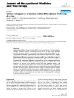
Figure 1a and b presents HR-TEM images of the Ag/Pt
nanoshells prepared by the galvanic displacement reaction.
The images clearly reveal that hexagonal Ag/Pt nanoshells
were successfully prepared via the galvanic displacement
reaction by adding Pt
2?
ions to a prepared solution of Ag
hexagonal nanotemplates, as shown in the inset of Fig. 1b.
It is noteworthy that prepared nanoshells with high order
assembly (shown in Fig. 1a) were clearly observed on the
copper grid with the supported carbon films. Compared to
the sacrificed Ag nanotemplates (see the inset of Fig. 1b),
the color of the nanoshell edge was significantly darker
than that of the nanoshell center. This means that the
prepared hexagonal nanoshells probably have hollow
structures. The EDX was utilized to ensure that the hex-
agonal Ag/Pt nanoshells were synthesized by this method.
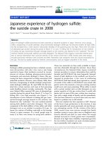
Figure 2 shows the line scanned EDX spectrum obtained
from the EDX analysis of a single nanoshell. The two
strong analysis signals from single nanoshells were iden-
tified as Pt and Ag. The exact composition of the prepared
nanomaterials was thus determined. On the basis of an
analysis of the HR-TEM image, the lattice spacing of a
hexagonal Ag/Pt nanoshell was measured, and is shown in
Fig. 1b. One spacing was detected at around 2.30 A
˚
. The
plane distance of (111) for the Pt and Ag nanoparticles was
*2.28 A
˚
[10] and *2.33 A
˚
[11], respectively. This indi-
cates that the distance of *2.30 A
˚
, which lies between
these two values, corresponds to a shell structure formed by
the Pt and Ag atoms. Similar results have been obtained
from HR-TEM experiments for mixtures of spherical Ag/Pt
nanoparticles [12].
Additional information on the nanoshell composition
was provided by the XRD pattern shown in Fig. 2b. The
four peaks, located at 38.25, 44.65, 64.85, and 77.55°, that
were detected for the dry Ag/Pt nanoshell powders were
attributed to the (111), (200), (220), and (311) diffraction
planes of the face-centered cubic (fcc) structure, respec-
tively. The diffraction peaks in the Ag standard spectrum
(JCPDS No. 089-3722) corresponding to the (111), (200),
(220), and (311) faces of the fcc structure are located at
38.12, 44.31, 64.46, and 77.41°, respectively. The (111),
(200), (220), and (311) peaks in the Pt standard XRD
Fig. 1 TEM images of Ag/Pt
hexagonal nanoshells: a high
order; b HR-TEM image. Inset:
hexagonal Ag nanotemplates
194 Nanoscale Res Lett (2009) 4:193–196
123
spectrum (JCPDS No. 087-0644) are located at 38.69,
44.97, 65.49, and 78.73°, respectively. The locations of the
peaks of the Ag/Pt nanoshells were between those of Ag
and Pt. This observation confirmed that the prepared
nanopowders were alloys of Ag and Pt.
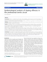
Figure 3 depicts LSV curves that compare the oxygen
reduction reactions in which the prepared hexagonal Ag/Pt
nanoshells, Ag/Pt nanoparticles, and Pt nanoparticles were
used as electrocatalysts. In contrast to the spherical Pt and
Ag/Pt nanocatalysts, the hexagonal Ag/Pt nanoshells
showed excellent activity starting from * 0.68 V in the
oxygen-saturated acid solution. In the TEM image (Fig. 1)
of the hexagonal Ag/Pd nanoshells, pores are observed to
form on the nanoshells’ surface. It is possible that the
trapping of the electrolyte species by pores with high-
surface areas resulted in the high activity of the nanoshells
for oxygen reduction. Recently, Pt-based alloy nanoparti-
cles with an optimized electronic structure were reported to
show high activity for the electroreduction of oxygen [13].
In the present study, another reason for the high activity of
the nanoshells is that Pt’s electronic structure was modified
by Ag during the formation of the alloy nanoshells.
In conclusion, hexagonal Ag/Pt nanoshells were suc-
cessfully synthesized using the galvanic displacement
reaction; in the reaction, the added Pt
2?
ions slowly reacted
with the prepared Ag nanoplates that were used as tem-
plates. The prepared hexagonal Ag/Pt nanoshells were
successfully used as electrocatalysts in an oxygen reduc-
tion process.
Acknowledgments The authors thank the National Science Council
of the Republic of China, Taiwan, for financially supporting this
research under Contract No. NSC 97-2221-E-151-028.
References
1. S.H. Joo, S.J. Choi, I. Oh, J. Kwak, Z. Liu, O. Terasaki, R. Ryoo,
Nature 412, 169 (2001). doi:10.1038/35084046
2. S. Cavaliere, F. Raynal, A. Etcheberry, M. Herlem, H. Perez,
Electrochem. Solid-State Lett. 7, A358 (2004). doi:10.1149/
1.1792259
3. H.M. Chen, R.S. Liu, M.Y. Lo, S.C. Chang, L.D. Tsai, Y.M.
Peng, J.F. Lee, J. Phys. Chem. C 112, 7522 (2008). doi:
10.1021/jp8017698
4. C.L. Lee, C.M. Tseng, S.C. Wu, R.B. Wu, Electrochem. Solid-
State Lett. 11, D27 (2008). doi:10.1149/1.2820903
5. C. Susut, T.D. Nguyen, G.B. Chapman, Y. Tong, Electrochim.
Acta 53, 6135 (2008). doi:10.1016/j.electacta.2007.12.016
30 40 50 60 70 80
Intensity (arbitrary unit)
(200)
(311)
(220)
(111)
2-Theta (deg.)
(A)
(B)
Fig. 2 Line scanned EDX spectrum and XRD pattern of Ag/Pt
hexagonal nanoshells: a line scanned EDX spectrum of single
nanoshells; b XRD pattern
0.4 0.5 0.6 0.7 0.8 0.9
-20
-15
-10
-5
0
5
Current density (mA/cm
2
)
Voltage (V) vs Ag/AgCl
Pt nanoparticles
Ag-Pt nanoparticles
Hexagonal Ag-Pt nanoshells
Fig. 3 The comparative LSV curves of the hexagonal Ag/Pt nano-
shells, spherical Ag/Pt nanoparticles and Pt nanoparticles for
electroreducing oxygen. Electrolyte: 1 M H
2
SO
4
(aq), Scan rate:
20 mV/s. The weight of hexagonal Ag/Pt nanoshells, spherical Ag/Pt
nanocatalysts and spherical Pt nanoparticles: 5 9 10
-2
mg
Nanoscale Res Lett (2009) 4:193–196 195
123
6. K.H. Park, K. Jang, H.J. Kim, S.U. Son, Angew. Chem. Int. Ed.
46, 1152 (2007). doi:10.1002/anie.200603961
7. W. Yang, X.L. Wang, F. Yang, C. Yang, X.R. Yang, Adv. Mater.
20, 2579 (2008). doi:10.1002/adma.200702949
8. C. Burda, X.B. Chen, R. Narayanan, M.A. El-Sayed, Chem. Rev.
105, 1025 (2005). doi:10.1021/cr030063a
9. J.N. Gao, X.L. Ren, D. Chen, F.Q. Tang, J. Ren, Scr. Mater. 57,
687 (2007). doi:10.1016/j.scriptamat.2007.06.049
10. J.S. Guo, G. Sun, S.G. Sun, S.Y. Yan, W.Q. Yang, J. Qi,
Y.S. Yan, Q. Xin, J. Power Sources 168, 299 (2007). doi:
10.1016/j.jpowsour.2007.02.085
11. C.L. Lee, Y.C. Huang, L.C. Kuo, J.C. Oung, F.C. Wu, Nano-
technology 17, 2390 (2006). doi:10.1088/0957-4484/17/9/053
12. K. Torigoe, Y. Nakajima, K. Esumi, J. Phys. Chem. 97, 8304
(1993). doi:10.1021/j100133a029
13. V. Stamenkovic, B.S. Mun, K. Mayrhofer, J. Rossmeisl,
J. Greeley, J. Nørskov, Angew. Chem. Int. Ed. 45, 2897 (2006).
doi:10.1002/anie.200504386
196 Nanoscale Res Lett (2009) 4:193–196
123