Báo cáo hóa học: " Magnetoresistance in Sn-Doped In2O3 Nanowires" ppt
Bạn đang xem bản rút gọn của tài liệu. Xem và tải ngay bản đầy đủ của tài liệu tại đây (544.12 KB, 5 trang )
NANO EXPRESS
Magnetoresistance in Sn-Doped In
2
O
3
Nanowires
Olı
´
via M. Berengue Æ Alexandre J. C. Lanfredi Æ
Livia P. Pozzi Æ Jose
´
F. Q. Rey Æ Edson R. Leite Æ
Adenilson J. Chiquito
Received: 12 February 2009 / Accepted: 24 April 2009 / Published online: 4 July 2009
Ó to the authors 2009
Abstract In this work, we present transport measurements
of individual Sn-doped In
2
O
3
nanowires as a function of
temperature and magnetic field. The results showed a
localized character of the resistivity at low temperatures as
evidenced by the presence of a negative temperature coef-
ficient resistance in temperatures lower than 77 K. The weak
localization was pointed as the mechanism responsible by
the negative temperature coefficient of the resistance at low
temperatures.
Keywords Oxide nanowires Á Weak localization Á
Electron transport Á Electron–electron scattering
Introduction
Quasi 1D metal oxide nanostructures have attracted con-
siderable interest in the last years for fundamental studies
and also for potential applications. In particular, they
present properties which range from metals to semicon-
ductors and insulators [1]: the performance of these devices
is strongly correlated to their structural and electronic
properties. These low-dimensional structures have been
used as building blocks in different devices and nanode-
vices [2] and they are important for both fundamental
research and applications because they have the potential to
reach high device integration. One of the most prominent
applications of these materials is the gas sensing devices
[3–5], but this and other potential electronic applications of
nanowires still require a detailed understanding of their
fundamental electronic properties [6].
Although these nanostructures be usually grown by
self-organized processes like the vapor–liquid–solid
mechanism (VLS) [7] and thus presenting a high crys-
talline quality, some disorder is always present. In this
way, electrons subjected to a random potential are not
able to move freely through the system if either potential
fluctuations due to disorder exceed a critical value or the
electron energy is lower than the characteristic potential
fluctuation [8, 9]. It is interesting to add that the carrier
localization should be evidenced in one-dimensional
structures as stated by the Anderson’s localization theory:
it predicts that disorder in these systems leads to carrier’s
localization.
In this work we present some transport measurements of
individual Sn-doped In
2
O
3
nanowires as a function of
temperature and magnetic field. The results showed a
localized character of the resistivity at low temperatures as
evidenced by the presence of a negative temperature
coefficient resistance in temperatures lower than 77 K.
This behavior was successfully associated to weak locali-
zation picture where the boundary scattering processes
provide the main inelastic scattering mechanism.
O. M. Berengue (&) Á L. P. Pozzi Á A. J. Chiquito
Departamento de Fı
´
sica, Universidade Federal de Sa
˜
o Carlos,
CEP 13565-905, CP 676 Sa
˜
o Carlos, Sa
˜
o Paulo, Brazil
e-mail:
A. J. C. Lanfredi Á J. F. Q. Rey
Centro de Engenharia, Modelagem e Cie
ˆ
ncias Sociais Aplicadas,
Universidade Federal do ABC, CEP 09210-170, Santo Andre
´
,
Sa
˜
o Paulo, Brazil
E. R. Leite
Laborato
´
rio Interdisciplinar de Eletroquı
´
mica e Cera
ˆ
micas,
Departamento de Quı
´
mica, Universidade Federal de Sa
˜
o Carlos,
CEP 13565-905, CP 676 Sa
˜
o Carlos, Sa
˜
o Paulo, Brazil
123
Nanoscale Res Lett (2009) 4:921–925
DOI 10.1007/s11671-009-9336-4
Experimental
The samples used here were grown by the well-known VLS
growth mechanism in association with a carbothermal
reduction process [10, 11]. For this purpose In
2
O
3
and
SnO
2
powders (purity [99.9%) were mixed with 10% in
weight of carbon black and each mixture was placed inside
a horizontal tube furnace in two separated alumina cruci-
bles. The synthesis was carried out at 1150 °C under a N
2
gas flux of 50 sccm for 4 h.
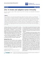
The wooly-like collected material was analyzed by X-
ray diffraction (XRD) as plotted in Fig. 1a. It was possible
to identify the cubic In
2
O
3
structure by the crystallographic
indices (PDF 6-416). Also, it could be identified the Sn and
SnO
2
structures as an evidence of the self-catalytic VLS
growth mechanism.
To improve the structural characterization of the sam-
ples, a transmission electron microscopy (TEM; Jeol model
JEM 2100 operating at 200 kV) was carried out on
individual ITO nanowires. Figure 1b shows a low-magni-
fication TEM image of an individual nanowire with
200 nm width. In order to study the orientation growth and
high crystallinity of the samples, a high resolution TEM
image (HRTEM) was performed at the end of the nanowire
and it is shown in Fig. 1c. After a fast Fourier transform
(FFT) of the HRTEM image, by using an image analysis
software, it was obtained a point matrix which is the fre-
quency spectrum, sketched in Fig. 1d. The points showed
in this equivalent SAED (selected-area electron diffraction)
image allow the indexing of the growth direction to be
[100] and are in agreement with interatomic distances
measured. This plane growth direction is in accordance
with the XRD pattern plotted in Fig. 1a, which shows the
[400] peaks intensity higher than the obtained from bulk
materials.
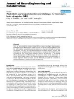
The Raman characterization was performed in order to
identify the ITO vibrational modes and confirms the XRD
results. The micro-Raman experiments were carried out at
room temperature with a T 64000 Jobin Yvon spectrometer
using the 514.5 nm line of an argon ion laser as excitation
source. The power was kept below 5 mW to avoid over-
heating. Figure 2 shows the Raman spectra of the wooly-
like material. It is known that the body-centered cubic
In
2
O
3
belongs to the space group Ia
3
,Th
7
[12]. For such a
structure, the vibrations with symmetry A
g
,E
g
, and T
g
are
Raman active giving rise to 22 Raman modes. Five Raman
peaks at 112, 133, 303, 491, and 616 cm
-1
were found to
belong to the vibrational modes of the bcc-In
2
O
3
, which
seems to be in good agreement with the reported values in
the literature [13]. The other peaks are probably related to
the presence of Sn atoms in In sites of In
2
O
3
leading to
different vibrational modes. The ITO vibrational features
are now under study and will be the subject of a new paper.
Fig. 1 a X-ray diffraction pattern of the as grown material; b low-
magnification TEM image of an ITO nanowire; c HRTEM image of
the end of the same nanowire; d fast Fourier transform of the image
shown in panel b
Fig. 2 Raman spectrum taken at room temperature showing the
expected phonon modes for bcc-In
2
O
3
922 Nanoscale Res Lett (2009) 4:921–925
123
After the structural characterization, the samples were
prepared for the contacts’ fabrication. The samples were
then ultrasonically dispersed in ethanol and were placed
onto an oxidized Si wafer (300 nm of SiO
2
layer) with
metallic (Au/Ni, 100 nm thickness) pads. The transport
measurements were carried out using standard low fre-
quency ac lock-in techniques, at 13 Hz. The low temper-
ature data were obtained using a closed cycle helium
cryostat at a base pressure 5 9 10
-6
Torr.
Results and Discussion
Samples with 68–70 nm width were used for transport
measurements. The resistance-temperature dependent
measurements were conducted with B = 0 and using dif-
ferent current levels (from 0.1 to 10 lA) but the results
remain unchanged (Fig. 3). In the high temperatures range,
77–300 K, the nanowires exhibit a positive temperature
coefficient of resistance and a weak temperature depen-
dence, a typical metal-like character. It is interesting to add
that the metallic behavior was found in other and larger
samples (see the inset in Fig. 3) and it is present even at the
lowest used temperature, T = 10 K. Since the ITO nano-
wires are expected to remain metallic, the carrier density is
a weak function of temperature and the resistance should
be mainly determined by the temperature dependence of
the various scattering mechanisms through electrons’
mobility [6]. At high temperatures (T [ 77 K), the phonon
scattering seems to be dominant and the resistance rises
with increasing temperature (metallic phase).
These resistance data were analyzed in the framework of
the Bloch–Gru
¨
neisen theory (based on the electron-acous-
tic phonon scattering mechanism) due to the observed
metallic character. In this way, the resistance is described
by [14]
RðTÞ¼R
0
þ A
T
H
D
n
Z
H
D
=T
0
z
n
e
z
e
z
À 1ðÞ
2
dz ð1Þ
where A is a parameter proportional to the electron–phonon
coupling and R
0
is the residual resistance; n usually ranges
from 3 to 5 when the electron–phonon interaction is mainly
responsible for the scattering events [14]; H
D
is the Debye
temperature. The fitting of the experimental data using Eq. 1
revealed n = 3.6 and H
D
= 1227 K: as the temperature
and phonon excitation increase, the amount of scattering
events experienced by the conduction electrons are
increased as well, resulting in a greater resistivity (theo-
retical value of H
D
= 1200 K). It is interesting to add that
the Bloch–Gru
¨
neisen theory can be only used in the range of
nanowires’ size where the electron-acoustic phonon scat-
tering remains unchanged as pointed in Ref. [15].
The analysis for the low temperature data is more
challenging: down from 77 K the sample’s resistance
increases indicating that a different transport and scattering
mechanisms are acting in this range of temperatures. The
observed negative temperature coefficient resistance could
not be fitted to an usual activation (exponential) law.
However, it preserves the localized character for electron
transport. As reported in literature for Zn [16] and Sb [17]
nanowires, we also found that the resistivity increase fol-
lows essentially a T
-1/2
law (the equation R ¼ 0:01 þ
6:7 Â10
À4
=
ffiffiffiffiffiffiffiffiffiffiffiffiffiffi
28 þT
p
fits well the temperature dependence
shown in Fig. 3). The observation of an exact T
-1/2
law
should be unambiguously attributed to a signature of the
presence of electron–electron inelastic scattering mecha-
nism [18]. Simple activated and one-dimensional hopping
laws were used and discarded because they lead us to
wrong results. Then, a more detailed analysis is needed
including, for instance, the presence of carriers’ localiza-
tion and other scattering mechanisms.
As observed in literature [6, 19–21] for small-dimension
nanowires, processes like collisions with the boundaries
provide disorder, which in turn randomizes the electron
energy and increases the electron–electron interaction.
These interactions are also expected to contribute to the
transport leading to an increase of the resistance since the
diffusive motion of the electrons enhances their interactions.
In our case, taking into account the nanowire’s cross
section, the boundary scattering (mostly temperature
independent) becomes an important inelastic scattering
Fig. 3 Resistance-temperature dependent measurements taken at B =
0 and using different current levels (only 1 lA is shown). In the high
temperatures range, the nanowires exhibit a positive temperature
coefficient of resistance and a weak temperature dependence. The
inset shows the results for other and larger samples showing the same
behavior
Nanoscale Res Lett (2009) 4:921–925 923
123
mechanism at low temperatures leading to a finite size
effect. As a result, a localized character for the electron’s
transport is achieved. Then, the observed negative tem-
perature coefficient can be interpreted as a result of the
mixture of the two scattering process (electron–electron
and boundary collisions) at low temperatures, both leading
to a localization character for the electron transport.
This effect can be studied by using magnetoresistance
experiments.
Independently of the main mechanism of inelastic
scattering of electrons, the phase-breaking time and length
are power functions of temperature leading to a quantum
correction for conductivity/resistivity which is quantified
by the weak localization in a form DR / ln T: Unfortu-
nately, the electron–electron interaction produces the same
dependence on the temperature. In order to establish the
mechanism responsible by the increase of the resistivity,
we conducted resistance measurements under different
magnetic fields. The weak localization is known by the
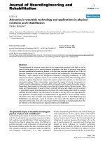
highly sensibility to a weak magnetic field. Figure 4a
shows the linear dependence of resistance as function of
the ln T and for different magnetic field intensities. From
these curves we see that the expected ln T dependence of
the resistivity is clearly observed until *0.3 T when the
magnetic field is strong enough to break the quantum
interference. For higher magnetic fields, the resistance does
not exhibit the negative temperature coefficient (also, the
larger samples do not show any dependence on the mag-
netic field, as expected).
Additional confirmation of weak localization effects was
obtained by magnetoresistance measurements as seen in
Fig. 4b. From these measurements we calculated the
phase-breaking length (L
/
= 72 nm). From the theory, the
critical B field needed to suppress the weak localization is
given by
B
C
¼
h
qWL
/
; ð2Þ
where W is the width of the wire [22]. Using the results
presented in Fig. 4, one finds B
C
= 0.84 T: below this
value the weak localization regime should determine the
behavior of the conductivity of the sample, as observed.
This result is twofold: first, the weak localization is
suppressed at high B fields as expected and confirming that
the negative temperature coefficient is a result of the dis-
order-induced localization. Second, it indirectly gives an
evidence of a transition from one-dimensional (weak field)
to three-dimensional localization (high field). In fact, for
weak fields the magnetic length L
B
ð¼
ffiffiffiffiffiffiffiffiffiffi
"h=eB
p
Þ is greater
than the width of the samples and L
/
: from the viewpoint
of the electrons, the nanowire is an one-dimensional sys-
tem. Otherwise, the nanowire behaves essentially like a
three-dimensional system. In both cases, the disorder
coming from the boundary scattering plays the funda-
mental role giving the main scattering mechanism for the
diffusive electron transport.
Conclusion
Electronic properties of self-assembled high crystalline
quality tin-doped indium oxide were studied. We report on
the experimental data and the related analysis on the
resistance and magnetoresistance of these single crystal
nanowires. The weak localization was pointed as the
mechanism responsible by the negative temperature coef-
ficient of the resistance at low temperatures. From the
magneto-resistance data we quantified the characteristic
phase-breaking length of the system; additionally, we
observed a three- to one-dimensional transition for the
localization character of the resistance.
Acknowledgment The authors thank the Brazilian research funding
Agencies FAPESP and CNPq for the financial support of this work.
(a)
(b)
Fig. 4 a The linear dependence of resistance as function of the ln T
and for different magnetic field intensities. The expected ln T
dependence of the resistivity is clearly observed until *0.3 T and for
higher magnetic fields, the resistance does not exhibit the negative
temperature coefficient. b The magnetoresistance measurements
providing additional confirmation of weak localization effects
924 Nanoscale Res Lett (2009) 4:921–925
123
References
1. Z. Pan, Z. Dai, Z. Wang, Science 291, 1947 (2001)
2. X. Duan, Y. Huang, Y. Cui, J. Wang, C.M. Lieber, Nature 409,
6649 (2001)
3. J. Kappler, A. Tomescu, N. Barsan, U. Weimar, Thin Solid Films
391, 186 (2001)
4. M. Radecka, J. Przewoznik, K. Zakrzewska, Thin Solid Films 39,
247 (2001)
5. C. Li, D. Zhang, S. Han, X. Liu, T. Tang, C. Zhou, Adv.
Mater.15, 143 (2003)
6. A.J. Chiquito, A.J.C. Lanfredi, R.F.M. Oliveira, L.P. Pozzi, E.R.
Leite, Nanoletters 7, 1439 (2007)
7. R.S. Wagner, W.C. Ellis, Appl. Phys. Lett. 4, 89 (1964)
8. Yu.A. Pusep, A.J. Chiquito, S. Mergulha
˜
o, A.I. Toropov, J. Appl.
Phys. 92, 3830 (2002)
9. A.J. Chiquito, Yu.A. Pusep, G.M. Gusev, A.I. Toropov, Phys.
Rev. B 66, 035323 (2002)
10. E.R. Leite, J.W. Gomes, M.M. Oliveira, E.J.H. Lee, E. Longo,
J.A. Varela, C.A. Paskocimas, T.M. Boschi, F. Lanciotti, P.S.
Pizani, P.C. Soares, J. Nanosci. Nanotechnol. 2, 125 (2002)
11. M.O. Orlandi, R. Aguiar, A.J.C. Lanfredi, E. Longo, J.A. Varela,
E.R. Leite, Appl. Phys. A 80, 23 (2005)
12. W.B. White, V.G. Keramidas, Spectrochim. Acta 28A, 501
(1972)
13. H. Sobotta, H. Neumann, G. Ku
¨
hn, V. Riede, Cryst. Res. Tech-
nol. 25, 61 (1990)
14. J.M. Ziman, Electrons and Phonons (Claredon Press, Oxford,
1960)
15. A. Bid, A. Bora, A.K. Raychaudhuri, Phys. Rev. B 74, 035426
(2006)
16. J.P. Heremans, C.M. Thrush, D.T. Morelli, M.C. Wu, Phys. Rev.
Lett. 91, 076804 (2003)
17. Y. Zhang, L. Li, G.H. Li, L.D. Zhang, Phys. Rev. B 73, 113403
(2006)
18. D.E. Beutler, N. Giordano, Phys. Rev. B 8, 38 (1988)
19. T.J. Thornton, M.L. Roukes, A. Scherer, B.P. Van der Gaag,
Granular Nanoelectronics, ed. by D.K. Ferry, J.R. Baker, C.
Jacobon, NATO ASI Series (Plenum Press, New York, 1991)
20. W. Wu, S.H. Brongersma, M. Van Hove, K. Maex, Appl. Phys.
Lett. 84, 2838 (2004)
21. H. Kind, H. Yan, B. Messer, M. Law, P. Yang, Adv. Mater. 14,
158 (2002)
22. S. Datta, Electronic Transport in Mesoscopic Systems (Cam-
bridge University Press, Cambridge, 1997)
Nanoscale Res Lett (2009) 4:921–925 925
123