Nanofibers Part 9 ppt
Bạn đang xem bản rút gọn của tài liệu. Xem và tải ngay bản đầy đủ của tài liệu tại đây (1.05 MB, 30 trang )
Nanofibers
230
catalysts. This mode combined with the CCVD method allows a significant decrease of
energy consumption and a shorter reaction time as compared with the heating mode with
outer furnace. CNFs have been synthesized by decomposition of pure ethylene over
Fe:Ni:Cu catalyst in a horizontal furnace. The catalyst was prepared from nitrate solutions
by co-precipitation with ammonium bicarbonate and was calcined at 400
0
C for 4 h. The
carbonaceous products were purified by extraction in HCl (37%) for 24 h, washed with
distilled water, and dried at 150
0
C for 3 h. A typical transmission electron microscope (TEM)
image (Figure 1) of the sample shows nanofibers with ‘‘herringbone’’ structure and
diameters ranging from 80 to 290 nm, similar to those reported in the literature. Their
specific area was determined by the BET method and the value was between 170-242 m
2
g
-1
.
The CNFs have been characterized by cyclic voltammetry and their adsorption properties
for biologically active substances have been closely followed (Pruneanu et al., 2006; Olenic et
al., 2009).
a b
Fig. 1. (a) HRTEM image of CNF (from ethylene at 600
0
C on Fe:Ni:Cu as catalyst); (b) SEM
image of CNFs. Reprinted from ref. Olenic et al., 2009 with kind permission of Springer
Science and Business Media.
In the synthesis of nanocarbon structures by CCVD method, the critical step is the catalyst
preparation. Metal nanoparticles catalyst (optimum size between 0.4–5 nm) favours the
catalytic decomposition of the carbon source gas in a temperature range of 600–1100
0
C. As
was shown in the literature, the amorphous carbon is deposited from the thermal
decomposition (pyrolysis) of the carbon source gas, whereas the carbon nanofibers are
grown from the catalytic decomposition of the carbon source gas (Teo et al., 2003).
According to the growth procedure, CVD method includes the seeded catalyst method (Li et
al., 1996) which uses the catalyst seeded on a substrate within a reactor (in this case the
interactions between the catalyst and support (alumina, silica, silicon) dictates the growth
mode (Randall et al., 2001); an advantageous one is the floating catalyst method which is a
method wherein the carbon vapour and the catalytic metal particles both get deposited in
the reaction chamber, without a substrate. (Martin-Gullon et al., 2006).
One of the CVD methods that has been developed is the synthesis of vertically aligned
nanofibers bundles for specific applications. The synthesis of VACNFs arrays were all
carried out in horizontal reactors (Cao et al., 2001). All the reported products by vertical
floating catalyst method were randomly arranged CNFs (Perez-Cabero et al., 2003). There
are few reports on aligned CNF bundles synthesized by floating catalyst procedure, in
vertical reactors (Cheng et al, 2004).
Electrochemical and Adsorption Properties of Catalytically Formed Carbon Nanofibers
231
VACNFs were also obtained by low-pressure inductively coupled PECVD (Caughman et al.,
2003); isolated VACNFs were synthesized by Melechko et al., 2003.
When CNFs are prepared, crystallized structures are generally desired (amorphous carbon-
free). The growth temperature affects the crystallinity: a too high temperature leads to the
formation of pyrolytic amorphous carbon. This is the reason for preferring the highest
deposition temperature without significant self-decomposition of the carbon source gas.
The growth mechanism leading to the formation of CNFs (reviewed by Teo et al., 2003) has
been studied by many groups. Baker et al., 1972 proposed a growth mechanism for both
nanofibers and nanotubes, which was later completed. Other models for growing CNFs were
proposed by Oberlin et al., 1976, Koch et al., 1985, Zheng et al., 2004. Formation mechanism of
large branched carbon nano-structures has been presented by Devaux et al., 2009.
Examination of synthesized CNFs by TEM and SEM reveals the basic microstructure of
graphitic CNFs. There are two types of carbon nanotubes: single-wall and multi-wall and
four types of carbon nanofibers that consist of stacked graphite layers, which can be
arranged parallel (tubular-adopting the structure of a “multi-walled faceted nanotube”),
perpendicular to the fiber axis (platelet-adopting the arrangement of a “deck of cards”), or
herringbone structure (the graphite platelets are at a particular angle to the fiber axis), and
amorphous type without crystalline structure. Most of carbon nanofibers and nanotubes
synthesized by CCVD method are crystalline or partially crystalline and only a few of them
are amorphous.
The herringbone structure seems to be favoured when the catalyst is an alloy. Herringbone-
type CNFs with large diameter and a very small or completely hollow core have been
synthesized through a CVD method (Terrones et al., 2001).
The only difference among the various forms of carbon nanofilaments is their chemical
structure. Martin-Gullon, et al., 2006, present in detail a classification of nanofilaments
depending on their structure.
The properties related to the morphology of CNFs depend on many factors, like: the
chemical nature of the catalyst and the conditions of its pretreatment (Huang et al., 2009;
Kovalenko et al., 2009 b), the composition and flow rate of a gas mixture and the
temperature and duration of the synthesis (Endo et al., 2003; Chuang et al., 2008).
On the other hand, the electrical and optical properties of carbon nanostructures are largely
dependent on their structures (Kataura et al., 1999; Yang et al., 2003).
The conducting properties of CNFs that can be varied from metal to semiconductor
(depending on the structural parameters and doping with heteroatoms) are very important
for practical applications (Ismagilov, 2009).
All CNFs products obtained by CCVD method contain impurities such as metal catalyst
particles, amorphous carbon and carbon nanoparticles depending on the reaction
conditions. Therefore, purification of carbon nanostructures is of great importance for
technological applications.
A purification step is usually required before carbon nanofilaments can be used, especially
for biomedical applications. Several purification methods are reported in the literature (Liu
et al., 2007). Graphitization (or heat treatment) is one of the most effective methods to
remove defects or impurities such as metallic compounds, which diminish the electrical and
mechanical properties of conventional carbon nanofibers.
Huang et al., 2009 demonstrated that high purity CNFs can be formed by varying the
synthesis temperature. Different types of CNFs were characterized by various techniques to
understand their crystal structure, morphology, graphitization degree and thermal stability.
Nanofibers
232
For more complex applications of carbon nanotubes, different functionalization methods
have been introduced. Investigation of the interaction between carbon nanotubes and
biological molecules are very important (Zhong et al., 2009).
McKnight et al., 2006 showed several approaches toward such site-specific functionalization
along the nanofiber length, including physical and electrochemical coating techniques,
chemical immobilization of DNA and enzyme species, and covalent attachment of biotin
followed by affinity-based capture of streptavidin-conjugated molecules.
4. Electrochemical properties of carbon nanofibers
For many electrochemical applications, carbon is a well known material of choice. Among
its practical advantages are: a wide potential window in aqueous solution, low background
current, lack of corrosion processes at positive potentials and low costs.
The advantages of CNFs in the construction of biosensors, relate to their small size with
large specific area, the promotion of electron transfer when used in electrochemical reactions
and easy bio-molecules immobilization. DNA molecules can be covalently bound on the
functionalized fiber surface (e.g. with carboxylic groups). In comparison with the classical
carbon electrodes, CNFs show better electrodic behaviour including good conducting ability
and high chemical stability. The electrochemical properties of CNFs paste electrodes have
been largely studied. In most cases, CNFs were prepared as composite electrodes.
It is of interest to explore the properties of carbon nanocomposite electrodes to see if they
might exhibit new properties, due to the high edge/surface area ratio of such materials.
Marken et al., 2001 have evaluated CNFs (obtained by ambient pressure CVD method) as
novel electrode materials for electrochemical applications (porous, pressed onto a glassy-
carbon substrate and non-porous, embedded in a solid paraffin matrix). They exhibit low
BET surface areas and high electrochemical capacitances due to the fact that the spaces
between the fibers allow the penetration of electrolyte solution. Capacitive currents tend to
mask voltammetric currents during cyclic voltammetry. By comparison, when the spaces
between CNFs are impregnated by an inert dielectric material (paraffin wax) the electrode
has good conductivity and low capacitance. These materials were compared with other
forms of nanostructured carbons: aerogel or activated charcoal.
Van Dijk et al 2001 prepared nanocomposite electrodes made of CNFs and black wax and
used them for anodic stripping voltammetry of zinc and lead.
Maldonado et al., 2005 have prepared nondoped and nitrogen-doped (N-doped) CNFs films
by the floating catalyst CVD method using precursors consisting of ferrocene and either
xylene or pyridine to control the nitrogen content. CNF coated nickel-mesh was used as
working electrode, to study the influence of nitrogen doping on the oxygen reduction
reaction. The electrodes have significant catalytic activity for oxygen reduction in aqueous
solutions (neutral to basic pH).
Yeo-Heung et al., 2006 tested the electrochemical actuation properties of carbon nanofiber–
polymethylmethacrylate (CNF–PMMA) composite material. They characterized the CNF-
PMMA actuator by impedance spectroscopy, at voltages up to 15V. The relationship
between displacement and applied voltage was determined.
Roziecka et al., 2006 prepared ITO electrodes modified with hydrophobic CNFs–silica film,
which was employed as support for liquid/liquid redox systems. The redox processes
within the ionic liquid is coupled to ionic transfer processes at the ionic liquid/water
Electrochemical and Adsorption Properties of Catalytically Formed Carbon Nanofibers
233
interface. Therefore, the CNFs electrode material was an excellent support for recording
both the Faradaic and capacitive currents. The efficiency of the electrode process increases
due to the use of the heterogeneous matrix.
Our group has studied the electrochemical properties of carbon nanofilaments (CNFs,
MWCNTs and SWCNTs- unpublished data). Paste electrodes were prepared by mixing the
carbon powder with silicon oil and then packing the resulting paste into the cavity of a PVC
syringe (2.5 mm diameter). The electrical contact was ensured by a Pt wire, tightly inserted
into the paste.
a b
c
Fig. 2. Cyclic voltammograms recorded in solution of 10
-2
M hydroquinone and 0.5M KCl
for: a) CNFs; b) MWCNTs; c) SWCNTs paste electrode; all voltammograms were recorded
with a sweep rate of 100 mVs
-1
.
The electrochemical behaviour of these types of electrodes was investigated by cyclic
voltammetry (100 mVs
-1
sweep rate) using as redox mediator a solution of 10
-2
M
hydroquinone (Figure 2 a,b,c). From Figure 2a one can see that carbon nanofibers showed
the best electrodic properties. The voltammograms exhibit two well-defined peaks, with the
peak potential separation, ∆Ep, around 150 mV. This value is higher than that generally
obtained for a reversible redox system (60 mV/n, where n is the number of electrons
transferred during the reaction).
Nanofibers
234
For MWCNTs and SWCNTs paste electrode, the peak potential separation, ∆Ep is
considerable larger (850 mV and respectively 1100 mV), indicating a lower conductivity and
a slow transfer of electrons.
Due to the excellent electrodic properties of CNFs paste electrode, Pruneanu et al., 2006 have
studied the oxidation of calf thymus DNA. The interest in this kind of research is due to the
fact that the electrochemical oxidation may mimic the biological oxidation mechanism,
involving enzymes. All the four bases of DNA can be chemically oxidized;
electrochemically, only guanine and adenine oxidation peaks can be recorded (thymine and
cytosine have oxidation potentials larger than 1.2V vs. Ag/AgCl). In order to establish the
exact position of purine oxidation potentials (adenine and guanine) the authors have
registered differential pulse voltammetry (DPV) curves, in solution containing 10
-3
M
adenine hemisulphate and 10
-3
M guanine hemisulphate (in 0.1M PBS pH 7+ 0.5M KCl,
Figure 3). The two peaks that appeared around 0.9V vs. Ag/AgCl and 1.18V vs. Ag/AgCl
were ascribed to guanine and adenine oxidation, respectively. The intensity of the peaks
decreased after successive recordings, due to the irreversible character of the oxidation
process.
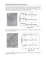
-0.2 0.0 0.2 0.4 0.6 0.8 1.0 1.2 1.4
-2.0x10
-6
0.0
2.0x10
-6
4.0x10
-6
6.0x10
-6
8.0x10
-6
1.0x10
-5
I(A)
E(V) vs Ag/AgCl
Fig. 3. DPVs recorded in a solution of 10
-3
M adenine hemisulphate and 10
-3
M guanine
hemisulphate, in 0.1M PBS (pH 7) + 0.5M KCl.
The signals obtained from guanine or adenine oxidation can be used for the construction of
a DNA biosensor. In Figure 4 one can see that the oxidation peak of adenine hemisulphate
increases with the increase of solution concentration (10
-7
….10
-3
M).
Oxidation of calf thymus DNA (single stranded or double stranded DNA) at carbon
nanofibers paste electrode was also studied by DPV (Figure 5). Prior experiments, calf
thymus DNA was physically adsorbed on the electrode surface, by immersing it in DNA
solution for about five minutes, under constant stirring. The two peaks corresponding to
guanine and adenine oxidation were clearly recorded for single stranded DNA (Figure 5,
straight line). In contrast, no signal was obtained when double stranded DNA was adsorbed
at the electrode surface (Figure 5, dashed line). This may be explained by the fact that in
Electrochemical and Adsorption Properties of Catalytically Formed Carbon Nanofibers
235
double stranded DNA the purine bases are hidden between the double helix, so they have
no free access to the electrode surface. In this case the transfer of electrons cannot take place.
-0.2 0.0 0.2 0.4 0.6 0.8 1.0 1.2 1.4
0.0
2.0x10
-6
4.0x10
-6
6.0x10
-6
8.0x10
-6
1.0x10
-5
1.2x10
-5
I(A)
E(V) vs Ag/AgCl
10
-7
M
10
-6
M
10
-5
M
10
-4
M
10
-3
M
Fig. 4. DPVs recorded in solutions of adenine hemisulphate of different concentration:
10
-7
10
-3
M in 0.1M PBS (pH 7) + 0.5M KCl.
-0.2 0.0 0.2 0.4 0.6 0.8 1.0 1.2 1.4 1.6 1.8
0.0
1.0x10
-6
2.0x10
-6
3.0x10
-6
4.0x10
-6
5.0x10
-6
I(A)
E(V) vs Ag/AgCl
Fig. 5. DPVs of single-stranded DNA (straight line) and double-stranded DNA (dashed line)
in solution of 0.1M PBS (pH 7) + 0.5M KCl (0.3 mgml
-1
DNA)
Zhang et al., 2004 performed I –V measurements on individual VACNFs. They fabricated
multiple Ti/Au ohmic contacts on individual fibers, having the contact resistance of only
few kOhm. The measurements demonstrated that VACNFs exhibit linear I –V behaviour at
room temperature. Between intergraphitic planes in VACNFs exists a dominant transport
mechanism of electrons, along the length of the fiber.
VACNFs are increasingly used in bioelectrochemistry, due to the fact that they exhibit fast
electron transfer to redox species from solution, or act as highly conducting substrates to
Nanofibers
236
connect redox enzymes to macro-sized electrodes. Their chemical stability combined with a
high degree of biologically accessible surface area and nanoscale dimension make VACNFs
ideal substrates for the development of scaffolds in biological detection. Additionally, their
mechanical strength and narrow diameter allow easy cell penetration, making them suitable
for intracellular electrochemical detection.
Baker et al., 2006a demonstrated the ability to use VACNFs as electrodes for biological
detection. He also emphasized the importance of the surface functionalization, in order to
control the overall electrochemical response. Functionalized VACNFs with the redox active
protein cytochrome c were characterized by cyclic voltammetry (CV) measurements.
Although the high surface area of the nanofibers allows the cytochrome c molecules to
produce an increase of the electrochemical current, the high capacitive currents partially
obscured this signal and partially offset the potential improvement in the signal-to-noise
ratio.
VACNT arrays were successfully grown on planar graphitic carbon substrates, using a bilayer
Al/Fe catalyst and water-assisted thermal CVD. Excellent voltammetric characteristics were
demonstrated after insulating the arrays with a dielectric material (Liu et al., 2009).
A method for the development of an amperometric biosensor for interference-free
determination of glucose was reported by Jeykumari & Narayan, 2009. The bienzyme-based
biosensor was constructed with toluidine blue functionalized CNTs. The electrochemical
behaviour of the sensor was studied by impedance spectroscopy, cyclic voltammetry and
chronoamperometry. The excellent electrocatalytic activity of the biocomposite film allowed
the detection of glucose under reduced over potential, with a wider range of determination
and with a very good detection limit. The sensor showed a short response time, good
stability and anti-interferent ability. The proposed biosensor exhibits good analytical
performance in terms of repeatability, reproducibility and shelf-life stability.
Sadowska et al., 2009, functionalized MWCNTs with azobenzene and anthraquinone
residues (chemical groups with redox activity) for potential application in catalysis and
memory storage devices. Using the Langmuir–Blodgett method, the nanotubes containing
electroactive substituents were transferred onto electrode substrates and characterized by
cyclic voltammetry. The amount of electroactive groups per mg of nanotubes was calculated
based on the cathodic current peak. A highly reproducible voltammetric response was
obtained with a single nanotube layer or multiple nanotube/octadecanol layers. It is
believed that such devices will be invaluable for future high-performance electrodes.
Minikanti et al., 2009 designed implantable electrodes as targets for wide frequency
stimulation of deep brain structures. They have demonstrated by cyclic voltammetry and
impedance spectroscopy, the enhanced performance of implantable electrodes coated with
multi-wall carbon nanotube. The results were compared with those obtained for the more
traditional stainless steel. They also investigated the surface morphology of aged electrodes
due to the fact that implantable electrodes have to be mechanically stable and present high
shelf life. The effect of superficial oxygen adsorption on the aged MWCNTs electrodes was
observed through a modified cyclic voltammetric spectrum.
In the past few years, considerable interest was focused on the application of carbon based
nanomaterials as electrodes for supercapacitors, due to their chemical inertness and easy
processability. The capacitive behaviour of the CNFs was studied in term of charge-
discharge curves and cyclic voltammetry.
Recently, carbon nanomaterials with various morphologies (carbon nanotubes, nanofibers,
nanowires and nanocoils) have been intensively studied as negative electrode materials in
Electrochemical and Adsorption Properties of Catalytically Formed Carbon Nanofibers
237
lithium-ion batteries (Zou et al., 2006). These nanofibers have low graphitic crystallinity. The
experimental results showed that CNF electrodes had high reversibility with small
hysteresis, in the insertion/extraction reactions of lithium-ion.
All these studies suggest that CNFs represent a new class of materials suitable for
electrochemical applications.
5. Adsorption properties of carbon nanofibers
The biologically active substances can be attached to CNFs surfaces by physical adsorption
(physisorption) or chemical immobilization.
For a long time, activated carbons (ACs) materials containing large surface area and well-
developed porosity were successfully applied in various industrial processes including
adsorption (gases and liquids), mixture separation, filtration, etc.
CNFs and activated CNFs have special properties, compared with activated carbon. Among
these, we mention the high chemical reactivity due to the large fraction of active sites,
available for chemical and physical interaction with different species.
Baker, 2007 noticed the use of nanofibers as adsorbents. He additionally emphasized that
the functionality of carbon nanofiber surface has an important role. The raw graphitic
materials are free of surface oxygen groups and therefore are hydrophobic in nature. CNFs
surface can have a hydrophilic character after a normal activation procedure. The control of
the acid-base properties of carbon nanofibers surface has an important impact on a variety
of potential applications. The structural characteristics e.g. the infinite number of graphite
layers and the weak Van der Waals forces are responsible for the high adsorption capacity
observed for these nanostructures.
Bououdina et al., 2006 presented a review on hydrogen absorbing materials. The hydrogen
is theoretically adsorbed on the surface of CNFs and then incorporated between the
graphitic sheets. The structure of CNFs allows the physisorption of large amounts of
hydrogen. The used catalyst was unsupported NiO powder. As regarding the catalyst, they
noticed that at low temperatures (400
0
C) Ni
3
C is formed while metallic Ni is formed at high
temperatures (500
0
C). The usage of high temperature (700
0
C) and Ni catalyst favour the
formation of crystalline structure. The Ni
3
C phase leads to the formation of herringbone
structure while Ni favours the formation of platelet structure. They also noticed that at low
temperature, the surface area of as-prepared CNFs increased about three times. The
microstructural modifications of obtained carbon nanostructures bring great benefits, by
correlating the catalytic phases (Ni
3
C or Ni metal) with hydrogen uptake.
Lupu et al., 2004 b used palladium catalyzed CNFs for hydrogen adsorption.
CNFs based electrodes, grown into a porous ceramic substrate, show promising properties
for applications in electrochemistry. Some aromatic compounds (hydroquinone,
benzoquinone, and phenol - Murphy et al., 2003) are strongly adsorbed on the surface of
carbon nanofiber composite electrode. The composite electrode has a high surface area due
to the carbon nanofiber and shows promising properties for applications in electroanalysis.
Diaz et al., 2007 evaluated the performance of different nonmicroporous carbon structures
(multi-wall carbon nanotubes, nanofibers, and high-surface-area graphites) as adsorbents
for volatile organic compounds, hydrocarbons, cyclic, aromatic and chlorinated compounds.
The evaluation was based on the adsorption isotherms, the values of heats of adsorption
and values of free energy of adsorption. They observed that the adsorption of n-alkanes and
Nanofibers
238
other polar probes on CNTs is less energetically favorable than the adsorption on flat
graphite.
Cuervo et al., 2008 have evaluated the effect of the chemical oxidation, on the adsorption
properties of CNFs. They discussed the adsorption of n-alkanes, cyclohexane and
chlorinated compounds. They showed that the adsorption is a complex process, where
morphological aspects are playing a key role. Both the capacity and adsorption strength
decreased after the oxidative treatment of carbon nanofibers, especially in the case of
chlorinated compounds. There is steric limitation in the adsorption process, after oxidation
of nanofiber. In the case of aromatic compounds, the steric limitation is compensated by the
interaction of aromatic rings with surface carboxylic groups. The absence of nucleophilic
groups in the chlorinated compounds hinders their adsorption on the activated nanofibers.
Kovalenko et al., 2001 investigated the adsorption properties of catalytic filamentous carbon
(CFC) with respect to biological adsorbates, like: L-tyrosine, bovine serum albumin,
glucoamylase and non-growing bacterial cells of Escherichia coli, Bacillus subtilis and
Rhodococcus sp. They have studied the influence of the surface chemical properties and
textural parameters of CFC, on the adsorption. They used three independent methods for
the calculation of the value of accessible surface area: comparative method, fractal method and
external geometrical surface of granules. The conclusion was that the adsorption of
biological adsorbates is mainly influenced by the accessible surface area. The roughness of the
surface also affects the efficiency of the adsorption/desorption of bacterial cells.
Wei et al., 2007 presented in a review the biological properties of carbon nanotubes (the
processing, chemical and physical properties, nucleic acid interactions, cell interactions and
toxicological properties). The unique biological and medical properties of carbon
nanostructured are of great interest in the last years. Finally, future directions in this area
are discussed.
Li et al., 2005 prepared herringbone nanofibers that were subsequently oxidized, in order to
create carboxylic acid groups on their surface. After that, they were functionalized with
reactive linker molecules derived from diamines and triamines.
Surface functionalization is an important step to enhance wettability, dispersibility and
surface reactivity of carbon nanostructures to help incorporation into composites and
devices. There are two known strategies currently employed to modify carbon
nanostructures surface: covalent functionalization and non-covalent wrapping of carbon
nanostructures with surfactants, polymers or ceramic coatings.
The successful surface functionalization of vapour-grown carbon nanofiber materials has
been extensively reported in literature. In particular, those having the platelet or
herringbone structures are especially suitable for surface functionalization, due to the
presence of edge-site carbon atoms.
A great advantage of carbon nanofibers is their compatibility with physiological cells and
tissues; additionally, these fibers have excellent conductivity and high strength to weight
ratios. The high conductivity is a promising property for electrical stimulation of neuronal
cells and can be beneficial for studying the nerve functions and regeneration. The excellent
electrical and mechanical properties of carbon nanofibers lead to promising potential
applications as central and peripheral neural biomaterials (McKenzie et al., 2004).
Many supports as powders, beads or chips (polymers and resins, silica and silica-alumina
composites and carbonaceous materials) have been studied for enzyme immobilization.
Electrochemical and Adsorption Properties of Catalytically Formed Carbon Nanofibers
239
Immobilized enzymes are used as catalysts in fine chemicals and chemicals production. The
immobilization of the enzymes on support brings important advantages over dissolved
enzymes, e.g. the possibility of recovery and reuse, simple operation and improved stability.
De Lathouder et al., 2004 functionalized ceramic monoliths with different carbon coatings
and the biocatalyst (enzyme lipase) was adsorbed on the supports. They found that CNFs
support have the highest adsorption capacity, preserve the activity of enzyme and have the
highest stability during storage. The pore volume, surface area and the nature of surface
groups of the supports influence the adsorption process of the different carbon types.
To investigate the interaction between carbon nanotubes and biomolecules, Bradley et al.,
2004 used compact transistor devices with carbon nanotubes being the conducting channel
and studied the interaction between nanotubes and streptavidin.
Olenic et al., 2009 have studied the adsorption properties of different bio-molecules onto the
surface of CNFs, synthesized by CCVD method (Lupu et al 2004a). Few amino acids
(alanine, aspartic acid and glutamic acid) and glucose oxidase (GOx) were adsorbed on
CNFs and activated carbon (AC). Hydrophilic and hydrophobic properties of CNFs and AC
surfaces were characterized by the pH value, the concentration of acidic/basic sites and by
naphthalene adsorption. Carbon nanofibers with the ‘‘herringbone’’ structure (Figure 1)
were purified in HCl. The specific area (170 m
2
g
-1
)
was determined by BET method. The
investigated carbon structures were weakly acidic mainly due to preparation and activation
methods. The adsorption properties of CNFs and AC were different for various amino acids,
depending on the molecular weight and acid–base functionalities of each amino acid. The
interaction between GOx and CNF support was complex, depending on factors like steric
hindrance or chemical groups attached to CNF surface. The filamentous morphology of
CNF was responsible for the greater stability of adsorbed enzyme, compared with the
enzyme used directly in solution.
Sample
BET
surface
(m
2
g
-1
)
pH
Acidic
values
(meq g
-1
)
Basic
values
(meq g
-1
)
Naphthalene
adsorption
(nmol m
-2
)
CNFs 170 6.20 0.15 0.6 51.17
AC 1400 6.52 0.04 0.28 27.8
Table 1. pH, hydrophilic and hydrophobic properties of CNFs and AC. Reprinted from ref.
Olenic et al., 2009 with kind permission of Springer Science and Business Media.
The data were fitted with the Langmuir adsorption isotherm. From the adsorption isotherms
(Figures 6, 7) one can see that the adsorption of amino acids onto CNFs increases from
alanine to aspartic acid; when the less hydrophobic AC was used as support, the adsorption
of amino acids increased from aspartic acid to alanine and to glutamic acid. Glutamic acid
adsorbed on CNFs doesn’t obey the Langmuir equation, due to its hydrophobicity. GOx
was also adsorbed on CNF and AC. In comparison with CNF, the adsorption process on AC
does not obey the Langmuir equation. This means that the intermolecular interactions
between adsorbate molecules are stronger than the interaction between the adsorbate
molecules and support.
Nanofibers
240
0.00 0.02 0.04 0.06 0.08 0.10 0.12 0.14
0.00
0.01
0.02
0.03
0.04
0.05
0.06
0.07
0.08
0.09
0.10
Adsorption (mg/g adsorbent)
Equilibrium concentration (mg/ml)
alanine/AC exp.
alanine/CNF exp.
0.00 0.02 0.04 0.06 0.08 0.10 0.12 0.14 0.16 0.18 0.20
0.0
0.1
0.2
0.3
0.4
0.5
0.6
0.7
0,000 0,005 0,010 0,015 0,020 0,025 0,030 0,035 0,040 0,045
0,00
0,01
0,02
0,03
0,04
0,05
Adsorption (mg/g adsorbent)
Equilibrium concentration (mg/ml)
Aspart ic Acid/AC
Adsorption (mg/g adsorbent)
Equilibrium concentration (mg/ml)
Aspartic acid/CNF exp
Aspartic acid/AC exp
a b
0.02 0.04 0.06 0.08 0.10 0.12 0.14 0.16 0.18 0.20 0.22
0.00
0.01
0.02
0.03
0.04
0.05
0.06
0.07
0.08
0.09
0.10
0.11
0.12
0.13
0.14
0.15
Adsorption (mg/g adsorbent)
Equilibrium concentration (mg/ml)
Glutamic acid/AC exp
Glutamic acid/CNF exp
c
Fig. 6. The adsorption isotherms of alanine (a) aspartic acid (b) and glutamic acid (c) on
CNFs and AC (error bars represent the standard deviation of the mean for 5 samples).
Reprinted from ref. Olenic et al., 2009 with kind permission of Springer Science and
Business Media.
Due to the fact that the accessible surface area (ASA) plays an important role in the adsorption
of various bio-molecules, we have determined the ratio of ASA
CNF
/ASA
AC
by comparative
method, for all adsorbate molecules. We have noticed that the adsorption of GOx on CNFs
reaches saturation earlier than on AC (unpublished data).
Bio-molecules Alanine Glutamic acid Aspartic acid
ASA
CNF
/ASA
AC
1.02 0.027 5.66
Table 2. The ratios of ASA
CNF
/ASA
AC
for adsorbate molecules
Electrochemical and Adsorption Properties of Catalytically Formed Carbon Nanofibers
241
0.0 0.2 0.4 0.6 0.8 1.0 1.2 1.4 1.6
2.5
3.0
3.5
4.0
4.5
5.0
5.5
6.0
6.5
GOx/CNF exp
Adsorption of GOx (mg/g adsorbent)
Equilibrium concentration of GOx(mg/ml)
3.4 3.6 3.8 4.0 4.2 4.4 4.6 4.8 5.0 5.2 5.4 5.6
0
20
40
60
80
100
120
Equilibrium concentration of GOx(mg/ml)
Adsorption of GOx (mg/g adsorbent)
GOx/AC exp.
a b
Fig. 7. The adsorption isotherms of GOx on a-CNF and b-AC (error bars represent the
standard deviation of the mean for 5 samples). Reprinted from ref. Olenic et al., 2009 with
kind permission of Springer Science and Business Media.
Carbon nanofibers as sensors
CNFs represent a promising material to assemble electrochemical sensors and biosensors.
The direct immobilization of enzymes onto the surface of CNFs was proved to be an
efficient method for the development of a new class of sensitive, stable and reproducible
electrochemical biosensors. Such sensors showed good precision, high sensitivity, acceptable
stability and reproducibility.
CNFs can efficiently immobilize antigen/antibody on their surfaces and can be used in the
preparation of amperometric immunosensors (Wohlstadter et al., 2003; O'Connor et al.,
2004; Yu et al., 2005; Viswanathan et al., 2006). An amperometric immunosensor for
separation-free immunoassay of carcinoma antigen-125, based on its covalent
immobilization coupled with thionine on carbon nanofiber was prepared by Wu et al., 2007.
The direct electrochemistry of NADH was studied at a glassy carbon electrode modified
using CNFs (Arvinte et al., 2007).
VACNFs were also used for biosensing applications (Baker et al., 2006 b). The use of highly
activated CNFs for the preparation of glucose biosensors, in comparison with SWCNT and
graphite powder, is presented by Vamvakaki et al., 2006. They demonstrated that CNFs are
far superior to carbon nanotubes or graphite powder as matrix for the immobilization of
proteins and enzymes and for the development of biosensors. They characterized the buffer
capacity and the electrochemical properties of supports. Carbon nanofiber-based glucose
biosensors provide higher sensitivity, reproducibility and longer lifetime. This is due to the
high surface area of nanofibers which together with the large number of active sites, offers
the grounds for the adsorption of enzymes. In addition, they allow for both the direct
electron transfer and increased stabilization of the enzymatic activity. These carbon
nanofiber materials are thus very promising substrates for the development of a series of
highly stable and novel biosensors.
Nanofibers
242
Metz et al., 2006 demonstrated a method for producing nanostructured metal electrodes, by
functionalization of CNFs with molecular layers bearing carboxylic acid groups, which then
serve as a template for electroless deposition of gold.
CNFs have been incorporated into composite electrodes for use with liquid|liquid redox
systems (Shul et al., 2005).
CNFs are very good materials for the interface between solid state electronics and biological
systems. Integrated VACNFs, grown on electronic circuits, were used in a multiplex
microchip for neural electrophysiology by Nguyen-Vu et al., 2005. The chip has multiple
nanoelectrode arrays with dual function: either as electrical stimulation electrodes or as
electrochemical-sensing electrodes. They tested the implantable electrodes in-vitro cell
culture experiments.
Lee et al., 2004 provided the fabrication of high-density arrays of biosensor elements using
functionalized VACNFs (with nitro groups). The surface of VACNFs was further modified
by an electrochemical reduction reaction (nitro groups on specific nanostructures were
reduced to amino groups). DNA was then covalently linked to only these nanostructures.
DNA-modified nanostructures have excellent biological selectivity for DNA hybridization.
MWCNTs inlaid nanoelectrode array have ultrahigh sensitivity in direct electrochemical
detection of guanine, in the nucleic acid target (Koehne et al., 2004).
Olenic et al., 2009 adsorbed the GOx on CNFs and prepared a glucose biosensor using
potassium ferrocyanide as redox mediator (Figure 8 a). In order to detect the changes in the
specific activity of GOx immobilized a long time on CNFs, an amperometric method was
used in an original manner (Figure 8 b). The specific activity was determined by taking into
consideration the decrease of the current in time. The proposed method is fast and very
simple and demonstrates that not all the enzyme immobilized on nanofibers can catalyze
the oxidation of glucose. The characteristics of biosensor are: linear range between 1.7 and 7
mM and sensitivity of 8.6 μA/mM. After 1 year, they have changed (linear range 1–3 mM
and sensitivity 1.5 μA/mM).
0,1 0,2 0,3 0,4 0,5 0,6 0,7 0, 8 0,9
0
2
4
6
8
10
12
14
16
18
I (μA)
C
gl u coz a
(M)
sensor GOx-2mg
0 20406080
12
13
14
15
16
17
sensor GOx-2mg
I(μΑ)
time (sec)
a b
Fig. 8. a-calibration curve of glucose biosensor; b- biosensor response during glucose
consumption (the points represent the media of five determinations). Reprinted from ref.
Olenic et al., 2009 with kind permission of Springer Science and Business Media.
The results presented in Table 3 shows that the enzymatic activity of GOx decreases in time.
Electrochemical and Adsorption Properties of Catalytically Formed Carbon Nanofibers
243
Time
Current
(µA)
Enzyme activity
(U mg
-1
)
Enzymatic activity
decreased (%)
After preparation 96 157 0
After 12 months 38 64 59
Table 3. The decrease of GOx activity in time. Reprinted from ref. Olenic et al., 2009 with
kind permission of Springer Science and Business Media.
We can conclude that the amount of enzyme required to prepare a high sensitive biosensor
has to be larger than that adsorbed on CNFs, due to the fact that some of it does not
participate to the reaction.
6. Conclusions and future research
A new synthesis technique of carbon nanofilaments in a cold wall reactor (CCVD method
with inductive heating) has been achieved and improved in the laboratory where the
authors are working. This method was a world premiere (Lupu et al., 2004).
Compared to the classical method, this technique is suitable for the synthesis of all types of
high quality carbon nanofilaments. Its efficiency was proved by the reduction of the global
synthesis time to one half and of the energetic consumption to a third. Nowadays, the
method is used in many laboratories from Japan, China, USA, etc.
The obtained CNF’s structures were electrochemically characterized by cyclic voltammetry.
Additionally, single stranded and double stranded calf thymus DNA was physisorbed on
the surface of a CNF’s electrode. The oxidation peaks of adenine and guanine were recorded
by differential pulse voltammetry. The authors also had in view the adsorbing properties of
these nanostructures, in the presence of some biologically active substances (amino-acids
and glucose oxidase). The nanomaterials have been used to obtain a glucose biosensor. A
new simple and trustful method has been finalized which helps to determine the enzymatic
activity of GOx. All the accomplished studies are genuine and they bring a great contibution
to the literature in the field. The adsorption studies can contribute to the development of
bio-technological processes, in the pharmaceutical industry and in clinical trials.
Further studies can be performed on CNFs with various morphological and structural
characteristics, in order to see their influence on the adsorption and electrochemical
properties. There is a possibility of enlarging the research area, by studying other
biologically active substances and by simulation of their adsorption on nanostructured
supports. Additionally, the study of direct oxidation (without redox mediator) of GOx and
DNA on CNFs electrodes, would help in improving the construction of new types of
biosensors.
Currently, the research in our laboratory is focused on the detection of new properties of the
functionalized carbon nanostructures, for treatment of human and animal pancreatic cancer
and other cancers in general.
7. Acknowledgements
Authors are thankful to the National Authority for Scientific Research, Romania for
providing financial support for the work.
Nanofibers
244
The authors are also thankful to Springer Science and Business Media for their kind
permission to use the published data in this review and to Dr. G. A. Kovalenko, for helping
gather relevant literature.
8. References
Arvinte, A.; Valentini, F.; Radoi, A.; Arduini, F.; Tamburri, E.; Rotariu, L.; Palleschi, G.&
Bala, C. (2007). The NADH Electrochemical Detection Performed at Carbon
Nanofibers Modified Glassy Carbon Electrode. Electroanalysis, Vol. 19, No. 14, (July
2007) pp. 1455 – 1459, Print ISSN: 1040-0397, Online ISSN: 1521-4109
Atwater, M. A.; Phillips, J.; Doorn, S. K.; Luhrs, C. C.; Fernandez, Y.; Menendez, J.A. &
Leseman, Z. C. (2009). The production of carbon nanofibers and thin films on
palladium catalysts from ethylene–oxygen mixtures. Carbon, Vol. 47, No.9, (August
2009) pp. 2269 –2280, ISSN 0008-6223
Baker, R.T.K.; Barber, M.A.; Harris, P.S.; Feates, F.S. & Waite, R.J. (1972). Nucleation and
growth of carbon deposits from the nickel catalyzed decomposition of acetylene, J.
Catal. Vol. 26, No.1, (July 1972) pp. 51–62. ISSN 0021-9517
Baker, R.T.K.; Kim, M.S.; Chambers, A.; Park, C. & Rodriguez, N. M. (1997). The relationship
between metal particle morphology and the structural characteristics of carbon
deposits, Proceedings of Catalyst Deactivation, Book Series: Studies in Surface Science and
Catalysis, Vol.111, pp. 99-109, ISBN 0444826033, Oct., 1997 Cancun, Mexico
Bartholomew C. H. & Fuentes G. A. (Eds.), Elsevier, Amsterdam, Netherlands
Baker, R. T. K. (2007). Nanofibers. In: Concise Encyclopedia of Composite Materials, Mortensen
A., (Ed.) pp. 96-104, Elsevier, ISBN13: 9780080451268 ISBN10: 0080451268,
Lausanne, Switzerland
Baker, S. E.; Tse, K.Y.; Hindin, E.; Nichols, B. M.; Clare, T. L. & Hamers, R. J. (2005). Covalent
functionalization for biomolecular recognition on vertically aligned carbon
nanofibers Chem. Mater., Vol.17, No.20, (October 2005) pp. 4971-4978, Print ISSN
0897-4756, Web Edition ISSN 1520-5002
(a) Baker, S.E.; Tse, K.Y.; Lee, C.S. & Hamers, R.J. (2006). Fabrication and characterization of
VACNFs electrodes for biosensing app.lications. Diamond & Related Materials, Vol.
15, No. 2-3, (February-March 2006) pp. 433 – 439, ISSN 0925-9635
(b) Baker, S. E.; Colavita, P. E.; Tse, K.Y. & Hamers, R. J. (2006). Functionalized Vertically
Aligned Carbon Nanofibers as Scaffolds for Immobilization and Electrochemical
Detection of Redox-Active Proteins. Chem. Mater., Vol. 18, No. 18, (September 2006)
pp. 4415-4422, Print ISSN: 0897-4756, Web Edition ISSN: 1520-5002
Baughman, R.H.; Zakhidov, A.A. & De Heer, W.A. (2002). Carbon nanotubes–the route
toward applications. Science, Vol. 297, No. 5582, (August 2002) pp. 787–792, Print
ISSN 0036-8075 Online ISSN 1095-9203
Bethune, D. S.; Kiang, C. H.; DeVries, M.S.; Gorman, G.; Savoy, R.; Vazquez, J. & Beyers, R.
(1993). Cobalt-catalyzed growth of carbon nanotubes with single-atomic layerwalls,
Nature, Vol. 363, No.6430, (June 1993) pp. 605-607, ISSN 0028-0836
Bououdina, M.; Grant, D. & Walker, G. (2006). Review on hydrogen absorbing materials—
structure, microstructure, and thermodynamic properties. Proceedings of
International Journal of Hydrogen Energy, Vol. 31, No. 2, pp. 177 – 182, ISSN 0360-3199
Donetsk, Ukraine, May 17-21, 2004 Pergamon-Elsevier Science LTD, Oxford,
England
Electrochemical and Adsorption Properties of Catalytically Formed Carbon Nanofibers
245
Bradley, K.; Briman, M.; Star, A & Gruner, G. (2004). Charge transfer from adsorbed
proteins. Nano Lett , Vol. 4, No. 2, (February 2004) pp. 253–256, Printed ISSN 1530-
6984, Online ISSN 1530-6992
Brown, S.; Jespersen, T.S. & Nygard J. (2008). A genetic analysis of carbon–nanotube-binding
proteins. Small, Vol. 4, No.4, (April 2008) pp. 416–420, ISSN 1613-6810
Cao, A.; Zhu, H.W.; Zhang, X.F.; Li, X.S.; Ruan, D.B.; Xu, C. L.; Wei, B.Q.; Liang, J. & Wu,
D.H., (2001). Hydrogen storage of dense-aligned carbon nanotubes Chem.Phys. Lett.,
Vol. 342, No. 5-6, (July 2001) pp. 510-514 , ISSN 0009-2614
Caughman, J. B. O.; Baylor, L. R.; Guillorn, M. A.; Merkulov, V. I.; Lowndes, D. H., & Allard,
L. F. (2003). Growth of vertically aligned carbon nanofibers by low-pressure
inductively coupled plasma-enhanced chemical vapour deposition, Appl.Phys. Lett.,
Vol. 83, No. 6, (August 2003) pp. 1207-1209, Print ISSN 0003-6951, Online ISSN
1077-3118
Charlier, J C. Eklund, P. C.; Zhu, J. & Ferrari, A.C. (2008). Electron and Phonon Properties of
Graphene: Their Relationship with Carbon Nanotubes: In: Carbon Nanotubes:
Synthesis, Structure, Properties and App.lications, Jorio, A., Dresselhaus, G &
Dresselhaus, M.S. (Eds.), pp. 673-709, Springer, ISBN 978-3-540-72864-1, Berlin
Heidelberg New York
Cheng, J.; Zhang, X.; Liu, F.; Tu, J.; Lu, H.; Sun, Y.& Chen, F. (2004). Long bundles of aligned
carbon nanofibers obtained by vertical floating catalyst method. Mater.Chem.Phys.,
Vol. 87, No. 2-3, (October 2004) pp. 241–245, ISSN 0254-0584
Chuang, C.C.; Liu, W.L.; Chen, W.J. & Huang, J.H. (2008). Temperature and substrate
dependence of structure and growth mechanism of carbon nanofiber. Applied
Surface Science, Vol. 254, No.15, (May 2008) pp. 4681–4687, ISSN 0169-4332
Cuervo, M. R. ; Asedegbega-Nieto, E.; Dýaz, E.; Vega, A.; Ordonez, S. ; Castillejos-Lopez, E.
& Rodrýguez-Ramos, I. (2008). Effect of carbon nanofiber functionalization on the
adsorption properties of volatile organic compounds. Journal of Chromatography A,
Vol. 1188, No.2, (April 2008) pp. 264–273, ISSN 0021-9673
(a) Cui, H.; Yang, X; Simpson, M. L.; Lowndes, D. H. & Varela M (2004). Initial growth of
vertically aligned carbon nanofibers. Appl Phys Lett, Vol. 84, No. 20, (May 2004) pp.
4077–4079, Print ISSN 0003-6951 Online ISSN 1077-3118
(b) Cui, H.; Kalinin, S.V.; Yang, X. & Lowndes, D. H., (2004). Growth of carbon nanofibers on
tipless cantilevers for high resolution topography and magnetic force imaging.
Nano Lett., Vol. 4, No. 11, (November 2004) pp. 2157-2161, Printed ISSN 1530-6984,
Online ISSN 1530-6992
Damnjanovic, M.; Milosevic, I.; Dobardzic, E.; Vukovic, T. & Nikolic, B. (2005). Symmetry
Based Fundamentals of Carbon Nanotubes, In: Applied Physics of Carbon Nanotubes:
Fundamentals of Theory, Optics and Transport Devices, Rotkin S.V. & Subramoney S.
(Eds.), pp. 41-88, Springer, ISBN 10: 3-540-23110-2 ISBN 13: 978-3-540-23110-3
Berlin Heidelberg New York
De Jong, K. P. & Geus, J. W. (2000). Carbon Nanofibers: Catalytic Synthesis and
Applications. Catalysis Reviews-Sci.Eng., Vol. 42, No. 4, (November 2000) pp. 481—
510, ISSN 0161-4940
De Lathouder, K. M.; Bakker, J. ; Kreutzer, M. T.; Kapteijn, F.; Moulijn, J. A. & Wallin, S. A.
(2004). Structured reactors for enzyme immobilization: advantages of tuning the
Nanofibers
246
wall morphology. Chem. Eng. Sci., Vol. 59, No.22-23, (September 2004) pp. 5027 –
5033, ISSN 0009-2509
Delzeit, L.; McAninch, I.; Cruden, B. A.; Hash, D.; Chen, B.; Han, J. & Meyyappan, M. (2002).
Growth of multiwall carbon nanotubes in an inductively coupled plasma reactor. J.
Appl. Phys. Vol. 91, No. 9, (May 2002) pp. 6027-6033, ISSN 0021-8979
Devaux, X.; Tsareva, S.Y.; Kovalenko, A.N.; Zharikov, E.V. & McRae E. (2009). Formation
mechanism and morphology of large branched carbon nano-structures. Carbon
Vol. 47, No. 5, (April 2009) pp. 1244-1250, ISSN 0008-6223
Díaz, E.; Ordóñez, S. & Vega A. (2007). Adsorption of volatile organic compounds onto
carbon nanotubes, carbon nanofibers, and high-surface-area graphites. Journal of
Colloid and Interface Science, Vol. 305, No. 1, (January 2007) pp. 7–16, ISSN 0021-9797
Eksioglu, B.& Nadarajah, A. (2006). Structural analysis of conical carbon nanofibers. Carbon,
Vol. 44, No. 2, (February 2006) pp. 360–373, ISSN 0008-6223
Endo, M.; Takeuchi, K.; Igarashi S.; Kobori, K.; Shiraishi, M. & Kroto, H. W. (1993). The
production and structure of pyrolytic carbon nanotubes (PCNTs), J. Phys. Chem.
Solids, vol. 54, no.12 , (December 1993) pp. 1841-1848 , ISSN 0022-3697
Endo, M.; Takeuchi, K.; Kobori, K.; Takahashi, K.; Kroto, H. W.& Sarkar, A. (1995). Pyrolytic
Carbon Nanotubes from Vapor-Grown Carbon Fibers, Carbon, Vol. 33, No.7,
(February 1995) pp. 873-881 , ISSN 0008-6223
Endo, M.; Kim, Y. A. , Hayashi, T.; Fukai, Y.; Oshida, K.; Terrones, M.; Yanagisawa, T.;
Higaki, S.; Dresselhaus, M. S. (2002). Structural characterization of cup-stacked–
type nanofibers with an entirely hollow core. Appl.Phys.Lett., Vol.80, No.7,
(February 2002) pp. 1267–1269, ISSN 1077-3118
Endo, M.; Kim, Y. A.; Hayashi, T.; Yanagisawa, T.; Muramatsu, H., Ezaka, M., Terrones, H.,
Terrones, M. & Dresselhaus, M. S. (2003). Microstructural changes induced in
‘‘stacked cup’’ carbon nanofibers by heat treatment. Carbon, Vol. 41, No. 10 (April
2003) pp. 1941– 1947, ISSN 0008-6223
Fan, S.; Chapline, M. G.; Franklin, N. R.; Tombler, T. W.; Cassell, A. M. & Dai H. (1999). Self-
oriented regular arrays of carbon nanotubes and their field emission properties.
Science, vol. 283, No. 5401 (January 1999) pp. 512–514, Print ISSN 0036-8075, Online
ISSN 1095-9203
Fletcher, B. L.; Hullander, E. D.; Melechko, A. V.; McKnight, T. E.; Klein, K. L.; Hensley, D.
K.; Morrell, J. L.; Simpson, M. L. & Doktycz, M. J. (2004). Microarrays of biomimetic
cells formed by the controlled synthesis of carbon nanofiber membranes. Nano Lett.,
Vol. 4, No. 10, (October 2004) pp. 1809-1814, Print ISSN 1530-6984, Online ISSN
1530-6992
Govindaraj, A. & Rao, C. N. R. (2006). Synthesis, growth mechanism and processing of
carbon nanotubes. In: Carbon Nanotechnology: Recent Developments in Chemistry,
Physics, Materials Science and Device App.lications, Dai L. (Ed.), pp. 15-52, Elsevier
ISBN 10: 0-444-51855-x, ISBN 13: 978-0-444-51855-2, Amsterdam, The Netherlands
Grobert, N.; Hsu, W. K.; Zhu, Y. Q.; Hare, J. P.; Kroto, H. W.; Walton, D. R. M.; Terrones, M.;
Terrones, H.; Redlich, P; Ruhle, M.; Escudero, R.& Morales, F. (1999). Enhanced
magnetic coercivities in Fe nanowires. App.l Phys Lett., Vol. 75, No. 21, (Nov.1999)
pp. 3363–3365, Print ISSN 0003-6951, Online ISSN 1077-3118
Guillorn, M. A.; McKnight, T. E.; Melechko, A.; Merkulov, V. I.; Britt, P. F.; Austin, D. W.;
Lowndes, D. H. & Simpson, M. L. (2002), Individually addressable vertically
Electrochemical and Adsorption Properties of Catalytically Formed Carbon Nanofibers
247
aligned carbon nanofiber-based electrochemical probes. J. App.l.Phys. Vol. 91, No. 6
(March 2002) pp. 3824 3828 ISSN 0021-8979
Helveg, S.; Lopez-Cartes, C.; Sehested, J.; Hansen, P.L.; Clausen, B.S.; Rostrup-Nielsen, J.R.;
Abild-Pedersen, F. & Norskov, J. K. (2004). Atomic-scale imaging of carbon
nanofibre growth. Nature, Vol. 427, No. 6973, (December 2003) pp. 426–429, ISSN
0028-0836
Hu, H.; Ni, Y.; Montana, V.; Haddon, C. & Parpura, V. (2004). Chemically functionalized
carbon nanotubes as substrates for neuronal growth. NanoLett. Vol. 4, No. 3,
(February 2004) pp. 507–511, ISSN 1530-6984
Huang, C W.; Wu, H C.; Lin, W H. & Li, Y Y. (2009). Temperature effect on the formation
of catalysts for growth of carbon nanofibers. Carbon, Vol. 47, No. 3, (March 2009)
pp. 795–803, ISSN 0008-6223
Huang, W.; Taylor, S.; Fu, K.; Lin, Y.; Zhang, D.; Hanks, T. W.; Rao, A. M. & Sun, Y-P. (2002).
Attaching proteins to carbon nanotubes via diimide-activated amidation. NanoLett.
Vol. 2, No. 4, (March 2002) pp. 311–314, ISSN 1530-6984
Hughes, T. V.& Chambers, C. R. (1889). Manufacture of carbon filaments. US Patent 405480
Iijima, S. (1991). Helical microtubules of graphitic carbon. Nature, Vol. 354, No. 6348,
(November 1991) pp. 56-58, ISSN 0028-0836
Iijima, S. & Ichihashi, T. (1993). Single-shell carbon nanotubes of 1-nm diameter. Nature, Vol.
363, No. 6430, (June 1993) pp. 603-605, ISSN 0028-0836
Ismagilov, Z. R.; Shalagina, A E.; Podyacheva, O. Yu.; Ischenko, A. V.; Kibis, L. S.; Boronin,
A. I.; Chesalov, Y. A.; Kochubey, D. I.; Romanenko, A. I.; Anikeeva O. B.; Buryakov,
T. I. & Tkachev E. N. (2009) Structure and electrical conductivity of nitrogen-doped
carbon nanofibers. Carbon, Vol. 47, No.8, (July 2009) pp. 1922–1929, ISSN 0008-6223
Jeykumari, D. R. S. & Narayanan, S. S. (2009). Functionalized carbon nanotube-bienzyme
biocomposite for amperometric sensing. Carbon, Vol. 47, No. 4, (April 2009) pp. 957-
966, ISSN 0008-6223
Ju, Y.W.; Choi, G.R.; Jung, H.R. & Lee, W.J. (2008). Electrochemical properties of electrospun
PAN/MWCNT carbon nanofibers electrodes coated with polypyrrole.
Electrochimica Acta, Vol. 53, No. 19, (August 2008) pp. 5796–5803, ISSN 0013-4686
Kam, N. W. S.; Jessop, T.C.; Wender, P. A. & Dai, H.J. (2004). Nanotube molecular
transporters: internalization of carbon nanotube-protein conjugates into
mammalian cells. J.Am. Chem.Soc., Vol. 126, No.22, (June 2004) pp.6850-6851, ISSN
0002-7863
Kataura, H.; Kumazawa, Y.; Maniwa, Y.; Umezu, I.; Suzuki, S.; Ohtsuka, Y. & Achiba, Y.
(1999). Optical properties of single-wall carbon nanotubes. Proceedings of Synth.
Metals, Vol. 103, No. 1-3, pp. 2555–2558, ISSN 0379-6779, Montpellier, France, July
12-18, 1999, Elsevier Science SA, Lausanne, Switzerland
Kavan, L. & Dunsch, L. (2008). Electrochemistry of Carbon Nanotubes, In: Carbon Nanotubes
Advanced Topics in the Synthesis, Structure, properties and App.lications, Jorio, A.,
Dresselhaus, G. & Dresselhaus, M. S. (Eds.), pp. 567-604, Springer, ISBN 978-3-540-
72864-1, Berlin Heidelberg New York
Koch, A. J. H. M.; Debokx, P. K.; Boellaard, E.; Klop, W. & Geus, J. W. (1985). The formation
of filamentous carbon on iron and nickel catalysts: II. Mechanism J. Catal., Vol. 96,
No.2, (December 1985) pp. 468-480, ISSN 0021-9517
Nanofibers
248
Koehne, J.; Li, J.; Cassell, A. M.; Chen, H.; Ye, Q.; Ng, H. T.; Han, J. & Meyyapp.an, M. (2004)
The fabrication and electrochemical characterization of carbon nanotube
nanoelectrode arrays J. Mater. Chem., Vol. 14, No. 4, (November 2003) pp. 676-684,
ISSN 0959-9428
Kovalenko, G. A.; Kuznetsova, E. V.; Mogilnykh, Y. I.; Andreeva, I. S.; Kuvshinov, D. G. &
Rudina, N. A. (2001). Catalytic filamentous carbons for immobilization of
biologically active substances and nongrowing bacterial cells. Carbon, Vol. 39, No.7,
(June 2001) pp. 1033-1043, ISSN 0008-6223
(a) Kovalenko, G.A.; Perminova, L.V.; Chuenko, T.V. & Rudina, N.A. (2009). Adsorptive
immobilization of enzymatic active substances on alumina–silica foam coated by
carbon nanofibers. Carbon, Vol. 47, No. 2, (February 2009) pp. 420–427, ISSN 0008-
6223
(b) Kovalenko, G. A.; Rudina, N.A.; Chuenko, T.V.; Ermakov, D. Y. & Perminova, L.V.
(2009). Synthesis of catalytic filamentous carbon by the pyrolysis of alkanes on
alumina–silica foam supporting nickel nanoparticles, Carbon, Vol. 47, No. 2,
(February 2009) pp. 428–435, ISSN 0008-6223
Kroto, H. W.; Hearth, J. R.; O`Brien, S. C.; Curl, R. F. & Smalley, R. E. (1985). C60
buckminsterfullerene. Nature, Vol. 318, No. 6042, (November 1985) pp. 162-163,
ISSN 0028-0836
Kymakis, E.; & Amaratunga, G. A. J. (2002). Single-wall carbon nanotube/conjugated
polymer photovoltaic devices. Appl. Phys. Lett., Vol. 80, No. 1, (January 2002) pp.
112-114, Print ISSN 0003-6951 Online ISSN 1077-3118
Lawrence, J. G., Berhan, L. M., & Nadarajah, A. (2008). Structural transformation of vapour
grown carbon nanofibers studied by HRTEM, J.Nanopart.Res., Vol. 10, No. 7,
(October 2008) pp. 1155–1167, Print ISSN 1388-0764, Online ISSN 1572-896X
Lee, C.S.; Baker, S. E.; Marcus, M. S.; Yang, W. S.; Eriksson, M. A. & Hamers, R. J. (2004).
Electrically Adresable Biomolecular Functionalization of Carbon Nanotube and
Carbon Nanofiber Electrodes, Nano Letters, Vol. 4, No. 9, (September 2004) pp. 1713-
1716, Print ISSN 1530-6984, Online ISSN 1530-6992.
Li, C. P.; Chen, Y. & Fitzgerald, J.F. (2006). Substitution reactions of carbon nanotube
template, App.l.Phys.Lett., Vol. 88, No. 22, (May 2006) pp. 223105-1-223105-4 Print
ISSN 0003-6951 Online ISSN 1077-3118
Li, J.; Vergne, M. J.; Mowles, E. D.; Zhong, W. H.; Hercules, D. M. & Lukehart, C. M. (2005).
Surface functionalization and characterization of graphitic carbon nanofibers
(GCNFs). Carbon, Vol. 43, No. 14 , (November 2005) pp. 2883–2893, ISSN 0008-6223
Li, W. Z.; Xie, S. S.; Qian, L. X.; Chang, B. H.; Zou, B. S.; Zhou, W. Y.; Zhao, R. A. & Wang, G.
(1996), Large-Scale Synthesis of Aligned Carbon Nanotubes, Science, Vol. 274, No.
5293, (December 1996) pp. 1701-1703, Print ISSN 0036-8075, Online ISSN 1095-9203
Ling-jun, G.; Dong-sheng, Z.; Ke-zhi, L.; He-jun, L.; Chuang, W. & Qian-gang, F. (2009).
Bamboo-shaped carbon nanofibers obtained in pyrolytic carbon by thermal
gradient chemical vapour deposition. Carbon, vol. 47, No. 10 (August 2009) pp. 2528
–2555, ISSN 0008-6223
Liu, P.F. & Hu, J. H. (2002), Carbon nanotube powder microelectrodes for nitrite detection.
Sensors Actuators B Chem., Vol. 84, No. 2-3, (May 2002) pp. 194-199, ISSN 0925-4005
Electrochemical and Adsorption Properties of Catalytically Formed Carbon Nanofibers
249
Liu, X.; Baronian, K. H. R. & Downard, A. J. (2009). Direct growth of vertically aligned
carbon nanotubes on a planar carbon substrate by thermal chemical vapour
deposition. Carbon, Vol. 47, No. 2, (February 2009) pp. 500–506, ISSN 0008-6223
Liu, Y.; Gao, L.; Sun, J., Zheng, S.; Jiang, L.; Wang, Y.; Kajiura, H., Li, Y. & Noda, K. (2007).
A multi-step strategy for cutting and purification of single-walled carbon
nanotubes. Carbon, Vol. 45, No. 10 ( September 2007) pp. 1972–1978, ISSN 0008-6223
(a) Lupu, D.; Biris, A. R.; Jianu, A.; Bunescu, C.; Burkel, E.; Indrea, E.; Mihailescu, G.;
Pruneanu, S.; Olenic, L. & Misan, I. (2004). Carbon nanostructures produced by
CCVD with induction heating. Carbon, Vol. 42, No. 3, (January 2004) pp. 503-507,
ISSN 0008-6223
(b) Lupu, D.; Biris, A.R.; Misan, I.; Jianu, A.; Holzhüter, G. & Burkel, E. (2004). Hydrogen
uptake by carbon nanofibers catalyzed by palladium. International Journal of
Hydrogen Energy Vol. 29, No. 1, (January 2004) pp. 97-102, ISSN 0360-3199
Maldonado, S. & Stevenson, K. J. (2005). Influence of Nitrogen Doping on Oxygen Reduction
Electrocatalysis at Carbon Nanofiber Electrodes. J.Phys.Chem. B, Vol. 109, No. 10
(March 2005) pp. 4707-4716, Printed ISSN 1520-6106
Marken, F.; Gerrard, M. L.; Mellor, I. M.; Mortimer, R. J.; Madden, C. E.; Fletcher, S.; Holt, K.;
Foord, J.S.; Dahm, R.H. & Page, F. (2001). Voltammetry at carbon nanofiber
electrodes. Electrochem. Commun., Vol. 3, No. 4 (April 2001) pp. 177-180, ISSN 1388-
2481
Martin-Gullon, I.; Vera, J.; Conesa, J. A.; Gonzalez, J. L. & Merino, C. (2006). Differences
between carbon nanofibers produced using Fe and Ni catalysts in a floating catalyst
reactor. Carbon, Vol. 44, No. 8 (July 2006) pp. 1572–1580, ISSN 0008-6223
McKenzie, J. L.; Waid, M.C.; Shi, R. Y. & Webster, T. J. (2004). Decreased functions of
astrocytes on carbon nanofiber materials. Biomaterials, Vol. 25, No. 7-8, (March-
April 2004) pp. 1309–1317, ISSN 0142-9612
McKnight, T. E.; Melechko, A. V.; Hensley, D. K.; Mann, D. G. J.; Griffin, G. D. & Simpson,
M. L. (2004). Tracking Gene Expression after DNA Delivery Using Spatially
Indexed Nanofiber Arrays Nano Lett., Vol. 4, No. 7, (July 2004) pp. 1213-1219, ISSN
1530-6984
McKnight, T.E.; Peeraphatdit, C.; Jones, S. W.; Fowlkes, J. D.; Fletcher, B. L.; Klein, K. L.;
Melechko, A. V.; Doktycz, M J. & Simpson, M. L. (2006). Site-Specific Biochemical
Functionalization along the Height of Vertically Aligned Carbon Nanofiber Arrays
Chem. Mater., Vol. 18, No. 14, (July 2006) pp. 3203-3211, ISSN 0897-4756
Melechko, A.V.; McKnight, T.E.; Hensley, D. K.; Guillorn, M. A.; Borisevich A. Y.; Merkulov,
V. I.; Lowndes, D.H. & Simpson, M. L. (2003). Large-scale synthesis of arrays of
high-aspect-ratio rigid vertically aligned carbon nanofibres, Nanotechnology, Vol.14,
No. 9, (September 2003) pp.1029–1035, Print ISSN 0957-4484, Online ISSN 1361-6528
Merkulov, V. I.; Hensley, D. K.; Melechko, A. V.; Guillorn, M. A.; Lownde,s D. H. &
Simpson, M. L. (2002). Control Mechanisms for the Growth of Isolated Vertically
Aligned Carbon Nanofibers, J. Phys. Chem. B, Vol. 106, No. 41, (September 2002) pp.
10570-10577, ISSN 1089-5647
Metz, K.M.; Tse, K. Y.; Baker, S. E.; Landis, E. C. & Hamers, R. J. (2006). Ultrahigh-Surface-
Area Metallic Electrodes by Templated Electroless Deposition on Functionalized
Carbon Nanofiber Scaffolds.
Chem Mater., Vol. 18, No. 23, (November 2006) pp.
5398-5400, ISSN 0897-4756
Nanofibers
250
Minnikanti, S.; Skeath, P. & Peixoto, N. (2009). Electrochemical characterization of multi-
walled carbon nanotube coated electrodes for biological applications. Carbon, Vol.
47, No. 3, (March 2009) pp. 884–893, ISSN 0008-6223
Murphy, M. A.; Wilcox, G. D.; Dahm, R. H. & Marken, F. (2003). Adsorption and redox
processes at carbon nanofiber electrodes grown onto a ceramic fiber backbone
Electrochemistry Communications, Vol. 5, No. 1, (January 2003) pp. 51–55, ISSN 1388-
2481
Nguyen-Vu, T D.B.; Chen, H.; Cassell, A.; Koehne, J.; Purewal, H.; Meyyapp.an, M.;
Andrews, R. & Li, J. (2005). Carbon Nanofiber Nanoelectrode Array for Closed-
Loop Electrical Stimulation, 10th Annual Conference of the International FES Society,–
Montreal, Canada, July 2005,
Oberlin, A., Endo, M. & Koyama, T. (1976). Filamentous growth of carbon through benzene
decomposition. J.Cryst.Growth, Vol. 32, No. 3, (March 1976) pp. 335–349, ISSN 0022-
0248
O'Connor, M.; Kim, S.N.; Killard, A.J.; Forster, R.J.; Smyth, M.R.; Papadimitrakopoulos, F. &
Rusling, J.F. (2004). Mediated amperometric immunosensing using single walled
carbon nanotube forests. Analyst, Vol. 129, No. 12, (November 2004) pp. 1176-1180,
ISSN 0003-2654
Olenic, L.; Mihailescu, G.; Puneanu, S.; Lupu, D.; Biris, A.R.; Margineanu, P.; Garabagiu, S.
(2009). Investigation of carbon nanofibers as supp.ort for bioactive substances. J.
Mat.Sci.: Mat.in Med. Vol.20, No.1, (August 2008) pp. 177-183, ISSN 0957-4530
Paredes, J. I.; Burghard, M.; Martinez-Alonso, A. & Tascon, J. M. D. (2005). Graphitization of
carbon nanofibers: visualizing the structural evolution on the nanometer and
atomic scales by scanning tunneling microscopy. Appl Phys A, Vol. 80, No. 4,
(February 2005) pp. 675–682, ISSN 0947-8396
Perez-Cabero, M.; Rodriguez-Ramos, I. & Guerrero-Ruiz, A. (2003). Characterization of
carbon nanotubes and carbon nanofibers prepared by catalytic decomposition of
acetylene in a fluidized bed reactor J. Catal., Vol. 215, No. 2, (April 2003) pp. 305-
316, ISSN 0021-9517
Peshnev, B. V.; Nikolaev, A. I.; Pilipeiko, A. Yu. & Estrin R. I. (2007). Structural and
Physicochemical Characterization of Carbon Nanofibers by the COMPAS Method.
Solid Fuel Chemistry ,Vol. 41, No. 1, (February 2007) pp. 52–57 ISSN 0361-5219
Poirier, E; Chahine, R & Bose, TK. (2001) Hydrogen adsorption in carbon nanostructures. Int
J Hydrogen Energy, Vol. 26, No. 8, (August 2001) pp. 831–835, ISSN 0360-3199
Pruneanu, S.; Ali, Z.; Watson, G.; Hu, S q.; Lupu, D.; Biris, A.R.; Olenic, L. & Mihailescu, G.
(2006). Investigation of Electrochemical properties of carbon nanofibres prepared
by CCVD method, Part. Sci. Technol., Vol. 24, No. 3, (July-September 2006) pp. 311-
320, ISSN 1548-0046
Radushkevich, L.V. & Lukyanovich, V.I. (1952). Carbon structure formed under thermal
decomposition of carbon monoxide on iron, Zh. Fiz. Khim., Vol. 26 , No. 1, (1952)
pp. 88-95, ISSN 0044-4537
Randall, L.; Wal, V.; Ticich, T. M. & Curtis, V. E. (2001). Substrate–supp.ort interactions in
metal-catalyzed carbon nanofiber growth. Carbon, Vol. 39, No.15, (December 2001)
pp. 2277–2289, ISSN 0008-6223
Ren, Z. F.; Huang, Z. P.; Wang, D. Z.; Wen, J. G.; Xu, J. W.; Wang, J. H.; Calvet, L. E.; Chen,
J.; Klemic, J. F. & Reed, M. A. (1999). Growth of a single freestanding multiwall
Electrochemical and Adsorption Properties of Catalytically Formed Carbon Nanofibers
251
carbon nanotube on each nanonickel dot. Appl. Phys. Lett., Vol. 75, No. 8, (August
1999) pp. 1086-1088, Print ISSN 0003-6951, Online ISSN 1077-3118
Rozniecka, E.; Niedziolka, J.; Sirieix-Plenet, J. ; Gaillon, L.; Murphy, M. A. ; Marken, F. &
Opallo, M. (2006). Ion transfer processes at the room temperature ionic liquid
vertical bar aqueous solution interface supp.orted by a hydrophobic carbon
nanofibers - silica composite film. Journal of Electroanalytical Chemistry, Vol. 587, No.
1, (February 2006) pp.133–139, ISSN 0022-0728
Sadowska, K.; Roberts, K. P.; Wiser, R.; Biernat, J. F.; Jablonowska, E. & Bilewicz, R. (2009).
Synthesis, characterization, and electrochemical testing of carbon nanotubes
derivatized with azobenzene and anthraquinone. Carbon, Vol. 47, No. 6, (May 2009)
pp. 1501-1510, ISSN 0008-6223
Shul, G.; Murphy, M. A.; Wilcox, G. D.; Marken, F. & Opallo, M. (2005). Effects of carbon
nanofiber composites on electrode processes involving liquid vertical bar liquid ion
transfer. J Solid State Electrochem, Vol. 9, No. 12, (December 2005) pp. 874–881, Print
ISSN 1432-8488, Online ISSN 1433-0768
Tennent, H. G. (1987). Carbon fibrils, method for producing same and compositions
containing same. US Patent 4,663,230 Publication date: May 1987
Teo, K. B. K.; Singh, C.; Chhowalla, M. & Milne, W. I. (2003). Catalytic synthesis of carbon
nanotubes and nanofibers In: Encyclopedia of Nanoscience and Nanotechnology.
Nalwa, H.S., (ed.) American Scientific Publishers, pp. 665-686. ISBN 1-58883-001-2
CA, USA
Terrones, H.; Hayashi, T.; Munoz-Navia, M.; Terrones, M.; Kim, Y. A.; Grobert, N.
Kamalakaran, R.; Dorantes-Davila, J.; Escudero, R.; Dresselhaus, M. S. & Endo, M.
(2001). Graphite cones in palladium catalysed carbon nanofibers. Chem Phys Lett.,
Vol. 343, No.3–4, (August 2001) pp. 241–250, ISSN 0009-2614
Tibbetts, G. G.; Gorkiewics, D. W. & Alig, R. L. (1993). A new reactor for growing carbon
fibers from liquid-and vapor-phase hydrocarbons. Carbon, Vol. 31, No.5, pp. 809–
814, ISSN 0008-6223
Tibbetts, G. G.; Bernado, C. A.; Gorkiewics, D. W. & Alig, R. L. (1994). Role of sulfur in the
production of carbon fibers in the vapour phase. Carbon, Vol. 32, No. 4, (1994) pp.
569–576, ISSN 0008-6223
Wei, W.; Sethuraman, A.; Jin, C.; Monteiro-Riviere, N. A. & Narayan, R. J. (2007). Biological
Properties of Carbon Nanotubes. Journal of Nanoscience and Nanotechnology, vol. 7,
No. 4-5 (April-May 2007) pp. 1284–1297, ISSN 1533-4880
Wohlstadter, J. N.; Wilbur, J. L.; Sigal, G. B.; Biebuyck, H. A.; Billadeau, M. A.; Dong, L. W.;
Fischer, A. B.; Gudibande, S. R.; Jameison, S. H.; Kenten, J. H.; Leginus, J.; Leland, J.
K.; Massey, R. J. & Wohlstadter, S. J. (2003). Carbon nanotube-based biosensor.
Adv. Mater., Vol. 15, No. 14, (July 2003) pp. 1184-1187, ISSN 0935-9648
Woo, Y. S.; Jeon, D. Y.; Han, I. T.; Lee, N. S.; Jung J. E. & Kim J. M. (2002). In situ diagnosis of
chemical species for the growth of carbon nanotubes in microwave plasma-
enhanced chemical vapor deposition. Diamond Relat. Mater., Vol. 11, No. 1, (January
2002) pp. 59-66, ISSN 0925-9635
Wu, L.; Yan, F. & Ju, H. (2007). An amperometric immunosensor for separation-free
immunoassay of CA125 based on its covalent immobilization coupled with
thionine on carbon nanofiber. Journal of Immunological Methods, Vol. 322, No. 1-2,
(April 2007) pp. 12–19, ISSN 0022-1759
Nanofibers
252
Van der Lee, M. K.; Van Dillen, A. J.; Bitter, J. H. & de Jong, K. P. (2005). Deposition
precipitation for the preparation of carbon nanofiber supported nickel catalysts. J.
Am. Chem. Soc., Vol. 127, No. 39, (October 2005) pp. 13573-13582, ISSN 0002-7863
Van Dijk, N.; Fletcher, S.; Madden, C. E. & Marken, F. (2001). Nanocomposite electrodes
made of carbon nanofibers and black wax. Anodic stripping voltammetry of zinc
and lead Analyst, Vol. 126, No. 11, (August 2001) pp. 1878–1881, ISSN 0003-2654
Vamvakaki, V.; Tsagaraki, K. & Chaniotakis, N. (2006). Carbon nanofiber-based glucose
biosensor. Anal. Chem., Vol. 78, No. 15, (August 2006) pp. 5538- 5542, ISSN 0003-
2700
Viswanathan, S.; Wu, L. C.; Huang, M.R. & Ho, J. A. A. (2006). Electrochemical
immunosensor for cholera toxin using liposomes and poly(3,4-
ethylenedioxythiophene)-coated carbon nanotubes. Anal. Chem., Vol. 78, No. 4,
(February 2006) pp. 1115- 1121, ISSN 0003-2700
Yang, X.; Guillorn, M. A.; Austin, D.; Melechko, A. V.; Cui, H.; Meyer H. M.; Merkulov V. I.;
Caughman, J. B. O.; Lowndes, D. H. & Simpson, M.L. (2003). Fabrication and
Characterization of Carbon Nanofiber-Based Vertically Integrated Schottky Barrier
Junction Diodes. Nanoletters, Vol. 3, No. 12, pp. 1751-1755, (November 2003) ISSN
1530-6984
Yeo-Heung, Y.; Miskin, A.; Kang, P.; Jain, S.; Narasimhadevara, S.; Hurd, D.; Shinde, V.;
Schultz, M. J.; Shanov, V.; He, P., Boerio, F. J.; Shi, D.& Srivinas, S. (2006). Carbon
Nanofiber Hybrid Actuators: Part I - Liquid Electrolyte-based; J. Intelligent Material
Systems and Structures, Vol. 17, No. 2, (2006) pp. 107-116, ISSN 1045-389X
Yu, X.; Kim, S. N.; Papadimitrakopoulos, F. & Rusling, J. F. (2005). Protein immunosensor
using single-wall carbon nanotube forests with electrochemical detection of
enzyme labels. Mol. Biosyst., Vol. 1, No. 1, (May 2005) pp. 70-78, ISSN 1742-206X
Zhang, L.; Austin, D.; Merkulov, V. I.; Meleshko, A. V.; Klein, K. L.; Guillorn, M. A.,
Lowndes, D. H. & Simpson, M. L. (2004). Four-probe charge transport
measurements on individual vertically aligned carbon nanofibers. Appl.Phys. Lett.,
vol. 84, no. 20, ( May 2004) pp. 3972-3974, Print ISSN 0003-6951, Online ISSN 1077-
3118
Zheng, G. B. ; Kouda, K. ; Sano H.; Uchiyama, Y.; Shi, Y. F. & Quan, H. J.(2004). A model for
the structure and growth of carbon nanofibers synthesized by the CVD method
using nickel as a catalyst. Carbon, Vol. 42, No. 3, (January 2004) pp. 635–640, ISSN
0008-6223
Zou, G.; Zhang, D.; Dong, C.; Li, H.; Xiong, K.; Fei, L. & Qian, Y. (2006). Carbon nanofibers:
Synthesis, characterization, and electrochemical properties. Carbon, Vol. 44, No. 5,
(April 2006) pp. 828–832, ISSN 0008-6223
13
Synthesis of Carbon Nanofibers
by a Glow-arc Discharge
Marquidia Pacheco, Joel Pacheco and Ricardo Valdivia
Instituto Nacional de Investigaciones Nucleares,
Mexico
1. Introduction
The carbon nanofibers (CNF) consist of graphite platelets arranged in diverse orientations
with respect to the fiber axis and present distinctive and special functional properties; these
structures have a large number of edges and remarked chemical interaction that favor the
absorption capacity [1], they also have a high-catalytic activity which can be used as solid
carbon supports for other catalytic reactions [2], [3].
Because all these remarkable features, CNF are quite appropriate for health [4,5] and
atmospheric pollutants treatment [6-8], on-chip interconnect integration [9,10] and they can
also be used like chemical or biochemical sensing on molecular scale [11] .
To appreciate, a little bit more, the vast world of carbon nanostructures, M.Monthioux and
V. Kuznetsov describe, from a carefully point of view [12], some amazing data about the
history of carbon nanostructures; in particular they mention a patent of Thomas Alba Edison
in 1892, dealing on the synthesis of carbon filaments for an incandescent lamp, employing a
thermal decomposition of gaseous methane. However, such patent can not be considered as
the first evidence for the growth of carbon nanotubes nor nanofibers, since the resolution of
the available optical microscopes were scarcely able to image filaments smaller than few
micrometers in diameter. Thanks to the subsequent invention of the transmission electron
microscope (TEM), in 1953 first TEM images of CNF were published [13].
At the end of the fifties and during the sixties, many laboratories and companies begin to be
interested on CNF, for example, R. Bacon had synthesized CNF of about 200nm by the
electric arc technique [14]. Later, during the 70’s, A. Oberlin, M. Endo and co-workers have
obtained CNF of about 7nm with the chemical vapour deposition (CVD) technique [15-17].
Afterward, new techniques of CNF synthesis were constantly reported in literature.
Nowadays, they exist a large quantity of methods to synthesize carbon nanofibers, the most
common is the CVD method; this gas-phase process, generally, operate at lower
temperatures, the experiment is carried out in a flow furnace at atmospheric pressure. In
perhaps the simplest experimental setup, the catalyst is placed in a ceramic boat which is
then put into a quartz tube. A reaction mixture consisting of, for example, acetylene and
argon is passed over the catalyst bed for several hours at temperatures ranging from 500 to
1100°C [18-23].
Another technique to vapour-grown CNF production is based on a ‘floating catalyst’ carried
in the gas stream inside a continuous flow reactor [24]. Supported catalysts have been used