Báo cáo hóa học: " The encapsulation effect of UV molecular absorbers into biocompatible lipid nanoparticles" pptx
Bạn đang xem bản rút gọn của tài liệu. Xem và tải ngay bản đầy đủ của tài liệu tại đây (628.5 KB, 8 trang )
NANO EXPRESS Open Access
The encapsulation effect of UV molecular
absorbers into biocompatible lipid nanoparticles
Ioana Lacatusu, Nicoleta Badea, Alina Murariu, Aurelia Meghea
*
Abstract
The efficiency of a cosmetic product depends not only on the active ingredients, but also on the carrier system
devoted to improve its bioavailability. This article aims to encapsulate two couples of UV molecular absorbers, with
a blocking action on both UV-A and UV-B domains, into efficient lipid nanoparticles. The effect of encapsulation on
the specific properties such as sun protection factor and photostability behaviour has been demonstrated. The lipid
nanoparticles with size range 30-350 nm and a polydispersity index between 0.217 and 0.244 are obtained using a
modified high shear homogenisation method. The nanoparticles had spherical shapes with a single crystallisation
form of lipid matrices characteristic for the least ordered crystal structure (a-form). The in vitro determination of
photoprotection has led to high SPF ratings, with values of about 20, which assure a good photoprotection and
filtering about 95% of UV radiation. The photoprotection effect after irradiation stage was observed to be increased
more than twice compared to initial samples as a result of isomerisation phenomena. All the results have shown
that good photoprotection effect and improved photostability could be obtained using such sunscreen couples,
thus demonstrating that UV absorbers-solid lipid nanoparticles are promising carriers for cosmetic formulations.
Introduction
The methodologies for nanoparticles synthesis represent
a promising approach which may be used to develop
new biocompatible carriersystemsforvariouscom-
pounds with lipophil character such as UV chemical
absorbers and applications in cosmetic field. The der-
mato-cosmetic products with photoprotective effect
have represented and continue to represent a real chal-
lenge for cosmetic industry.
The protection against UV radiation became a promi -
nent problem for human hea lth because of harmful
effects of UV radiation on skin such as: skin drying,
spots emergence, erythema, rapid ageing of skin (wrin-
kles, photoageing) and induction of skin cancer [1].
Photoprotection is an essential prophylactic and thera-
peutic element which is very important in order to
avo id all th ese undesirable effects [2]. The most reliable
indicator for evaluating the photoprotection degree is
the sun protection fact or (SPF) rating. The SPF corre-
sponds to the mult iple of time during which the sunsc-
reen will prevent obvious reddening of the skin, over
the exposure time that causes unprotected skin to exhi-
bit reddening.
The substances with SPF have been widely used as
photoprotective agents for a long time in the cosmetic
industry, but their encapsulation in biocompatible lipid
nanoparticles with enhanced properties was not fully
elucidated; only a few publications for this research area
being presented in the literature [3,4]. As a result, the
solar protection agent formulation, which aimed at
improving UV protective effect, is a subject of great
importance in order to avoid exposure to harmful ultra-
violet radiation and the response injury induced by UV
photons in skin [5], simul taneously with minim ising of
local adverse effects.
The efficiency of a cosmetic produ ct depends not only
on the active ingredients, but also on the carri er system
with the aim to improve its bioavailability . The real effi-
cacy of new or old active compounds is not enough for
obtaining a cosmetic product really efficient. The pro-
duct depends not only on used active principles, but
also on the penetrating degree into the skin layers
which is strongly d ependent on the used carriers. The
nanodisperse sys tems represent a mild way in order to
enhance the penetration degree and increase the perfor-
mance of a cosmetic product [6,7]. In this context, lipid
* Correspondence:
Faculty of Applied Chemistry and Materials Science, University POLITEHNICA
of Bucharest, Polizu Street No. 1, 011061 Bucharest, Romania.
Lacatusu et al. Nanoscale Research Letters 2011, 6:73
/>© 2011 Lacatusu et al; licensee Springer. This is an Open Access article distribu ted under the t erms of the Creative Commons
Attribution License ( which permits unrestricted use, distribution, and re production in
any medium, provided t he original work is properly cited.
nanoparticles are attractive colloidal carrier systems for
cosmetics and dermatologic formulations due to their
beneficial effects on skin, compared to other colloidal
car rier systems [7], being based on nontoxic and nonir -
ritant lipids [8]. This is the most remarkable advantage
of these systems - the lipid matrix being composed of
well-tolerated and physiological lipids, thus leading to
minimise the danger of acute and chronic toxicity.
Due to the lipid biocompatibility, the self-assembling
capacity, versatility of the particle size and low cost, sys-
tems based on lipid nanoparticles have become the sub-
ject of many topics of research, most of them developed
by Müller [who discovered solid lipid nanoparticles
(SLN) systems in 1991 and later the nanostructured
lipid carriers systems, 1999] for cosmetics formulations
used mainly for local treatment of skin diseases [9,10].
After 2005 the lipid nanoparticles systems have
gained attention in a continuous growth amongst
researchers in cosmetic sector due to their ability to
prevent the deficiencies of both systems existent up to
their occurrence: microcapsules and classic colloidal
delivery systems [11,12]. The lipid nanoparticles sys-
tems present some features and in the same time
advantages that recommend them as promising carrier
systems for cosmetic applications [13]: provide an
improved stability of chemical labile active ingredients
[14]; are able to provide a carrier system with con-
trolled release [15]; show occlusive properties which
help in formation of film on skin [15]; present a high
potential to block UV radiation [16].
The use of lipid nanopartic lesasanewgenerationof
carrier systems for UV absorbers has be en introduced
only a few years ago [17]. It was shown that these lipid
nanopar tic les present a high potential to inhibit the UV
radiation, they may act as a specific physical UV sunsc-
reen by efficient scattering of light, being thus able to
improve the sun protection effect [11]. The first article
that has opened the development of distribution systems
based on lipid nanoparticles for UV absorbers was
drawn up by Müller in 2002 [18]. The improved
efficiency of lipid carrier, based on in vitro investiga-
tions, was demonstrated by encapsulation of a classic
sunscreen - 3-benzophenone in crystalline lipid nanopar-
ticles. Similarly, Wissing and Muller [13] have
conducted several in vitro release studies of another lipo-
phil sunscreen widely u sed in cosmetic formulations -
oxybenzone. The preparation and characterisation of
SLNs with cetyl palmitate loaded with an absorber with
broad spectrum of action on both UV-A and UV-B
domain (Ethylhexyloxyphenol methoxyphenyl triazine),
was described in a research published three years ago [4].
Therefore, this investigation will focus on the study of
the behaviour of two couples of UV molecular absor-
bers, two of the constituents having a blocking action
on UV-B (2-ethylhexyl-2-cyano-3,3-diphenylacrylate,
OCT and 2-ethylhexyl trans-4-methoxycinnamate,
OMC) and one manifesting a broad action on both
UV-A and UV-B domains (Bis-ethylhexyloxyphenol
methoxyphenyl triazine, BEMT), after encapsulation into
efficient lipid nanoparticles. Moreover, their specific
properties: photoprotective index and photostability
behaviour, have been characterised. Finally, for exploring
the potential of SLNs in improving the photostability in
mild irradiation conditions, some cosmetic formulations
were developed and evaluated, based on a combination
between a cream base with OMC-OCT - SLN and
BEMT-OCT - SLN.
Experimental
Materials
Polyethylene glycol sorbitan monooleate (Tween 80) was
purchased f rom Merck (Germany); Synperonic PE/F68
(block copolymer of polyethylene and polypropylene gly-
col), L-a- Phosphatidylcholine (Lecithin), OCT, 97% and
OMC, 98% were obtained from Sigma Aldrich Chemie
GmbH (Munich, Germany); n-hexadecyl palmitate (CP),
95% was purchased from Acros Organics (USA); glyceryl
stearate (GS), Bis-BEMT and the cream base (which
contains stearates, glycerine, fatty alcohols, emulsifier,
emollients and a n antioxidant - butylhydroxyanisole)
were supplied by Elmiplant S.A. Company, Romania.
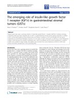
Synthesis of sunscreen nanoparticles embedded into lipid
matrices
Different GS:CP nanosuspensions were produced by a
modified melt homogenisation method. The steps fol-
lowed in synthesis of lipid nanoparticles loaded with
both couples of molecular sunscreens (OMC-OCT-SLN
and BEMT-OCT-SLN) are presented in Figure 1. The
lipid mixture (hexadecyl palmitate:GS = 1:1, w/w) was
melted at the temperature of 85°C. In the melted lipids
that represent 10% from the total SLN dispersion, an
amount of 1% sunscreen mixture was added. A solution
of polyet hylene glycol sorbitan monooleate, synperonic
PE and lecithin (1:0.25:0.25, w/w) in deionised water
was heated to the same temperature. Before the forming
of lipid pre-emulsion, the aqueous surfactant solution
was processed by high shear homogenisation (using a
Lab High-Shear Homo geniser SAII-20 type; 0-28,000
rpm and power o f 300 W, Shanghai Sower Mechanical
& Electrical Equipment Co., Ltd., China) for 2 min at
25,000 rpm in order to destroy the multilamellar lipo-
some formed by lecithin. The hot pre-emulsion was
further processed by applying 25,000 rpm for 15 min.
The lipid nanoparticles dispersion obtained by adding
50 mL water was exposed to lyophilisation in order to
increase the loaded-SLN concentration (using a Christ
Delta 2-24 KD lyophiliser, Germany). The sunscreen
Lacatusu et al. Nanoscale Research Letters 2011, 6:73
/>Page 2 of 8
loaded lipid nanoparticles have been analysed by
dynamic light scattering (DLS), TEM, DSC, UV-Vis
techniques and SPF analyses.
Methods and equipment for lipid nanoparticle
characterisation
DLS technique
Particle size (z-average) and polydispersity index (PI) of
each SLN dispersion were determined after 1 day of pre-
paration and few months later, using dynamic light scat-
tering technique (Zetasizer Nano ZS, Malvern Instruments
Ltd., UK), at a scattering angle of 90° and 25°C. Disper-
sions were analy sed after appropriate dilution with deio-
nised water to an adequate scattering intensity prior to the
measurement. T he particle size analysis data were evalu-
ated using intensity distribution. The zeta potential of the
SLN dispersions was evaluated with the same DLS techni-
que. For each sample, the hydr odynamic radius and zeta
potential have been measured in triplicate.
Transmission electronic microscopy
The morphology of OMC-OCT - S LN and BEMT-OCT
- SLN was examined using a transmission electron
microscope (Philips 208 S, Netherlands). A drop of the
diluted lipid nanoparticle solution was placed onto a
carbon-coated copper grid and kept for 15 min before
the samples were viewed and photographed.
Differential scanning calorimetry
In order to investigate the changes in the crystallinity of the
lipid matrix, DSC analysis was performed. Thermograms
were recorded w ith a d ifferen tial scanning calorimeter Jupi-
ter, STA 449C (from Netzsch Instruments N.A. LLC).
Samples were heated at the scanning rate of 3°C/min over
a temperature range between 30 and 100°C.
In vitro determination of SPF
The determination of SPF ratings was realised using
UV-Vis V670 Spectrophotometer equipped with inte-
grated sphere and the adequate soft. For SPF evaluation,
an amount of 2 mg/cm
2
cream is applied onto T rans-
pore™ 3M support (a synthetic skin) and the sample
spectrum is registered on 290-400 nm, using a reference
support - Transpore™ 3M without cream. The method
for in vitro determination of SPF of sunscreen s is based
on Diffey and Robson theory [19]:
SPF
MPF
()
()
400 290
400 290
EB
EB
where E
l
sun radiation extinction for Earth (between
20° and 40° N latitude); B
l
relative extinction for each
wavelength; MPF
l
the monochromatic protection factor
for selected wavelength (the difference between the
spectrum of measured sample applied on support and
support spectrum).
UV-A and UV-B irradiation
The photostability of UV-absorber couples-SLN has
been evaluated by irradiation on UVA-UVB with an
Aqueous Phase
(surfactants mixture, 3%)
Lipid Phase
(GS:CP mixture, 10%; sunscreen mixture, 1%)
1
. Magnetic stirring, 1/2h, 85
o
C
2. High shear homogenisation,
25.000, 2 min.
Magnetic stirring, 1/2h, 85
o
C
Lipid pre-emulsion
1. Magnetic stirring, 85
o
C, 2h
2. High shear homogenisation, 25.000, 15 min.
3. Add deionised water
Lipid nanoparticles
dispersion
-40
o
Cl
y
o
p
hilisation
,
,
8h
Lyophilised lipid
nanoparticles
Physico-chemical characterization
(DLS, TEM, DSC, UV-VIS, SPF)
Cosmetic formulation (SPF
evaluation, photochemical stability)
Figure 1 Synthesis procedure of some couples of UV molecular chemical absorbers encapsulated into lipid nanoparticles.
Lacatusu et al. Nanoscale Research Letters 2011, 6:73
/>Page 3 of 8
energy of 19.5 J/cm
2
,attwowavelengths:365nm
(UVA) and 312 nm (UVB) on a short period (1 h on
UVA and 2 h on UVB - irradiation I ) and prolonged
period of time (2 h on UVA and 4 h on UVB - irradia-
tion II), using Irradiation System BioSun, Vilver Lour-
mat, France. The extent of photodegradation was
monitored by recording the absorption spectra in the
wavelength range 290-400 nm on a UV-Vis V670 Spec-
trophotometer (Jasco, Japan), using the accessory with
integrated sphere.
Results and discussion
Size distribution and stability of UV absorbers couples -
SLN
The SLNs suspension is a heterogenou s system with co-
existence of additional colloidal structures (micelles,
liposomes, supercooled melts) which caused a specific
size distribution [11,20], depending on the selected pre-
paration procedure. For this reason, even in the litera-
ture there are some preparation procedures [e.g. high
pressure homogenisation (HPH), microemulsion, solvent
diffusion, high shear homogenisation coupled with ultra-
sound technique], t he most used technique for produc-
tion of SLNs is HPH which allows obtaining of a
narrow size distribution of nanoparticles.
In this st udy is demonstrated the possibility to obtain
lipid nanoparticles with relatively narrow size distribu-
tion and no micron particles using a modified-HSH
technique, without an additional ultrasound treatment.
Due to the use of lecithin that is not able to form
micelles in aqueous solut ion, it for ms only liposomes, a
supplementary shear homogenisation of surfactant aqu-
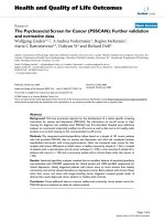
eous solution has led to expected results. All the nano-
particles formulated in this study were completely
distributed in the size range 20-350 nm (Figure 2). The
results obtained by DLS evidenced that for both couples
of UV absor bers encapsulated into lipid nanopar ticles, a
relatively narro w size distribution was o bserved, with a
polydispersity ranging between 0.217 and 0.244. The
average size of lipid nanoparticles after 1 day of prepara-
tion was about 96.5 nm (for OMC-OCT - SLN) and
about 79.5 nm (for OCT-BEMT - SLN).
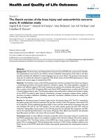
The measurement of zeta potential allows predictions
about the storage s tability of colloidal systems. In gen-
eral, the particles aggregation is unlikely to appear if the
particles are charged and present high zeta potential
values due to the electrostatic repulsions. The zet a
potential distribution for both OMC-OCT - SLN and
BEMT-OCT-SLNisshowninFigure3.Thezeta
potentialvaluesstartfrom-50mVforOMC-OCT-
SLN (with an average potential of -85 mV) and from
-25 mV f or BEMT-OCT - SLN (with an average poten-
tial of -67 mV), respectively. These highly electronega-
tive values demonstrate that using this method a high
stability of SLN systems and good size distribution are
obtained.
In Table 1 are collected the data of particle size o f
lipid nanoparticles loaded with molecular UV-absorber
after 1 day of preparation and after a few months of sto-
rage at 4°C. The SLN suspensions show sufficient long-
term stability with only slight particle size increase after
storage.
Morphologic and crystalline characteristics of molecular
absorbers loaded into SLN
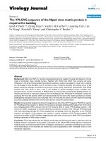
TEM images of SLN loaded with both couples of UV
absorbers which are shown in Figure 4 indicated that
the particles ha d nanometre size and spherical shapes
and no irregular crystallisation with the majority of nee-
dle crystals visible. This last aspect underlines the higher
content in the least ordered crystal structure (a-form) in
the lipid phase, whilst the perfect crystals manifest a
Figure 2
0
5
10
15
0.1 1 10 100 1000 1000
0
Intensity (%)
Size (r.nm)
Size Distribution by Intensity
Record 771: SLN_BEMT_OCT_1 Record 772: SLN_BEMT_OCT_2
Record 773: SLN_BEMT_OCT_3 Record 799: SLN_OMC_OCT_1
Record 800: SLN_OMC_OCT_2 Record 801: SLN_OMC_OCT_3
Figure 2 Size distribution of lipid nanoparticles evaluated by dynamic light scattering.
Lacatusu et al. Nanoscale Research Letters 2011, 6:73
/>Page 4 of 8
0
200000
400000
600000
800000
1000000
1200000
-200 -100 0 100 20
0
Total Counts
Zeta Potential (mV)
Zeta Potential Distribution
Record 780: SLN_OCT_OMC_1 Record 781: SLN_OCT_OMC_2
Record 782: SLN_OCT_OMC_3 Record 783: SLN_OCT_BEMT_1
Record 784: SLN_OCT_BEMT_2
Record 785: SLN_OCT_BEMT_3
Figure 3 Zeta potential distribution for OMC-OCT - SLN and EMT-OCT - SLN.
Table 1 The size evolution/stability of OMC-OCT - SLN and BEMT-OCT - SLN in time
OCT-OMC - SLN
After 24 h After 2 months After 9 months After 12 months
Z
average
[nm] Pdl Z
average
[nm] Pdl Z
average
[nm] Pdl Z
average
[nm] Pdl
96.0 0.240 94.9 0.271 98.7 0.211 101.6 0.242
94.5 0.240 96.2 0.273 97.4 0.244 100.1 0.233
94.4 0.231 98.5 0.273 98.8 0.228 98.9 0.235
OCT-BEMT - SLN
After 24 h After 2 months After 9 months After 12 months
Z
average
[nm] Pdl Z
average
[nm] Pdl Z
average
[nm] Pdl Z
average
[nm] Pdl
79.2 0.244 81.3 0.220 85.4 0.235 88.2 0.203
79.4 0.219 81.9 0.204 84.8 0.218 86.7 0.200
79.7 0.217 81.9 0.216 83.3 0.229 83.2 0.223
B
A
Figure 4 TEM images of lipid nanoparticles: OCT-OMC - SLN (a) and OCT-BEMT - SLN (b).
Lacatusu et al. Nanoscale Research Letters 2011, 6:73
/>Page 5 of 8
typical elongated, needle-shaped crystals characteristic to
amoreorderedstructure(b modification) [21,22]. The
most stable b form is not desired due to the expulsion
in time of UV absorbers. This observation is also con-
firmed by DSC analysis where there is a single crystalli-
sation form of lipid matrices (Figure 5).
Figure 5 shows the allure of the melting process of
bulk lipid matrix (physical mixture of CP and GS), free
lipid nanoparticles and lipid nanoparticles loaded with
UV absorbers. From DSC curves it is observed that the
crystallinity was different in the bulk lipid mixture, free-
SLN and loaded-SLN, due to the presence of surfactants
and molecular UV absorb ers in their compositions. The
lipid mixture exhibits a broad melting range, whilst the
lipid nanoparticles have a narrow peak at 50.2°C (for
empty SLN), 49.5°C (for OCT-OMC - S LN) and 51.4°C
(for OCT-BEMT - SLN), respect ively. The narrow of
melting range in the case of SLNs is a proof of surfac-
tants presence inside the lipid network that confers a
more ordered arrangement. Moreover, by comparing the
free SLN with SLN loaded with OCT-OMC and OCT-
BEMT, it may be observed th at the incorporation of UV
absorbers inside the solid l ipid matrix has led to a
decrease of crystallin arrangement, pointed out by the
decrease of endothermal peak intensity.
Photoprotective effect. In vitro determination of SPF
SPF is the most reliable indicator of the e fficacy of
sunscreen filters, defined as the sun radiation dose
required producing the minimum erythemal do se after
application of 2 mg/cm
2
of sunscreen on unprotected
skin [23]. The UV-Vis spect ra of lyophilised SLN con-
taining 8.3% mixture of OCT-OMC and 7.14% OCT-
BEMT (referring to the lipid matrix and surfactant
composition of SLN) are presented in Figure 6. The
protection regions are clear evidenced for both SLN
types when c ompare to the base cream. As expected,
due to the BEMT presence, the absorption region of
BEMT-OCT is larger than OMC-OCT, this covering a
wide UV domain, between 290 and 375 nm. In both
prepared sunscreen - SLNs, the encapsulation led to a
synergistic UV blocking effect due to the size effect
induced by the op timised surfactant co mposition and
lipid matrix which are known to manifest an anti
UV-effect [24].
The in vitro determination of SPF, based on Diffey
method for the empty base cream was SPF = 1, whilst for
lyophilised OMC-OCT - SLN and BEMT-OCT - SLN
were 19.9 and 19.3, respectively, which assure a good
photoprotection, filtering about 95% of UV radiation.
Photostability behaviour of UV absorbers - SLNs
incorporated into a cosmetic carrier
In order to facilitate the spreading onto a synthetic
skin, the lyophilised SLNs have been inco rporated in
an appropriate cosmetic carrier (a base cream) that
does not induce the dissolution or aggregation of lipid
nanoparticles. The cream formulations have been pre-
pared by dispersing various amounts of lyophilised
sunscreen-SLNs in the cream base, so that the final
cream formulations contained 0.5, 1.25 and 4.5%
sunscreens mixture (w/w), which means less than half
[#] I t t
Fil
Dt
Id tit
Sl
M/
St
R
At h
C
40 50 60 70 80 90 100
Temperature /°C
-0.1
0.0
0.1
0.2
0.3
0.4
0.5
0.6
0.7
DSC /(μV/mg)
Main 2009-10-09 10:23 User: o.oprea
[1]
p
exo
[3]
[4]
[2]
Figure 5 Thermal behaviour of: (1) bulk lipid matrix; (2) unloa ded SLN; (3) SLN loaded with mixture of OMC and OCT; (4) SLN loaded
with mixture of BEMT and OCT.
Lacatusu et al. Nanoscale Research Letters 2011, 6:73
/>Page 6 of 8
of maximum concentration recommended by Food
Drug Administration regulations (for OCT is 7.5% and
for OMC and BEMT is 10%).
The composition of the lipid core influences signifi-
cantly the specific properties of developed formulatio ns.
Thus, the UV absorber type loaded into SLN led to dif-
ferent behaviours at irradiation on short time. The effect
of irradiation conditions on SPF values of the cosmetic
formulations (Figure 7) was examined on wavelength
range 290-400 nm, by irradiation in simulated tanning
conditions. The irradiati on conditions have been chosen
to mimic the low energy existent in the middle day
(19.5 J/cm
2
) [25]. The results shown in Figure 6 have
demonstrated that after UV irradiation, the photoprotec-
tive effec t has be en significantly increase d regardless of
content of UV absorbers, as comparing with formula-
tions before irradiation. For comparison purpose, the
same amount of SLN without UV absorbers has been
subjected t o UV irradiation, but the initial SPF value of
1.2 has been almost unchanged (SPF after first irradia-
tion period was 1.3 and after second period was 1.2).
Even the BEMT has a broad UV-A and UV-B b lock-
ing action, the SPF values for OCT-OMC couple are
higher (SPF = 16 after irradiation I and 20.8 after irra-
diation II), as comparing to the BEMT-OCT couple
(SPF = 8.7 after irradiation I and 12.3 after irradiation
II), for a content of 4.5% molecular sunscreens. Having
in view the fact that these UV absorbers manifest a
good photostability [26], they do not undergo significant
chemical change/photodegradation, allowing them to
retain the UV-absorbing capacity [27]. The main reason
for this behaviour may be explained based on the struc-
ture of UV molecular absorbers, OCT and OMC have
double bonds conjugated with carbonyl groups, which
upon exposure to UV light undergo i somerisation phe-
nomena and are tra nsformed into keto- enolic form. The
BEMT molecule does not present carbonyl groups, thus
avoiding such phenomena.
Conclusion
The encapsulation of both OMC + OCT and OCT +
BEMT UV couples into lipid matrices led to average
particle size less than 100 nm, with a relatively narrow
particle distribution (PI <0.244), using an efficient high
shear homogenisation method. All the collo ida l systems
of nanoparticles have presented zeta potent ial values
less than -50 mV, which assure a high stability of pre-
pared SLNs dispersions.
The crystallisation phenomena of the lipid phase
coupled with microscopy images emphasise the presence
of the less ordered crystal structure of spherical shape
( a-form), whilst avoiding the a ppearance of undesired
0
1
.
9
0.5
1
1.5
290 40
0
300 350
A
bs
Wavelen
g
th [nm]
1
2
3
Figure 6 Wavelength scans of (1) lyophilised OMC-OCT - SLN;
(2) lyophilised BEMT-OCT - SLN; (3) empty base cream.
0.50%
1.25%
4.50%
initial
irradiation I
irradiation II
0
5
10
15
20
25
SPF
UV absorbers amount
initial
irradiation I
irradiation II
A.
0.50%
1.25%
4.50%
initial
irradiation I
irradiation II
0
2
4
6
8
10
12
14
SPF
UV absorbers amount
initial
irradiation I
irradiation II
B.
Figure 7 Effect of irradiation o n SPF value for three cream
formulations which contain: (a) OCT-OMC - SLN; (b) OCT-BEMT
- SLN.
Lacatusu et al. Nanoscale Research Letters 2011, 6:73
/>Page 7 of 8
perfect crystals of needle shape, characteristic for
b modification.
The in vitro deter mination of photoprotection has led
to high SPF ratings, with values of 19.9 and 19.3, respec-
tively, for OMC-OCT - SLN and BEMT-OCT - SLN
(with 8.3% mixture of OCT-OMC and 7.14% O CT-
BEMT), which assure a good photoprotection, filtering
about 95% of UV radiation.
The photostability of developed cosmetic formula-
tions based on sunscreen-SLNs has been evaluated by
exposure to a photochemical UV irradiat ion at a low
energy. The photoprotection effect after irradiation
stage of molecular sunscreens into lipid nanoparticles
was observed to be increased more than twofold com-
pared to initial samples. The incorporation of two
sunscreen couples into SLN leads to a further advan-
tage - penetration of UV absorbers into the skin is
thereby reduced, resulting in a positive effect on the
toxicological potential of the UV absorbers. Thus, it is
possible to obtain a good photoprotection effect, an
improved photostability and a lower allergenic poten-
tial using these sunscreen couples, thus demonstrating
that UV absorbers-SLNs are promising carrier systems
for cosmet ic formulations.
Acknowledgements
This study was supported by CNCSIS - UEFISCSU, project number PNII - IDEI
ID_1050/2007.
Authors’ contributions
IL conceived of the study, performed the synthesis of the lipid nanoparticles
loaded with different UV absorbers, investigate the changes in the
crystallinity of the lipid matrix and carried out the TEM analysis. NB carried
out the in vitro determination of SPF ratings and the evaluation of absorber
couples - lipid nanoparticles photostability by a UV-A and UV-B irradiation
study. AMu participed in the synthesis step and in the size distribution
evaluation by DLS technique. AMe participated in the drafting of the study
and its coordination.
Competing interests
The authors declare that they have no competing interest s.
Received: 25 May 2010 Accepted: 12 January 2011
Published: 12 January 2011
References
1. Teeranachaideekul V, Müller RH, Junyaprasert VB: Encapsulation of ascorbyl
palmitate in nanostructured lipid carriers (NLC)–effects of formulation
parameters on physicochemical stability. Int J Pharm 2007, 340:198.
2. González S, Fernández-Lorente M, Gilaberte-Calzada Y: The latest on skin
photoprotection. Clin Dermatol 2008, 26 :614.
3. Bernd Herzog GW: Formulation of UV absorbers by incorporation in solid
lipid nanoparticles, US Patent No. 7,147,841 B2, Date of Patent: Dec. 12.
2006.
4. Lee GS, Lee DH, Kang K, Lee C, Pyo HB, Choi TB: Preparation and
characterization of bis-ethylhexyloxyphenolmethoxyphenyltriazine
(BEMT) loaded solid lipid nanoparticles (SLN). J Ind Eng Chem 2007,
13:1180.
5. Velasco MVR, Sarruf FD, Salgado-Santos IMN, Haroutiounian-Filho CA,
Kaneko TM, Baby AR: Broad spectrum bioactive sunscreens. Int J Pharm
2008, 363:50.
6. Küchler S, Radowski MR, Blaschke T, Dathe M, Plendl J, Haag R, Schafer-
Korting M, Kramer KD: Nanoparticles for skin penetration enhancement–a
comparison of a dendritic core-multishell-nanotransporter and solid lipid
nanoparticles. Eur J Pharm Biopharm 2009, 71:243.
7. Pardeike J, Hommoss A, Müller RH: Lipid nanoparticles (SLN, NLC) in
cosmetic and pharmaceutical dermal products. Int J Pharm 2009, 366:170.
8. Wissing SA, Müller RH: The influence of solid lipid nanoparticles on skin
hydration and viscoelasticity in vivo study. Int J Pharm 2003, 254:65.
9. Weyenberg W, Filev P, Van den Plas D, Vandervoort J, Smet K, Sollie P,
Ludwig A: Cytotoxicity of submicron emulsions and solid lipid
nanoparticles for dermal application. Int J Pharm 2007, 307:291.
10. Wissing SA, Saupe A, Müller RH: Nanostructured lipid carriers (NLC) for
topical use - an overview. Euro Cosmetics 2003, 5:14.
11. Müller RH, Mäder K, Gohla S: Solid lipid nanoparticles (SLN) for controlled
drug delivery–a review of the state of the art. Eur J Pharm Biopharm
2000, 50:161.
12. Schubert MA, Müller-Goymann CC: Characterisation of surface-modified
solid lipid nanoparticles (SLN): Influence of lecithin and nonionic
emulsifier. Eur J Pharm Biopharm 2005, 61:77.
13. Wissing SA, Muller RH: Solid lipid nanoparticles as carrier for sunscreens:
in vitro release and in vivo skin penetration. J Controlled Release 2002,
81:225.
14. Wissing SA, Kayser O, Muller RH:
Solid lipid nanoparticles for parenteral
drug delivery. Adv Drug Deliv Rev 2004, 56:1257.
15. Müller RH, Mehnert W, Lucks JS, Schwarz C, zur Mühlen A, Weyhers H,
Freitas C, Rühl D: Solid lipid nanoparticles (SLN)–an alternative colloidal
carrier system for controlled drug delivery. Eur J Pharm Biopharm 1995,
41:62.
16. Wissing SA, Lippacher A, Müller RH: Investigations on the occlusive
properties of solid lipid nanoparticles (SLN™). J Cosmet Sci 2001, 52:313.
17. Mandawgade SD, Patravale VB: Development of SLNs from natural lipids:
application to topical delivery of tretinoin. Int J Pharm 2008, 363:132.
18. Wissing SA, Müller RH: The development of an improved carrier system
for sunscreen formulations based on crystalline lipid nanoparticles. Int J
Cosmet Pharm 2002, 242:373.
19. Diffey BL, Robson J: A new substrate to measure sunscreen protection
factors throughout the ultraviolet spectrum. J Soc Cosmet Chem 1989,
40:127.
20. Li H, Zhao X, Ma Y, Zhai G, Li L, Lou H: Enhancement of gastrointestinal
absorption of quercetin by solid lipid nanoparticles. J Controlled Release
2009, 133:238.
21. Weiss J, Decker EA, McClements DJ, Kristbergsson K, Helgason T, Awad TS:
Solid lipid nanoparticles as delivery systems for bioactive food
components. Food Biophysics 2008, 3:146.
22. Helgason T, Awad TS, Kristbergsson K, McClements DJ, Weiss J: Effect of
Surfactant Surface Coverage on Formation of Solid Lipid Nanoparticles
(SLN). J Colloid Interface Sci 2009, 334:75.
23. Lautenschlager S, Wulf HC, Pittelkow MR: Photoprotection. Lancet 2007,
370:528.
24. Müller RH, Radtke M, Wissing SA: Solid lipid nanoparticles and
nanostructured lipid carriers in cosmetic and dermatological
preparations. Adv Drug Deliv Rev 2002, 54:S131.
25. Kullavanijaya P, Lim HW: Photoprotection. J Am Acad Dermatol 2005,
52:937.
26. Gaspar LR, Maia P, Campos PM: Evaluation of the photostability of
different UV filters associations in a sunscreen. Int J Pharm 2006, 307:123.
27. Camile L, Hexsel MD, Scott D, Bangert MD: Current sunscreen issues: 2007
Food and Drug Administration sunscreen labelling recommendations
and combination sunscreen/insect repellent products. J Am Acad
Dermatol 2008,
59:316.
doi:10.1186/1556-276X-6-73
Cite this article as: Lacatusu et al.: The encapsulation effect of UV
molecular absorbers into biocompatible lipid nanoparticles. Nanoscale
Research Letters 2011 6:73.
Lacatusu et al. Nanoscale Research Letters 2011, 6:73
/>Page 8 of 8