Báo cáo hóa học: " A brachialis muscle rupture diagnosed by ultrasound; case report" pdf
Bạn đang xem bản rút gọn của tài liệu. Xem và tải ngay bản đầy đủ của tài liệu tại đây (377.23 KB, 3 trang )
CAS E REP O R T Open Access
A brachialis muscle rupture diagnosed by
ultrasound; case report
Titus JA Schönberger
1*†
and Miranda F Ernst
2†
Abstract
Trauma to the elbow caused by lifting heavy objects frequently involves rupture of the tendon of the biceps
brachii muscle. Less frequently a rupture of the brachialis muscle occurs. To our knowledge, only five cases
involving traumatic rupture of the brachialis muscle were described in the past 20 y ears. We will briefly report
these cases.
To demonstrate and evaluate muscle injuries, magnetic resonance imaging (MRI) is considered the most sensitive
and specific method of choice. We report an isolated brachialis muscle rupture caused by resisted flexion and
pronation of the lower arm. Physical examination combined with ultrasound evaluation confirmed the diagnosis of
ruptured brachialis muscle. Treatment was non-operative with full restoration of function.
Background
Trauma to the elbow caused by lifting heavy objects fre-
quently involves rupture of the tendon of the biceps bra-
chii muscle. Less frequently a rupture of the brachialis
muscle occurs. After an extensive online search, we found
only five cases involving traumatic rupture of the brachia-
lis muscle had been described in the past 20 years. To
demonstrate and evaluate muscle injuries, magnetic reso-
nance imaging (MRI) is considered the most sensitive and
specific method of choice. We report an isolated brachialis
muscle rupture caused by resisted flexion and pronation
of the lower arm. Physical examination combined with
ultrasound evaluation confirmed the diagnosis of ruptured
brachialis muscle. Treatment was non-operative.
Case presentation
A 45-year-old male, right-handed, amateur bodybuilder
and metalworker presented to our emergency depart-
ment with pain in the left elbow after lifting his motor-
cycle. At the time of injury, he noticed a sudden snap in
his left elbow and felt immediate pain and weakness.
There were no previous injuries to the elbow, but the
patient reported a visible dell on the medial surface of
the proximal brachial portion of the arm. There were no
paresthesias of the left upper extremity. The patient
denied the use of medication, drugs or food supplements,
and denied smoking or excessive alcohol use as well.
On physical examination, maximum pain was elicited
on active flexion and pronation of the lower arm. Pas-
sive extension and resisted flexion o f the elbow
enhanced the pain on the medial side of the elbow.
Movement of the palm and fingers did not increase
pain. The biceps and triceps brachii tendons were intact,
and the proximal portion of the ulna and the lateral side
of the distal upper arm were pa inful to pa lpation. There
were no neurological or vascular abnormalities of the
arm.
Conventional radiographs of the elbow revealed no
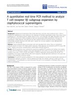
fracture, dislocation or elbow joint effusion. Ultrasound
imaging demonstrated an inhomogeneous structure of
low echogenicity at the ulnar attachment of the brachialis
muscle and direct distally to the coronoid. The brachialis
muscle itself revealed another inhomogeneous structure
with low echogenicity (Figure 1). The pronator teres
muscle was intact.
The diagnosis of a brachialis muscle rupture was made.
The affected arm was immobilized for 1 week using a
plaster cast. After 1 week, the patient was instructed to
gradually exert effort with his arm to maximum tolerable
pain. Out-patient follow-up showed a gradual decrease in
pain and an improvement in function and strength of the
left arm. Near-normal function and strength of the elbow
were achieved 10 weeks after the traumatic event.
* Correspondence:
† Contributed equally
1
Emergency Department, Jeroen Bosch Hospital, ‘s-Hertogenbosch, The
Netherlands
Full list of author information is available at the end of the article
Schönberger and Ernst International Journal of Emergency Medicine 2011, 4:46
/>© 2011 Schön berg er and Ernst; licensee Springer. This is an Open Access article distribute d under the terms of the Creative Commons
Attribution License ( licenses/by/2.0), which permits unrestr icted use, distribution, and reprod uction in
any medium, provided the original work is properly cited .
Discussion
Injury to the brachialis muscle is a rare phenomenon and
is infrequently described in literature [1,2]. This may pro-
mote misdiagnosis of this injury. Furthermore, there are
conflicting thoughts on the anatomy and the precise func-
tion of the brachialis muscle. Gray’s Anatomy describes a
normal variant with two or more parts [3], while Leonello
et al. suggest that all brachialis muscles consist of a super-
ficial and a deep head [4]. The rarity of brachialis muscle
injury and the conflicting thoughts on the normal mor-
phology and function of the muscle make diagnosing and
treating a brachialis muscle injury a real challenge. The
first case report, by Van Den Berghe, presented a male
who was clinically diagnosed with a tear of the biceps bra-
chii muscle after lifting a heavy object. However, a MRI
revealed a tear in the distal aspect of the brachialis muscle.
He was treated conservatively in an outpatient setting and
regained full function in 10 months [5].
Nishida et al. described two cases in which the patients
were referred for evaluation of a possible muscular neo-
plasm. Both patients complained of pain and a loss of
active extension in the elbow 1 week after the injury. MRI
showed a brachialis muscle tear, mimicking an intramus-
cular tumor. Active mobilization was initiated on both
patients with eventual full restoration of function after 3
months [6].
The fourth patient, reported by Winblad et al, was
diagnosed with a brachialis muscle tear after a hyperex-
tension injury of the elbow. MRI sequencing confirmed
the diagnosis. The patient was treated conservatively
with full restoration of function [7].
The final case report was published by Wasserstein
and involves a hyperextension injury of the elbow,
resulting in a brachialis muscle rupture, confirmed by
MRI. Their patient was treated non-operatively and
regained full function [8].
To summarize, expensive diagnostic modalities, such
as MRI, are too often felt to be needed to definitely
diagnose brachi alis muscle injury. In our hospital, ultra-
sound is the first modality of choi ce if additional studies
are needed for diagnosing tendon or muscle ruptures In
the case of equivocal findings from the ultrasound ima-
ging, a MRI sequencing is done for the definit ive diag-
nosis. In our experience , we believe that most brachialis
muscle ruptures can be treated conservatively with early
active mobilization.
Conclusions
To diagnose peripheral muscle ruptures, ultrasound exam-
ination can be an adequate, easy to perform and cost-
effective alternative for MRI sequencing in visualizing ten-
domuscular ruptures. Moreover, we believe that most
cases of ruptured brachialis muscle can be treated
conservatively.
Consent
Written informed consent was obtained from the patient
for publication of this case report and any accompany-
ing images. A cop y of the written consent is available
for review by the Editor-in-Chief of this journal.
Author details
1
Emergency Department, Jeroen Bosch Hospital, ‘s-Hertogenbosch, The
Netherlands
2
Department of Surgery, Jeroen Bosch Hospital, ‘s-
Hertogenbosch, The Netherlands
Figure 1 Inhomogeneous structure with low echogenicity: brachial muscle rupture.
Schönberger and Ernst International Journal of Emergency Medicine 2011, 4:46
/>Page 2 of 3
Authors’ contributions
TS treated the patient and wrote the case report. ME revised the manuscript
critically. Both authors read and approved the final manuscript.
Competing interests
The authors declare that they have no competing interests.
Received: 7 February 2011 Accepted: 26 July 2011
Published: 26 July 2011
References
1. Chavan PR, Duquin TR: Repair of the ruptured distal biceps tendon: a
systematic review. Am J Sports Med 2008, 36(8):1618-24.
2. Safran MR, Graham SM: Distal biceps tendon ruptures: incidence,
demographics, and the effect of smoking. Clin Orthop Relat Res 2002,
404:275-83.
3. Gray H: Gray’s Anatomy. 29 edition. Philadelphia: Lea & Febiger; 1985.
4. Leonello DT, Galley IJ: Brachialis muscle anatomy. A study in cadavers. J
Bone Joint Surg Am 2007, 89(6):1293-7.
5. Van den Berghe GR, Queenan JF: Isolated rupture of the brachialis: a case
report. J Bone Joint Surg Am 2001, 83-A:1074-5.
6. Nishida Y, Tsukushi S: Brachialis muscle tear mimicking an intramuscular
tumor: a report of two cases. J Hand Surg Am 2007, 32:1237-41.
7. Winblad JB, Escobedo E, Hunter JC: Brachialis Muscle Rupture and
Hematoma. Radiology Case Reports 2008, 3:251.
8. Wasserstein D, White L: Traumatic brachialis muscle injury by elbow
hyperextension in a professional hockey player. Clin J Sport Med 2010,
20:211-2.
doi:10.1186/1865-1380-4-46
Cite this article as: Schönberger and Ernst: A brachialis muscle rupture
diagnosed by ultrasound; case report. International Journal of Emergency
Medicine 2011 4:46.
Submit your manuscript to a
journal and benefi t from:
7 Convenient online submission
7 Rigorous peer review
7 Immediate publication on acceptance
7 Open access: articles freely available online
7 High visibility within the fi eld
7 Retaining the copyright to your article
Submit your next manuscript at 7 springeropen.com
Schönberger and Ernst International Journal of Emergency Medicine 2011, 4:46
/>Page 3 of 3