Báo cáo toán học: " Visual Diagnosis: Pearling: a case study" doc
Bạn đang xem bản rút gọn của tài liệu. Xem và tải ngay bản đầy đủ của tài liệu tại đây (788.77 KB, 3 trang )
CAS E REP O R T Open Access
Visual Diagnosis: Pearling: a case study
David P Nguyen, Bobby K Desai
*
and Michael Falgiani
Abstract
We present the case of a patient who attempted to perform a type of body modification known as “pearling” or
“genital beading” while in prison. This patient unfortunately caused severe trauma to his penis, requiring surgical
intervention. Photographs of the traumatic injuries are presented.
Background
“Pearling,” also known as “genital beading” is the prac-
tice of permanently inserting small beads made of var-
ious materials beneath the skin of the genitals [1]. As
well as being an aesthetic practice, this is usually
intended to enhance the pleasure of partners during sex-
ual intercourse by increasing physical stimulation. It is
most commonly done on the dorsal surface of the shaft
of the penis where small, superficial incisions are made
and beads are placed under the skin surface. Most
implants are made of small inert metal beads (stainless
steel, titanium) or plastic beads (nylon, silicone).
This form of body modification is still practiced in
various world cultures. Historically, the Yakuza of Japan,
an organized crime syndicate, is the most well known
for “pearling .” Each pearl supposedly symbolizes each
year that was spent in prison. Interestingly, “pearling”
has become more commonplace in the United States,
especially in the US prison system.
Case presentation
A 19-year-old male inmate presented to our Emergency
Department (ED) after attempting to purposefully cut
the dorsal surface of his penis with a brand-new razor
blade for self-performed “pearling.” He made two hori-
zontal incisions on the shaft, one proximal and close to
the base of the pen is, and one distal near the g lans
pen is. This was performed approx imately 6-7 h prior to
arrival at the ED. The pati ent alerted the prison staff to
request medical evaluation after he noted worsening
pain, swelling and ecchymosis to his penis, as well as a
significant amount of blood when urinating. Upon
arrival, the patient appeared to be in no acute distress,
without obvious active bleeding. He denied dysuria.
In the Emergency Department, the patient’ sinitial
vital signs were: temperature of 37°C, pulse of 84 beats
per minute, respiratory rate of 16, and blood pressure of
141/88 mmHg. His airway was patent with clear, bilat-
eral breath sounds and unlabored breathing. On cardiac
exam the patient had a regular rate and rhythm. His
abdomen was soft, non-tender, and non-distended. Neu-
rological exam revealed no gross motor or sensory
deficits.
After removal of bandaging placed by prison medical
staff, his genitouri nary exam revealed an uncircumci sed
penis with two horizontal lacerations on the dorsal
shaft, one about 1.5 cm from the base of the penis and
about 1 cm in width, a nd the other about 1 cm from
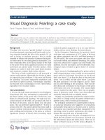
the glans and about 1 cm wide (Figures 1 and 2). There
was no active bleeding to the lacerations. There was dif-
fus e edema and ecchymosis on the dorsum of the penis
with blood clots over the wounds. The wound depth
was not explored at that point. There was no paraphi-
mosis or phimosis noted. Testes were descended and
nontender bilaterally with no palpable masses.
Urology was emergently consulted for surgical evalua-
tion. Prior to Urology arrival, the patient urinated into a
portable urinal, which revealed gross hematuria.
Per urological assessment, his marked penile ecchymo-
sis and gross hematuria were suggestive of a hematoma
and possible deep injury to the penis and or urethra.
The pati ent was consented and taken emergently to the
operating room for penile exploration and repair. A
tetanus shot was given prior to leaving the ED.
In the operating room, the penis was degloved. It was
found that the patient’stwolacerationsinvolvedonly
the subcutane ous tissue and dartos fascia. There was no
injury to Buck’ s fas cia or to the tunica albuginea. A
* Correspondence:
Department of Emergency Medicine, University of Florida, PO Box 100186,
Gainesville 32610, FL, USA
Nguyen et al. International Journal of Emergency Medicine 2011, 4:74
/>© 2011 Nguyen et al; licensee Springer. This is an Ope n Access article distributed under the terms of the Creative Commons
Attribution License ( which permits unrestricted use, dis tribution, and reproduction in
any medium , provided the original work is properly cited.
small subcutaneous hematoma was also evacuated from
the proximal laceration. Irrigation of the wounds
revealed several bleeding vessels within each wound,
and they were cauterized with Bovie electrocautery. The
postoperative diagnosis listed in the operative report
was low velocity sharp penile injury.
The patient was subsequently brought to the surgical
recovery room (PACU) in st able condition, and when
fully recovered, he was discharged back to law enforce-
ment custody. He re ceived instructions to remove the
postoperative dressings the next day, and was discharged
with 5 days of cephalexin and pain medication. He was
toreturntotheclinicin2weeksforapostoperative
check.
Discussion
Penile injuries, especially self-inflicted, are uncommon
complaints in the ED. This case highlights a body modi-
fication practice known as “pearling” or “genital bead-
ing.” In contemporary societies, this procedure is usually
performed by professional body piercers where it is rela-
tively safe and without major complications. However,
“pearling” has apparently gained increasing popularity in
the prison system where inmates have been doing this
on their own with limited tools and knowledge of penile
anatomy. This can lead to disastrous outcomes that
need emergency and surgical care, as seen in this case.
Other known complications due to pearling include
penile abscess and pain on erection [2]. Long-term com-
plications can include scar tissue formation causing
chronic pain and/or erectile dysfunction. This is an
uncommon injury in the ED, and if there is any suspi-
cion of injury to deep penile structures, including the
urethra, a urologic consultation is recommended.
Conclusions
“Pearlin g,” while intended to increase the sexual plea-
sure of partners, can cause significant morbidity to indi-
viduals themselves during object placement.
Consent
Written informed consent was obtained from the patient
for publication of this case report and any accompany-
ing images. A copy of the written consent is available
for review from the Editor-in-Chief of this journal.
Figure 1 Laceration to dorsal surface of penis.
Figure 2 Close-up view of lacerations.
Nguyen et al. International Journal of Emergency Medicine 2011, 4:74
/>Page 2 of 3
Abbreviations
PACU: Post-Anesthesia Care Unit.
Authors’ contributions
DN and BD saw the patient and obtained consent; DN wrote the initial
report; BD and MF edited and revised the report, and added the discussion.
All authors read and approved the final manuscript.
Competing interests
The authors declare that they have no competing interest s.
Received: 14 September 2011 Accepted: 8 December 2011
Published: 8 December 2011
References
1. Fischer N, Hauser S, Brede O: Implantation of artificial penile nodules–a
review of literature. J Sex Med 2010, 7(11):3565-3571.
2. Marzouk E: Implantation of beads into the penile skin and its
complications. Scand J Urol Nephrol 1990, 24(3):167-169.
doi:10.1186/1865-1380-4-74
Cite this article as: Nguyen et al.: Visual Diagnosis: Pearling: a case
study. International Journal of Emergency Medicine 2011 4:74.
Submit your manuscript to a
journal and benefi t from:
7 Convenient online submission
7 Rigorous peer review
7 Immediate publication on acceptance
7 Open access: articles freely available online
7 High visibility within the fi eld
7 Retaining the copyright to your article
Submit your next manuscript at 7 springeropen.com
Nguyen et al. International Journal of Emergency Medicine 2011, 4:74
/>Page 3 of 3