báo cáo hóa học:" Increased androgen receptor expression in serous carcinoma of the ovary is associated with an improved survival" doc
Bạn đang xem bản rút gọn của tài liệu. Xem và tải ngay bản đầy đủ của tài liệu tại đây (1.05 MB, 6 trang )
Nodin et al. Journal of Ovarian Research 2010, 3:14
/>Open Access
RESEARCH
© 2010 Nodin et al; licensee BioMed Central Ltd. This is an Open Access article distributed under the terms of the Creative Commons
Attribution License ( which permits unrestricted use, distribution, and reproduction in
any medium, provided the original work is properly cited.
Research
Increased androgen receptor expression in serous
carcinoma of the ovary is associated with an
improved survival
Björn Nodin
1
, Nooreldin Zendehrokh
1
, Jenny Brändstedt
1,2
, Elise Nilsson
1
, Jonas Manjer
2,3
, Donal J Brennan
4
and
Karin Jirström*
1
Abstract
Background: Altered androgen hormone homeostasis and androgen receptor (AR) activity have been implicated in
ovarian carcinogenesis but the relationship between AR expression in ovarian cancer and clinical outcome remains
unclear.
Methods: In this study, the prognostic impact of AR expression was investigated using immunohistochemistry in
tissue microarrays from 154 incident cases of epithelial ovarian cancer (EOC) in the prospective, population-based
cohorts Malmö Diet and Cancer Study and Malmö Preventive Project. A subset of corresponding fallopian tubes (n =
36) with no histopathological evidence of disease was also analysed.
Results: While abundantly expressed in the majority of fallopian tubes with more than 75% positive nuclei in 16/36
(44%) cases, AR was absent in 108/154 (70%) of EOC cases. AR expression was not related to prognosis in the entire
cohort, but in the serous subtype (n = 90), AR positivity (> 10% positive nuclei) was associated with a prolonged
disease specific survival in univariate (HR= 0.49; 95% CI 0.25-0.96; p= 0.038) and multivariate (HR= 0.46; 95% CI 0.22-
0.97; p= 0.042) analysis, adjusted for age, grade and clinical stage.
Conclusions: AR expression is considerably reduced in EOC as compared to fallopian tubes, and in EOC of the serous
subtype, high AR expression is a favourable prognostic factor. These results indicate that assessment of AR expression
might be of value for treatment stratification of EOC patients with serous ovarian carcinoma.
Background
Epithelial ovarian carcinoma (EOC) is the second most
common and the most lethal malignancy of the female
reproductive tract [1]. Etiological factors involved in
ovarian carcinogenesis remain poorly defined, and effec-
tive treatment protocols are limited. Alterations in andro-
gens and androgen receptor homeostasis have been
implicated in ovarian carcinogenesis and progression [2-
5].
While several immunohistochemistry (IHC)-based
studies have confirmed widespread AR expression in
EOC [6-8], data describing it as a prognostic biomarker
are relatively sparse. One study describing a large series of
tumors (n = 322), found no association between AR pro-
tein expression and clinical outcome [8], however indi-
vidual histological subtypes were not examined.
Increased levels of AR mRNA have been described in
cells from normal ovarian surface epithelium as com-
pared to ovarian cancer cells, the majority of which were
derived from serous tumors [9]. We are, however,
unaware of any studies describing AR expression in fallo-
pian tubes, from which a substantial but not yet not fully
appreciated proportion of serous ovarian carcinomas are
thought to arise [10].
The purpose of this study was to analyze the prognostic
impact of AR expression in 154 EOCs collected from two
population-based, prospective cohorts. Based on the in
vitro data described above [9], our hypothesis was that
AR protein expression may be down-regulated in EOC
compared to fallopian tubes and the prognostic value of
* Correspondence:
1
Center for Molecular Pathology, Department of Laboratory Medicine, Lund
University, and Skåne Regional Laboratories, Malmö, Sweden
Full list of author information is available at the end of the article
Nodin et al. Journal of Ovarian Research 2010, 3:14
/>Page 2 of 6
AR would become more obvious when tumors were strat-
ified into serous and non-serous histological subtypes.
Methods
Patients
Tumors (n = 154) from all incident cases of invasive EOC
that had occurred in two prospective, population-based
cohorts, the Malmö Diet and Cancer Study (MDCS)[11]
and Malmö Preventive Project (MPP) cohorts [12] up to
Dec 31
st
2007 were collected and histopathologically re-
evaluated. The MDCS was initiated in 1991 and enrolled
17035 healthy women [11]. The MPP was established in
1974 for screening with regard to cardiovascular risk fac-
tors and enrolled 10.902 women[12].
The standard surgical management was a total abdomi-
nal hysterectomy, bilateral salpingo-oophorectomy and
omentectomy with cytological evaluation of peritoneal
fluid or washings. Routine pelivic lymphadenectomy was
not performed. Residual disease was resected to less than
1 cm where possible. Volume of residual disease was not
availabe. Standard adjuvant therapy was combination of
paclitaxel and platinum-based chemotherapy.
Median age at diagnosis was 62 (range 47-83). Informa-
tion on cause of death was obtained by matching with the
Swedish Cause-of-Death Registry. After a median follow-
up of 2.67 years (Range 0-21.14 years) 105 patients were
dead, 98 from ovarian cancer. Approval was obtained
from the Ethics committee at Lund University (Ref no
335-08) Study design, methodological and technical con-
siderations, as well as data presentation were based on
the REMARK criteria [13]
Tissue microarrays and immunohistochemistry
TMAs were constructed as previously described[14].
Two 1.0 mm cores were taken from viable, non-necrotic
tumor areas, when possible from both ovaries, and from
concomitant peritoneal metastases (n = 33). Fallopian
tubes with no evidence of histological disease were also
sampled from 38 cases.
Four μm TMA-sections and 3μm full-face sections
were deparaffinised and rehydrated. Heat mediated anti-
gen retrieval (pH = 9) was performed using the PT-link
system and IHC was performed in the DAKO
Autostainer system (Dako, Glostrup, Denmark) using
mouse monoclonal anti-AR antibody (1:200 dilution; AR
441, LAB VISION, Warm Springs, CA), anti-ER antibody
(1:50 dilution; M 7047 Dako), and anti-PR antibody
(1:400 dilution; M 3569 Dako).
To control for heterogenous expression patterns, IHC
was also performed on full-face sections from 15 ran-
domly selected cases and compared to corresponding
cores. AR expression was also examined on full-face sec-
tions from fallopian tubes obtained from 10 patients who
had undergone hysterectomy for benign disease.
Statistics
Spearman's Rho correlation and the χ
2
test were used to
estimate the relationship between AR expression and
clinicopathological parameters. Kappa-statistics were
used as a measure of agreement between scoring of tissue
cores and full-face sections. Kaplan-Meier analysis and
log rank test were used to illustrate differences in ovarian
cancer specific survival (OCSS) between strata. Cox
regression proportional hazards models were used to
estimate the relationship between survival and AR status,
age, stage and grade. All calculations were performed
using SPSS version 17.0 (SPSS Inc, Chicago, IL). P values
< 0.05 were considered statistically significant.
Results
AR expression in fallopian tubes, primary and metastatic
EOC
Thirty-six of the 38 fallopian tubes were suitable for anal-
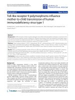
ysis. AR protein expression was evident in the majority of
fallopian tubes with > 75% positivity seen in 44% (n = 16)
of cases (Figure 1). AR was also abundantly expressed (>
50%) in 10 fallopian tubes with a benign diagnosis (data
not shown).
All primary tumors (n = 154) and metastatic deposits (n
= 33) were suitable for analysis. Compared to tubal epi-
thelium, AR protein expression was lower in primary
tumors and metastases, with absent expression in 70% (n
= 108) of primary tumors and 67% (n = 22) of metastatic
deposits (Figure 1). AR expression in primary tumors cor-
related with expression in metastases (R= 0.95, p < 0.001)
particularly when serous carcinomas (n = 90) were ana-
lyzed separately (R = 0.97, p < 0.001). No correlation was
seen between tubal AR expression and expression in
either primary or metastatic tumors. As samples from all
three locations were only available for six patients, this
study did not allow for a meaningful analysis of AR
expression related to individual tumor progression.
AR expression in full-face sections correlated with
TMA-based scoring (kappa-value 0.87, p = 0.001, n = 15),
suggesting that AR is a suitable protein for TMA-based
analysis.
Correlation between AR expression and clinicopathological
parameters
No significant association was evident between AR
expression in primary tumors and conventional clinico-
pathological parameters in the entire cohort (n = 154)
(Table 1). In primary tumors, AR expression was associ-
ated with ER and PR positivity (Table 1). Subset analysis
of serous carcinoma's (n = 90), revealed that the associa-
tion between AR and ER positivity remained significant,
whereas the relationship with PR expression was lost
(Table 1). AR expression was also associated with well-
differentiated serous tumors (Table 1).
Nodin et al. Journal of Ovarian Research 2010, 3:14
/>Page 3 of 6
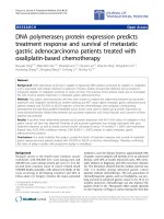
Figure 1 Immunohistochemical AR staining and distribution in fallopian tubes, ovarian cancer and omental metastases. AR nuclear staining
was assessed as the percentage of positive tumor cells (grading 0-1%, 2-10%, 11-50%, 51-75%, >75%).Examples of tumors with low AR expression are
visualized in the left panels and tumors with high expression in the right panels. Bars in the middle represent the distribution of positive cases in ab-
solute numbers.
Nodin et al. Journal of Ovarian Research 2010, 3:14
/>Page 4 of 6
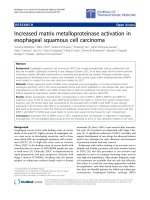
AR expression in relation to survival
Analysis of the entire cohort (n = 154) revealed no rela-
tionship between increased AR expression (> 10%) in pri-
mary tumors and outcome (Figure 2A). However, subset
analysis in serous carcinomas (n = 90) revealed that
increased AR expression was associated with a prolonged
OCSS (p = 0.034) (Figure 2B). Cox univariate analysis
confirmed the association between AR and OCSS in
serous carcinomas (HR= 0.49; 95% CI 0.25-0.96; p=
0.038) and this association remained significant in a mul-
tivariate model controlling for age, grade and stage (HR=
0.46; 95% CI 0.22-0.97; p= 0.042). AR was not prognostic
in non-serous carcinomas (data not shown).
Table 1: Correlations between androgen receptor status and patient and tumour characteristics in all tumours and serous carcinomas
respectively
All tumours Serous carcinoma
AR low AR high AR low AR high
126 28 p-value ± 71 19 p-value ±
Age
median(range) 62(47-83) 63(50-79) 0.800 63(47-83) 64(50-79) 0.514
Histological subtype
Serous 71 19 0.207
Endometroid 28 7
Other 27 2
Stage
I 22 4 0.667 4 2 0.136
II 16 2 5 2
III 57 18 41 13
IV 19 3 13 1
missing 12 1 8 1
Differentiation grade
High/intermediate 37 10 0.511 14 7 0.015
low 8918 5710
ER
Negative 62 5 0.003 28 2 0.024
Positive 60 21 40 15
missing 4 2 3 2
PR
Negative 103 18 0.033 61 15 0.676
Positive 19 9 9 3
AR = androgen receptor, ER= estrogen receptor, PR= progesterone receptor
< 10% positive nuclei used as cutoff for AR, ER and PR positivity
± Mann Whitney u-test for comparison of medians and Chi-square test for categorized variables
Cases with missing values were not included in the analysis
Nodin et al. Journal of Ovarian Research 2010, 3:14
/>Page 5 of 6
Discussion
Evaluation of AR protein expression in 154 EOC cases
from two large, prospective population-based studies
demonstrated frequent expression of AR in fallopian tube
epithelium irrespective of the presence of ovarian cancer
and decreased AR expression in primary ovarian tumors
and metastatic deposits. While not conferring a prognos-
tic value within the entire cohort, reduced AR expression
was an independent predictor of decreased OCSS in
serous tumors. The main limitation of this study is the
absence of data on residual disease and future studies of
AR expression in EOC should incorporate this in any
multivariate analysis.
While associated with AR expression, neither ER nor
PR expression correlated with survival in this study. Such
findings contrast with Lee et al. who reported PR, but not
ER or AR expression as an independent predictor of good
prognosis [8]. In their study, however, the prognostic
value of hormone receptors was not analysed in strata
according to different histological subtypes, an approach
that has been deemed an essential component of EOC
biomarker studies[15]. These findings further highlight
the heterogeneity of ovarian cancer, which should not be
considered as a single disease, but rather several distinct
entities with different clinical behaviours. These entities
are in part reflected in histopathological characteristics
and therefore, to obtain better prognostic and predictive
information biomarkers should not only be assessed
across entire cohorts, but also in histological subgroups.
Although androgen receptors are expressed in normal
ovarian surface epithelium[16], we are not aware of any
previous reports describing AR expression in tubal epi-
thelium. Recent reports have suggested that a significant
proportion of serous carcinomas arise within the fimbrial
tubal epithelium [10,17,18]. Our findings indicate that
malignant transformation could involve a downregula-
tion of AR in certain EOC cases. AR expression in pri-
mary ovarian tumors and metastases was quite similar,
suggesting that downregulation of AR occurs early in
ovarian carcinogenesis.
This is to our knowledge the first report on AR expres-
sion in EOC from population-based cohorts, potentially
representing a selected part of the background popula-
tion. Nevertheless, as established prognostic parameters,
i.e. clinical stage and histological grade, are highly signifi-
cant indicators of survival in this cohort, its use for
assessment of investigative prognostic markers is justi-
fied.
Conclusions
These data demonstrate that AR is an independent
marker of prolonged OCSS in patients with serous carci-
noma of the ovary, and thus a potentially relevant bio-
marker for treatment stratification in this subgroup. Our
findings also highlight the need for further studies inves-
tigating the influence of both lifestyle-related and genetic
factors in relation to ovarian cancer risk in general and to
AR-defined subtypes in particular.
Abbreviations
AR: Androgen Receptor; EOC: Epithelial Ovarian Cancer; OCSS: Ovarian Cancer
Specific Survival; TMA: Tissue Microarray; IHC: Immunohistochemistry; ER:
Estrogen Receptor; PR: Progesterone Receptor
Competing interests
The authors declare that they have no competing interests.
Authors' contributions
BN carried out the immunohistochemical analysis, performed the statistical
analysis, and drafted the manuscript. NZ and EN carried out the immunohis-
tochemical analysis and drafted the manuscript. JB collected the clinical data.
JM participated in collection of clinical data and drafted the manuscript. DB
performed the statistical analysis and drafted the manuscript. KJ participated
in the conception and design of the study, performed the histopathological re-
Figure 2 Impact of androgen receptor expression on ovarian can-
cer specific survival. Kaplan-Meier curves visualizing OCSS according
to AR expression in all tumors (A) and serous carcinomas (B), using a
threshold of 10% positive nuclei to define low and high AR expression.
The total number of events was 52/71 (73%) in AR high serous tumors
and 11/19 (58%) in AR low serous tumors.
Nodin et al. Journal of Ovarian Research 2010, 3:14
/>Page 6 of 6
evaluaton and drafted the manuscript. All authors read and approved the final
manuscript.
Acknowledgements
This work was supported by grants from the Swedish Cancer Society, Gunnar
Nilsson's Cancer Foundation, the Crafoord Foundation and the Research funds
of Skåne University Hospital, Malmö. The UCD Conway Institute is funded by
the Programme for Third Level Institutions (PRTLI), as administered by the
Higher Education Authority (HEA) of Ireland.
Author Details
1
Center for Molecular Pathology, Department of Laboratory Medicine, Lund
University, and Skåne Regional Laboratories, Malmö, Sweden,
2
Department of
Clinical Sciences, Division of Surgery, Lund University, Skåne University
Hospital, Malmö, Sweden,
3
The Malmö Diet and Cancer Study, Skåne University
Hospital, Malmö, Sweden and
4
UCD School of Biomolecular and Biomedical
Science, UCD Conway Institute, University College Dublin, Dublin, Ireland
References
1. Jemal A, Siegel R, Ward E, Hao Y, Xu J, Thun MJ: Cancer statistics, 2009.
CA Cancer J Clin 2009, 59(4):225-49.
2. Helzlsouer KJ, Alberg AJ, Gordon GB, Longcope C, Bush TL, Hoffman SC,
Comstock GW: Serum gonadotropins and steroid hormones and the
development of ovarian cancer. Jama 1995, 274:1926-1930.
3. Silva EG, Tornos C, Fritsche HA Jr, el-Naggar A, Gray K, Ordonez NG, Luna
M, Gershenson D: The induction of benign epithelial neoplasms of the
ovaries of guinea pigs by testosterone stimulation: a potential animal
model. Mod Pathol 1997, 10:879-883.
4. Ludwig A, Murawska M, Panek G, Timorek A, Kupryjanczyk J: Androgen,
progesterone and FSH receptor polymorphisms and ovarian cancer
risk and outcome. Endocr Relat Cancer 2009.
5. Schildkraut JM, Murphy SK, Palmieri RT, Iversen E, Moorman PG, Huang Z,
Halabi S, Calingaert B, Gusberg A, Marks JR, Berchuck A: Trinucleotide
repeat polymorphisms in the androgen receptor gene and risk of
ovarian cancer. Cancer Epidemiol Biomarkers Prev 2007, 16:473-480.
6. Cardillo MR, Petrangeli E, Aliotta N, Salvatori L, Ravenna L, Chang C,
Castagna G: Androgen receptors in ovarian tumors: correlation with
oestrogen and progesterone receptors in an immunohistochemical
and semiquantitative image analysis study. J Exp Clin Cancer Res 1998,
17:231-237.
7. Chadha S, Rao BR, Slotman BJ, van Vroonhoven CC, Kwast TH van der: An
immunohistochemical evaluation of androgen and progesterone
receptors in ovarian tumors. Hum Pathol 1993, 24:90-95.
8. Lee P, Rosen DG, Zhu C, Silva EG, Liu J: Expression of progesterone
receptor is a favorable prognostic marker in ovarian cancer. Gynecol
Oncol 2005, 96:671-677.
9. Lau KM, Mok SC, Ho SM: Expression of human estrogen receptor-alpha
and -beta, progesterone receptor, and androgen receptor mRNA in
normal and malignant ovarian epithelial cells. Proc Natl Acad Sci USA
1999, 96:5722-5727.
10. Dubeau L: The cell of origin of ovarian epithelial tumours. Lancet Oncol
2008, 9:1191-1197.
11. Berglund G, Elmstahl S, Janzon L, Larsson SA: The Malmo Diet and Cancer
Study. Design and feasibility. J Intern Med 1993, 233:45-51.
12. Berglund G, Eriksson KF, Israelsson B, Kjellstrom T, Lindgarde F, Mattiasson
I, Nilsson JA, Stavenow L: Cardiovascular risk groups and mortality in an
urban swedish male population: the Malmo Preventive Project. J Intern
Med 1996, 239:489-497.
13. McShane LM, Altman DG, Sauerbrei W, Taube SE, Gion M, Clark GM:
REporting recommendations for tumour MARKer prognostic studies
(REMARK). Eur J Cancer 2005, 41:1690-1696.
14. Kononen J, Bubendorf L, Kallioniemi A, Barlund M, Schraml P, Leighton S,
Torhorst J, Mihatsch MJ, Sauter G, Kallioniemi OP: Tissue microarrays for
high-throughput molecular profiling of tumor specimens. Nat Med
1998, 4:844-847.
15. Kobel M, Kalloger SE, Boyd N, McKinney S, Mehl E, Palmer C, Leung S,
Bowen NJ, Ionescu DN, Rajput A, et al.: Ovarian carcinoma subtypes are
different diseases implications for biomarker studies. PLoS Med 2008,
5:e232.
16. Edmondson RJ, Monaghan JM, Davies BR: The human ovarian surface
epithelium is an androgen responsive tissue. Br J Cancer 2002,
86:879-885.
17. Carlson JW, Miron A, Jarboe EA, Parast MM, Hirsch MS, Lee Y, Muto MG,
Kindelberger D, Crum CP: Serous tubal intraepithelial carcinoma: its
potential role in primary peritoneal serous carcinoma and serous
cancer prevention. J Clin Oncol 2008, 26:4160-4165.
18. Lee Y, Miron A, Drapkin R, Nucci MR, Medeiros F, Saleemuddin A, Garber J,
Birch C, Mou H, Gordon RW, et al.: A candidate precursor to serous
carcinoma that originates in the distal fallopian tube. J Pathol 2007,
211:26-35.
doi: 10.1186/1757-2215-3-14
Cite this article as: Nodin et al., Increased androgen receptor expression in
serous carcinoma of the ovary is associated with an improved survival Jour-
nal of Ovarian Research 2010, 3:14
Received: 27 April 2010 Accepted: 17 June 2010
Published: 17 June 2010
This article is available from: 2010 Nodin et al; licensee BioMed Central Ltd. This is an Open Access article distributed under the terms of the Creative Commons Attribution License ( ), which permits unrestricted use, distribution, and reproduction in any medium, provided the original work is properly cited.Journal of Ovarian Research 2010, 3:14