Báo cáo hóa học: " Green fluorescent protein as a reporter of prion protein folding" potx
Bạn đang xem bản rút gọn của tài liệu. Xem và tải ngay bản đầy đủ của tài liệu tại đây (951.01 KB, 9 trang )
BioMed Central
Page 1 of 9
(page number not for citation purposes)
Virology Journal
Open Access
Research
Green fluorescent protein as a reporter of prion protein folding
Snezana Vasiljevic
†
, Junyuan Ren
†
, YongXiu Yao, Kevin Dalton,
Catherine S Adamson and Ian M Jones*
Address: School of Animal and Microbial Sciences, The University of Reading, Reading RG6 6AJ, UK
Email: Snezana Vasiljevic - ; Junyuan Ren - ; YongXiu Yao - ;
Kevin Dalton - ; Catherine S Adamson - ; Ian M Jones* -
* Corresponding author †Equal contributors
Abstract
Background: The amino terminal half of the cellular prion protein PrP
c
is implicated in both the
binding of copper ions and the conformational changes that lead to disease but has no defined
structure. However, as some structure is likely to exist we have investigated the use of an
established protein refolding technology, fusion to green fluorescence protein (GFP), as a method
to examine the refolding of the amino terminal domain of mouse prion protein.
Results: Fusion proteins of PrP
c
and GFP were expressed at high level in E.coli and could be purified
to near homogeneity as insoluble inclusion bodies. Following denaturation, proteins were diluted
into a refolding buffer whereupon GFP fluorescence recovered with time. Using several truncations
of PrP
c
the rate of refolding was shown to depend on the prion sequence expressed. In a variation
of the format, direct observation in E.coli, mutations introduced randomly in the PrP
c
protein
sequence that affected folding could be selected directly by recovery of GFP fluorescence.
Conclusion: Use of GFP as a measure of refolding of PrP
c
fusion proteins in vitro and in vivo proved
informative. Refolding in vitro suggested a local structure within the amino terminal domain while
direct selection via fluorescence showed that as little as one amino acid change could significantly
alter folding. These assay formats, not previously used to study PrP folding, may be generally useful
for investigating PrP
c
structure and PrP
c
-ligand interaction.
Background
The cellular prion protein PrP
c
is a glycosylinositol phos-
pholipid (GPI) anchored glycoprotein present on neuro-
nal and other cells [1,2] with a demonstrable ability to
bind and transport copper ions [3-6]. The protein is essen-
tial for susceptibility to the Transmissible Spongiform
Encephalopathies (TSEs) where the accumulation of a dis-
ease associated conformational variant, PrP
Sc
, is depend-
ent on the presence of the cellular PrP
c
isoform (for
reviews [7-9]). A role for prion protein in copper metabo-
lism may be linked to cell resistance to oxidative stress
and, thereby, to pathology [10-16]. The C-terminal
domain of mouse PrP
c
, whose structure has been deter-
mined by NMR, has three α-helices and a short section of
antiparallel β-sheet [17]. It folds quickly in vitro to a stable
structure largely unaffected by amino acid substitution
[18,19]. By contrast, the N-terminal domain of PrP
c
is flex-
ibly disordered in the full-length molecule [20,21]. This
region encodes the octarepeat motifs (residues 23–90)
responsible for low affinity copper binding [3,4,22-24]
and the central hydrophobic region of PrP
c
observed to be
toxic to cells in culture [25], that also binds copper
Published: 29 August 2006
Virology Journal 2006, 3:59 doi:10.1186/1743-422X-3-59
Received: 28 June 2006
Accepted: 29 August 2006
This article is available from: />© 2006 Vasiljevic et al; licensee BioMed Central Ltd.
This is an Open Access article distributed under the terms of the Creative Commons Attribution License ( />),
which permits unrestricted use, distribution, and reproduction in any medium, provided the original work is properly cited.
Virology Journal 2006, 3:59 />Page 2 of 9
(page number not for citation purposes)
[6,15,26] and is involved in the conversion of PrP
c
to PrP
Sc
[27-29]. Prion diseases have been proposed to be essen-
tially diseases of protein folding [30-32] in which mis-
folded PrP
c
, triggered by the presence of PrP
Sc
, forms
aggregates associated with toxicity. Equally, misfolded
PrP
c
could be linked to disease through failure to fulfil its
normal function, possibly in copper transport [6,33,34].
In keeping with these models, antibodies or tagged PrP
c
that compete for prion protein interaction prevent the
accumulation of PrP
Sc
[35,36] and subsequent pathology
[37,38]. Pathology could also result from aberrant or
amplified signalling, leading to apoptosis, a situation
mimicked by the binding of antibodies that cross link cell
surface PrP
c
[39]. Interestingly, antibodies that cause
apoptosis map to the unstructured domain (residues 95–
105) while those binding to the structured C-terminal half
of PrP
c
are not active [39]. Thus, methods that address
prion protein folding may help describe the exact link
between folding and the various properties ascribed to the
PrP
c
molecule. We have investigated a methodology
developed originally to improve the expression of pro-
teins for structural studies [40-42] to report on prion pro-
tein folding. Using constructs with endpoints reported
previously to alter expression levels [43] we show that
PrP
c
-GFP fusions protein can be refolded in vitro and that
folding is related to the sequence of the PrP
c
expressed. In
addition, mutations that directly affect folding can be
selected from a random expression libraries based of the
recovery of GFP fluorescence. The use of a co-folding part-
ner thus offers an indirect measure of prion protein fold-
ing both in vivo and in vitro.
Results
Establishment of PrP
c
-GFP refolding in vitro
The chromophore of GFP is made up of the tripeptide
sequence Ser-Tyr-Gly that cyclizes in the folded form of
the protein [44,45]. Denaturation and reduction abolish
fluorescence but it can be recovered by dilution into a
refolding buffer where the rate of fluorescence re-acquisi-
tion parallels protein folding [46,47]. GFP extended at the
N-terminus can also be refolded with a similar recovery of
fluorescence [40,41]. To assess this technology as a meas-
ure of PrP
c
folding, we expressed GFP appended at the N-
terminus with the complete mature prion protein (resi-
dues 23–231) and a short fragment of PrP
c
, residues 76–
156, as a control for the effect of size of the amino termi-
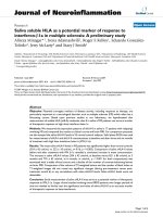
nal extension on refolding (Fig 1A). Expression of the PrP
c
23–231
-GFP and PrP
c
76–156
-GFP fusion protein in E.coli led
to the accumulation of non-fluorescent insoluble inclu-
sion bodies that were purified to ~90% (Fig 1B) and then
denatured before dilution into refolding buffer. GFP fluo-
rescence (510 nm) rose with time to a maximum refold-
ing level of ~6 fold for PrP
c
23–231
-GFP and ~25 fold for
PrP
c
76–156
-GFP over background within 3 hrs under the
conditions of the experiment (Fig 1C). Ranging experi-
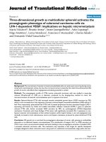
Establishment of the Prp-GFP refolding assayFigure 1
Establishment of the Prp-GFP refolding assay. A. Fragments
of the mouse prnp a allele whose structure is shown were
amplified by PCR and positioned at the N terminus of GFP in
a E.coli expression vector under transcriptional control of the
T7 promoter. B. Purified PrP-GFP fusion proteins were ana-
lysed by 10% SDS-PAGE before (lanes 1 & 2) and after (lanes
3 & 4) the refolding reaction. The lanes are: M-Molecular
weight markers as shown; 1&3-PrP
23–231
-GFP; 2&4-PrP
76–156
-
GFP. The lower staining intensity of the refolded samples is
due to dilution in the refolding buffer. C. Recovery of fluo-
rescence with time following dilution of the solublised PrP-
GFP fusion proteins into refolding buffer. In this experiment
the increase in fluorescence units was 6 fold (ᮀ) and 27 fold
(●) for PrP
23–231
-GFP and PrP
76–156
-GFP respectively. Assays
were done in duplicate and the average fluorescent units
plotted against time.
Virology Journal 2006, 3:59 />Page 3 of 9
(page number not for citation purposes)
ments showed optimal refolding to occur at >pH8 and at
21°C (not shown). We conclude from this data that 1)
PrP
c
-GFP fusion proteins can refold in vitro to regenerate
the GFP chromophore and 2) the level of refolding is
related to the PrP
c
sequence fused to GFP as alteration of
the fragment size altered the rate of fluorescence recovery.
The observed fluorescence was directly attributable to
refolding of PrP
c
-GFP as re-examination of the fusion pro-
teins after the refolding assay showed full length protein
in solution with no evidence of breakdown to release free
GFP (Fig. 1B). As PrP
c
binds both copper [3,4,26,48,49]
and RNA [50-52] the effect of both of these ligands on the
refolding reaction of full length prion protein present in
construct PrP
c
23–231
-GFP was assessed. However, neither
addition of copper (100 nM) nor RNA, prepared as
described [51], significantly altered the rate of fluores-
cence recovery for the full length prion protein, which
remained slow when compared to the shorter variant (see
Additional file 1).
Use of GFP refolding to assess the role of the extreme N
terminus
Previous expression of PrP
c
-GFP fusion proteins within
eukaryotic cells indicated a marked effect of the extreme
N-terminal basic residues 23–28 on prion protein
processing [53,54] and further studies have suggested an
interaction between the extreme amino terminus and the
C terminal folded domain [43] extending an earlier anti-
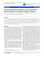
body binding study [55]. In order to assess directly if the
N terminal sequence affects folding per se, amino terminal
truncations were made in which PrP
c
residues 23–156,
29–156, 23–169 and 29–169 (see 1) were appended to
the N terminus of GFP and the fusion proteins purified as
an insoluble fraction prior to dilution into the refolding
reaction (Fig. 2A). When equimolar amounts of each
fusion protein were subjected to the refolding assay, the
rates of fluorescence reacquisition were found to vary con-
siderably (Fig. 2B). The presence of residues 23–28 at the
extreme N-terminus of PrP
c
severely limited refolding in
the context of a fragment truncated at residue 156 with
overall refolding little better than the complete 23–231
PrP
c
sequence despite being a considerably shorter frag-
ment (cf Fig. 1). Deletion of residues 23–28 (construct
PrP
c
29–156
-GFP) enhanced fluorescence recovery ~4 fold
when compared to PrP
c
23–156
-GFP (Fig. 2B). However, a
fragment starting at residue 23 but with an extended C-ter-
minal truncation point at residue 169 (PrP
c
23–169
-GFP),
refolded far more efficiently than PrP
c
23–156
-GFP (Fig. 2B)
and deletion of the amino terminal 6 residues in PrP
c
29–
169
-GFP failed to improve the level of fluorescence
observed. Fluorescence recovery was associated with
equivalent quantities of soluble full-length fusion protein
as no free GFP was apparent when the refolded samples
were analysed by SDS-PAGE after removal from the
refolding reaction (Fig. 2A). Thus, recovery of fluores-
cence by PrP
c
-GFP fusion proteins in vitro following dena-
turation and renaturation measures a direct role for
residues 23–28 and 156–169 in folding and mirrors the
expression patterns observed for prion protein fragments
of the same endpoints in vivo [43].
Use of GFP for direct selection of folding variants
That GFP fluorescence recovered in vitro reflected proper-
ties measured in eukaryotic cells suggested that PrP
c
-GFP
fusions retained a degree of physiological significance. We
sought therefore to use fluorescence for the direct selec-
tion of prion mutants with altered folding properties. To
do this we used the plasmid encoding PrP
c
23–231
-GFP as
template for error prone PCR based mutagenesis [56] of
the PrP
c
sequence followed by substitution of the degen-
erate amplified material for the wild type sequence in
order to generate a library of random PrP
c
mutations fused
to GFP (see 1). Nucleotide sequencing of several library
members picked at random showed a variety of sequence
changes causing premature stop codons as well as single
or multiple amino acid changes within the PrP
c
coding
region (not shown). To select altered folding variants the
library was plated at high density, replicated to agar plates
containing IPTG and colonies were screened for fluores-
cence following irradiation with ultraviolet light. The
overall number of fluorescent colonies was low and after
eradication of false positives three mutants (M17, M22
and M25), which showed particularly strong fluorescence,
(Fig. 3) were isolated and characterised further. As a recov-
ery in fluorescence could indicate a change in folding and
solubility bacterial cultures of the parental construct and
each fluorescent variant were induced, harvested and
lysed and the level of PrP
c
-GFP fusion protein present in
the soluble and insoluble fractions was assessed by west-
ern blot using the PrP
c
monoclonal antibody 6H4
(epitope 144–152). As noted the parental sequence was
wholly insoluble but significant amounts of the fusion
protein from variants M22 and M25 and approximately
50% of the protein from mutant M17 were found in the
supernatant fraction (Fig. 4). One variant (M22) showed
substantial proteolysis leading to loss of full length anti-
body reactive material in the supernatant fraction, a char-
acteristic of soluble PrP
c
expression in E.coli [57]. DNA
sequencing of each variant revealed that M22 and M25
each had two amino acid changes, E152V+N48S and
Y149H+G228E respectively while variant M17 showed
only a single amino acid change at H84Q (Figure 5). Thus,
changes of as little as one or two amino acids throughout
the PrP
c
polypeptide chain can cause significant alteration
in protein folding. None of the mutations selected by this
procedure occurred in the prion hydrophobic sequence
(amino acids 111–133) rather, as suggested, change of
charge was the predominant feature observed [58].
Virology Journal 2006, 3:59 />Page 4 of 9
(page number not for citation purposes)
Discussion
The use of protein fusions as reporters of protein folding
and solubility has emerged rapidly and includes use of
chloramphenicol acetyltransferase (CAT) [59], β-galactos-
idase [60,61] and secretion by defined bacterial transloca-
tion systems [62]. The most well defined system however
has been fusion to the N terminus of GFP [40,42]
although fusions within the loops of the folded structure
have also been reported [63]. The requirement for
increased folding and solubility has been largely driven by
the production of proteins for structural studies [64] but
studies with known misfolding proteins such as Alzhe-
imer's amyloid beta peptide have shown that they can be
equally applied to the study of folding per se [60,62]. Here
we showed that fusion of GFP to the C-terminus of the
mouse prion protein or fragments thereof can provide a
measure of the role of prion sequence in folding in vitro
and that direct selection of fluorescence in vivo results in
PrP
c
-GFP fusion proteins with altered proprieties of solu-
bility. Refolding of PrP
c
-GFP fusions was found to be
robust and not to result in degradation but marked varia-
tion in efficiency was noted when the refolding of individ-
ual fragments of PrP
c
was investigated. In particular, the
presence or not of residues 23–28 (KKRPKP), highly con-
served in prion sequences [65], substantially affected
refolding in vitro and mirrored their affect on PrP
c
-GFP
fusion protein expression in vivo [43]. The diverse biolog-
ical properties of this region, including binding of prion
protein to charged molecules such as Heparin and GAGs
[66-69], suramin [70] and cellular routing [53,54] would
be consistent with a role on the overall structure of the
prion protein. Indeed, restricting movement by N-termi-
nal tethering of PrP
c
to the cell surface abrogates the only
known function of the protein, cellular resistance to oxi-
dative stress [71]. Previous antibody binding studies have
suggested that the prion N-terminus may contact the car-
boxyl domain [72] and we have previously suggested this
interaction may occur between the basic amino terminus
and the acidic patch in helix-1 (
143
DWED
146
) [43]. Matsu-
naga et al., using an N-terminally truncated PrP
c
molecule,
previously proposed a model in which the free N-terminal
amine of PrP
c
residue 90 (the truncation point) interacted
with the acidic charge cluster in helix-1 following the
observation that cryptic epitopes for monoclonal anti-
Refolding of PrP
c
-GFP fusion proteins containing fragments from the prion amino terminal domainFigure 2
Refolding of PrP
c
-GFP fusion proteins containing fragments from the prion amino terminal domain. A. 10% SDS-PAGE analysis
of purified PrP-GFP fusion proteins encoding fragments from the N-terminus before (lanes 1–4) and after (5–8) refolding.
Lanes 1 & 5, PrP
c
23–156
-GFP; lanes 2&6, PrP
c
29–156
-GFP; lanes 3&7, PrP
c
23–169
-GFP; lanes 4&8, PrP
c
29–169
-GFP. B. In vitro refold-
ing kinetics of purified recombinant PrP-GFP fusion proteins; PrP
c
23–156
-GFP (᭜); PrP
c
29–156
-GFP (●); PrP
c
23–169
-GFP(■) and
PrP
c
29–169
-GFP(▲). Assays were done in duplicate and the average fluorescent units plotted against time. Fluorescence units
are as recorded by the plate reader. The lower staining intensity of the refolded samples is due to dilution in the refolding
buffer.
Virology Journal 2006, 3:59 />Page 5 of 9
(page number not for citation purposes)
body 3F4 within the N-terminus are revealed by titration
of acidic residues around Glu 152 [55]. The GFP fluores-
cence recovery assay described here supports this model
but suggests it is residues 23–28 that have a direct role in
folding, consistent with binding to the carboxyl domain
described elsewhere [43]. While various properties have
been ascribed to this short section of charged residues
[43,53,54,67,68,70,73] use of refolding in vitro indicates
for the first time that these observations could be the
result of a role in the overall folding of the molecule.
A corollary of prion sequence identity affecting refolding
in vitro is that direct selection of fluorescence from the
non-fluorescent PrP
c
23–231
-GFP should result in altered
solubility. To assess this we carried out forced evolution of
the PrP
c
sequence and used GFP to screen for a fluorescent
outcome. Model experiments have suggested that as little
a change as one amino acid can have a profound effect on
the physiochemical properties of complete proteins such
as α-synuclein but the effect of mutations associated with
PrP
c
has been only tested on isolated peptides [74] mak-
ing the same conclusion for the complete prion protein
uncertain. Three mutants isolated by virtue of their fluo-
rescence had either one or two residue changes when
compared to the parental sequence. Changes at residue 84
(mutant 17) and 47 (part of mutant M22) were outside of
the known prion structure [17] but in the case of residue
M17 changed the character of the residue from charged to
neutral. Of particular interest however is that one each of
the double mutations, E151V (mutant M22) and Y148H
(mutant M25) lie in the first alpha helix suggested to
interact with the N terminus [43,55] and mapped to be
the site of interaction of a major PrP
c
ligand, the laminin
receptor [75] (Figure 6). In addition the majority of
changes identified were charged residues (Figure 5).
Change of net charge, particularly among the familial
forms of amyloid disease proteins has been suggested to
have a major effect on protein solubility [58,74]. None of
the mutations associated with improved solubility coin-
cide directly with known prion polymorphisms although
interestingly residue 84 (mutant M17) is the point of sev-
eral octarepeat insertions associated with Gerstmann-
Sträussler-Scheinker Syndrome [76,77]. However,
although our data add direct experimental support to the
notion that prion protein folding is very susceptible to
minor changes of sequence, it does not directly address
the role of prion protein solubility in the pathogenicity of
prion disease.
Mutants M17, 22 and 25, selected by recovery of fluores-cence, were grown and PrP
c
-GFP fusion protein present in the soluble (S) and insoluble (I) fractions of each induced cul-ture after detergent lysis were resolved by 10% SDS-PAGE and probed with the prion monoclonal antibody 6H4Figure 4
Mutants M17, 22 and 25, selected by recovery of fluores-
cence, were grown and PrP
c
-GFP fusion protein present in
the soluble (S) and insoluble (I) fractions of each induced cul-
ture after detergent lysis were resolved by 10% SDS-PAGE
and probed with the prion monoclonal antibody 6H4. Reac-
tion with mutant M22 has been largely lost due to degrada-
tion in the soluble phase and only residual insoluble material
is detected.
Direct selection of P
c
23–231
-GFP mutants with increased fluo-rescenceFigure 3
Direct selection of P
c
23–231
-GFP mutants with increased fluo-
rescence. Fluorescence of the three prion mutants (17, 22
and 25) isolated by the procedures described. Each was
grown overnight on agar plates and a heavy inoculum trans-
ferred to a sectored agar plate supplemented with IPTG to
induce expression of the fusion protein. After three hours at
37 degrees the plate was photographed under UV light.
Virology Journal 2006, 3:59 />Page 6 of 9
(page number not for citation purposes)
Conclusion
Prion protein misfolding is thought to underlie its
involvement with the TSE diseases and its study, directly
or indirectly, may help determine the molecular mecha-
nisms involved. Use of GFP as a folding reporter has been
well described but its use as a probe of prion innate fold-
ing rather than cellular targeting has not been previously
reported. The GFP fluorescence assay we have described
may be useful for assessing a number of prion mutations
and the interaction of PrP
c
with its various reported lig-
ands [78].
Methods
E.coli strains
E.coli Top 10 (Invitrogen) was used throughout for clon-
ing. Plasmids were transformed into E.coli BL21 DE3
(pLysS) (Novagen) for T7 driven protein production.
Plasmid construction
Mouse Prnpa allele (accession A23544) and enhanced
green fluorescence protein (accession AAC53663) were
used throughout. cDNA fragments encoding amino acids
23–231 and the N-terminal residues 23–156, 29–156,
76–156, 23–169 and 29–169 were amplified by the
polymerase chain reaction (PCR) to be flanked by restric-
tion sites for Bam H1 and first cloned into baculovirus
transfer vector pAcVSV
GTM
GFP [79] for expression in
insect cells [43]. Each construct was then used as a tem-
plate to amplify the sequence encoding the fusion of PrP
c
and eGFP flanking the sequence with restriction sites
Nde1 and Xho1 at the 5' at the 3' ends respectively. Frag-
ments were digested with the same enzymes and cloned
into pET23a (Novagen) through the same sites to produce
PrP
c
-GFP gene fusions under the control of the T7 pro-
moter.
Expression libraries
A degenerate library of prion sequences was created by
error prone PCR [56] and cloned en masse into pET23a
upstream of, and in frame with, a sequence encoding
eGFP. Several library members were picked at random for
nucleotide sequencing to ensure errors had been intro-
duced. The library was maintained in E.coli BL21 pLysS in
an un-induced state and induced for fluorescence screen-
ing by replica plating to agar containing 2 mM IPTG. Col-
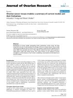
Location of the mutations selected by fluorescence recovery in the three dimensional structure of the prion proteinFigure 6
Location of the mutations selected by fluorescence recovery
in the three dimensional structure of the prion protein. The
unstructured amino terminus up to residue 90 is represented
by the grey oval. The amino and carboxyl termini of the
solved structure and the location of helix 1 are indicated.
Sequence alignment of mutants M17, 22 and 25 compared to the mouse wild type sequenceFigure 5
Sequence alignment of mutants M17, 22 and 25 compared to the mouse wild type sequence. Only those areas showing changes
are shown. Amino acid changes that cause a change of net charge are indicated by the asterisk below the aligned sequence.
Virology Journal 2006, 3:59 />Page 7 of 9
(page number not for citation purposes)
onies were screened visually after a further 5 hours
incubation and positives re-streaked to ensure positivity
bred true. Once confirmed, uninduced colonies were re-
streaked from the master plate and DNA isolated for
sequencing.
Purification of inclusion bodies (IBs)
IBs were prepared by a modified differential solubility
regime [80]. Following inoculation of a single colony into
Luria broth cultures were induced with IPTG (0.2 mM) at
an OD600 of 0.5. Cultures were grown for a further 2
hours and bacteria harvested by centrifugation at 4500
rpm for 20 minutes at 4°C. The pellet was resuspended in
10 ml PBS and the IBs released by sonication on ice for 10
minutes, 1% triton X-100 (v/v) was added to complete
solublization and the IBs collected by centrifugation at
4500 rpm for 10 minutes. The pellet was washed repeat-
edly with 1% Triton X-100 until the purity of the IBs was
at least 90 % as judged by SDS-PAGE.
Protein refolding
IBs were denatured and reduced at 95°C for 5 minute in
4 M Urea and then clarified by ultracentrifugation. Refold-
ing was initiated by a single 7× dilution step into a buffer
containing 50 mM Tris.HCl pH8.5, 1 mM KCl, 2 mM
MgCl
2
. Recovery of fluorescence over time was monitored
by periodic fluorescence measurement at 510 nm in a
Genios microplate reader (Tecan). Assays were done in
duplicate and the average fluorescent units plotted against
time. To assess the role of metal ions in refolding buffers
were depleted for ions my mixing with chelex-100 (Bio-
Rad) as described [25] and filtering prior to constitution
of the assay. Ranging studies showed that the addition of
copper above 10 micromolar was found to be generally
inhibitory (i.e. inhibited the refolding of GFP only).
Purification of RNA
Total RNA for inclusion in the refolding assay was pre-
pared from SNB cells as described for RNA that stimulates
PrP
c
-PrP
Sc
conversion [51]. Briefly, cells were washed with
PBS and resuspended in 1 ml of Trizol (Invitrogen). The
lysate was extracted with chloroform and the RNA recov-
ered by precipitation with isopropanol. The pellet was
washed with 75% ethanol, air dried, resuspended in RNA-
ase free water and quantitated by A
260
.
Protease K digestion
RNA stimulated partial protection of PrP
c
was assessed by
digestion of the reaction products after refolding with pro-
tease K as described [52].
Western blotting
Protein samples to be analyzed were separated on pre-cast
10% Tris-HCl SDS-polyacrylamide gels (Bio-Rad) and
transferred onto Immobilon-P transfer membrane (Milli-
pore). Western blotting was performed as described (Bur-
nette, 1981) except that sensitivity was increased through
the use of a biotin conjugated secondary antibody fol-
lowed by extravidin-peroxidase (Sigma). The membrane
was finally developed with BM Chemiluminescence
(Roche). The primary antibodies used were prion mono-
clonal antibodies 6H4 (Prionics) and anti-GFP (Clon-
tech).
Competing interests
The author(s) declare that they have no competing inter-
ests.
Authors' contributions
SV and JYR developed the in vitro refolding and random
library mutagenesis protocols respectively. YY, KD and
CSD contributed various constructs and assays and IMJ
conceived, planned and advised throughout the study. All
authors contributed to the writing of the manuscript.
Additional material
Acknowledgements
We thank Barbara Konig for technical assistance and the UK Medical
Research Council and Department for Environment, Food and Rural Affairs
(DEFRA) for grant support.
References
1. Basler K, Oesch B, Scott M, Westaway D, Walchli M, Groth DF,
McKinley MP, Prusiner SB, Weissmann C: Scrapie and cellular PrP
isoforms are encoded by the same chromosomal gene. Cell
1986, 46:417-428.
Additional File 1
Additional figure 1. Shows the role of copper ions and RNA on in vitro
refolding of PrP
23–231
-GFP using the standard assay described in the man-
uscript.
Click here for file
[ />422X-3-59-S1.tiff]
Additional File 2
Additional figure 2. Cartoon representation of the PrP
c
expression con-
structs used to investigate the role of the N terminal sequence on refolding
in vitro.
Click here for file
[ />422X-3-59-S2.tiff]
Additional File 3
Additional figure 3. Cartoon and flow diagram of the process for random
selection of soluble variants of PrP
c
-GFP by virtue of mutations that allow
fluorescence in vivo.
Click here for file
[ />422X-3-59-S3.tiff]
Virology Journal 2006, 3:59 />Page 8 of 9
(page number not for citation purposes)
2. Safar J, Prusiner SB: Molecular studies of prion diseases. Prog
Brain Res 1998, 117:421-434.
3. Stockel J, Safar J, Wallace AC, Cohen FE, Prusiner SB: Prion protein
selectively binds copper(II) ions. Biochemistry 1998,
37:7185-7193.
4. Wong BS, Venien-Bryan C, Williamson RA, Burton DR, Gambetti P,
Sy MS, Brown DR, Jones IM: Copper refolding of prion protein.
Biochem Biophys Res Commun 2000, 276:1217-1224.
5. Quaglio E, Chiesa R, Harris DA: Copper converts the cellular
prion protein into a protease-resistant species that is distinct
from the scrapie isoform. J Biol Chem 2001, 276:11432-11438.
6. Jackson GS, Murray I, Hosszu LL, Gibbs N, Waltho JP, Clarke AR,
Collinge J: Location and properties of metal-binding sites on
the human prion protein. Proc Natl Acad Sci U S A 2001,
98:8531-8535.
7. Horwich AL, Weissman JS: Deadly conformations protein mis-
folding in prion disease. Cell 1997, 89:499-510.
8. Liemann S, Glockshuber R: Transmissible spongiform encepha-
lopathies. Biochem Biophys Res Commun 1998, 250:187-193.
9. Prusiner SB, Scott MR: Genetics of prions. Annu Rev Genet 1997,
31:139-175.
10. Brown DR, Schulz-Schaeffer WJ, Schmidt B, Kretzschmar HA: Prion
protein-deficient cells show altered response to oxidative
stress due to decreased SOD-1 activity. Exp Neurol 1997,
146:104-112.
11. Guentchev M, Voigtlander T, Haberler C, Groschup MH, Budka H:
Evidence for oxidative stress in experimental prion disease.
Neurobiol Dis 2000, 7:270-273.
12. Milhavet O, McMahon HE, Rachidi W, Nishida N, Katamine S, Mange
A, Arlotto M, Casanova D, Riondel J, Favier A, Lehmann S: Prion
infection impairs the cellular response to oxidative stress.
Proc Natl Acad Sci U S A 2000, 97:13937-13942.
13. Wong BS, Pan T, Liu T, Li R, Petersen RB, Jones IM, Gambetti P,
Brown DR, Sy MS: Prion disease: A loss of antioxidant function?
Biochem Biophys Res Commun 2000, 275:
249-252.
14. Kim JI, Choi SI, Kim NH, Jin JK, Choi EK, Carp RI, Kim YS: Oxidative
stress and neurodegeneration in prion diseases. Ann N Y Acad
Sci 2001, 928:182-186.
15. Turnbull S, Tabner BJ, Brown DR, Allsop D: Copper-dependent
generation of hydrogen peroxide from the toxic prion pro-
tein fragment PrP106-126. Neurosci Lett 2003, 336:159-162.
16. Rachidi W, Vilette D, Guiraud P, Arlotto M, Riondel J, Laude H, Leh-
mann S, Favier A: Expression of prion protein increases cellular
copper binding and antioxidant enzyme activities but not
copper delivery. J Biol Chem 2003, 278:9064-9072.
17. Riek R, Hornemann S, Wider G, Billeter M, Glockshuber R, Wuthrich
K: NMR structure of the mouse prion protein domain
PrP(121-321). Nature 1996, 382:180-182.
18. Wildegger G, Liemann S, Glockshuber R: Extremely rapid folding
of the C-terminal domain of the prion protein without
kinetic intermediates. Nat Struct Biol 1999, 6:550-553.
19. Glockshuber R: Folding dynamics and energetics of recom-
binant prion proteins. Adv Protein Chem 2001, 57:83-105.
20. Donne DG, Viles JH, Groth D, Mehlhorn I, James TL, Cohen FE,
Prusiner SB, Wright PE, Dyson HJ: Structure of the recombinant
full-length hamster prion protein PrP(29-231): the N termi-
nus is highly flexible [see comments]. Proc Natl Acad Sci U S A
1997, 94:13452-13457.
21. Zahn R, Liu A, Luhrs T, Riek R, von Schroetter C, Lopez Garcia F, Bil-
leter M, Calzolai L, Wider G, Wuthrich K: NMR solution structure
of the human prion protein. Proc Natl Acad Sci U S A 2000,
97:145-150.
22. Brown DR, Qin K, Herms JW, Madlung A, Manson J, Strome R, Fraser
PE, Kruck T, von Bohlen A, Schulz-Schaeffer W, Giese A, Westaway
D, Kretzschmar H: The cellular prion protein binds copper in
vivo. Nature 1997, 390:684-687.
23. Perera WS, Hooper NM: Ablation of the metal ion-induced
endocytosis of the prion protein by disease-associated muta-
tion of the octarepeat region. Curr Biol 2001, 11:519-523.
24. Kramer ML, Kratzin HD, Schmidt B, Romer A, Windl O, Liemann S,
Hornemann S, Kretzschmar H: Prion protein binds copper
within the physiological concentration range. J Biol Chem 2001,
276:16711-16719.
25. Jobling MF, Huang X, Stewart LR, Barnham KJ, Curtain C, Volitakis I,
Perugini M, White AR, Cherny RA, Masters CL, Barrow CJ, Collins SJ,
Bush AI, Cappai R: Copper and zinc binding modulates the
aggregation and neurotoxic properties of the prion peptide
PrP106-126. Biochemistry 2001, 40:8073-8084.
26. Qin K, Yang Y, Mastrangelo P, Westaway D: Mapping Cu(II) bind-
ing sites in prion proteins by diethyl pyrocarbonate modifica-
tion and matrix-assisted laser desorption ionization-time of
flight (MALDI-TOF) mass spectrometric footprinting. J Biol
Chem 2002, 277:1981-1990.
27. Flechsig E, Shmerling D, Hegyi I, Raeber AJ, Fischer M, Cozzio A, von
Mering C, Aguzzi A, Weissmann C: Prion protein devoid of the
octapeptide repeat region restores susceptibility to scrapie
in PrP knockout mice. Neuron 2000, 27:399-408.
28. Leclerc E, Peretz D, Ball H, Sakurai H, Legname G, Serban A, Prusiner
SB, Burton DR, Williamson RA: Immobilized prion protein
undergoes spontaneous rearrangement to a conformation
having features in common with the infectious form. Embo J
2001, 20:1547-1554.
29. Peretz D, Williamson RA, Legname G, Matsunaga Y, Vergara J, Burton
DR, DeArmond SJ, Prusiner SB, Scott MR: A change in the confor-
mation of prions accompanies the emergence of a new prion
strain. Neuron 2002, 34:921-932.
30. Dobson CM: Getting out of shape. Nature 2002, 418:729-730.
31. Bucciantini M, Giannoni E, Chiti F, Baroni F, Formigli L, Zurdo J, Tad-
dei N, Ramponi G, Dobson CM, Stefani M: Inherent toxicity of
aggregates implies a common mechanism for protein mis-
folding diseases. Nature 2002, 416:507-511.
32. Dobson CM: Protein folding and misfolding. Nature 2003,
426:884-890.
33. Rachidi W, Mange A, Senator A, Guiraud P, Riondel J, Benboubetra
M, Favier A, Lehmann S: Prion Infection Impairs Copper Binding
of Cultured Cells. J Biol Chem 2003, 278:14595-14598.
34. Mani K, Cheng F, Havsmark B, Jonsson M, Belting M, Fransson LA:
Prion or amyloid-b-derived Cu(II)- or free Zn(II)-ions sup-
port S-nitroso-dependent autocleavage of glypican-1
heparan sulfate. J Biol Chem 2003:M300394200.
35. Peretz D, Williamson RA, Kaneko K, Vergara J, Leclerc E, Schmitt-
Ulms G, Mehlhorn IR, Legname G, Wormald MR, Rudd PM, Dwek
RA, Burton DR, Prusiner SB: Antibodies inhibit prion propaga-
tion and clear cell cultures of prion infectivity. Nature 2001,
412:739-743.
36. Enari M, Flechsig E, Weissmann C: Scrapie prion protein accumu-
lation by scrapie-infected neuroblastoma cells abrogated by
exposure to a prion protein antibody. Proc Natl Acad Sci U S A
2001, 98:9295-9299.
37. White AR, Enever P, Tayebi M, Mushens R, Linehan J, Brandner S,
Anstee D, Collinge J, Hawke S: Monoclonal antibodies inhibit
prion replication and delay the development of prion dis-
ease. Nature 2003, 422:80-83.
38. Meier P, Genoud N, Prinz M, Maissen M, Rulicke T, Zurbriggen A,
Raeber AJ, Aguzzi A: Soluble Dimeric Prion Protein Binds
PrP(Sc) In Vivo and Antagonizes Prion Disease. Cell 2003,
113:49-60.
39. Solforosi L, Criado JR, McGavern DB, Wirz S, Sanchez-Alavez M,
Sugama S, DeGiorgio LA, Volpe BT, Wiseman E, Abalos G, Masliah E,
Gilden D, Oldstone MB, Conti B, Williamson RA: Cross-linking cel-
lular prion protein triggers neuronal apoptosis in vivo. Science
2004, 303:1514-1516.
40. Waldo GS, Standish BM, Berendzen J, Terwilliger TC: Rapid pro-
tein-folding assay using green fluorescent protein. Nat Biotech-
nol 1999, 17:691-695.
41. Pedelacq JD, Piltch E, Liong EC, Berendzen J, Kim CY, Rho BS, Park
MS, Terwilliger TC, Waldo GS: Engineering soluble proteins for
structural genomics. Nat Biotechnol 2002, 20:927-932.
42. Waldo GS: Improving protein folding efficiency by directed
evolution using the GFP folding reporter. Methods Mol Biol
2003, 230:343-359.
43. Yao Y, Ren J, Jones IM: Amino terminal interaction in the prion
protein identified using fusion to green fluorescent protein.
J Neurochem
2003, 87:1057-1065.
44. Cubitt AB, Woollenweber LA, Heim R: Understanding structure-
function relationships in the Aequorea victoria green fluo-
rescent protein. Methods Cell Biol 1999, 58:19-30.
45. Heim R, Prasher DC, Tsien RY: Wavelength mutations and post-
translational autoxidation of green fluorescent protein. Proc
Natl Acad Sci U S A 1994, 91:12501-12504.
46. Reid BG, Flynn GC: Chromophore formation in green fluores-
cent protein. Biochemistry 1997, 36:6786-6791.
Publish with BioMed Central and every
scientist can read your work free of charge
"BioMed Central will be the most significant development for
disseminating the results of biomedical research in our lifetime."
Sir Paul Nurse, Cancer Research UK
Your research papers will be:
available free of charge to the entire biomedical community
peer reviewed and published immediately upon acceptance
cited in PubMed and archived on PubMed Central
yours — you keep the copyright
Submit your manuscript here:
/>BioMedcentral
Virology Journal 2006, 3:59 />Page 9 of 9
(page number not for citation purposes)
47. Fukuda H, Arai M, Kuwajima K: Folding of green fluorescent pro-
tein and the cycle3 mutant. Biochemistry 2000, 39:12025-12032.
48. Viles JH, Cohen FE, Prusiner SB, Goodin DB, Wright PE, Dyson HJ:
Copper binding to the prion protein: Structural implications
of four identical cooperative binding sites. Proc Natl Acad Sci U
S A 1999, 96:2042-2047.
49. Brown DR: Prion protein expression modulates neuronal cop-
per content. J Neurochem 2003, 87:377-385.
50. Gabus C, Derrington E, Leblanc P, Chnaiderman J, Dormont D, Swi-
etnicki W, Morillas M, Surewicz WK, Marc D, Nandi P, Darlix JL: The
prion protein has RNA binding and chaperoning properties
characteristic of nucleocapsid protein NCP7 of HIV-1. J Biol
Chem 2001, 276:19301-19309.
51. Deleault NR, Lucassen RW, Supattapone S: RNA molecules stim-
ulate prion protein conversion. Nature 2003, 425:717-720.
52. Adler V, Zeiler B, Kryukov V, Kascsak R, Rubenstein R, Grossman A:
Small, highly structured RNAs participate in the conversion
of human recombinant PrP(Sen) to PrP(Res) in vitro. J Mol
Biol 2003, 332:47-57.
53. Nunziante M, Gilch S, Schatzl HM: Essential role of the prion pro-
tein N terminus in subcellular trafficking and half-life of cel-
lular prion protein. J Biol Chem 2003, 278:3726-3734.
54. Sunyach C, Jen A, Deng J, Fitzgerald KT, Frobert Y, Grassi J, McCaf-
frey MW, Morris R: The mechanism of internalization of glyc-
osylphosphatidylinositol-anchored prion protein. Embo J
2003, 22:3591-3601.
55. Matsunaga Y, Peretz D, Williamson A, Burton D, Mehlhorn I, Groth
D, Cohen FE, Prusiner SB, Baldwin MA: Cryptic epitopes in N-ter-
minally truncated prion protein are exposed in the full-
length molecule: dependence of conformation on pH. Proteins
2001, 44:110-118.
56. Crameri A, Whitehorn EA, Tate E, Stemmer WP: Improved green
fluorescent protein by molecular evolution using DNA shuf-
fling. Nat Biotechnol 1996, 14:315-319.
57. Hornemann S, Korth C, Oesch B, Riek R, Wider G, Wuthrich K,
Glockshuber R: Recombinant full-length murine prion protein,
mPrP(23-231): purification and spectroscopic characteriza-
tion. FEBS Lett 1997, 413:277-281.
58. Chiti F, Calamai M, Taddei N, Stefani M, Ramponi G, Dobson CM:
Studies of the aggregation of mutant proteins in vitro pro-
vide insights into the genetics of amyloid diseases. Proc Natl
Acad Sci U S A 2002, 99 Suppl 4:16419-16426.
59. Maxwell KL, Mittermaier AK, Forman-Kay JD, Davidson AR: A sim-
ple in vivo assay for increased protein solubility. Protein Sci
1999, 8:1908-1911.
60. Wigley WC, Stidham RD, Smith NM, Hunt JF, Thomas PJ: Protein
solubility and folding monitored in vivo by structural com-
plementation of a genetic marker protein. Nat Biotechnol 2001,
19:131-136.
61. Stidham RD, Wigley WC, Hunt JF, Thomas PJ: Assessment of pro-
tein folding/solubility in live cells. Methods Mol Biol 2003,
205:155-169.
62. Fisher AC, Kim W, DeLisa MP: Genetic selection for protein sol-
ubility enabled by the folding quality control feature of the
twin-arginine translocation pathway. Protein Sci 2006,
15:449-458.
63. Cabantous S, Pedelacq JD, Mark BL, Naranjo C, Terwilliger TC,
Waldo GS: Recent advances in GFP folding reporter and split-
GFP solubility reporter technologies. Application to improv-
ing the folding and solubility of recalcitrant proteins from
Mycobacterium tuberculosis. J Struct Funct Genomics 2005,
6:113-119.
64. Waldo GS: Genetic screens and directed evolution for protein
solubility. Curr Opin Chem Biol 2003, 7:33-38.
65. Wopfner F, Weidenhofer G, Schneider R, von Brunn A, Gilch S,
Schwarz TF, Werner T, Schatzl HM: Analysis of 27 mammalian
and 9 avian PrPs reveals high conservation of flexible regions
of the prion protein. J Mol Biol 1999, 289:1163-1178.
66. Snow AD, Wight TN, Nochlin D, Koike Y, Kimata K, DeArmond SJ,
Prusiner SB: Immunolocalization of heparan sulfate proteogly-
cans to the prion protein amyloid plaques of Gerstmann-
Straussler syndrome, Creutzfeldt-Jakob disease and scrapie.
Lab Invest 1990, 63:
601-611.
67. Warner RG, Hundt C, Weiss S, Turnbull JE: Identification of the
heparan sulfate binding sites in the cellular prion protein. J
Biol Chem 2002, 277:18421-18430.
68. Pan T, Wong BS, Liu T, Li R, Petersen RB, Sy MS: Cell surface prion
protein interacts with glycosaminoglycans. Biochem J 2002,
368:81-90.
69. Gonzalez-Iglesias R, Pajares MA, Ocal C, Espinosa JC, Oesch B, Gas-
set M: Prion protein interaction with glycosaminoglycan
occurs with the formation of oligomeric complexes stabi-
lized by Cu(II) bridges. J Mol Biol 2002, 319:527-540.
70. Gilch S, Winklhofer KF, Groschup MH, Nunziante M, Lucassen R,
Spielhaupter C, Muranyi W, Riesner D, Tatzelt J, Schatzl HM: Intra-
cellular re-routing of prion protein prevents propagation of
PrP(Sc) and delays onset of prion disease. Embo J 2001,
20:3957-3966.
71. Zeng F, Watt NT, Walmsley AR, Hooper NM: Tethering the N-
terminus of the prion protein compromises the cellular
response to oxidative stress. J Neurochem 2003, 84:480-490.
72. Li R, Liu T, Wong BS, Pan T, Morillas M, Swietnicki W, O'Rourke K,
Gambetti P, Surewicz WK, Sy MS: Identification of an epitope in
the C terminus of normal prion protein whose expression is
modulated by binding events in the N terminus. J Mol Biol
2000, 301:567-573.
73. Brimacombe DB, Bennett AD, Wusteman FS, Gill AC, Dann JC, Bos-
tock CJ: Characterization and polyanion-binding properties
of purified recombinant prion protein. Biochem J 1999, 342 Pt
3:605-613.
74. Chiti F, Stefani M, Taddei N, Ramponi G, Dobson CM: Rationaliza-
tion of the effects of mutations on peptide and protein aggre-
gation rates. Nature 2003, 424:805-808.
75. Hundt C, Peyrin JM, Haik S, Gauczynski S, Leucht C, Rieger R, Riley
ML, Deslys JP, Dormont D, Lasmezas CI, Weiss S: Identification of
interaction domains of the prion protein with its 37-kDa/67-
kDa laminin receptor. Embo J 2001, 20:5876-5886.
76. Goldfarb LG, Brown P, Vrbovska A, Baron H, McCombie WR,
Cathala F, Gibbs CJJ, Gajdusek DC: An insert mutation in the
chromosome 20 amyloid precursor gene in a Gerstmann-
Straussler-Scheinker family. J Neurol Sci 1992, 111:189-194.
77. Laplanche JL, Hachimi KH, Durieux I, Thuillet P, Defebvre L, Delas-
nerie-Laupretre N, Peoc'h K, Foncin JF, Destee A: Prominent psy-
chiatric features and early onset in an inherited prion disease
with a new insertional mutation in the prion protein gene.
Brain 1999, 122 ( Pt 12):2375-2386.
78. Gauczynski S, Hundt C, Leucht C, Weiss S: Interaction of prion
proteins with cell surface receptors, molecular chaperones,
and other molecules. Adv Protein Chem 2001, 57:229-272.
79. Chapple SD, Jones IM: Non-polar distribution of green fluores-
cent protein on the surface of Autographa californica nucle-
opolyhedrovirus using a heterologous membrane anchor. J
Biotechnol 2002, 95:269-275.
80. Chan DC, Fass D, Berger JM, Kim PS: Core structure of gp41
from the HIV envelope glycoprotein. Cell 1997, 89:263-273.