Báo cáo hóa học: " HIV-1 gp120 N-linked glycosylation differs between plasma and leukocyte compartments" pdf
Bạn đang xem bản rút gọn của tài liệu. Xem và tải ngay bản đầy đủ của tài liệu tại đây (636.59 KB, 10 trang )
BioMed Central
Page 1 of 10
(page number not for citation purposes)
Virology Journal
Open Access
Research
HIV-1 gp120 N-linked glycosylation differs between plasma and
leukocyte compartments
Yung Shwen Ho
1,4
, AnaBAbecasis
2
, Kristof Theys
2
, Koen Deforche
2
,
Dominic E Dwyer
3
, Michael Charleston
4
, Anne Mieke Vandamme
2
and
Nitin K Saksena*
1
Address:
1
Retroviral Genetics Laboratory, Centre for Virus Research, Westmead Millennium Institute, Westmead Hospital, University of Sydney,
Westmead NSW. 2145 Sydney. Australia,
2
Clinical and Epidemiological Virology, Rega Institute for Medical Research, Leuven, Belgium,
3
Centre
for Infectious Diseases and Microbiology Laboratory Services, ICPMR, Westmead Hospital, Westmead NSW 2145, Australia and
4
School of
Information Technologies, University of Sydney, Camperdown NSW 2006, Australia
Email: Yung Shwen Ho - ; Ana B Abecasis - ;
Kristof Theys - ; Koen Deforche - ;
Dominic E Dwyer - ; Michael Charleston - ;
Anne Mieke Vandamme - ; Nitin K Saksena* -
* Corresponding author
Abstract
Background: N-linked glycosylation is a major mechanism for minimizing virus neutralizing
antibody response and is present on the Human Immunodeficiency Virus (HIV) envelope
glycoprotein. Although it is known that glycosylation changes can dramatically influence virus
recognition by the host antibody, the actual contribution of compartmental differences in N-linked
glycosylation patterns remains unclear.
Methodology and Principal Findings: We amplified the env gp120 C2-V5 region and analyzed
305 clones derived from plasma and other compartments from 15 HIV-1 patients. Bioinformatics
and Bayesian network analyses were used to examine N-linked glycosylation differences between
compartments. We found evidence for cellspecific single amino acid changes particular to
monocytes, and significant variation was found in the total number of N-linked glycosylation sites
between patients. Further, significant differences in the number of glycosylation sites were
observed between plasma and cellular compartments. Bayesian network analyses showed an
interdependency between N-linked glycosylation sites found in our study, which may have immense
functional relevance.
Conclusion: Our analyses have identified single cell/compartment-specific amino acid changes and
differences in N-linked glycosylation patterns between plasma and diverse blood leukocytes.
Bayesian network analyses showed associations inferring alternative glycosylation pathways. We
believe that these studies will provide crucial insights into the host immune response and its ability
in controlling HIV replication in vivo. These findings could also have relevance in shielding and
evasion of HIV-1 from neutralizing antibodies.
Published: 23 January 2008
Virology Journal 2008, 5:14 doi:10.1186/1743-422X-5-14
Received: 18 December 2007
Accepted: 23 January 2008
This article is available from: />© 2008 Ho et al; licensee BioMed Central Ltd.
This is an Open Access article distributed under the terms of the Creative Commons Attribution License ( />),
which permits unrestricted use, distribution, and reproduction in any medium, provided the original work is properly cited.
Virology Journal 2008, 5:14 />Page 2 of 10
(page number not for citation purposes)
Introduction
The HIV-1 envelope (env) gp120 region plays a crucial
role in the entry of HIV-1 into target cells through the
fusion of viral envelope with the target cell membrane.
Variable regions (V1-V5) in env are spaced between the
conserved regions (C1-C5). Both N-linked and O-linked
glycans are present on the HIV envelope glycoprotein. O-
linked glycans are present on several unidentified serine
or threonine residues in env gp120, but very little is
known about their actual role in governing the viral phe-
notype of both HIV and simian immunodeficiency virus
(SIV) [1,2]. In contrast, N-linked glycans comprise about
50% of the mass of the env gp160 [3]. These sugar moie-
ties are involved in various activities such as metabolism,
transport, structural maintenance of the cell and protein,
protein folding, recognition of particular cell types and
adhesion to other cells. The N-linked glycosylation (NLG)
of viral envelope proteins, through the formation of a
"glycan shield", is one of the major mechanisms for
blocking or minimizing virus neutralizing antibody
response [4]. which promotes viral persistence and
immune evasion. This has been demonstrated in SIV
[5,6], HIV-1 [4,7] influenza virus [8], hepatitis B virus [9]
and the Lactate Dehydrogenase-elevating Virus [10].
Despite considerable genetic variation in HIV strains, the
number of NLG are often found to be around 25 sites in
the HIV-1 env gp120 region [11], suggesting that strong
selective pressures maintain this number [4]. The HIV
envelope "glycan shield" is known to evolve in response
to host antibodies [4] and it is thought that the density of
gp120 NLG is a significant obstacle to the design of effec-
tive vaccine and elicitation of humoral immune
responses. Any alteration or positional shift of a glycosyla-
tion site (commonly seen in HIV and SIV glycoproteins)
can have dramatic consequences for the virus and its rec-
ognition by the antibody.
Although recent studies have shown compartmentaliza-
tion of HIV-1 NLG sites between viral populations in
plasma and the female genital tract [12,13], the critical
issue of possible differences in NLG of HIV-1 strains
derived from cell-associated and cell-free compartments
remains unexplored. Such differences are important to
future drug development because the drugs used in highly
active antiretroviral therapy (HAART) primarily target
plasma or cell-free virus. Cell-free virus has a high turno-
ver rate (< 6 hours) [4] and therefore has a strong need to
maintain viral integrity through constant shielding from
host antibodies. In contrast, cell-associated virus are kept
away from neutralizing antibodies and can remain inte-
grated in the human genome indefinitely. They can pro-
duce viral progeny upon activation in vivo and this acts as
an impediment to the success of therapy. The integrated
provirus concealed in diverse blood leukocyte popula-
tions is one strategy HIV uses to avoid immune detection.
Given the incessant virus trafficking between cellfree and
cell-associated compartments, a clear determination of
differences of HIV populations in plasma and diverse cell
types is needed to understand critical molecular determi-
nants for viral survival, turnover, evasion and adaptation
in vivo. The relevance of NLG is also known for many
other viruses [14-16]. Together, these studies imply that
the virus-producing cell type is an important factor, which
may be crucial in viral tropism and transmissibility in vivo.
The role of single amino acid residue changes in the HIV-
1 env in its adaptation to cellular compartments remains
similarly unexplored. Given that different cellular com-
partments have different immune functions in our body,
we suspect that the virus populations within them are sub-
jected to distinct selection pressures, as opposed to freely
circulating virus in plasma [17]. These distinct selective
forces may further exert influence on the make-up of NLG,
depending on the cell type. This evolutionary make-up
may, in turn, define biological and functional aspects of
viral variants in a given environment. Different glycosyla-
tion sites have been shown to offer variable sensitivity to
antibody-mediated neutralization [4], such as sites in the
env hypervariable C3 and C5 regions. The sites around the
base of the V3 loop have been consistently found to be
associated with neutralization sensitivity, especially in
HIV-1 subtype B viruses [4]. As the majority (17 of 25) of
NLG sites are concentrated in the env C2-V5 region of
gp120, and given the functional relevance of glycans in
HIV pathogenesis, we chose this region for the study of
HIV-1 glycosylation patterns in cell-free and cell-associ-
ated compartments.
We analyzed 305 clones of HIV-1 variants derived from
plasma and diverse blood leukocytes (whole Peripheral
Blood Mononuclear Cell (PBMC), CD4
+
T cells, CD8
+
T
cells and monocytes) from 15 HIV-1 infected patients on
HAART, each displaying different levels of plasma viremia
and T cell counts. In addition to the patient specific
changes observed in our analyses, we further found evi-
dence in favor of compartmental NLG differences and dis-
tinct cell-specific molecular changes in the env C2-V3
region of HIV-1.
Results
Phylogenetic analysis
Phylogenetic tree reconstruction using a maximum likeli-
hood heuristic search algorithm showed patient-specific
clustering of virus from all compartments, confirming the
patient origin of HIV-1 variants and the absence of cross-
patient contamination. Further, within each patient there
was distinct clustering of HIV-1 sequences from each com-
partment, confirming the purity of diverse blood leuko-
cytes and plasma samples (Figure 1). This provided us
with a platform to compare our data both at the level of
Virology Journal 2008, 5:14 />Page 3 of 10
(page number not for citation purposes)
single amino acid mutations and NLG differences across
cell-free and cellassociated compartments.
Signature pattern analysis
From our signature pattern analysis, we found seven sin-
gle amino acid differences from the 305 HIV-1 sequences
across five different compartments (Table 1). These posi-
Phylogenetic tree analysis showing patient sequence purityFigure 1
Phylogenetic tree analysis showing patient sequence purity. Phylogenetic analysis on the 305 HIV-1 env gp120 C2-V5
region sequences from plasma, peripheral blood mononuclear cells, CD4
+
T cells, CD8
+
T cells and monocytes. Using the
ProML program of the PHYLIP software package, a maximum likelihood phylogenetic tree was calculated for our patient
sequences. The branch lengths are scaled to distance. A single asterisk represents each sequence from our dataset. Individual
sequences are not identified as it is only the broad pattern of clustering that is of interest here. Distinct clustering of patient-
related sequences can be seen from the phylogenetic tree.
Virology Journal 2008, 5:14 />Page 4 of 10
(page number not for citation purposes)
tions are referenced to the HIV-1 HXB2 prototype using
the referencing guidelines available from the Los Alamos
HIV sequence database website [18]. The columns in
Table 1 categorize our data into plasma and blood leuko-
cyte compartments, while the rows in Table 1 represent
the amino acid differences at each of the identified sites.
As shown in Table 1, CD4
+
T cells, CD8
+
T cells and mono-
cyte-derived sequences were found with the amino acid
asparagine (N) at position 279, whereas the PBMC and
plasma-derived sequences were found to have aspartic
acid (D) at the same position. The amino acid lysine (K)
was uniquely seen in monocyte-derived HIV sequences at
positions 335 and 350, whereas other compartments
showed arginine (R) at that position. Further amino acid
differences across compartments were found at positions
320, 336, 360 and 440 as illustrated in Table 1. Statistical
analysis confirmed a significant association (p = 0.044) in
the observed single amino acid differences between CD4
+
T cell and plasma-derived sequences at position 360.
However a lost of significance was found (p = 0.22) when
we correct for multiple comparisons.
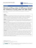
N-linked glycosylation analysis
N-linked glycosylation analysis using the N-Glycosite pro-
gram [11] identified 17 NLG sites from our 305 HIV-1 env
gp120 gap-stripped protein sequences (Figure 2). The
positions of the sites are referenced using the HXB2 proto-
type sequence as described above. On the whole, the NLG
frequencies were found to vary greatly from site to site.
Positions 276, 295, 301, 332, 339, 386 and 448 had a
high frequency of ≥70% in our sequences. Positions 293,
302, 317, 334, 338, 340, 363, 368 and 444 had a low fre-
quency (<16%) in our sequence population. The number
of glycosylation sites varied significantly between patients
(p < 2.2 × 10
-16
) in our inter-patient analysis. Among the
NLG sites identified, the one at position 338 from patient
13 was unique to the HIV-1 strain and had not previously
been recorded in the Los Alamos HIV database.
Empirical statistical analysis
The χ
2
test for the comparison of frequencies across five
different compartment using a 5 × 2 contingency table
gave a p-value of 0.0015 for position 295. Fisher's exact
test for the comparison of frequencies observed from
plasma versus all cell-types (CD4, CD8, Monocytes,
PBMC) showed significant difference (p = 0.04) in the fre-
quencies observed at position 448. We are aware that
these values might be affected by our treatment of several
clones from the same patient and from the same compart-
ment, as the different sequences are not truly independent
events. Hence, we extended this analysis further to include
only the median number of NLG sites per patient per
compartment and not each individual count of NLG sites
per sequence. While this procedure eliminates the effect of
non-independence, it also lessens the number of observa-
tions and possibly the statistical power. When we ana-
lysed the median number of NLG sites between
compartments with the gap-stripped sequences (Tables
2), the Kruskal-Wallis test confirmed a significantly higher
median number of NLG sites observed in plasma than in
cellular compartments (p = 0.022) when sequences from
PBMC, CD4
+
T cells, CD8
+
T cells, monocytes were
grouped together. Moreover, this difference was slightly
more pronounced when we compared plasma and mono-
cyte sequences (p = 0.017; Table 2). Repeating the statisti-
cal analysis with Bonferroni correction for multiple
comparisons gave us a p-value of 0.114 and 0.085 respec-
tively. No statistical differences in the median number of
N-linked glycosylation sites were found in the gap-inclu-
sive alignment.
Bayesian network analysis
We found a strong statistical significance between patient-
specific sequences and the presence/absence of certain
glycosylation sites at positions 295, 339, 362, 386 and
448 (Figure 3). This analysis also showed an interesting
dependency between NLG sites at different positions,
shown in Figure 3. While some of those dependencies are
expected from N-linked glycosylation motif (like
293–295 and 332–334), the associations found between
other sites 301–444, 293–362, 295–334, and 301–332
respectively, are novel.
To deduce the possible biological and functional rele-
vance of these findings, we also compared the 17 poten-
tial NLG sites found in our study to functionally analyze
N-linked glycosylation sites published by Wei et al [4].
Most of the sites noted in our study matched the N-linked
glycosylation sites (from GenBank U21135
) of functional
relevance described by Wei et al [4] (Figure 2). Due to
sequence variability, some glycosylation sites were very
close to each other, we have chosen to associate more than
one site to the NLG positions reported by Wei et al [4]. For
example, positions 301 and 302, being 1 amino acid away
Table 1: Single amino acid differences found across plasma and
diverse blood leukocyte population in vivo.
HXB2 CD4
+
CD8
+
Monocytes PBMC Plasma
279 Asparagine (N) Aspartic acid (D)
336 Threonine (T) Alanine (A)
335 Arginine (R) Lysine (K) Arginine (R)
350 Arginine (R) Lysine (K) Arginine (R)
320 Alanine (A) Threonine (T) Alanine (A)
440 Arginine (R) Serine (S) Arginine (R)
360 Isoleucine (I) Alanine Valine(v) Alanine (A)
Each column represents a particular compartment from which
sequences were derived. Distinct signature pattern differences are
shown in the rows with the corresponding amino acid variations. The
locations of these variations are aligned with those of the HIV-1 HXB2
reference strain using the guidelines available on the HIV Database
website, Los Alamos, NM, USA [18].
Virology Journal 2008, 5:14 />Page 5 of 10
(page number not for citation purposes)
from one another, are likely to represent NLG 305N from
their study [4]. We took the same view for position pair
332 and 334, corresponding to NLG 335N, and position
pair 339 and 340, corresponding to NLG 342N from Wei
et al [4].
Searching for the best Bayesian network representation for
this dataset is an extremely difficult task considering our
relatively small sample size against the number of varia-
bles (potential NLG sites) we have account for. This pitfall
was overcome by combining the Bayesian network analy-
sis with a bootstrap approach. The strength of the arcs is
proportional to its bootstrap support and not to the the
importance of the conditional independencies to the joint
probability of the network. Overall, these analyses have
allowed us to obtain a detailed profile of potential N-
N-linked glycosylation frequency observed in the env gp120 C2-V5 region across plasma and cellular viral sequencesFigure 2
N-linked glycosylation frequency observed in the env gp120 C2-V5 region across plasma and cellular viral
sequences. Frequency of HIV-1 N-linked glycosylation sites in plasma and diverse blood leukocyte populations. The X-axis
represents the potential N-linked glycosylation sites identified in our study. The Y-axis shows the percentage frequency of the
sequences for each compartment found with NLG at the relevant position. Below the bar chart is the env gp120 sequence of
our reference HIV-1 HXB2 strain. Lines from the X-axis to the reference sequence illustrate where the observed NLG in our
data would be on HXB2. NLG sites observed were matched with those reported by Wei et al [4] using the GenBank sequence
U21135
. The numbers below each red oval indicates where you might associate our NLG site with those from the study by
Wei et al [4] in the GenBank sequence U21135
. Positions with no visual correlation are indicated with a dash '-'.
Table 2: Statistical comparison on the number of glycosylation
sites between compartments using the gap-stripped sequences.
Number of NLG
sites
CD4
+
CD8
+
Monocytes PBMC Plasma
CD4
+
- 0.8713 0.1711 0.7645 0.08367
CD8
+
- - 0.2908 0.9132 0.0962
Monocytes - - - 0.3412 0.01696
PBMC 0.0818
Grouped cells - - - - 0.02281
Above are the p-values resulting from the Kruskal-Wallis test when
we compared for statistical differences in the observed number of
glycosylation sites across different compartments using the gap-
stripped sequences. The category "Grouped cells" represents
sequences from cellular compartments (CD4
+
, CD8
+
T cells,
Monocytes and PBMC). The gap-inclusive results are not shown as no
statistical significant difference was observed (see text).
Virology Journal 2008, 5:14 />Page 6 of 10
(page number not for citation purposes)
linked glycosylation distribution and possible inter-rela-
tionship between NLG sites in HIV-1 env gp120 sequences
between different blood leukocyte and plasma-derived
HIV-1 populations in vivo.
Discussion
In order for HIV to be successful in evading the immune
system, both cell-free and cell-associated forms of HIV
must adopt distinct molecular strategies to adapt and sur-
vive in vivo. Such adaptation includes the modulation of
N-linked glycosylation and cell-specific single amino acid
changes in HIV. Given the importance of the envelope
glycoprotein in neutralization, pathogenesis, tropism and
viral evasion, we analysed the C2-V5 region of 305 HIV-1
env gp120 sequences from plasma and diverse blood leu-
kocytes of 15 patients on HAART. Single amino acid dif-
ferences specific to plasma and cellular compartments
were observed. Notable was the presence of lysine (K) in
monocytes at positions 335 and 350 whereas these posi-
tions have arginine (R) in the other compartments. This
suggests that positions 335 and 350 have the greatest need
to maintain a basic amino acid residue (lysine) in mono-
cytes, compared with the other compartments. This differ-
ence may have a role in viral adaptation to monocytes and
possibly relevant to monocyte tropism. In addition, the
isoleucine at position 360 was unique to CD4
+
T cells (p <
0.044). Additionally the presence of valine was unique to
PBMC (and absent in plasma and the three other cell
types) at position 360. Although the monocyte compart-
ment is a subset of the PBMC compartment, the majority
of PBMC sequences were found with valine (V) and the
monocyte sequences with alanine (A). We believe that
although all the cellular compartments we analyzed were
derived from the whole PBMC, the valine may be specific
to a leukocyte subset, which was not analyzed in this
study, due to the limitation of human bleed obtained
from each patient. Nonetheless, this difference between
monocytes and PBMC supports our observation that com-
partmental-specific amino acid changes in HIV-1 env
gp120 are present. Our results agree with previous work
on single amino acid changes in the env gene in associa-
tion with viral tropism and pathogenicity [19]. A recent
study by Clevestig et al [20], shows the V3 loop glycan
(especially the sequon motif NNT) to be critical for CCR5
use, which may have a direct role in HIV tropism, further
supporting our conclusions. We believe that these single
amino acid differences could be vital to HIV and are
acquired through intra-host evolution in order to success-
fully adapt and thrive in different in vivo environments.
The modulation of N-linked glycosylation sites in the
HIV-1 envelope has been known to facilitate viral evasion
from the host immune system. Similar to the signature
pattern analysis, the N-linked glycosylation analysis yields
interesting results showing compartmental variations and
possible associations among glycosylation sites. An exam-
ination of the NLG frequencies along the C2-V5 region
(Figure 2) revealed that plasma virus had a similar or
higher percentage of its sequences glycosylated at the
identified sites, compared to the other compartmental
viruses. A possible explanation for this is that the differ-
ences in N-linked glycosylation sites in plasma may be
required for the maintenance of infectious potential.
Through the suppression of HIV-1 in CD4
+
T cells and
plasma in our HAART patient pool, glycosylation change
in the virus may enable it to overcome the selection pres-
sures from the antiretroviral regimen. Further examina-
tion of the NLG sites using Bayesian networks found
unique associations between sites 301–444, 293–362,
295–334, and 301–332. These associations could indicate
alternative pathways for glycosylation and possible simul-
taneous co-selection of glycosylation sites. Similar to the
study of evolutionary interactions between NLG sites by
Poon Art F, Y [21], we believe that these evolutionary
mechanisms is important to HIV for its struggle against
host immune selection pressure. Since all our work was
performed directly on ex vivo-collected cells from each
patient, our results reflect the closest possible snapshot to
the in vivo situation.
Bayesian network associations found between observed N-linked glycosylation sitesFigure 3
Bayesian network associations found between
observed N-linked glycosylation sites. Representation
of the Bayesian network analysis results, generated as
described in the methods section. Only arcs with a bootstrap
support of at least 70% are presented. Associations between
patient ID and glycosylation sites are dashed. Associations
between glycosylation sites that can be structurally explained
are colored grey. Associations between glycosylation sites
that cannot be structurally explained are colored black.
Virology Journal 2008, 5:14 />Page 7 of 10
(page number not for citation purposes)
We examined the variation in the number of glycosylation
sites among compartments (gap-free and gap-inclusive)
to infer a global picture of HIV-1 in vivo. The trade-off
between them is an important issue to consider in this
analysis. While using the gap-inclusive alignment might
include unavoidable interference due to the uncertainty of
the alignment in the hypervariable region of env, the use
of the gap-stripped alignment might also introduce an
oversight of the observed glycosylation due to the poten-
tial removal of important glycosylation sites from the
alignment. Therefore, we consider both analyses in the
results section. The results were twofold: no statistical sig-
nificance in the total number of glycosylation sites
between plasma and different cellular compartment
sequences was found using the gap-inclusive alignment.
However when we performed the gap-stripped analysis,
we observed a significant difference (p = 0.022) between
plasma and cellular compartment sequences. This is con-
sistent with the notion that the number of glycosylation
sites is highly conserved in HIV-1 [4]. The disparity
between these results suggests that there is a difference in
the number of glycosylation sites between plasma virus
and cellular virus in the less conserved regions of HIV-1
env. Despite the loss of statistical significance when we
applied the Bonferroni correction for multiple compari-
son from P = 0.022 to P = 0.115, we believe that it is still
an important finding together with the rest of our statical
analysis. This is because by correcting for multiple com-
parisons, we are also increasing the risk of making a type
II error which might lead us to not report a correlation
when there is one. Between both gap-stripped and gap-
inclusive analysis, the gap-inclusive alignment gives us the
actual number of glycosylation sites, as no glycosylation
sites were omitted in the evaluation. Not finding any sig-
nificant differences in this gap-inclusive alignment indi-
cates that possible selective pressure to remove
glycosylation sites from the more conserved part of env
was compensated by the creation of new glycosylation
sites in those parts of env that better tolerate substitutions,
insertions and deletions.
Even though we found evidence for compartmentaliza-
tion of HIV-1 across plasma and diverse blood leukocyte
populations in vivo, it is important to note that the NLG
observed in our sequences are distinctly patient-specific (p
≤ 2.2 × 10
-16
). This supports our belief that the selection
of NLG sites are primarily dependent on the patient's
immune response. The choice to examine 305 sequences
from 15 independent patients with a range of CD4
+
T cell
counts and plasma viral load in our study has allowed us
to infer a balanced macroscopic perspective of HIV-1
adaptation in vivo during therapy. These analyses are the
first to provide a detailed comparison of cell-free and cel-
lassociated virus, especially in individual leukocyte types.
Previous studies by Hanna et al [14] and Lin et al [15] sup-
port the validity of our studies on diverse cell types and
add further credence to the functional basis of different
NLG frequencies in plasma and cell-associated virus.
Together, these findings might provide a new direction
and perspective into the role of glycosylation in diverse
compartments in vivo and these data may have relevance
in HIV immune recognition, viral adaptation, vaccine
strategies and HIV pathogenesis in general.
Conclusion
This study examined single cell/compartment-specific
amino acid variations and unique differences in N-linked
glycosylation patterns between plasma and diverse blood
leukocytes. It has provided deeper insights into how HIV
may evade antibodies and maintain its pathogenic poten-
tial. Bayesian analysis has shown associations that suggest
possible glycosylation pathways. We believe that these
analyses provide useful insights into the host immune
response and its ability in controlling HIV replication in
vivo. Further, this enhance our understanding of pathoge-
nous differences between cell-free and cell-associated
HIV-1. Consequently, these analyses will allow further
biological and functional assessment of such molecular
changes in the context of viral escape, adaptation and res-
ervoir establishment in vivo. A better understanding of
diverse N-linked glycosylation sites and their functional
role may provide useful strategies for choosing and elimi-
nating the "right" N-linked glycosylation site(s), thus
facilitating the design of more effective envelope-based
immunogens that elicit broad neutralization antibody
responses.
Methods
Consent
This work is carried out in accordance with the human
ethics guidelines and scientific principles set out by the
National Health and Medical Research Council of Aus-
tralia (NHMRC). All patients have given written consent
on the study and understand that the study will be con-
ducted in a manner conforming to the ethical and scien-
tific principle set out by the NHMRC.
Patient selection
Fifteen HIV-1 infected patients receiving HAART from the
Westmead Hospital in Sydney, Australia, were enrolled in
this study after prior consent. These patients exhibited var-
ying plasma viral loads and T cell counts as shown in
Table 3. Patients 6, 11, 12, and 13, who were successful in
therapy, had plasma viral loads below detectable levels
(<50 copies/ml plasma). Patient 9, who was at an
advanced stage of therapy, had low viremia. Patients 2, 4,
8, 10, 14 and 15 had varying degrees of plasma viremia
after being on HAART for 4 weeks. Patients 1, 3, 5 and 7
had high plasma viral load (>100,000 copies/ml plasma)
and were resistant to antiretroviral drugs [22]. No
Virology Journal 2008, 5:14 />Page 8 of 10
(page number not for citation purposes)
untreated patients could be recruited in this study as every
HIV patient in Australia receives treatment for their condi-
tion.
Cell purification and sequence generation
50 ml of blood was collected from each patient. Individ-
ual cell types (CD4
+
T cells, CD8
+
T cells and CD14
+
monocytes) were separated from PBMC using magnetic
beads coated with monoclonal antibodies (Dynal, Oslo,
Norway) using the procedure described and developed by
Potter et al [17]. FACS analysis of separated cellular frac-
tions showed an average purity of 99.6%. Proviral DNA
was extracted from PBMC and individually sorted into cel-
lular fractions using the Qiagen Blood kit (Qiagen, Ger-
many) as per the manufacturer's protocol. A nested
polymerase chain reaction (PCR) was used to amplify a
600 bp fragment in the C2-V5 region of env gene. Reverse
transcription-PCR (RT-PCR) of HIV-1 RNA extracted from
plasma using the Qiagen RNA Extraction Kit (Qiagen,
Germany) was carried out to amplify viral populations
from the cell-free plasma fraction [17]. Independent PCR
experiments were performed in triplicate on each sepa-
rated fraction, and pooled products were used to generate
compartment-specific clones (five clones per compart-
ment). To analyse cell-free and cell-associated viral popu-
lations from each patient in parallel, the HIV-1
populations from their whole PBMC, CD4
+
, CD8
+
T cells,
monocytes and plasma were cloned. In each case, the
major amplicon population without cloning was first
obtained to assess diversity within a patient. Following
that, cloning and sequencing were performed as described
previously [17] to analyse diversity in each compartment
at the quasispecies level, using the major population from
the same patient as a comparison. This was performed to
derive a clear estimate of intrapatient genetic diversity.
Reverse transcription-PCR from plasma was unsuccessful
from patient 6, 10 and 13 as these patients had plasma
viremia below detectable levels. PCR amplification of
CD8
+
T cells was not successful for patient 6 and 9; neither
was it successful for monocytes for patients 6, 7, 9, 14 and
15 or PBMC for patients 14 and 15. A total of 305 HIV-1
cloned sequences were generated from 15 patients. BLAST
searches and phylogenetic analyses (Figure 1) were done
to rule out any evidence of laboratory contamination
through PCR. Further, manual inspection of all sequences
was performed to ensure that the sequenced region was
in-frame and there were no significant gene alterations
(insertions, deletions and nonsense mutations) in both
major populations and clones. All sequences derived from
the 15 different patients were identified as subtype B.
All 305 HIV-1 nucleotide sequences from the C2-V5
region of the env gp120 region were translated into their
protein equivalent using Transeq (EMBOSS) [23]. From
the protein sequences, a "gap-inclusive" alignment was
created with a multiple sequence alignment program (in
this case CLUSTALW [24]) and verified visually. Gaps
introduced in sequences by this process correspond to
hypothesized insertion or deletion (indel) events, and the
alignment is therefore referred to as gap-inclusive. As fre-
quent indels render the alignment very difficult, and in
some cases ambiguous, we also created a "gap-stripped"
alignment, by removing from the gap-inclusive alignment
all sites that contain a gap in any sequence. All alignments
mentioned in this paper are considered gap-inclusive
unless stated otherwise. The HIV-1 HXB2 envelope
sequence was used as our reference.
Phylogenetic analysis
Phylogenetic reconstructions were performed to confirm
the purity of viral sequences at the level of individual
patients and each compartment analyzed. Phylogenetic
analysis was performed based on the gap-stripped amino
acid alignment using maximum likelihood on the ProML
program (version 3.66) of the PHYLIP package [25] with
the Jones, Taylor and Thornton model of amino acid
replacement with a constant rate of change.
Signature pattern analysis
Signature pattern analysis was performed on the trans-
lated protein sequences using the Viral Epidemiology Sig-
nature Analysis (VESPA) software [26]. The VESPA
software examines single amino acid differences between
Table 3: Patients plasma viral load, CD4
+
and CD8
+
T cell counts.
Patient CD4
+
Count/μl
blood
CD8
+
Count/μl
blood
Plasma Viral Load
(RNA c/ml)
1 200 790 > 100,000
2 437 950 5730
3 60 900 > 100,000
4 16 N/A 87700
5 8 172 > 100,000
6 105 675 < 50
7 14 420 > 100,000
8 302 800 60200
9 195 1110 1510
10 135 1647 64500
11 580 870 < 50
12 360 460 < 50
13 1012 2806 < 50
14 180 510 97700
15 340 750 46000
Plasma viral load, CD4
+
and CD8
+
T cell counts were performed on
the samples obtained from 15 patients. The patients showed varied
level disease stage according to the T cell numbers and corresponding
viral load presented. Patients 1,3,5,7 and 14 were at the late stage of
the HIV infection with high plasma viral load (at least 100,000 copies of
viral RNA per ml of plasma). Patients 4,8,10 and 15 had intermediate
levels of plasma viral load. Patients 2,6,9,11,12 and 13 had low plasma
viral load, indicating that they were at a earlier stage of their HIV
infection.
Virology Journal 2008, 5:14 />Page 9 of 10
(page number not for citation purposes)
groups of sequences by creating consensus sequences for
each group. In our study, the sequences were grouped into
their original host cell types (PBMC, CD4
+
T cells, CD8
+
T
cells, monocytes and plasma) and compared inter-com-
partmentally. Given the extensive inter-strain/inter-
patient variation in our 305 sequences, the majority con-
sensus parameter was used. The "no fixed rates" option
was chosen because a fixed rate would not capture the
range of diversity observed.
N-linked glycosylation analysis
N-linked glycosylation analysis was performed on all 305
protein sequences from the C2-V5 region of the env gp120
protein to examine for NLG differences between plasma
and cell types (CD4
+
T cells, CD8
+
T cells, PBMC and
monocyte) virus in vivo. Our data was analysed using the
program N-Glycosite [11], available from the Los Alamos
National Laboratory HIV Database website [27] (NM,
USA). A NLG site is identified in the amino acid sequence
by the motif (or "sequon") NX[S/T] with N-Glycosite. The
sequon has to begin with an asparagine (N) followed by
any amino acid except Proline. The next amino acid resi-
due has to be either a threonine (T) or a serine(S). Both
the gap-stripped and gap-inclusive alignments were used
in this analysis. Through the gapstripping process, sepa-
rate regions in the alignment could come together and
consequently form an unintentional NX[ST] sequon. This
would cause the alignment to register a false NLG site.
Through careful examination, we confirmed that no glyc-
osylation sites were accidentally created from the gap
stripping process.
Empirical statistical analysis
The frequency of NLG sites in the sequences found in both
plasma and diverse leukocytes (PBMC, CD4
+
T cells, CD8
+
T cells and monocytes) was examined. The χ
2
test was
used to compare the frequencies observed across five dif-
ferent compartments for each NLG site identified. A 5 × 2
contingency table was used to evaluate their statistical sig-
nificance. Next, Fisher's exact test was used to compare the
frequencies observed from plasma versus all cell-types
together for each NLG site. We further analysed the differ-
ences in the mean and median number of NLG sites both
across compartments and between patients using the non-
parametric Kruskal-Wallis test, as the hypothesis of a nor-
mal distribution for the number of glycosylation sites was
rejected by the Shapiro-Wilks test. All statistical analyses
were performed with the R package [28] (ver. 2.4.1).
These statistical analyses allowed us to derive a true snap-
shot of glycosylation distribution in the 305 HIV-1 env
gp120 protein sequences from different blood leukocyte
populations and plasma in vivo.
Bayesian network analysis
A Bayesian network describes a set of direct dependencies
that together explain as much as possible the observed
correlations in a dataset [29]. As there could be interde-
pendencies and statistical correlation between individu-
ally observed NLG sites, patient grouped sequences and/
or compartment categorized sequences, we generated
Bayesian networks of all 305 HIV-1 env gp120 protein
sequences using the procedure developed by Deforche et
al [30].
For each patient, we created compartmental (PBMC,
CD4
+
T cells, CD8
+
T cells, monocytes and plasma) con-
sensus sequences from our clones. These five consensus
sequences for each patient were then used for the Bayesian
network analysis. This approach allowed us to remove any
false positive associations caused because multiple cloned
sequences from the same patient of the same compart-
ment are likely to be highly similar. If the consensus
sequences were not used, the arcs of the network could be
artificially strengthened and we might overestimate of the
significance of some associations. The most probable
Bayesian network is the one that maximizes the posterior
probability of the model given the data, subject to a prior
distribution of model, which we assumed was uniform.
We used a simulated annealing heuristic to search in the
space of all possible Bayesian network structures [30,31].
To measure the reliability of each of the arcs, a bootstrap-
ping method was used in which 100 replicates of the orig-
inal dataset were generated by random sampling with
replacement, and the most probable Bayesian network re-
inferred. Only arcs that occurred in at least 70% of these
networks were considered significant for their inclusion in
our result.
Competing interests
The author(s) declare that they have no competing inter-
ests.
Authors' contributions
YSH: Carried out the entire study, designed the frame-
work, developed in-house bioinformatic tools for analysis
and wrote the manuscript; ABA, MC, KD, KT and AMV
provided highly coordinated help with statistical analysis
and Bayesian network analysis; DD provided patients and
their enrolment for this study, and provided all the clini-
cal information needed and NKS conceived of the study,
and participated in its design and coordination and
helped to draft the manuscript. All authors read and
approved the final manuscript.
Acknowledgements
The authors are thankful to all the patients for their consent to give samples
and their participation. YSH is thankful to USYD for the Australian Post-
graduate Award. ABA was supported by Fundação para a Ciência e Tecno-
logia (Grant nr SFRH/BD/19334/2004). This work has been presented
Publish with BioMed Central and every
scientist can read your work free of charge
"BioMed Central will be the most significant development for
disseminating the results of biomedical research in our lifetime."
Sir Paul Nurse, Cancer Research UK
Your research papers will be:
available free of charge to the entire biomedical community
peer reviewed and published immediately upon acceptance
cited in PubMed and archived on PubMed Central
yours — you keep the copyright
Submit your manuscript here:
/>BioMedcentral
Virology Journal 2008, 5:14 />Page 10 of 10
(page number not for citation purposes)
orally at the International Aids Society (IAS) 2007 conference in Sydney,
Australia in the HIV diversity, tropism and compartmentalization session of
the basic science track.
References
1. Chackerian B, Rudensey LM, Overbaugh J: Specific N-linked and
O-linked glycosylation modifications in the envelope V1
domain of simian immunodeficiency virus variants that
evolve in the host alter recognition by neutralizing antibod-
ies. J Virol 1997, 71:7719-7727.
2. Huang X, Barchi JJ Jr, Lung FD, Roller PP, Nara PL, Muschik J, Garrity
RR: Glycosylation affects both the three-dimensional struc-
ture and antibody binding properties of the HIV-1IIIB GP120
peptide RP135. Biochemistry 1997, 36:10846-10856.
3. Leonard CK, Spellman MW, Riddle L, Harris RJ, Thomas JN, Gregory
TJ: Assignment of intrachain disulfide bonds and characteri-
zation of potential glycosylation sites of the type 1 recom-
binant human immunodeficiency virus envelope
glycoprotein (gp120) expressed in Chinese hamster ovary
cells. J Biol Chem 1990, 265:10373-10382.
4. Wei X, Decker JM, Wang S, Hui H, Kappes JC, Wu X, Salazar-
Gonzalez JF, Salazar MG, Kilby JM, Saag MS, et al.: Antibody neutral-
ization and escape by HIV-1. Nature 2003, 422:307-312.
5. Reitter JN, Means RE, Desrosiers RC: A role for carbohydrates in
immune evasion in AIDS. Nat Med 1998, 4:679-684.
6. Pikora C, Wittish C, Desrosiers RC: Identification of two N-
linked glycosylation sites within the core of the simian
immunodeficiency virus glycoprotein whose removal
enhances sensitivity to soluble CD4. J Virol 2005,
79:12575-12583.
7. McCaffrey RA, Saunders C, Hensel M, Stamatatos L: N-linked glyc-
osylation of the V3 loop and the immunologically silent face
of gp120 protects human immunodeficiency virus type 1
SF162 from neutralization by anti-gp120 and anti-gp41 anti-
bodies. J Virol 2004, 78:3279-3295.
8. Skehel JJ, Stevens DJ, Daniels RS, Douglas AR, Knossow M, Wilson IA,
Wiley DC: A carbohydrate side chain on hemagglutinins of
Hong Kong influenza viruses inhibits recognition by a mono-
clonal antibody. Proc Natl Acad Sci USA 1984, 81:1779-1783.
9. Lee J, Park JS, Moon JY, Kim KY, Moon HM: The influence of glyc-
osylation on secretion, stability, and immunogenicity of
recombinant HBV pre-S antigen synthesized in Saccharomy-
ces cerevisiae. Biochem Biophys Res Commun 2003, 303:
427-432.
10. Chen Z, Li K, Plagemann PG: Neuropathogenicity and sensitivity
to antibody neutralization of lactate dehydrogenase-elevat-
ing virus are determined by polylactosaminoglycan chains on
the primary envelope glycoprotein. Virology 2000, 266:88-98.
11. Zhang M, Gaschen B, Blay W, Foley B, Haigwood N, Kuiken C, Kor-
ber B: Tracking global patterns of N-linked glycosylation site
variation in highly variable viral glycoproteins: HIV, SIV, and
HCV envelopes and influenza hemagglutinin. Glycobiology
2004, 14:1229-1246.
12. Kemal KS, Foley B, Burger H, Anastos K, Minkoff H, Kitchen C, Phil-
pott SM, Gao W, Robison E, Holman S, et al.: HIV-1 in genital tract
and plasma of women: compartmentalization of viral
sequences, coreceptor usage, and glycosylation. Proc Natl Acad
Sci USA 2003, 100:12972-12977.
13. Overbaugh J, Anderson RJ, Ndinya-Achola JO, Kreiss JK: Distinct
but related human immunodeficiency virus type 1 variant
populations in genital secretions and blood. AIDS Res Hum Ret-
roviruses 1996, 12:107-115.
14. Hanna SL, Pierson TC, Sanchez MD, Ahmed AA, Murtadha MM,
Doms RW: N-linked glycosylation of west nile virus envelope
proteins influences particle assembly and infectivity. J Virol
2005, 79:13262-13274.
15. Lin G, Simmons G, Pohlmann S, Baribaud F, Ni H, Leslie GJ, Haggarty
BS, Bates P, Weissman D, Hoxie JA, Doms RW: Differential N-
linked glycosylation of human immunodeficiency virus and
Ebola virus envelope glycoproteins modulates interactions
with DC-SIGN and DC-SIGNR. J Virol 2003, 77:1337-1346.
16. Wojczyk B, Shakin-Eshleman SH, Doms RW, Xiang ZQ, Ertl HC,
Wunner WH, Spitalnik SL: Stable secretion of a soluble, oligo-
meric form of rabies virus glycoprotein: influence of N-gly-
can processing on secretion. Biochemistry 1995, 34:2599-2609.
17. Potter SJ, Lemey P, Achaz G, Chew CB, Vandamme AM, Dwyer DE,
Saksena NK: HIV-1 compartmentalization in diverse leuko-
cyte populations during antiretroviral therapy. J Leukoc Biol
2004, 76:562-570.
18. Numbering Positions in HIV Relative to HXB2CG
[http://
www.hiv.lanl.gov/content/sequence/HIV/REVIEWS/HXB2.html]
19. Wang B, Jozwiak R, Ge YC, Saksena NK: A unique, naturally
occurring single-amino acid mutation in HIV type 1 V3 loop
can discriminate between its cytopathicity and replication in
vivo and in vitro. AIDS Res Hum Retroviruses 1998, 14:1019-1021.
20. Clevestig P, Pramanik L, Leitner T, Ehrnst A: CCR5 use by human
immunodeficiency virus type 1 is associated closely with the
gp120 V3 loop N-linked glycosylation site. Journal of General
Virology 2006, 87:607-612.
21. Poon AFY, Lewis FI, Pond SLK, Frost SDW: Evolutionary Interac-
tions between Nlinked Glycosylation Sites in the HIV-1
Envelope. PLoS Computational Biology 2007, 3:e11.
22. Potter SJ, Dwyer DE, Saksena NK: Differential cellular distribu-
tion of HIV-1 drug resistance in vivo: evidence for infection
of CD8+ T cells during HAART. Virology 2003, 305:339-352.
23. Rice P, Longden I, Bleasby A: EMBOSS: the European Molecular
Biology Open Software Suite. Trends Genet 2000, 16:276-277.
24. Thompson JD, Higgins DG, Gibson TJ: CLUSTAL W: improving
the sensitivity of progressive multiple sequence alignment
through sequence weighting, position-specific gap penalties
and weight matrix choice. Nucleic Acids Res 1994, 22:4673-4680.
25. Felsenstein J: PHYLIP (Phylogeny Inference Package). 3.6th
edition. Department of Genome Science, University of Washington,
Seattle; 2005.
26. Korber B, Myers G: Signature pattern analysis: a method for
assessing viral sequence relatedness. AIDS Res Hum Retroviruses
1992, 8:1549-1560.
27. N-GlycoSite [ />COSITE/glycosite.html]
28. Team RDc: R: A language and environment for statistical
computing. R foundation for Statistical Computing 2004.
29. Heckerman D: A Tutorial on Learning with Bayesian Networks MIT press;
1999.
30. Deforche K, Silander T, Camacho R, Grossman Z, Soares M, Van Lae-
them K, Kantor R, Moreau Y, Vandamme AM: Analysis of HIV-1
pol sequences using Bayesian networks: implications for
drug resistance. Bioinformatics 2006.
31. Abecasis AB, Deforche K, Snoeck J, Bacheler LT, McKenna P, Car-
valho AP, Gomes P, Camacho RJ, Vandamme AM: Protease muta-
tion M89I/V is linked to therapy failure in patients infected
with the HIV-1 non-B subtypes C, F or G. Aids 2005,
19:1799-1806.