báo cáo hóa học: " Reactive oxygen species drive herpes simplex virus (HSV)-1-induced proinflammatory cytokine production by murine microglia" ppt
Bạn đang xem bản rút gọn của tài liệu. Xem và tải ngay bản đầy đủ của tài liệu tại đây (616.9 KB, 9 trang )
RESEARC H Open Access
Reactive oxygen species drive herpes simplex
virus (HSV)-1-induced proinflammatory cytokine
production by murine microglia
Shuxian Hu, Wen S Sheng, Scott J Schachtele and James R Lokensgard
*
Abstract
Background: Production of reactive oxygen species (ROS) and proinflammatory cytokines by microglial cells in
response to viral brain infection contributes to both pathogen clearance and neuronal damage. In the present
study, we examined the effect of herpes simplex virus (HSV)-1-induced, NADPH oxidase-derived ROS in activating
mitogen-activated protein kinases (MAPKs) as well as driving cytokine and chemokine expression in primary murine
microglia.
Methods: Oxidation of 2’,7’-dichlorodihydrofluorescin diacetate (H
2
DCFDA) was used to measure production of
intracellular ROS in microglial cell cultures following viral infection. Virus-induced cytokine and chemokine mRNA
and protein levels were assessed using real-time RT-PCR and ELISA, respectively. Virus-induced phosphorylation of
microglial p38 and p44/42 (ERK1/2) MAPKs was visualized using Western Blot, and levels of phospho-p38 were
quantified using Fast Activated Cell-based ELISA (FACE assay). Diphenyleneiodonium (DPI) and apocynin (APO),
inhibitors of NADPH oxidases, were used to investigate the role of virus-induced ROS in MAPK activation and
cytokine, as well as chemokine, production.
Results: Levels of intracellular ROS were found to be highly elevated in primary murine microglial cells following
infection with HSV and the majority of this virus-induced ROS was blocked following DPI and APO treatment.
Correspondingly, inhibition of NADPH oxidase also decreased virus-induced proinflammatory cytokine and
chemokine production. In addition, microglial p38 and p44/42 MAPK s were found to be phosphorylated in
response to viral infection and this activation was also blocked by inhibitors of NADPH oxidase. Finally, inhibition of
either of these ROS-induced signaling pathways suppressed cytokine (TNF-a and IL-1b) production, while
chemokine (CCL2 and CXCL10) induction pathways were sensitive to inhibition of p38, but not ERK1/2 MAPK.
Conclusions: Data presented herein demonstrate that HSV infection induces proinflammatory responses in
microglia through NADPH oxidase-dependent ROS and the activation of MAPKs.
Background
Microglia, like other phagocytic cells, generate reactive
oxygen species (ROS) as a mechanism to eliminate
invading pathogens. Oxygen-containing free radicals
such as superoxide (O
2
-
), the hydroxyl radical (
.
OH),
and hydrogen peroxide (H
2
O
2
)arehighlyreactive.ROS
production by microglial cells, while beneficial in clear-
ing invading pathogens from the brain , may also induce
irreparable harm through bystander damage to crucial
host neural cells. The imbalance between the generation
of ROS and the cell’s ability to detoxify these same med-
iators produces a state known as oxidative stress [1]. It
is well-established that oxidative stress is an important
contributing factor to many pathologic and neurodegen-
erative processes in the c entral nervous system (CNS)
including HIV-associated neurocognitive disease
(HAND), Alzheimer’ sdisease,Parkinson’sdisease,and
Amyotrophic lateral sclerosis [2,3].
It is becoming increasingly clear that ROS are also
responsible for mediating many of the secondary
mechanisms of tissue damage during and subsequent to
viral encephalitis [4]. Herpes simplex virus (HSV)-1
* Correspondence:
Neuroimmunology Laboratory, Center for Infectious Diseases and
Microbiology Translational Research, Department of Medicine, University of
Minnesota, Minneapolis, MN, USA
Hu et al. Journal of Neuroinflammation 2011, 8:123
/>JOURNAL OF
NEUROINFLAMMATION
© 2011 Hu et al; lice nsee BioMed Central Ltd. This is an Open Access article distributed under the terms of the Creative Commons
Attribution License ( which permits unrestricted use, distribution, and reproduction in
any medium, provid ed the original work is properly cited.
infection o f the brain is the leading cause of sporadic
viral encephalitis with known etiology [5]. It results in
devastating necrotizing acute encephalitis, but may also
develop into a chronic inflammatory brain disease with
associated neurodegeneration [6,7]. As a result, many of
the cytopathic effects observed during viral encephalitis
may not simply be due to viral replication, but may also
result from host-mediated secondary mechanisms of
damage associated with viral clearance including oxida-
tive stress.
In the membrane of phagocytic cells, such as micro-
glia, ROS are generated by the activity of the NADPH
oxidase family of enzymes. These NADPH oxidases gen-
erate ROS by carrying electrons across membranes from
NADPH in the cytosol to an electron acceptor (i.e., oxy-
gen) in the extracellular space or phagosome [8]. This
results in toxicity being directed towards the invading
pathogen. In addition to their direct toxic effects on
invading microbes, ROS are also important second mes-
sengers in signal transduction (a phenomenon known as
redox signaling). In several models, ROS generated from
NADPH oxidase have been demonstrated to affect the
redox signaling pathways which stimulate cytokine and
chemokine production by microglia [9-11]. NADPH oxi-
dase activity has also been linked to HIV Tat-induced
cytokine and chemokine production by microglia, as
well as Tat-induced transactivation of the HIV LTR
[12,13].
We have previously reported that both human and
murine microglial cells are the primary brain cell type
responsible for cytokine and chemokine production in
response to infection with HSV-1 [14,15] . In the present
study, we examined the effect of the inhibition of
NADPH oxidase on HSV-induced intracellular signal
transduction pathways, as well a s downstream cytokine
and chemokine production.
Methods
Reagents
The following reagents were purchased from the indi-
cated sources: Dulbecco’ s modified Eagle’ smedium
(DMEM), Hanks’ balanced salts (HBSS), penicillin,
streptomycin, trypsin, Tween 20, phosphate buffered sal-
ine (PBS), poly-L-lysine, Tris, bovine serum albumin
(BSA), diphenylene iodonium (DPI), apocyn in (APO,
Sigma-Aldrich,St.Louis,MO);Iba1(ionizedcalcium
binding adaptor molecule 1) and Mac-1 antibodies (BD
Biosceneces, San Diego, CA); acrylamide/bis-acrylamide
gel (Bio-Rad, Hercules, CA); CDP-Star substrate
(Applied Biosystems, Foster City, CA); K-Blue substrate
(Neogen, Lexington, KY); heat-inactivated fetal bovine
serum (FBS, Hyclon e, Logan, UT); anti-p38 and -extra-
cellular signal-regulated kinase 1 and 2 (ERK1/2 or p44/
42) MAPK antibodies (Cell Signaling, Beverly, MA);
recombinant murine interleukin (IL)-1b, tumor necrosis
factor (TNF)-a, CCL2 CXCL10, ant i-murine TNF-a, IL-
1b, CCL2 and CXCL10 antibodies (R&D Systems, Min-
neapolis, MN); RNase inhibitor, SuperScript™ III
reverse transcriptase (Invitrogen, Carlsbad, CA); DNase
(Ambion, Austin, TX); random hexmer, and oligo (dT)
12-18
(Gene Link, Hawthorne, NY); SYBR
®
Advantage
®
qPCR premix (ClonTech, Mountain View, CA); dNTPs
(GE Healthcare, Piscataway, NJ); 2’ ,7’ -dichlorodihydro-
fluorescein diacetate (H
2
DCFDA), SB203580 (an inhibi-
tor of p38 MAPK), SB202474 (a negative control for
SB203580), U0126 (an inhibitor of MAP kinase kinase
[MEK ]1/2, upstr eam of ERK1/ 2), and U0124 (a negative
control for U0126) (EMD Chemicals, Gibbstown, NJ).
Animals
Female and male BALB/c mice, 8 to 10 weeks old, were
purchased from Charles River (Wilmington, MA). These
mice were housed in a specific pathogen free room (12-
hr light-dark cycle) and had open access to a commer-
cial diet and water. This study was approved by the Uni-
versity of Minnesota Institutional Ani mal Care, Use, and
Research Committee.
Microglial cell cultures
Microglial cells were prepared as previously described
[6,15]. In brief, murine cerebral cortical brain tissues
from 1 d-old mice were dissociated after a 30-min tryp-
sinization (0.25%) and plated in 75-cm
2
Falcon culture
flasks in DMEM containing 10% heat-inactivated FBS
and antibiotics. The medium was replenished 1 and 4
days after plating. On day 12 of culture, flo ating micro-
glial cells were harvested, plated into 96-well (4 × 10
4
cells/well) or 12-well (1 × 10
6
cells/well) plates, and
incubated at 37°C. Purified microglial cell cultures were
comprised of a cell population in which > 98% stained
positively with Mac-1 and Iba-1 antibodies and < 2%
stained positively with antibodies specific to glial fibril-
lary acidic protein (GFAP), an astrocyte marker.
Virus
HSV-1 strain 17 syn
+
was propagated and titrated using
plaque assay on rabbit skin fibroblasts (CCL68; Ameri-
can Type Culture Collection, Manassas, VA).
Intracellular ROS assay
Production of intracellular ROS was measured using
H
2
DCFDA oxidation. Murine microglial cultures seeded
(4 × 10
4
/well) in 96-well plates or 4-well chamber slides
were infected with HSV-1 (MOI = 2.5). At designated
time points, cells were washed and incubated with
HBSS (with Ca
2+
)containingH
2
DCFDA (20 μM) for 45
min (a voiding light exposure). After incubation, cell cul-
ture plates were re ad using a fluorescence plate reader
Hu et al. Journal of Neuroinflammation 2011, 8:123
/>Page 2 of 9
at Ex
485
and Em
530
or viewed and photographed under a
fluorescence microscope. Each sample was run in tripli-
cate and sample means were normalized to their respec-
tive controls (% of control).
Real-time PCR
One μg of total RNA extracted from microglia after treat-
ment was treated with DNase and reverse transcribed to
cDNA with oligo (dT)
12-18
,randomhexmer,dNTPs,
RNase inhibitor and SuperScript™ III reverse transcrip-
tase. Mixtures of diluted cDNA, p rimers and SYBR
®
Advantage
®
qPCR premix were subjected to real-time
PCR (Stratagene, La Jolla, CA) according to manufac-
turer’ s protocol. Primer sequences were sense 5’ -
TGCTCGAGATGTCATGAAGG-3’ and antisense 5’ -
AATCCAGCAGGTCAGCAAAG-3’ for HPRT; sense 5’-
GCCTCTTCTCATTCCT GCTTGT-3’ ,antisense5’ -
CACTTGGTGGTTTGCTACGAC-3 ’ for TNF-a;sense
5’ -AGACTTCCATCCAGTTGCCTTC-3’ and antisense
5’-C ATTTCCACGATTTCCCAGAG-3’ for IL-6; sense
5’ - AGGCTGGAGAGCTACAAGAGGA-3’ and anti-
sense 5’ -GACCTTAGGGCAGATGCAGTTT-3’ for
CCL2; sense 5’-GTCATTTTCTGCCTCATCCTGCT-3’
and antisense 5’-GGATTCAGACATCTCTGCTCATCA-
3’ for CXCL10. The relative mRNA expression levels
were quantified using the 2
(-ΔΔCT)
method [16] and were
normalized to the housekeeping gene hypoxanthine
phosphoribosyl transferase (HPRT; NM_013556).
ELISA
In brief, 96-well ELISA plate s pre-coated with goat or
rabbit anti-mouse cytokine/chemokine antibody (2 μg/
ml) overnight at 4°C were blocked with 1% BSA in PBS
for 1 h at 37°C. After washing with PBS containing
Tween 20 (0.05%), culture supernatants and a series of
dilution of cytokines/chem okines (as standards) were
added to w ells for 2 h at 37°C. Anti -mouse cytokine/
chemokine det ection antibodies were a dded for 90 m in
followed by addition of anti-IgG horseradish peroxidase
conjugate (1:10, 000) for 45 min. The chromogen sub-
strate K-Blue was added at room temperature for color
development which was termina ted with 1 M H
2
SO
4
.
The plate was read at 450 nm and cytokine/chemokine
concentrations were extrapolated from the standard
concentration curve.
Western Blot
Cell lysates collect ed after trea tment were elec trophor-
esed in 12% acrylamide/bis-acrylamide, electrotrans-
ferred onto nitrocellulose membrane and probed with
ant ibodies for phospho-p38 (Thr180/Tyr182) and phos-
pho-p44/42 (Thr202/Tyr204) MAP kinase followed by
alkaline phosphatase-conjugat ed secondary antibodies
with chemiluminescence detection using Kodak Image
Station (Carestream Health (formerly Kodak), New Hea-
ven, CT). Levels of phosphor-p38 (T180/Y182) and total
p38 MAPK were measured using a Fast Activated Cell-
based ELISA (FACE™), in-cell Western analysis accord-
ing to the manufacturer’s instructions (Active Motif,
Carlsbad, CA).
MAPK inhibition
Microglial cell cultures were pretreated with SB203580,
SB202474, U0126 or U0124 for 1 h prior to viral infec-
tion followed by collection of cell culture supernatants
for ELISA.
Statistical analysis
Data are expressed as mean ± SD or SEM as indicated.
For comparison of means of multiple groups analysis of
variance (ANOVA) was used followed by Scheffe’s test.
Results
Viral infection induces intracellular ROS generation by
murine microglia
To determine the role of redox responses in virus-
induced cytokine and chemokine production, we first
examined ROS production by HSV-stimulated microglia.
Purified murine microglial cell cultures were infected
with HSV at an MOI = 2.5. Virus-induced changes in
intracellular ROS levels were assessed through loading
the cells with the ROS fluorescence indicator H
2
DCFDA
and examination by fluorescence microscopy. In these
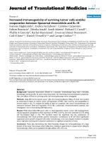
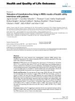
studies, viral infection was found t o induce rapid gen-
eration of microglial cell-produced ROS, as early as 3 h,
with robust levels evident in most cells by 24 h p.i. (Fig-
ure 1). The concentration of H
2
DCFDA used in these
experiments (i.e., 20 μM) did not induce microglial cell
toxicity as determined by MTT assay and trypan blue
staini ng. In addition, MTT assay was used to check cell
viability following viral infection and showed approxi-
mately 15% and 40% decreases at 24 and 48 h p.i.,
respectively.
Inhibition of NADPH oxidase blunts virus-induced ROS
production
We then went on to examine virus-induced ROS pro-
duction over a time-course o f infection. In these experi-
ments, microglial cells were stimulated with HSV for
the designated time, followed by quantification of
H
2
DCFDA oxidation using a fluorescence plate reader.
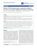
Using this microplate assay, ROS levels in microglial cell
cultures were found to be elevated by 24 h p.i., and
reached maximal levels by 48 h (Figure 2A). We went
on to investigate the effect of inhibition of NADPH oxi-
dase on the production of this HSV-induced ROS. In
these experiments, microglia were pretre ated with the
NADPH oxidase inhibitors DPI or APO for 1 h prior to
Hu et al. Journal of Neuroinflammation 2011, 8:123
/>Page 3 of 9
viral stimulation . HSV-induced ROS production was sig-
nificantly d ecreased by DPI in a concentration-depen-
dent manner and by APO at 300 μMfollowingthe
inhibition of NADPH oxidase (Figure 2B). The concen-
trations of DPI or A PO used did not themselves induce
microglial cell toxicity as determined by MTT ass ay and
trypan blue staining.
ROS drive cytokine and chemokine expression in virus-
infected microglia
We have previously reported that HSV stimulation of
both human and murine microglial cells initiates robust
cytokine and chemokine production [14,15]. Data pre-
sented here demonstrate that ROS production by micro-
glial cells o ccurs within 3 h following HSV infection.
We’ve previously reported that cytokine and chemokine
mRNA is first detectable using RT-PCR by 5 h p.i. and
protein is first detectable by ELISA within 8 h p .i. [15].
The involveme nt of ROS in drivi ng virus-in duced
expression of these immune mediators was investigated
by pretreatment of microglial cells with DPI (0.03 - 1
μM) and APO (10 - 300 μM) and then using real-time
RT-PCR to assess gene expression for select cytokines
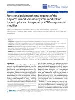
and chemokines. Treatment with either inhibitor of
NADPH oxidase (i.e., DPI or APO) was found to inhibit
TNF-a,interleukin(IL)-1b, CCL2, and CXCL10 mRNA
expression at 5 h p.i. (Figure 3A-D). We went on to
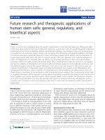
assess the involvement of NADPH oxidase and ROS in
cytokine and chemokine production using ELISA to
measur e protein levels in cell culture supernatants. Cor-
responding to our f indings at the mRNA level, both
inhibitors of NADPH oxidase blunted cytokine (TNF-a
and IL-1b) and chemokine (CCL2 and CXCL10) protein
production in virus-infected microglial cultures (Figure
4A-D).
Viral infection activates p38 and p44/42 (ERK1/2) MAPKs
in primary microglia cells
Activation of MAPKs plays an essential role in the cyto-
kine response of microglial cells to inflammatory stimuli.
p38 MAPK has recently been shown to be critical for
the neurotoxic phenotype of monocytic cells following
exposure to HIV gp120 [17]. For this reason, we exam-
ined whether HSV infection activated p38 and p44/42
MAPKs in our primary murine microglia. Using Wes-
tern Blot, viral infection of primary microglial cells was
found to stimulate phosphorylation of both kinases by 2
h p .i. (Figure 5A). These results were confir med using a
more quantifiable FACE in-cell Western assay over a 24
h time-course of infection. Using this assay, significant
phosphorylation of p38 MAPK in response to viral
infection was detected as early as 1 h p.i., with pro-
longed activation evident at 24 h p.i. (Figure 5B).
Redox signaling drives the p38 MAPK activation
We went on to examine the effect of NADPH oxidase
and ROS production on MAPK activation i n response
3h
24h
Control HSV-1
Figure 1 Intracellular ROS generation in response to HSV-1
infection of primary microglia. Purified murine microglial cell
cultures were either left uninfected (Control) or infected with HSV-1
(MOI = 2.5) for 3 or 24 h prior to loading with H
2
DCFDA (20 μM, 45
min) for visualization using fluorescence microscopy. Data shown
are representative of five individual experiments using microglial
cells obtained from different animals.
0
50
100
150
200
250
300
DPI (
P
M) - 1 0.03 0.1 0.3 1
APO (
P
M) - - 300 - - - 10 30 100 300
HSV
- - -+++ + + + + + +
**
††
††
††
††
††
††
B
0
100
200
300
400
3h 8h 24h 48h 72h
HSV p.i.
**
**
**
Intracellular R
OS
(% of Control, OD at Ex
485
Em
538
A
Figure 2 Inhibition of NADPH oxidase blunts virus-induced
ROS production. Microglia were A) infected with HSV-1 for the
designated time or B) left untreated or pretreated with the NADPH
oxidase inhibitors DPI (0.03 - 1 μM) or APO (10 - 300 μM) at the
indicated concentrations for 1 h prior to viral infection for 36 h,
followed by addition of H
2
DCFDA (20 μM) for 45 min and
quantification using a fluorescent microplate reader. Data are
presented as mean ± SEM from 6-8 separate experiments. **p <
0.01 vs. control;
†
p < 0.05 and
††
p < 0.01 vs. HSV alone.
Hu et al. Journal of Neuroinflammation 2011, 8:123
/>Page 4 of 9
to viral infection. In these studies, treatment of micro-
glial cells with either DPI or APO prior to viral infection
blunted HSV-induced MAPK phosphorylation as
detected using Western Blot at 2 h p.i. (Figure 6A).
Additionally, F ACE assay analysis at 2 h p.i. confirmed
that either DPI or APO treatment significantly reduced
phosphorylation of p38 MAPK (Figure 6B).
MAPK inhibition blocks cytokine and chemokine
production
In the la st set of experiments, we examined the inv olve-
ment of these two ROS-driven MAPK signaling path-
ways in cytokine and chemokine production by
micro glia in response to viral infection. In these studies,
inhibition of the p38 MAPK signaling pathway using
SB203580 (0.1 to 10 μM) was found to suppress both
cytokine (TNF-a and IL-1b) and chemokine (CCL2 and
CXCL10) production (Figure 7). In contrast, inhibition
of p44/42 MAPK signaling using U0126 (0.1 to 10 μM)
inhibited cytokine (Figure 7A, B), but not chemokine
production (Figure 7C, D). Additional assays tested
whether MAPK inhibition affected HSV-induced ROS
production itself. Data generated from these s tudies
showed that the ERK1/2 (p4 4/p42) inhibitor U0126 par-
tially suppressed ROS production b y 11.1%, 18.1%, and
20.9%, at 0.1, 1.0, and 10 μM, respectively. Correspond-
ingly, the p38 MAPK inhibitor SB203580 also partially
suppressed ROS production by 16.3%, 21.1%, and 42.4%,
at 0.1, 1.0, and 10 μM, respectively.
Discussion
We have recently reported that HSV-induced ROS p ro-
duction by microglial cells is responsible for lipid perox-
idation, oxidative damage, and toxicity to neurons in
culture, and that viral recognition is mediated, at least
in part, through Toll-like receptor (TLR)-2 [18]. In sev-
eral other systems, engagement of TLRs has been
demonstrated to induce NADPH oxidase activation,
with corresponding ROS generation, which subsequently
activates NF-B to induce proinflammatory cytokine
production [19-21]. Following up on ou r previous work,
the present study examined the effect of HSV-1-
induced, NADPH oxidase-derived ROS in activating
mitogen-activated protein kinases (MAPKs) and driving
cytokine, as well as chemokine, expression in prima ry
murine microglia. Data obtained during these studies
clearly demonstrate that intracell ular ROS are generated
following viral infection of murine microglia and are
associated with a m arked increase in the expression of
NADPH oxidase mRNA. Viral infection was found to
induce microglial cell-produced ROS as early as 3 h in
individual cells, however, additional time was required
to reach statistical significance when the entire culture
was assessed.
ROS are important second messengers in redox sig-
naling. Viral brain infection initiates robust inflamma-
tory r esponses pivoting on t he production of cyto kines
and chemokines by microglial cells [15]. We have pre-
viously reported that microglial cells undergo an abor-
tive, non-productive infection with HSV-1 in which
immediate early gene (e.g., IC P4) expres sion occurs, but
late gene expression (e.g., such as glycoprotein D, gD)
and viral replication are blocked [15]. These cells
respond t o HSV infection by inducing a burst of cyto-
kine and chemokine pr oduction, followed b y apoptotic
death. It has previously been reported that microglial
ROS, produced largely through the action of NADPH
oxidases, precedes cytokine and chemokine production
in response to HIV Tat or M. tuberculosis 30-kDa Ag
[12,22]. In the present study, inhibition of NADPH oxi-
dase with either DPI or APO was also found to decrease
0
5
10
15
20
25
TNF-
D
mRNA expression
(fold change vs. Control)
0
200
400
600
800
CXCL10 mRNA expression
(fold change vs. Control)
DPI APO
DPI APO
-2
-1
0
1
2
3
4
IL-1
E
mRNA expression
(fold change vs. Control)
DPI APO
0
2
4
6
8
10
12
14
16
CC
L2 mRNA express
i
on
(fold change vs. Control)
DPI APO
HSV
HSV
H
SV
H
SV
AB
CD
Figure 3 ROS drive cytokine and chemokine mRNA ex pression
in virus-infected microglia. Microglial cell cultures were pre-
treated with the NADPH oxidase inhibitors DPI or APO for 1 h prior
to a 5 h exposure to HSV. Following viral infection, RNA was
extracted and cDNA synthesized to assess mRNA expression
through quantitative real-time PCR for A) TNF-a; B) IL-1b; C) CCL2;
and D) CXCL10. mRNA levels were normalized to the housekeeping
gene HPRT and are presented as fold induction over uninfected
controls. Data shown are representative of three individual
experiments using microglial cells obtained from different animals.
Hu et al. Journal of Neuroinflammation 2011, 8:123
/>Page 5 of 9
subsequent HSV-induced cytokine and c hemokine pro-
duction. These data d emonstrate that NADPH-derived
ROS drive cy tokine and chemokine expre ssion by
microglia in response to viral infection.
Phosphorylation of p38 and p44/p42 ERK1/2 MAPK is
commonly associated with T LR signaling and has been
implicated in TLR-associated ROS production
[11,19,23,24]. Because these MAPKs play an important
role in regulating the expression of immune mediators
following stimulation with viruses, viral proteins, and
other inflammatory factors [9,14,17,25-27], we next
investigated the role of p38 and p44/p42 ERK1/2 activa -
tion i n HSV-infected microglia. In these studies, we first
found that viral infection induced the phosphorylation
C DPI APO 0.03 0.1 0.3 1 10 30 100 300
DPI
(
P
M)
APO
(
P
M)
HSV
0
20
40
60
80
100
120
140
160
180
IL-1
E
(ng/ml)
††
††
**
††
††
B
0.0
0.2
0.4
0.6
0.8
1.0
DPI
(
P
M)
APO
(
P
M)
HSV
C DPI APO 0.03 0.1 0.3 1 30 100 300
††
††
††
††
††
††
**
TNF-
D
(ng/ml)
A
0
.0
0
.2
0
.4
0
.6
0
.8
1.0
1.2
1.4
1.6
1.8
DPI
(
P
M)
APO
(
P
M)
HSV
C DPI APO 0.03 0.1 0.3 1 10 30 100 300
CCL2 (ng/ml)
††
††
††
†
**
C
0.0
0.2
0.4
0.6
0.8
DPI
(
P
M)
APO
(
P
M)
H
SV
C DPI APO 0.03 0.1 0.3 1 10 30 100 300
CXCL10 (ng/ml)
††
††
**
††
††
D
Figure 4 ROS contribute to cytokine and chemokine production by microglia in response to viral infection. Supernatants were collected
from murine microglial cell cultures pretreated with DPI or APO at the indicated concentrations for 1 h prior to viral exposure for 36 h (or 16 h
for TNF-a) and cytokine and chemokine levels were assessed using ELISA for A) TNF-a; B)IL-1b; C) CCL2; and D) CXCL10. Data are presented as
mean ± SD of 3 replicates from 3 separate experiments. **p < 0.01 vs. uninfected control;
†
p < 0.05 and
††
p < 0.01 vs. HSV alone.
Hu et al. Journal of Neuroinflammation 2011, 8:123
/>Page 6 of 9
of both MAPKs. We then went on to perform experi-
ments using the inhibitors DPI and APO to determine
whether NADPH oxidase-derived ROS drive viral activa-
tion of p38 and p44/p42 ERK1/2 MAPKs. In these stu-
dies, treatment of microglial cells with the NADPH
oxidase inhibitors was found to blunt HSV-induced
MAPK phosphorylatio n by Western Blot (p38 and p44/
p42 ERK1/2) and FACE (p38) assay.
In our last set of experiments we investigated the
effect of blocking specific MAPK pathways on HSV-
induced cytokine and chemokine production. Using
human microglia, we have previously reported that
while an inhibitor of p38 MAPK (SB202190) blocked
both HSV-induce d cytokine and chemokine production,
treatment with the ERK1/2 inhibitor (U0126) inhibited
the induction of cytokines (i.e., TNF-a,IL-1b), but not
chemokines (i. e., CCL5 and CXCL10), [14]. In the pre-
sent study, very similar differential cytokine and chemo-
kine results are found using HSV-i nfected murine
microglia. HSV-induced TNF-a and IL-1b production
was found to be susceptible to inhibition by both the
p38 MAPK inhibitor SB203580 and the p44/p42 ERK1/2
inhibitor U0126, while virus-induced CXCL10 and
CCL2 was suppressed by SB2 03580, but the p44/p42
ERK1/2 inhibitor had no inhibitory effect at any concen-
tration tested. Taken together, it is likely that insuffi-
cient activation of these MAPK pathways following the
inhibition of NADPH oxidase, and decreased ROS gen-
eration, is responsible for the attenuated cytokine
production.
A number of studies have shown that beneficial neu-
roimmune responses, for example those needed to
purge infectious virus from the brain, can develop into
chronic pathological inflammation with progressive
A
B
C 15m 30m 1h 2h 3h 4h 5h 6h 10h 18h 24h
RLU
(% of Control)
0
200
400
600
800
1000
Total p38
Phospho p38
H
SV
**
**
**
**
**
**
**
**
*
p44/42
p38
Phospho p38
Phospho p44/42
E
-Actin
+HSV
C 15’ 30’ 1h 2h 6h 10h
Figure 5 Activation of p38 and p44/42 (ERK1/2) MAPKs in
response to viral infection of primary microglia. A) Control
uninfected (C) or virus-infected (+HSV) microglial cell culture lysates
were collected at the indicated time points to assess MAPK
activation using Western Blot. B) The kinetics of p38 MAPK
activation were quantified in microglial cell cultures infected with
HSV-1 using a FACE™ p38 Chemi, in-cell Western assay (Active
Motif, Carlsbad, CA). Data presented are representative of mean ±
SD with 3 replicates from 2 separate experiments. *p < 0.05 and** p
< 0.01 vs. uninfected control.
RLU (% of Control)
0
50
100
150
200
250
300
350
400
450
Total p38
Phospho p38
C 1
P
M 300
P
M 0.3
P
M 1
P
M 100
P
M 300
P
M
DPI AP
O
DPI AP
O
††
**
††
††
††
HSV
A
B
p44/42
p38
Phospho p38
Phospho p44/42
E
-Actin
C DPI APO
+HSV
Figure 6 Redox signaling drives p38 MAPK activation.A)Cell
lysates from uninfected control (C) or virus-infected (+HSV)
microglial cells, pretreated with either DPI (1 μM) or APO (300 μM),
were collected at 2 h post-infection and MAPK activation was
assessed using Western Blot. B) The effect of NADPH oxidase
inhibitors (1 h pretreatment) on virus-induced activation of p38
MAPK was quantified 2 h post-infection using a FACE assay. Data
are presented as mean ± SD of triplicates and are representative of
2 separate experiments. **p < 0.01 vs. uninfected control;
††
p < 0.01
vs HSV alone.
Hu et al. Journal of Neuroinflammation 2011, 8:123
/>Page 7 of 9
0
20
40
60
80
100
120
140
160
180
SB202474 (
P
M) 0 10 0 0 0.1 1 10 0 0 0
SB203580
(
P
M) 0 0 10 0 0 0 0 0.1 1 10
HSV
- - - + + + + + + +
IL-1
E
(pg/ml)
††
††
**
0
20
40
60
80
100
120
U0124 (
P
M) 0 10 0 0 0.1 1 10 0 0 0
U0126
(
P
M) 0 0 10 0 0 0 0 0.1 1 10
HSV
- - - + + + + + + +
††
††
**
††
0.0
0.2
0.4
0.6
0.8
1.0
TNF-
D
(ng/ml)
SB202474 (
P
M) 0 10 0 0 0.1 1 10 0 0 0
SB203580
(
P
M) 0 0 10 0 0 0 0 0.1 1 10
HSV
- - - + + + + + + +
††
††
**
†
0.0
0.2
0.4
0.6
0.8
1.0
U0124 (
P
M) 0 10 0 0 0.1 1 10 0 0 0
U0126
(
P
M) 0 0 10 0 0 0 0 0.1 1 10
HSV
- - - + + + + + + +
††
**
††
A
B
0.0
0.2
0.4
0.6
0.8
1.0
1.2
1.4
1.6
1.8
0.0
0.2
0.4
0.6
0.8
1.0
1.2
1.4
CCL2 (ng/ml)
SB202474 (
P
M) 0 10 0 0 0.1 1 10 0 0 0
SB203580
(
P
M) 0 0 10 0 0 0 0 0.1 1 10
HSV
- - - + + + + + + +
††
††
**
U0124 (
P
M) 0 10 0 0 0.1 1 10 0 0 0
U0126
(
P
M) 0 0 10 0 0 0 0 0.1 1 10
HSV
- - - + + + + + + +
**
C
0.0
0.2
0.4
0.6
0.8
0.0
0.2
0.4
0.6
0.8
1.0
SB202474 (
P
M) 0 10 0 0 0.1 1 10 0 0 0
SB203580
(
P
M) 0 0 10 0 0 0 0 0.1 1 10
HSV
- - - + + + + + + +
U0124
(
P
M) 0 10 0 0 0.1 1 10 0 0 0
U0126
(
P
M) 0 0 10 0 0 0 0 0.1 1 10
HSV
+ + + + + + +
CXCL10 (ng/ml)
††
††
**
**
D
Figure 7 Involvement of p38 and p44/42 (ERK1/2) in cytokine and chemokine production by virus-infected primary murine microglia.
Microglial cell cultures pretreated with inhibitors of p38 (SB203580 or its negative control SB202474) or ERK1/2 (U1026 or its negative control
U0124) MAPKs for 30 min prior to viral infection. At 16 h p.i., supernatants were collected and assessed for A) TNF-a or 36 h for B) IL-1b,C)
CCL2, and D) CXCL10 production using ELISA. Data presented are representative of mean ± SD with 3 replicates of 2 separate experiments. **p
< 0.01 vs. uninfected control;
†
p < 0.05 and
††
p < 0.01 vs. HSV alone.
Hu et al. Journal of Neuroinflammation 2011, 8:123
/>Page 8 of 9
neurodegeneration [28]. Restoration of redox balan ce
may be an important determinant i n returning activated
microglia back to a resting st ate following viral infection
and neuroinflammation. The findings presented herein
support the idea that ROS-driven microglial cell activa-
tion, and its associated neurotoxicity, may be a target
for therapeutic modulation through the stimulation of
opposing anti-oxidative responses.
Acknowledgements
This project was supported by Award Number MH-066703 from the National
Institute of Mental Health. The content is solely the responsibility of the
authors and does not necessarily represent the official views of the National
Institute of Mental Health or the National Institutes of Health.
Authors’ contributions
SH co-conceived of the study, and designed and performed experiments.
WS performed experiments and analyzed data. SJS participated in study
design. JRL co-conceived of the study, participated in its design, and wrote
the manuscript. All authors have read and approved the final manuscript.
Competing interests
The authors declare that they have no competing interests.
Received: 9 May 2011 Accepted: 26 September 2011
Published: 26 September 2011
References
1. Valko M, Leibfritz D, Moncol J, Cronin MT, Mazur M, Telser J: Free radicals
and antioxidants in normal physiological functions and human disease.
Int J Biochem Cell Biol 2007, 39:44-84.
2. Block ML, Zecca L, Hong JS: Microglia-mediated neurotoxicity: uncovering
the molecular mechanisms. Nat Rev Neurosci 2007, 8:57-69.
3. Reynolds AD, Glanzer JG, Kadiu I, Ricardo-Dukelow M, Chaudhuri A,
Ciborowski P, Cerny R, Gelman B, Thomas MP, Mosley RL, Gendelman HE:
Nitrated alpha-synuclein-activated microglial profiling for Parkinson’s
disease. J Neurochem 2008, 104:1504-1525.
4. Milatovic D, Zhang Y, Olson SJ, Montine KS, Roberts LJ, Morrow JD,
Montine TJ, Dermody TS, Valyi-Nagy T: Herpes simplex virus type 1
encephalitis is associated with elevated levels of F2-isoprostanes and
F4-neuroprostanes. J Neurovirol 2002, 8:295-305.
5. Khetsuriani N, Holman RC, Anderson LJ: Burden of encephalitis-associated
hospitalizations in the United States, 1988-1997. Clin Infect Dis 2002,
35:175-182.
6. Marques CP, Cheeran MC, Palmquist JM, Hu S, Urban SL, Lokensgard JR:
Prolonged microglial cell activation and lymphocyte infiltration
following experimental herpes encephalitis. J Immunol 2008,
181:6417-6426.
7. Armien AG, Hu S, Little MR, Robinson N, Lokensgard JR, Low WC,
Cheeran MC: Chronic cortical and subcortical pathology with associated
neurological deficits ensuing experimental herpes encephalitis. Brain
Pathol 2010, 20:738-750.
8. Bedard K, Krause KH: The NOX family of ROS-generating NADPH oxidases:
physiology and pathophysiology. Physiol Rev 2007, 87:245-313.
9. Qin L, Liu Y, Wang T, Wei SJ, Block ML, Wilson B, Liu B, Hong JS: NADPH
oxidase mediates lipopolysaccharide-induced neurotoxicity and
proinflammatory gene expression in activated microglia. J Biol Chem
2004, 279:1415-1421.
10. Block ML, Hong JS: Microglia and inflammation-mediated
neurodegeneration: multiple triggers with a common mechanism. Prog
Neurobiol 2005, 76:77-98.
11. Yang CS, Lee HM, Lee JY, Kim JA, Lee SJ, Shin DM, Lee YH, Lee DS, El-
Benna J, Jo EK: Reactive oxygen species and p47phox activation are
essential for the Mycobacterium tuberculosis-induced pro-inflammatory
response in murine microglia. J Neuroinflammation 2007, 4:27.
12. Turchan-Cholewo J, Dimayuga VM, Gupta S, Gorospe RM, Keller JN, Bruce-
Keller AJ: NADPH oxidase drives cytokine and neurotoxin release from
microglia and macrophages in response to HIV-Tat. Antioxid Redox Signal
2009, 11:193-204.
13. Zhang HS, Sang WW, Ruan Z, Wang YO: Akt/Nox2/NF-kappaB signaling
pathway is involved in Tat-induced HIV-1 long terminal repeat (LTR)
transactivation. Arch Biochem Biophys 2011, 505:266-272.
14. Lokensgard JR, Hu S, Sheng W, vanOijen M, Cox D, Cheeran MC,
Peterson PK: Robust expression of TNF-alpha, IL-1beta, RANTES, and IP-
10 by human microglial cells during nonproductive infection with
herpes simplex virus. J Neurovirol 2001, 7:208-219.
15. Aravalli RN, Hu S, Rowen TN, Palmquist JM, Lokensgard JR: Cutting edge:
TLR2-mediated proinflammatory cytokine and chemokine production by
microglial cells in response to herpes simplex virus. J Immunol 2005,
175:4189-4193.
16. Livak KJ, Schmittgen TD: Analysis of relative gene expression data using
real-time quantitative PCR and the 2(-Delta Delta C(T)) Method. Methods
2001, 25:402-408.
17. Medders KE, Sejbuk NE, Maung R, Desai MK, Kaul M: Activation of p38
MAPK is required in monocytic and neuronal cells for HIV glycoprotein
120-induced neurotoxicity. J Immunol 2010, 185:4883-4895.
18. Schachtele SJ, Hu S, Little MR, Lokensgard JR: Herpes simplex virus
induces neural oxidative damage via microglial cell Toll-like receptor-2. J
Neuroinflammation 2010, 7:35.
19. Yang CS, Shin DM, Lee HM, Son JW, Lee SJ, Akira S, Gougerot-Pocidalo MA,
El-Benna J, Ichijo H, Jo EK: ASK1-p38 MAPK-p47phox activation is
essential for inflammatory responses during tuberculosis via TLR2-ROS
signalling. Cell Microbiol 2008, 10:741-754.
20. Park HS, Jung HY, Park EY, Kim J, Lee WJ, Bae YS: Cutting edge: direct
interaction of TLR4 with NAD(P)H oxidase 4 isozyme is essential for
lipopolysaccharide-induced production of reactive oxygen species and
activation of NF-kappa B. J Immunol 2004, 173:3589-3593.
21. Lee IT, Wang SW, Lee CW, Chang CC, Lin CC, Luo SF, Yang CM:
Lipoteichoic acid induces HO-1 expression via the TLR2/MyD88/c-Src/
NADPH oxidase pathway and Nrf2 in human tracheal smooth muscle
cells. J Immunol 2008, 181:5098-5110.
22. Lee HM, Shin DM, Kim KK, Lee JS, Paik TH, Jo EK: Roles of reactive oxygen
species in CXCL8 and CCL2 expression in response to the 30-kDa
antigen of Mycobacterium tuberculosis. J Clin Immunol 2009, 29:46-56.
23. Lee JG, Lee SH, Park DW, Yoon HS, Chin BR, Kim JH, Kim JR, Baek SH: Toll-
like receptor 9-stimulated monocyte chemoattractant protein-1 is
mediated via JNK-cytosolic phospholipase A2-ROS signaling. Cell Signal
2008, 20:105-111.
24. Reed-Geaghan EG, Savage JC, Hise AG, Landreth GE: CD14 and toll-like
receptors 2 and 4 are required for fibrillar A{beta}-stimulated microglial
activation. J Neurosci 2009, 29:11982-11992.
25. Lee YB, Schrader JW, Kim SU: p38 map kinase regulates TNF-alpha
production in human astrocytes and microglia by multiple mechanisms.
Cytokine 2000, 12:874-880.
26. Lin W, Tsai WL, Shao RX, Wu G, Peng LF, Barlow LL, Chung WJ, Zhang L,
Zhao H, Jang JY, Chung RT: Hepatitis C virus regulates transforming
growth factor beta1 production through the generation of reactive
oxygen species in a nuclear factor kappaB-dependent manner.
Gastroenterology 2010, 138:2509-2518, 2518 e2501.
27. Mendez-Samperio P, Perez A, Alba L: Reactive oxygen species-activated
p38/ERK 1/2 MAPK signaling pathway in the Mycobacterium bovis
bacillus Calmette Guerin (BCG)-induced CCL2 secretion in human
monocytic cell line THP-1. Arch Med Res
2010, 41:579-585.
28. Gao HM, Hong JS: Why neurodegenerative diseases are progressive:
uncontrolled inflammation drives disease progression. Trends Immunol
2008, 29:357-365.
doi:10.1186/1742-2094-8-123
Cite this article as: Hu et al.: Reactive oxygen species drive herpes
simplex virus (HSV)-1-induced proinflammatory cytokine production by
murine microglia. Journal of Neuroinflammation 2011 8:123.
Hu et al. Journal of Neuroinflammation 2011, 8:123
/>Page 9 of 9