báo cáo hóa học: " Platelet-activating factor enhancement of calcium influx and interleukin-6 expression, but not production, in human microglia" pptx
Bạn đang xem bản rút gọn của tài liệu. Xem và tải ngay bản đầy đủ của tài liệu tại đây (302.48 KB, 8 trang )
BioMed Central
Page 1 of 8
(page number not for citation purposes)
Journal of Neuroinflammation
Open Access
Research
Platelet-activating factor enhancement of calcium influx and
interleukin-6 expression, but not production, in human microglia
Prasongchai Sattayaprasert
†1,3
, Hyun B Choi
†1,2
,
Sukumal Chongthammakun
3
and James G McLarnon*
1
Address:
1
Department of Pharmacology and Therapeutics, Faculty of Medicine, University of British Columbia, Vancouver, BC, Canada,
2
Division
of Neurology, Department of Medicine, University of British Columbia, Canada and
3
Department of Anatomy, Mahidol University, Bangkok,
Thailand
Email: Prasongchai Sattayaprasert - ; Hyun B Choi - ;
Sukumal Chongthammakun - ; James G McLarnon* -
* Corresponding author †Equal contributors
Microgliaplatelet-activating factorinterleukin-6store-operated channels
Abstract
Calcium-sensitive fluorescence microscopy and molecular biology analysis have been used to study
the effects of platelet-activating factor (PAF) on intracellular calcium [Ca
2+
]
i
and IL-6 expression in
human microglia. PAF (applied acutely at 100 nM) elicited a biphasic response in [Ca
2+
]
i
consisting
of an initial rapid increase of [Ca
2+
]
i
due to release from internal stores, followed by a sustained
influx. The latter phase of the [Ca
2+
]
i
increase was blocked by SKF96365, a non-selective store-
operated channel (SOC) inhibitor. RT-PCR analysis showed PAF treatment of microglia induced
expression of the pro-inflammatory cytokine IL-6 in a time-dependent manner which was blocked
in the presence of SKF96365. However, ELISA assay showed no production of IL-6 was elicited at
any time point (1–24 h) for microglial exposures to PAF. These findings suggest that PAF
stimulation of human microglia induces expression, but not production, of IL-6 and that SOC-
mediated [Ca
2+
]
i
influx contributes to the enhanced expression of the cytokine.
Background
Microglia are resident, immunocompetent cells in the
brain. They show functional plasticity and can be acti-
vated by a diversity of inflammatory stimuli including
ones associated with neurodegenerative diseases [9,18].
The functional responses of microglia following activa-
tion include proliferation, phagocytosis and secretion. In
the latter case microglia can secrete pro- and anti-inflam-
matory cytokines, chemokines, neurotrophic factors and
excitotoxins such as glutamate [20].
One important inflammatory agent is platelet-activating
factor (PAF), an alkyl ether phospholipid compound,
which both stimulates and is produced by microglia [13].
PAF contributes to inflammatory responses in the brain
and is reported to be upregulated in CNS pathophysiol-
ogy [2,17]. Acute application of PAF to human microglia
induces a biphasic change in levels of intracellular Ca
2+
([Ca
2+
]
i
) with an initial rapid phase due to intracellular
release from endoplasmic reticulum (ER) stores and a sec-
ondary phase due to influx through store operated chan-
nels (SOC) [15,31]. Importantly, SOC has been shown to
Published: 15 April 2005
Journal of Neuroinflammation 2005, 2:11 doi:10.1186/1742-2094-2-11
Received: 19 January 2005
Accepted: 15 April 2005
This article is available from: />© 2005 Sattayaprasert et al; licensee BioMed Central Ltd.
This is an Open Access article distributed under the terms of the Creative Commons Attribution License ( />),
which permits unrestricted use, distribution, and reproduction in any medium, provided the original work is properly cited.
Journal of Neuroinflammation 2005, 2:11 />Page 2 of 8
(page number not for citation purposes)
exhibit sustained activation following stimulation of
human [31] and rodent [29] microglia. Prolonged entry
of Ca
2+
through SOC in stimulated microglia could consti-
tute a coupling signal between an activating stimulus and
cellular functional response. Indeed, the involvement of
sustained Ca
2+
responses has been reported as a factor in
the production of arachidonic acid by rat microglia [23].
The pro-inflammatory cytokine IL-6 is released from acti-
vated microglia and mediates inflammatory responses in
brain. Levels of IL-6 in serum and cerebrospinal fluid have
been found to be elevated in stroke patients [8,28] and the
cytokine has also been implicated in the etiopathology of
neurodegenerative disorders such as Alzheimer's disease
(AD), Parkinson's disease (PD) and HIV encephalopathy
[3,14,25]. Interestingly, some evidence is also available
suggesting that under some conditions elevated levels of
IL-6 in brain may actually be beneficial [27].
In this study we have examined a role for SOC mediated
[Ca
2+
]
i
influx in mediating actions of the inflammatory
stimulus PAF to induce IL-6 in human microglia.
Materials and methods
Preparation of cells
The procedures for the isolation of human microglia have
been previously reported [24]. In brief, human embryonic
brain tissues were dissected into small blocks, incubated
in phosphate-buffered saline (PBS) containing 0.25%
trypsin and 40 µg/ml DNase and then dissociated into
single cells by repeated pipetting. Cells were plated in T75
flasks in a medium consisting of Dulbecco's modified
Eagle's medium (DMEM) containing 5% horse serum, 5
mg/ml glucose, 25 µg/ml gentamicin, and 2.5 µg/ml
amphotericin B. Freely floating microglia were harvested
from a medium of mixed cell cultures after 7–10 days of
growth in culture flasks and plated on aclar coverslips for
identification, on poly-L-lysine-coated glass coverslips for
calcium spectrofluorometry and plated on six-well multi-
plates for RT-PCR or ELISA. CD11b and ricinus communis
agglutinin (RCA), specific markers for microglia, were
used to confirm purity of the culture which was in excess
of 98% [24,30].
Calcium spectrofluorometry
The procedures used for measurement of intracellular
Ca
2+
have been reported [6,31]. Microglia were incubated
with 1 µM fura-2/AM (acetoxymethyl ester, Molecular
Probes, Eugene, OR) plus 1 µM pluronic acid in normal
physiological saline solution (PSS) for 30 min. PSS solu-
tion contained (in mM): NaCl (126), KCl (5), MgCl
2
(1.2), HEPES (10), D-glucose (10) and CaCl
2
(1); pH of
7.4. All reagents were obtained from Sigma (St. Louis,
MO).
Following a 20 min wash in dye-free solution, coverslips
were placed on the stage of a Zeiss Axiovert inverted
microscope employing a ×40 quartz objective lens. Cells
were exposed to alternating wavelengths of 340/380 nm
at 6 s intervals and emission light passed through a 510
nm filter. An imaging system (Empix Imaging, Missis-
sauga, ON) was used to record fluorescence ratios using a
CCD camera (Retiga 1300i, Burnaby, BC). Fluorescence
ratios were determined and converted to values of [Ca
2+
]i
using published procedures [11]. All experiments were
done at room temperature (20–22°C).
Reverse transcription-PCR and ELISA assay
IL-6 expression was detected with the reverse-transcriptase
polymerase chain reaction (RT-PCR). Isolation of RNAs
was performed using TRIzol (Gibco-BRL, Gaithersburg,
MD, USA) and DNA contamination was eliminated using
DNase. cDNA synthesis was done using M-MLV reverse
transcriptase (Gibco-BRL). The sequences for the human
specific primers for IL-6 as follows: sense primer: 5'-
GTGTGAAAGCAGCAAAGAGGC-3'; antisense primer: 5'-
CTGGAGGTACTCTAGGTATAC-3'. Human-specific IL-6
signals were generated with the GeneAmp thermal cycler
and Amplitaq Gold DNA polymerase (Applied Biosys-
tems, Foster City, CA). The conditions for PCR were as fol-
lows: initial denaturation at 95°C for 6 min followed by
28 cycles of denaturation at 95°C for 45 sec, annealing at
56°C for 1 min and extension at 72°C for 1 min. A final
extension step at 72°C for 10 min was carried out. PCR
products (159 bp) were identified using 1.5% agarose gels
containing ethidium bromide and visualized under UV
light. GAPDH was used as a reaction standard and human
specific primer sequences were as follows: sense primer:
5'-CCATGTTCGTCATGGGTGTGAACCA-3'; antisense
primer: 5'-GCCAGTAGAGGCAGGGATGATGTTC-3'. The
intensities of each band were measured using NIH image
J 1.24 software (National Institutes of Health, Bethesda,
MD). Relative mRNA levels for each treatment were nor-
malized to GAPDH.
Enzyme-linked immunosorbent assays (ELISA) were per-
formed according to manufacturer instructions (R & D
systems, Minneapolis, MN). Cells were plated on multi-
well plates (≈10
5
cells/well) and treated with PAF (100
nM) in the absence or presence of SKF96365 (20 µM for 8
hr). The cell-free supernatants were used for analysis of IL-
6 production (kit detects IL-6 as low as 0.7 pg/ml). Values
were expressed as means ± SEM and statistical significance
(p < 0.05) was determined using one-way ANOVA and
Newman-Keuls multiple comparison post-test.
Journal of Neuroinflammation 2005, 2:11 />Page 3 of 8
(page number not for citation purposes)
Results
Effects of SKF96365 on SOC-mediated [Ca
2+
]
i
influx by
PAF
PAF-induced changes in [Ca
2+
]i from human microglia
have previously been reported [15,21,31]. Initial study
showed a transient increase in SOC [31] but more recent
work has shown PAF application to evoke a sustained
phase of SOC following an initial component due to
depletion of Ca
2+
from intracellular stores [15,21]. The
differences in PAF responses is considered in the
Discussion.
A representative response to acute application of PAF
(applied at 100 nM) is presented in Fig 1A (n = 18 cells).
A plateau level of [Ca
2+
]i was sustained for a duration
exceeding 2 min after removal of PAF. Following estab-
lishment of a clearly defined plateau phase, the bath solu-
tion was replaced with Ca
2+
-free PSS. This procedure
caused an immediate decline in [Ca
2+
]i to baseline levels
(Fig 1A). Long durations of SOC-mediated influx of Ca
2+
have also been documented in mouse microglial cells
[29].
The results of application of the SOC inhibitor SKF96365
(at 20 µM) to the plateau phase of a PAF response is
shown in the representative recording of Fig 1B (n = 21
cells). SOC-mediated entry of Ca
2+
was reduced to base-
line values by SKF96365. Amplitude of Ca
2+
influx
through SOC was measured as the difference between
baseline and plateau levels and in five independent exper-
iments (n = 107 cells) the amplitude prior to SKF96365
was 140 ± 21 nM and after SKF96365 was at baseline lev-
els. Previous work has shown SKF96365 pretreatment of
human microglia (50 µM for 5 min) abolished a transient
SOC in the cells [31].
Effects of SKF96365 on microglial expression of IL-6
We next examined effects of PAF on expression of the pro-
inflammatory cytokine IL-6 in the absence and presence
of SOC inhibition. The time-dependence of PAF stimula-
tion (100 nM) of human microglia on IL-6 are presented
in Fig 2A. The representative RT-PCR showed no constitu-
tive expression of IL-6 in unstimulated microglia (lane 1
of Fig 2A). IL-6 was maximally expressed at 1 h of expo-
sure to PAF then declined to lower levels at longer treat-
ment times (longest exposure of 6 h). A similar time-
dependence for IL-6 expression was exhibited in a total of
four experiments.
A one hour exposure of human microglia to PAF was cho-
sen for subsequent RT-PCR analysis. As shown in Fig 2B,
constitutive expression of IL-6 was absent (lane 1). PAF
treatment was effective in stimulating expression of the
cytokine (Fig 2B, lane 2). The expression of IL-6 was abol-
ished when SKF96365 was included with the PAF applica-
tion (Fig 2B, lane 3). No evident IL-6 expression was
observed for PAF application in Ca
2+
-free PSS (Fig 2B, lane
4). SKF96365, applied alone in PSS solution, did not
cause any increase in IL-6 (Fig 2B, lane 5).
It was of interest to compare PAF as an inducer of micro-
glial IL-6 to that of LPS (lipopolysaccharide) a potent
inflammatory stimulus of cells. The results of exposure of
human microglia to LPS (100 ng/ml for 6 h) is presented
in Fig 2B (lane 6) showing LPS stimulation caused an
intense band for IL-6. Altering the number of PCR cycles
had no apparent effect on intensity (data not shown) sug-
gesting IL-6 band saturation with LPS (Fig 2B, lane 6).
Comparison of band intensity indicated LPS was a more
effective inducer of IL-6 relative to PAF. Interestingly, a
partial inhibition of LPS-induced IL-6 mRNA was
observed when SKF96365 was applied with LPS (Fig 2B,
lane 7).
Semi-quantitative RT-PCR analysis is presented in Fig 2C
and shows PAF as an effective stimulator of IL-6 expres-
sion (n = 3). However, expression of IL-6 was considera-
bly lower with PAF as a stimulus compared with LPS (Fig
2B,C). Inclusion of SKF96365 with PAF or application of
PAF in Ca
2+
-free PSS eliminated expression of IL-6 (n = 3).
Although LPS was not the subject of this study, the
decrease in LPS induction of IL-6 with SKF96365 is of
interest and is discussed below.
ELISA assay for effects of PAF on microglial production of
IL-6
We next investigated production of IL-6 from PAF-treated
human microglia using an exposure time of 8 h. No pro-
duction of IL-6 was evident in four experiments (data not
shown); levels of IL-6 were below the detection levels for
ELISA assay (≤ 1 pg/ml). In order to determine if the treat-
ment time was a limiting factor in IL-6 production, a series
of experiments using different microglial times of expo-
sure to PAF were undertaken (from 1–24 h). The results
are presented in Fig 3; no significant production of IL-6 (n
= 4) was found for any treatment time (PAF applied for
1,2,8 or 24 h).
We also examined if a ten-fold increase in PAF concentra-
tion (to 1 µM) would be effective in producing IL-6. As
shown in Fig 3, this higher concentration of PAF also had
no effect to induce IL-6 production for treatment times of
8 or 24 h (n = 3 independent experiments). The effects of
LPS stimulation were also determined in these experi-
ments (using 100 ng/ml for 8 h). Microglia, treated with
LPS, produced high concentrations of IL-6 to levels
exceeding 400 pg/ml (n = 4 independent experiments).
Journal of Neuroinflammation 2005, 2:11 />Page 4 of 8
(page number not for citation purposes)
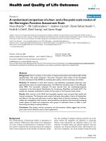
PAF-induced Ca
2+
responsesFigure 1
PAF-induced Ca
2+
responses. A: Representative trace (n = 18 cells) showing change in [Ca
2+
]i induced by PAF (100 nM).
Following a prolonged level of SOC-mediated influx of Ca
2+
, the perfusion of Ca
2+
-free PSS abolished the response. B: Results
from a separate experiment showing effects of SKF96365 (20 µM) on a PAF-induced increase in [Ca
2+
]i (n = 21 cells).
SKF96365 application, during a sustained entry of Ca
2+
through SOC, effectively reduced [Ca
2+
] to baseline levels.
Journal of Neuroinflammation 2005, 2:11 />Page 5 of 8
(page number not for citation purposes)
Expression of IL-6 in PAF treated human microgliaFigure 2
Expression of IL-6 in PAF treated human microglia. A: RT-PCR analysis for different exposure times of microglia to
PAF (applied at 100 nM). B: Effects of PAF, PAF plus SKF96365, PAF plus Ca
2+
-free and SKF96365 applied alone (1 h treat-
ments). Also shown are effects of LPS and LPS plus SKF96365 (6 hr treatments). GAPDH was used as a reaction standard. C:
Semi-quantitative RT-PCR for effects of the different treatments. * P < 0.05 compared with unstimulated control; # P < 0.05
compared with PAF treated microglia.
Journal of Neuroinflammation 2005, 2:11 />Page 6 of 8
(page number not for citation purposes)
Discussion
The results from this work indicate that PAF-mediated
changes in [Ca
2+
]
i
are involved in the cellular expression
of the pro-inflammatory agent, IL-6 in human microglia.
In essence, activation of SOC acts as a transcriptional con-
trol for expression of IL-6. Our results show that inhibi-
tion of SOC with SKF96365 blocked both the influx of
Ca
2+
and microglial expression of IL-6. However, PAF-
induced expression of IL-6 (Fig 2) did not translate into
production of the cytokine (Fig 3). This result could sug-
gest that an additional signal or factor may be required for
microglial secretion of IL-6.
As found for other types of unexcitable cells, microglia do
not normally express voltage-dependent Ca
2+
channels
[7]. The sustained entry of Ca
2+
through SOC is likely an
important pathway for microglial responses to specific
inflammatory stimuli [15,22,26]. Although opening of
SOC is required for re-filling of ER stores, other roles for
this influx pathway have not been well established. Acti-
vation of SOC is necessary for expression of IL-6 but an
additional signal is required to produce the pro-inflam-
matory cytokine in human microglia. The activation state
of human microglia may influence the extent of Ca
2+
influx through SOC. Microglia showing an ameboid mor-
phology are considered representative of an activated state
whereas cells with a ramified morphology are considered
quiescent. We have found sustained SOC responses from
PAF-stimulated microglia in cells demonstrating ameboid
morphology [15,21] and also in the present work. How-
ever, an initial study using a mixture of ameboid and ram-
ified shaped cells, showed a transient SOC response with
stimulation by PAF [31]. Further work will be useful to
correlate expression of SOC with cell activation.
A recent review has provided a detailed overview of ATP as
an inducer of IL-6 expression and production in MG-5
microglial cell line [12]. ATP and the purinergic agonist
BzATP were both effective in increasing expression of IL-6
with effects involving activation of the p38 MAPK path-
way. However, ATP (activator of both metabotropic P2YR
and ionotropic P2XR) but not BzATP (activator of the
ionotropic subtype P2X
7
R), was found to induce produc-
tion of the cytokine. The role of SOC in MG-5 cell
responses is unclear since ATP evokes a monophasic
change in [Ca
2+
]i due to P2YR dependent release from
intracellular stores. In human microglia we have attrib-
uted the lack of a SOC phase of [Ca
2+
]i due to concomi-
tant ATP binding to some P2XR (not P2X
7
R) causing
cellular depolarization and block of Ca
2+
influx [6].
PAF induction of IL-6 was found to be time-dependent
(Fig 2A) in addition to the dependence on the presence of
extracellular Ca
2+
and SOC (Fig 2B). We observed no IL-6
expression at one-half hour and a maximal level at one
hour of microglial exposure to PAF. Little or no IL-6 was
expressed with longer PAF treatments of microglia. Inhibi-
tion of endoplasmic reticulum Ca
2+
ATPase (SERCA) has
been reported to increase IL-6 mRNA expression in rodent
macrophages within 15 min [4,19]. Blockade of SERCA,
by compounds such as thapsigargin, and subsequent
depletion of intracellular stores is a stimulatory protocol
for activation of SOC. However, SOC-mediated entry of
Ca
2+
was not determined in the rodent studies.
Although PAF was an effective stimulator of IL-6 expres-
sion in human microglia, LPS elicited a higher expression
of the cytokine. Indeed, bands for IL-6 appeared saturated
(Fig 2B) and showed no change in intensity with
increased number of PCR cycles (data not shown). Satura-
tion with LPS would prevent a quantitative comparison
between PAF and LPS as activating stimuli for microglial
expression of IL-6 (Fig 2C). An interesting observation
was that SKF96365 partially inhibited the LPS-induced
expression of IL-6 (Fig 2C). Although LPS has been
reported to act in a Ca
2+
-independent manner on macro-
phages [19], several studies have found the bacterial com-
pound evokes changes in [Ca
2+
]i in microglia/
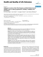
ELISA assays for production of IL-6 in human microgliaFigure 3
ELISA assays for production of IL-6 in human micro-
glia. PAF (at 100 nM) induced no significant production of IL-
6 from microglia following exposures from 1–24 h (n = 4 for
each time points). PAF (at 1 µM) induced no significant pro-
duction of IL-6 (following exposures for 8 h and 24 h; n = 3
for both time points); these values are near the lower limits
for sensitivity of the ELISA kits. LPS was used as a positive
control in these experiments (n = 4); note the change of
scale for the ordinate (from 10 to 400 pg/ml). * P < 0.05
compared with unstimulated control.
Journal of Neuroinflammation 2005, 2:11 />Page 7 of 8
(page number not for citation purposes)
macrophages [1,5,16,32] suggesting possible involve-
ment of SOC in LPS induction of cytokines.
The present results may have relevance to roles of IL-6 in
aging. Several studies have provided evidence for age-
dependent increases in levels of IL-6 in rodent brain
[reviewed in [10]]. For example, one finding was that
brains from older mice showed considerable elevations in
expression and production of IL-6 compared with brains
from younger animals [33]. This result was correlated
with microglial production of the cytokine [33]. It will be
of interest to determine if PAF-stimulated adult human
microglia are more potent producers of IL-6 compared
with fetal human cells.
List of abbreviations
PAF: platelet-activating factor; SOC: store-operated chan-
nels; IL-6; interleukin-6; PSS: physiological saline solu-
tion; PBS: phosphate-buffered saline; [Ca2+]i:
intracellular calcium; DMEM: Dulbecco's modified
Eagle's medium
Competing interests
The author(s) declare that they have no competing
interests.
Authors' contributions
PS and HBC contributed equally to calcium imaging, RT-
PCR and ELISA experiments. HBC also carried out
isolation of microglia. SC participated in the design of
experiments and reviewed and edited the manuscript.
JGM designed and supervised all experiments, interpreted
the data and finalized the manuscript. All authors read
and approved the final manuscript.
Acknowledgements
This work was supported by grants from the Heart and Stroke Foundation
of British Columbia and Yukon and Alzheimer's Society of Canada (to JGM)
and a doctoral research award from the Heart and Stroke Foundation of
Canada (to HBC).
References
1. Bader MF, Taupenot L, Ulrich G, Aunis D, Ciesielski-Treska J: Bacte-
rial endotoxin induces [Ca2+]i transients and changes the
organization of actin in microglia. Glia 1994, 11:336-344.
2. Bielenberg GW, Wagener G, Beck T: Infarct reduction by the
platelet activating factor antagonist apafant in rats. Stroke
1992, 23:98-103.
3. Blum-Degen D, Muller T, Kuhn W, Gerlach M, Przuntek H, Riederer
P: Interleukin-1 beta and interleukin-6 are elevated in the
cerebrospinal fluid of Alzheimer's and de novo Parkinson's
disease patients. Neurosci Lett 1995, 202:17-20.
4. Bost KL, Mason MJ: Thapsigargin and cyclopiazonic acid initi-
ate rapid and dramatic increases of IL-6 mRNA expression
and IL-6 secretion in murine peritoneal macrophages. J
Immunol 1995, 155:285-296.
5. Choi HB, Khoo C, Ryu JK, van Breemen E, Kim SU, McLarnon JG:
Inhibition of lipopolysaccharide-induced cyclooxygenase-2,
tumor necrosis factor-alpha and [Ca2+]i responses in human
microglia by the peripheral benzodiazepine receptor ligand
PK11195. J Neurochem 2002, 83:546-555.
6. Choi HB, Hong SH, Ryu JK, Kim SU, McLarnon JG: Differential acti-
vation of subtype purinergic receptors modulates Ca
2+
mobi-
lization and COX-2 in human microglia. Glia 2003, 43:95-103.
7. Eder C: Ion channels in microglia (brain macrophages). Am J
Physiol 1998, 275:C327-C342.
8. Ferrarese C, Mascarucci P, Zoia C, Cavarretta R, Frigo M, Begni B,
Sarinella F, Frattola L, De Simoni MG: Increased cytokine release
from peripheral blood cells after acute stroke. J Cereb Blood
Flow Metab 1999, 19:1004-1009.
9. Gao MH, Jiang J, Wilson B, Zhang W, Hong JS, Liu B: Microglial acti-
vation-mediated delayed and progressive degeneration of
rat nigral dopaminergic neurons: relevance to Parkinson's
disease. J Neurochem 2002, 81:1285-1297.
10. Godbout JP, Johnson RW: Interleukin-6 in the aging brain. J
Neuroimmunol 2004, 147:141-144.
11. Grynkiewicz G, Poenie M, Ysien R: A new generation of Ca
2+
indi-
catiors with greatly improved fluorescence properties. J Biol
Chem 1985, 260:3440-3450.
12. Inoue K: Microglial activation by purines and pyrimidines. Glia
2002, 40:156-163.
13. Jaranowska A, Bussolino F, Sogos V, Arese M, Lauro GM, Gremo F:
Platelet-activating factor production by human fetal micro-
glia. Effect of lipopolysaccharides and tumor necrosis factor-
alpha. Mol Chem Neuropathol 1995, 24:95-106.
14. Jones S, Horiuchi S, Topley N, Yamamoto N, Fuller G: The soluble
interleukin 6 receptor: mechanisms of production and impli-
cations in disease. FASEB J 2001, 15:43-58.
15. Khoo C, Helm J, Choi HB, Kim SU, McLarnon JG: Inhibition of
store-operated Ca
2+
influx by acidic extracellular pH in cul-
tured human microglia. Glia 2001, 36:22-30.
16. Letari O, Nicosia S, Chiavaroli C, Vacher P, Schlegel W: Activation
by bacterial lipopolysaccharide causes changes in the
cytosolic free calcium concentration in single peritoneal
macrophages. J Immunol 1991, 147:980-983.
17. Lindsberg PJ, Yue TL, Frerichs KU, Hallenbeck JM, Feuerstein G: Evi-
dence for platelet-activating factor as a novel mediator in
experimental stroke in rabbits. Stroke 1990, 21:1452-1457.
18. Lue LF, Rydel R, Brigham EF, Yang LB, Hampel H, Murphy GM Jr, Bra-
chova L, Yan SD, Walker DG, Shen Y, Rogers J: Inflammatory rep-
ertoire of Alzheimer's disease and nondemented elderly
microglia in vitro. Glia 2001, 35:72-79.
19. Marriott I, Bost KL, Mason MJ: Differential kinetics for induction
of IL-6 mRNA expression in murine peritoneal macro-
phages: Evidence for calcium-dependent and independent-
signalling pathways. J Cell Physiol 1998, 177:232-240.
20. McGeer PL, McGeer EG: The inflammatory response system of
brain: implications for therapy of Alzheimer and other neu-
rodegenerative diseases. Brain Res Rev 1995, 21:195-218.
21. McLarnon JG, Helm J, Goghari V, Franciosi S, Choi HB, Nagai A, Kim
SU: Anion channels modulate store-operated calcium influx
in human microglia. Cell Calcium 2000, 28:261-268.
22. Moller T: Calcium signaling in microglial cells. Glia 2002,
40:184-194.
23. Mori M, Aihara M, Kume K, Hamanoue M, Kohsaka S, Shimizu T: Pre-
dominant expression of platelet-activating factor receptor in
the rat brain microglia. J Neurosci 1996, 16:3590-3600.
24. Nagai A, Nakagawa E, Hatori K, Choi HB, McLarnon JG, Lee MA, Kim
SU: Generation and characterization of immortalized human
microglial cell lines: expression of cytokines and
chemokines. Neurobiol Dis 2001, 8:1057-1068.
25. Neuroinflammation Working Group: Inflammation and Alzhe-
imer's disease. Neurobiol Aging 2000, 21:383-421.
26. Parekh AB, Penner R: Store depletion and calcium influx. Physiol
Rev 1997, 77:901-930.
27. Pavelko KD, Howe CL, Drescher KM, Gamez JD, Johnson AJ, Wei T,
Ransohoff RM, Rodriguez M: Interleukin-6 protects anterior
horn neurons from lethal virus-induced injury. J Neurosci 2003,
23:481-492.
28. Tarkowski E, Rosengren L, Blomstrand C, Wikkelso C, Jensen C,
Ekholm S, Tarkowski A: Early intrathecal production of inter-
leukin-6 predicts the size of brain lesion in stroke. Stroke 1995,
26:1393-1398.
29. Toescu EC, Moller T, Kettenmann H, Verkhratsky A: Long-term
activation of capacitative Ca
2+
entry in mouse microglial
cells. Neuroscience 1998, 86:925-935.
Publish with BioMed Central and every
scientist can read your work free of charge
"BioMed Central will be the most significant development for
disseminating the results of biomedical research in our lifetime."
Sir Paul Nurse, Cancer Research UK
Your research papers will be:
available free of charge to the entire biomedical community
peer reviewed and published immediately upon acceptance
cited in PubMed and archived on PubMed Central
yours — you keep the copyright
Submit your manuscript here:
/>BioMedcentral
Journal of Neuroinflammation 2005, 2:11 />Page 8 of 8
(page number not for citation purposes)
30. Walker DG, Kim SU, McGeer P: Complement and cytokine gene
expression in cultured microglia derived from postmortem
human brains. J Neurosci Res 1995, 40:478-493.
31. Wang X, Bae JH, Kim SU, McLarnon JG: Platelet-activating factor
induced Ca
2+
signaling in human microglia. Brain Res 1999,
842:159-165.
32. Xie YC, Schafer R, Barnett JB: Inhibitory effect of 3,4-dichloro-
propionaniline on cytokine production by macrophages is
associated with LPS-mediated signal transduction. J Leukoc
Biol 1997, 61:745-752.
33. Ye SM, Johnson RW: Increased interleukin-6 expression by
microglia from brain of aged mice. J Neuroimmunol 1999,
93:139-148.