Photodiodes Communications Bio Sensings Measurements and High Energy Part 10 potx
Bạn đang xem bản rút gọn của tài liệu. Xem và tải ngay bản đầy đủ của tài liệu tại đây (1.16 MB, 20 trang )
Photodiode Array Detection in Clinical Applications;
Quantitative Analyte Assay Advantages, Limitations and Disadvantages
171
measured simultaneously on the array of fixed photodiodes. The speed of scanning the
spectrum is thus determined by the speed of data acquisition. In modern diode-array UV
detectors equipped with powerful computers the time necessary to take the full spectrum
from 190 to 600 nm can be reduced to as short as about 10 msec. This speed is more than
sufficient in the overwhelming majority of cases in pharmaceutical analysis when the half-
band width of peaks separated by HPLC is usually in the order of 1 min and it is only very
rarely in the order of 1-10 sec in fast HPLC systems and especially in capillary
electrophoresis where the peaks are in general narrower.
The quality of the UV spectrum of the separated impurities obtained by the diode-array
detector is influenced by several of photodiodes. For example, the number of diodes in a
DAD of the HPLC instrument is only 205 while in the other it is 1024. If the spectrum has
fine structure, better quality spectra are obtainable with the latter. In addition to this the
quality of the spectra of especially the low level impurities greatly depends on the baseline
noise. This can be reduced by using a light source with high intensity, by selecting a suitable
reference wavelength (which is as close to the cut-off wavelength of the separated analyte as
possible and a suitable slit width. Generally speaking the sensitivity of the new generation
of diode-array detectors is much higher than that of the older ones.
There are three main areas within drug impurity profiling where the advantages of diode-
array detectors can contribute to the success of the HPLC (CE) analysis (see Figures 5-7).
(a)Peak purity determination. The determination of peak homogeneity is an integral part of the
protocol in the validation of any kind of HPLC (and CE) analysis of pharmaceuticals. In the
course of impurity profiling studies it is especially important to check the peak of the main
component for its homogeneity from the simple and most widely used absorbance ratio
method [Drouen et al.,1984; Wilson et al.,1989 ] to more sophisticated deconvolution,
spectral suppression, spectrum subtraction and other chemometric methods[Huber &
George, 1993]. If any kind of peak in-homogeneity is found (impurity on the leading or
tailing edges of the main peak or fused impurity peaks, conveniently demonstrated in the
three-dimensional mode) the diode-array spectra themselves furnish further information for
the identification of the unresolved impurities.
Fig. 5. Peak purity measurement
Photodiodes – Communications, Bio-Sensings, Measurements and High-Energy Physics
172
Fig. 6. Maximum impurity detection
(b) Spectral matching. Matching the diode-array spectra of components separated by HPLC
with those taken by computer search from spectral libraries is a widely used method [Huber
& George, 1993] especially in toxicological analysis . This approach is of limited value in
drug impurity profiling since it is unlikely that impurities of especially new drugs are
included in spectrum libraries. However, matching the diode-array spectra of the separated
impurities with standard materials can greatly support the identification of the impurities
on the basis of retention matching.
(c) Structure elucidation of the separated impurities. It is reasonable to begin the search for the
structure unknown impurity separated by HPLC or CE with drawing as many conclusions
from its diode-array UV spectrum as possible.
Fig. 7. Determination of peak purity
The short-wavelength parts of the (diode-array) UV spectra can be subject of several
distorting effects, moreover even false maxima can occur. In addition to this, short-
Photodiode Array Detection in Clinical Applications;
Quantitative Analyte Assay Advantages, Limitations and Disadvantages
173
wavelength UV bands can originate from different chromophoric functional groups and for
this reason they are of limited value in the structure elucidation of organic compounds. As a
consequence of these factors it is a prerequisite of drawing useful conclusions from the UV
spectrum of an impurity that it should have at least one maximum above 210-220 nm.
Another limitation is that the difference between the structures of the drug material and the
impurity should be at or near the chromophoric part of the molecule in order that the
difference between their spectra can be of diagnostic value in the structure elucidation of the
impurity. For example, the chromophoric group of various steroids is the 4-ene-3-oxo group
with an absorption maximum around 240 nm. As it will be shown later, the position of this
band is influenced by substituents in the B and C ring of the steroid nucleus but by no
means by substituents at C-17. For this reason various esters of 17-hydroxy-4-ene-3-oxo
steroids (testosterone, 19-nortestosterone, 17-hydroxyprogesterone, etc.) cannot be
differentiated on the basis of their UV spectra.
HPLC with photodiode array detection (HPLC-PDA or HPLC-DAD) is regularly employed
for substance identification in the context of Systematic Toxicological Analysis [Koves,1995;
Gaillard & Pépin,1997; Herre & Pragst,1997]. With HPLC-PDA the most important
parameters in identifying a compound are its retention time and its UV spectrum. Critics of
the method often question the specificity of UV detection because of poorly structured
spectra and broad absorption bands. Therefore a systematic investigation into the selectivity
of PDA detection was carried out by analyzing large numbers of UV spectra with respect to
their correlation with chemical structure.
For data analysis the following tools are needed:
1. A spectra library ; the library is embedded into the chromatography software in a way
that spectral similarity is compared nm by nm and a “hit list” is returned to the
operator.
2. A database of retention times and specific peak areas.
3. A database of all molecular structures with an ability for substructure searches.
4. A structural database of all registered chromophores.
As an alternative to Mass Spectrometers, absorbance detectors (including PDA) are much
less expensive and relatively simple to use. LC-DAD is a fast and robust method for
screening biological samples in conjunction with a library search algorithm to quickly
identify those samples that require confirmatory testing. Numerous methods for using LC-
PDA as a screening method have been published and were recently reviewed by Pragst et
al. [Pragst et al.,2004]. Because a PDA detector can collect an entire spectrum at each time
point in a chromatogram, the data are information rich and more selective than single
wavelength chromatograms. Herzler et al. [Herzler et al.,2003] showed that PDA data could
be used to selectively identify abused substances in spectrochromatograms based on
comparison to a library of over 2500 “toxicologically relevant” substances. Their method
relied on the calculation of a ‘similarity index’ (related to the correlation coefficient) to
determine the similarity between a spectrum in an unknown chromatogram and a library
spectrum. In addition to spectral matching, a relative retention time was also used to
identify the substances of interest.
1.1.4.3 Medical chemistry applications of HPLC-PDA
High performance liquid chromatography (HPLC) with photodiode array detection has
been proved to be the demanded method of systematic analysis for unknown drugs in
biological sample because of separation efficiency, sensitivity, flexibility and identification
Photodiodes – Communications, Bio-Sensings, Measurements and High-Energy Physics
174
potential. HPLC can be an easy way of quantitation as well. Ultraviolet spectra acquired
with photodiode array detector together with retention data are used to identify unknown
or suspected drugs and metabolites in various biological material. These analytical systems
are suitable for toxicological examinations of forensic cases, acute poisonings, drug abuse.
They are convenient to subsequent monitoring of serum drug levels during treatment of
intoxication as well.
High-performance liquid chromatography coupled with diode array detection (HPLC-DAD)
has been widely used as a powerful means for the analysis of multi-component medicines,
which can provide a UV chromatogram and comprehensive data about the compounds in
complex mixtures [Han et al.,2007; Su etal.,2010; Wei et al.,2010; Zhang et al.,2010]. This
technology facilitates identification of unknown components in the matrices system
remarkably with high sensitivity and accuracy.
Photodiode array (PDA) detectors record light absorption at different wavelengths and can
provide spectra of the analytes. This is useful in identifying unknowns. Mass spectrometry
(MS) is a better detector for unknowns. It gives an unambiguous molecular weight of an
analyte and provides structural information. When coupled with CE or HPLC, MS can
separate co-eluting analytes with different mass to charge ratios. But the Mass spectrometer
is an expensive instrument and the possibility of using it is not available in all laboratories.
Of course, if possible HPLC/ESI-MS/UV-DAD analysis gives the best sensitivity
[Cuyckens& Claeys,2002; Beretta et al.,2009; Christiansen et al.,2011].
The potentials and limitations of high-performance liquid chromatography-photodiode
array detection are highlighted in respect to its use in the analysis of different biological
matrices followed by the identification of unknowns. The logical analytical approach used in
clinical and forensic toxicology, vital for the identification of one or more toxic substances as
a cause of intoxication, is largely based on both simple and fast "general unknown
screening" methods which cover most relevant drugs and potentially hazardous chemicals.
In this field of systematic toxicological analysis, a literature overview shows that HPLC can
play a substantial role. Both column packing material and eluent composition have their
impact on intra- and inter laboratory reproducibility. In view of the sometimes different
retention characteristics of various HPLC columns, several possibilities are addressed to
enhance the discriminating power of primary retention parameters. The advantages of
photodiode array detection as compared to UV detection have been of paramount
importance to the success of HPLC in toxicological analysis. Dedicated libraries with
spectral information and searching software are powerful tools in the process of
identification of an unknown substance. In the present section, these aspects are also
verified in a number of real cases.
HPLC-DAD used as a general unknown screening tool should cover as many drugs and
toxicants as possible, but should be also very selective, sensitive and reliable. Liquid
chromatography is used in forensic laboratories for numerous applications including
examination of drugs. LC with photodiode array detection (PDA) is a hybrid technique
which can provide complete UV-visible spectral information on a given peak in a
chromatogram, enabling determinations of peak purity to be made, and identification of
unknown peaks to be assigned by library searches of spectral information in combination
with retention behavior. These are valuable features normally associated with gas
chromatography-mass spectrometry. The additional information available on each peak
makes LC-PDA a particularly attractive technique for the forensic laboratory where higher
levels of certainty are often demanded in test results. This paper reviews some of those
Photodiode Array Detection in Clinical Applications;
Quantitative Analyte Assay Advantages, Limitations and Disadvantages
175
applications for LC-PDA in the forensic sciences, including drug screening, drug and
pharmaceutical analysis, idenfication of pesticides, fungi, quality control testing and
profiling of cosmetics, street drugs and profiling of other complex mixtures. The practical
and technical limitations of the technique are explored and its place in the hierarchy of
methods available in forensic laboratories is evaluated [Proença et al.,2003; Madej et al.,2003;
Proenc et al.,2004; Nieddu et al., 2007; Es’haghi et al.,2010; ; Vosough et al.,2010].
HPLC-DAD offers many advantages in terms of specificity, sensitivity, speed and ruggedness.
The data produced, comprising both retention behavior and absorption spectra of eluting
chemical entities, result in an identification power at low cost and with widened availability
through many laboratories. In addition, the examples showed a great versatility in application
fields and excellent quantitative potential. The fast progress in DAD detector technology,
computer and software power and HPLC packing material quality have led to an exponential
rise of the number of reports on the use of HPLC-DAD. The advent of routine use of HPLC-
MS will probably promote HPLC as a viable if not better alternative to GC-MS.
We examined that combined with a sample preparation method; HPLC-PDA can be easy
achieved to very low detection limits [Es’haghi et al., 2009, 2010]. In a research, we used of
direct suspended droplet microextraction (DSDME) method, based on a three-phase
extraction system which is compatible with HPLC-PDA for determination of ecstasy;
MDMA (3,4methylendioxy-N-methylamphetamine) in human hair samples. After the
extraction, pre-concentrated analyte was directly introduced into HPLC for further analysis.
In concentration range between 1.0 and 15,000 ng mL
-1
calibration curve is drowned.
Linearity was observed with r = 0.9921 for analyte. Limit of detection (LOD) were calculated
as the minimum concentration providing chromatographic signals three times higher than
background noise. Limit of quantification (LOQ) was estimated as the minimum
concentration preparing chromatographic signals ten times higher than background noise.
Thus, LOD obtained was 0.1 and LOQ was 1.0 ng mL
-1
too [Es’haghi et al., 2010].
In the other work we successfully used of DSDME method combined with HPLC-PDA for
determination of low-residue benzodiazepine, diazepam and lorazepam, in the
environmental water samples [Es’haghi et al., 2009, 2009]. After the optimized extraction
conditions, the suspended micro-droplet is withdrawn by a HPLC microsyringe, injected to
and analyzed by HPLC-DAD. Method was evaluated and enrichment factor 839.8, linearity
range from 25 to 5000 ng mL
-1
with an average of relative standard deviation (n=5) 5.62% for
diazepam using a photodiode array detector were determined. HPLC-PDA has good
matches with complex matrices such as hair.
A method combining liquid–liquid–liquid microextraction and automated movement of the
acceptor and donor phases (LLLME/AMADP) with ion-pair HPLC/DAD has been
developed to detect trace levels of chlorophenols in water [Lin etal.,2008] . The extracted
chlorophenols, present in anionic form, were then separated, identified, and quantitated by
ion-pair high-performance liquid chromatography with photodiode array detection
(HPLC/DAD). For trace chlorophenol determination using HPLC/DAD, the
chlorophenolate anion provides a better ultraviolet spectrum for quantitative and
qualitative analyses than does uncharged chlorophenol. The proposed method was capable
of identifying and quantitating each analyte to 0.5 ng mL
-1
, confirming the HPLC/DAD
technique to be quite robust for monitoring trace levels of chlorophenols in water samples.
HPLC/DAD could simultaneously detect UV absorptions at multiple wavelengths and
extract the UV spectra of separated analytes in a chromatogram. Absorbance measurements
at the band maxima of UV spectra obey the linear Beer’s law more accurately than
Photodiodes – Communications, Bio-Sensings, Measurements and High-Energy Physics
176
measurements off the band maxima, and UV spectra of the separated analytes can be
utilized to identify target analytes in HPLC/DAD. Accordingly, each extracted
chlorophenolate anion after ion-pair liquid chromatography separation was quantitated by
the maximum adsorption of its own red shift characteristic band, and each target
chlorophenlate anion was identified by its own red shift characteristic band as well as its
enhanced B band. The chlorophenols were determined under selected experimental
conditions to assess repeatability, linearity, coefficient of determination, and detection limit.
A HPLC-DAD method for drug screening in plasma were developed by M. A. Alabdalla
[Alabdalla,2005]. This analytical method extracted and tested a number of drugs of different
classes. The method included; an acidic and basic Solid Phase Extraction (SPE) of plasma
with C18 cartridges, a gradient elution of a modified cyano column with acidic
buffer/acetonitrile eluent and a photodiode array ultraviolet (UV) detection. The drug
screening procedure applied used retention index and UV spectral data for the identification
of compounds, may be appropriate in particular laboratory settings.
Continuous administration of polyphenols from aqueous rooibos (Aspalathus linearis)
extract ameliorates dietary-induced metabolic disturbances in hyperlipidemic mice was
studied by HPLC-DAD and introduced by R. Beltrán-Debón et al. [Beltrán-Debón et al.,
2011]. In this biological matrices and they could find good results.
In a recent study neurons from the olfactory system of the fish crucian carp, Carassius
carassius L. were used as components in an in-line neurophysiologic detector (NPD) to
measure physiological activities following the separation of substances by high-performance
liquid chromatography (HPLC). The skin of crucian carp, C. carassius L. contains
pheromones that induce an alarm reaction in conspecifics. Extra-cellular recordings were
made from neurons situated in the posterior part of the medial region of the olfactory bulb
known to mediate this alarm reaction. The nervous activity of these specific neurons in the
olfactory bulb of crucian carp was used as an in-line neurophysiologic detector. HPLC was
performed with a diode array detector (DAD) [Brondz et al.,2004].
UV spectral detection was performed at 214, 254 and 345 nm, and scans (190–400 nm) were
collected continuously. This system enabled the selection of peaks in the chromatogram
with fish alarm pheromone activity. Neurophysiologic detectors (NPDs) in-line with diode
array detectors (DADs) are able to provide the physiologically active substances and their
spectral characteristics.
Li-wei Yang et al. were developed a method using high-performance liquid
chromatography–photodiode array detection (HPLC–DAD) for the quality control of
Hypericum japonicum thunb (Tianjihuang), a Chinese herbal medicine. For the first time,
the feasibility and advantages of employing chromatographic fingerprint were investigated
for the evaluation of Tianjihuang by systematically comparing chromatograms with a
professional analytical. The results revealed that the chromatographic fingerprint combining
similarity evaluation could efficiently identify and distinguish raw herbs of Tianjihuang
from different sources. The effects resulted from collecting locations; harvesting time and
storage time on herbal chromatographic fingerprints were also examined [Yang et al.,2005].
1.1.4.4 Photo diode array detector in kinetic study
In kinetic experiments, transient optical absorption is recorded versus time to evaluate rate
constants related to the species under investigation. In addition, the recording of a spectrum
sometimes becomes necessary in order to identify the species. In most cases, the spectrum is
constructed from point-to-point recordings of kinetic curves at selected wavelengths. This
procedure is time consuming, and becomes boring especially at long recording times in the
Photodiode Array Detection in Clinical Applications;
Quantitative Analyte Assay Advantages, Limitations and Disadvantages
177
second and minute time domain. The use of a device, which enables the recording of a
complete spectrum, can be very helpful as it reduces experiment time remarkably.
Unwanted side effects, such as photolysis during long recording times, can also be
prevented. The application of optical multichannel analyzers which use either a linear
charge coupled device (CCD) or a linear photodiode array (PDA) in kinetic experiments was
reported by some laboratories [Hunter et al .,1985; Sedlmair et al.,1986; Johnson et al.,1994].
The advantage of using such a detector is the ability to immediately record a complete
spectrum from UV to IR with one measurement.
The PDA detector has the ability to record a spectrum over a large range of wavelengths.
The uniformity of the analyzing light intensity over the whole range is important because
the dynamics and the sensitivity of the measurements depend largely on the intensity. The
spectral distribution of the analyzing light, as recorded by the multichannel detector is
shown in Figure .8.
Fig. 8. Light intensity vs. wavelength of an xenon lamp, recorded by the multichannel
detector.
The source of the analyzing light is an xenon lamp. The light intensity is attenuated tenfold
as compared to kinetic experiments. Although, the recorded intensity of the analyzing light
decreases drastically below 350 nm, a spectral range from 300 to 800 nm can be covered.
Below 300 nm, recording should be accomplished in small segments and with the help of
band-pass filters in order to adjust for the reduced level of analyzing light and for the
decreased sensitivity of the detector, and, in addition, to avoid scattered light effects. The
measurement depends largely on proper focusing of the light path, i.e., how well the lamp
arc is imaged onto the diode array.
Each spectrum is the average of some (for example five) individual measurements; each
irradiation consists of a train of ten pulses. The interval between the recordings of the
individual spectra or between the pulses in each pulse train was set to zero. The recording at
time zero, i.e. before irradiation, shows a straight line. The change in absorption increases with
increasing irradiation. In general, kinetic trace scan be constructed from the recorded spectra at
selected wavelengths. Similar to the construction of spectra from kinetic traces [Janata,1994].
Photodiodes – Communications, Bio-Sensings, Measurements and High-Energy Physics
178
At measurements in the UV region, Cerenkov emission is a common problem at short
measuring times. The intensity of the Cerenkov emission increases with decreasing
wavelength and can be much larger than the kinetic signal itself, but probably will not exceed
the intensity of the analyzing light. Although this apparatus makes data at longer time scale
available, overdriving of the photodiodes and long recovery times are conceivable.
The use of an optical multichannel detector consisting of a linear diode array embedded in
the instrumentation for kinetic spectroscopy, as well as the highlights of the computer
program used for controlling the gathering and the evaluation of data are described.
Complete spectra can be recorded and irradiation can be triggered according to a preset
timetable. Due to the read-out time of the photodiode array and the time required by the
computer to control the experiment, this apparatus is suitable for application starting in the
millisecond time domain and extending up to very long time periods.
1.1.4.5 Chemometrics investigations using photo diode array detection
Chemometrics is a statistical approach to the interpretation of patterns in multivariate data.
When used to analyze instrument data, chemometrics often results in a faster and more
precise Assessment of composition of a product or even physical or sensory properties. For
example, composition of drugs can be quickly measured using LC and chemometrics. Food
properties can also be monitored on a continuous basis. In all cases, the data patterns are
used to develop a model with the goal of predicting quality parameters for future data. The
two general applications of chemometrics technology to predict a property of interest; and
to classify the sample into one of several categories (e.g., good versus bad, Type A versus
Type B versus Type C etc.). Chemometrics can be used to speed methods development and
make routine the use of statistical models for data analysis. Keeping in view of the
complexity of the chromatographic fingerprint and the irreproducibility of chromatographic
and spectral instruments and experimental conditions, several chemometric approaches
such as variance analysis, peak alignment, correlation analysis and pattern recognition were
employed to deal with the chromatographic fingerprint. Many mathematical algorithms are
used for data processing in chemometric approaches. The basic principles for this approach
are variation determination of common peaks/regions and similarity comparison with
similarity index and linear correlation coefficient. Similarity index and linear correlation
coefficient can be used to compare common pattern of the chromatographic fingerprints
obtained. In general, the mean or median of the chromatographic fingerprints under study
is taken as the target and both are considered to be reliable [Brereton,1987].
The rapid scanning detectors, as diode array detection, present an alternative technology for
rapid, multi-wavelength detection in HPLC. If hyphenated chromatography is further
combined with chemometric approaches, clear pictures might be developed for
chromatographic fingerprints obtained. A chemical fingerprint obtained by hyphenated
chromatography, out of question, will become the primary tool for quality control of
medicines.
The full UV-Vis spectrum became accessible as a three-dimensional (3D) data matrix (A, A,
t). Data are available in the time, concentration and wavelength domains. This allows the
simultaneous use of more than two wavelengths for detection or for the full application of
detector information to the analytical problem by means of available chemometric
techniques to data from second-order bilinear instruments, as chromatographic and
excitation-emission data.
As an alternative to MS, absorbance detectors (including PDA) are much less expensive and
relatively simple to use. LC-DAD is a fast and robust method for screening biological samples
Photodiode Array Detection in Clinical Applications;
Quantitative Analyte Assay Advantages, Limitations and Disadvantages
179
in conjunction with a library search algorithm to quickly identify those samples that require
confirmatory testing. Numerous methods for using LC-DAD as a screening method have been
published and were recently reviewed by Pragst et al. [Pragst et al., 2004]. Because a DAD can
collect an entire spectrum at each time point in a chromatogram, the resultant data are
information rich and more selective than single wavelength chromatograms.
For the above reasons could be adopted PDA detectors with the various chemometric
methods to match spectra contained within a spectrochromatogram to a library.
In a research, triply coupled diode array detection high performance liquid chromatography
mass spectroscopy was applied to a complex mixture of at least eight chlorophyll degradation
products. Derivatives were employed to determine parts of the chromatogram of composition
one. Mass selection was performed on the mass spectroscopic data. Principal components
analysis was performed on both the raw and simultaneously normalised/standardised data;
three dimensional projections of the data were obtained and compared to conventional two
dimensional graphs. Angular plots between diode array loadings characteristic of individual
compounds and scores of the diode array data were described. In mass spectra, angular plots
between loadings characteristic of individual compounds and the remaining diagnostic
masses revealed further mass spectral structure [Zissis et al.,1999].
Liquid chromatography–chemometric methods [LC-Partial least squares (LC-PLS), LC-
principle component regression (LC-PCR) and LC-artificial neural network (LC-ANN)] were
developed for the determination of anomalin (ANO) and deltoin (DEL) in the root by Alev
Tosun et al.[ Tosun et al.,2007]. Firstly, chemometric conditions were optimized by testing
different mobile phases at various proportions of solvents with various flow rates in
different wavelengths by using a normal phase column to obtain the best separation and
recovery results. As a result, a mobile phase consisting of n-hexane and ethyl acetate (75:25
v/v) at a constant flow rate of 0.8 mL min
-1
on the at ambient temperature were found to be
the optimal chromatographic conditions for good separation and determination of ANO and
DEL in samples. Multi-chromatograms for the concentration set containing ANO and DEL
compounds in the concentration range of 50–400 ng mL
-1
were obtained by using a diode
array detector (DAD) system at selected wavelength sets, 300 (A), 310 (B), 320 (C), 330 (D)
and 340 (E). Three LC-chemometric approaches were applied to the multichromatographic
data to construct chemometric calibrations. As an alternative method, traditional LC at
single wavelength was used for the analysis of the related compounds in the plant extracts.
All of the methods were validated by analyzing various synthetic ANO–DEL mixtures.
After the above step, traditional and chemometric LC methods were applied to the real
samples consisting of extracts from roots and aerial parts of analytes.
In a recent research, metabolism disorders in Kunming mice induced by two tumor cells
were characterized. Metabolic fingerprint based on high performance liquid
chromatography-diode array detector (HPLC-DAD) was developed to map the disturbed
metabolic responses. Based on 27 common peaks, principal component analysis (PCA) and
partial least squares-discriminant analysis (PLS-DA) were used to distinguish the abnormal
from control and to find significant endogenous compounds which have significant
contributions to classification. The tumor growth inhibition ratios of Taxol groups were
used to validate the predictive accuracies of the PLS-DA models. The predictive accuracies
of PLS-DA models for tumors model groups were 97.6 and 100%, respectively. Nine and
seven of two models tumors were discovered, including uric acid and cytidine. In addition,
the correlations between relative tumor weights and chromatographic data were significant
(p < 0.05). Investigations on the stability and precision of the established metabolic
Photodiodes – Communications, Bio-Sensings, Measurements and High-Energy Physics
180
fingerprints demonstrate that the experiment is well controlled and reliable. This work was
shown that the platform of HPLC-DAD coupled with chemometric methods provides a
promising method for the study of metabolism disorders [Sun et al., 2011].
2. References
Alabdalla, M.A.,(2005),HPLC-DAD for analysis of different classes of drugs in plasma,J.
Clinical Forensic Med., Vol.12, No.6, (December 2005 ), pp.310-315, ISSN: 13531131
Beltrán-Debón, R., Rull, A., Rodríguez-Sanabria, F., Iswaldi, I., Herranz-López, M.,
Aragonès, G., Camps, J., Alonso-Villaverde, C., Menéndez, J.A., Micol,V., Segura-
Carretero, A., Aragonès,G., Joven, J.(2011), Continuous administration of
polyphenols from aqueous rooibos (Aspalathus linearis) extract ameliorates
dietary-induced metabolic disturbances in hyperlipidemic mice, Phytomedicine
,Article in press,ISSN: 09447113
Beretta, G., Artali, R., Caneva, E., Orlandini, S., Centini, M., Facino, R.M.(2009), Quinoline
alkaloids in honey: Further analytical (HPLC-DAD-ESI-MS, multidimensional
diffusion-ordered NMR spectroscopy), theoretical and chemometric studies, J.
Pharm. Biomed. Anal., Vol.50,No. 3,( 15 October 2009), pp. 432-439, ISSN: 07317085
Bothe, E., Janata, E.(1994), Instrumentation of kinetic spectroscopy-12. Software for data
acquisition in kinetic experiments, Radiat. Phys. Chem. Vol.44, No. 4, (November
1994), pp. 449–454, ISSN: 0969806X
Brereton, R.G.,(1987), Chemometrics in analytical chemistry :a review, Analyst, Vol. 112,
No.12, (December 1987), pp. 1635-1657, ISSN: 00032654
Brondz, I., Hamdani, E.H., Døving, K.(2004), Neurophysiologic detector - A selective and
sensitive tool in high-performance liquid chromatography, J.Chromatogr. B, Vol.800,
No.1-2,( 5 February 2004),pp. 41-47, ISSN: 15700232
Christiansen, A., Backensfeld, T., Kühn, S., Weitschies, W.(2011), Investigating the stability
of the nonionic surfactants tocopheryl polyethylene glycol succinate and sucrose
laurate by HPLC-MS, DAD, and CAD,J.Pharm. Sci, Vol.100, No.5,( May 2011),pp.
1773-1782 , ISSN: 00223549
Cuyckens, F., Claeys, M.(2002), Optimization of a liquid chromatography method based on
simultaneous electrospray ionization mass spectrometric and ultraviolet
photodiode array detection for analysis of flavonoid glycosides, Rapid. Comm. Mass.
Spectrom., Vol.16,No. 24,( nd), pp.2341-2348, ISSN: 09514198
Drouen, A.C.J.H. , Billiet, H.A.H. , De Galan,L. (1984), Dual-wavelength absorbance ratio for
solute recognition in liquid chromatography, Anal. Chem., Vol.56, No.6, (May 1984),
pp. 971–978, ISSN: 00032700
Es'Haghi, Z., Bandegi, S., Daneshvar, L., Salari, P.(2009),Analysis of diazepam residue from
water samples by triple phase-suspended droplet microextraction coupled to high
performance liquid chromatography and diode array detection. Asian J. Chem. , Vol.
21, No. 8, (October 2009), pp. 6392-6402, ISSN: 09707077
Es'Haghi, Z., Daneshvar, L., Salari, P., Bandegi, S. (2009), Determination of low-residue
benzodiazepine, lorazepam, in the environmental water samples by suspended
droplet icroextraction and high performance liquid hromatography-diod array
detector. Chemija, Vol.20, No.3, (nd), pp.181-186, ISSN: 0235-7216
Es'haghi, Z., Mohtaji, M., Hasanzade-Meidani, M., Masrournia, M.(2010), The measurement
of ecstasy in human hair by triple phase directly suspended droplet
microextraction prior to HPLC-DAD analysis, J. Chromatogr. B, Vol.878, No. 1, (1
April 2010),pp. 903–908, ISSN: 15700232
Photodiode Array Detection in Clinical Applications;
Quantitative Analyte Assay Advantages, Limitations and Disadvantages
181
Gaillard, Y. , Pépin, G. (1997), Use of high-performance liquid chromatography with
photodio de-array UV detection for the creation of a 600-compound library.
Application to forensic toxicology, J. Chromatogr. A, vol. 763,No. 1-2,( 28 February
1997), pp. 149–163, ISSN: 00219673
Han, J., Ye, M., Guo, H., Yang, M., Wang, B r., Guo, D a.(2007), Analysis of multiple
constituents in a Chinese herbal preparation Shuang-Huang-Lian oral liquid by
HPLC-DAD-ESI-MS, J. Pharm. Biomed. Anal. Vol.44, No.2, (28 June 2007), pp. 430–
438, ISSN: 07317085
Herre, S . , Pragst, F. (1997), Shift of the high performance liquid chromatographic retention
ti mes of metabolites in relation to the original drug at an RP-8 column with acidic
mobile phase, J. Chromatogr. B, vol. 692, No. 1, (25 April 1997), pp. 111–126, ISSN:
15726495
Herzler,M., Herre,S., Pragst,F. (2003), Selectivity of substance identification by HPLC-DAD
in toxicological analysis using a UV spectra library of 2682 compounds, J. Anal.
Toxicol. Vol.27, No. 4, (May 2003),pp. 233-242, ISSN: 01464760
Huber, L. ; George, S.A.(2003). Diode Array Detection in HPLC (Second edition), Marcel
Dekker, , ISBN: 0-8247-4 ,New York
Hunter, E.P.L., Simic, M.G., Michael, B.D.,(1985), Use of an optical multichannel analyzer for
recording absorption spectra of short-lived transients, Rev. Sci. Instrum. Vol.56,
No.12 (1985), pp. 2199–2204, ISSN: 00346748
Johnson, J.B., Edwards, G., Mendenhall, M.,(1994), A low-cost, high-performance array
detector for spectroscopy based on a charged-coupled photodiode, Rev. Sci.
Instrum.,Vol.65, No.5,(1994), pp. 1782-1783 , ISSN: 00346748
Koves, E. M.(1995), Use of high-performance liquid chromatography-diode array detection
in forensic toxicology, J. Chromatogr. A, vol. 692, No. 1-2,(nd) , pp. 103–119, ISSN:
00219673
Lambert, W. E., Van-Bocxlaer, J.F. , De Leenheer, A. P .( 1997), Potential of high-performance
liquid chromatography with photodiode array detection in forensic toxicology, J.
Chromatogr. B, vol. 689,No.1, (7 February 1997), pp. 45–53, ISSN: 15726495
Lin, C Y., Huang, S D.(2008),Application of liquid–liquid–liquid microextraction and ion-
pair liquid chromatography coupled with photodiode array detection for the
determination of chlorophenols in water, J. Chromatogr. A, vol. 1193, No. 1-2, (6 June
2008) , pp. 79–84, ISSN: 00219673
Madej, K., Parczewski, A., Kała, M.(2003), HPLC/DAD Screening Method for Selected
Psychotropic Drugs in Blood, Toxicol. Mech. Meth., Vol.13, No.2,( April 2003),pp.
121-127, ISSN: 15376524
Nieddu, M., Boatto, G., Serra, D., Soro, A., Lorenzoni, S., Lubinu, F. (2007), HPLC-DAD
determination of mepivacaine in cerebrospinal fluid from a fatal case. J. Forensic
Sci., Vol.52, No.5, (September 2007), pp.1223-1224, ISSN: 00221198
Pragst, F., Herzler, M., Erxleben, B T.,(2004), Systematic toxicological analysis by high-
performance liquid chromatography with diode array detection (HPLC-DAD),
Clin. Chem. Lab. Med. Vol. 42 , No.11,(nd), pp. 1325-1340,ISSN:14346621
Proença, P., Pinho Marques, E., Teixeira, H., Castanheira, F., Barroso, M., Ávila, S., Vieira,
D.N.(2003), A fatal forensic intoxication with fenarimol: Analysis by
HPLC/DAD/MSD, Forensic Sci. Int.,Vol.133, No. 1-2,( 23 April 2003),pp. 95-100,
ISSN: 03790738
Proença, P., Teixeira, H., Pinheiro, J., Marques, E.P., Vieira, D.N. (2004), Forensic intoxication
with clobazam: HPLC/DAD/MSD analysis, Forensic Sci. Int.,
Vol.143, No.2-3, (16
July 2004), pp. 205–209, ISSN: 03790738
Photodiodes – Communications, Bio-Sensings, Measurements and High-Energy Physics
182
Sedlmair, J., Ballard, S.G., Mauzerall, D.C.,(1986), Diode-array spectrometer (DAPS) for
visible and near-IR absorption measurements with 10-ns time resolution, Rev. Sci.
Instrum, Vol.57, No.12, (nd), pp. 2995–3003, ISSN: 00346748
Skoog, D. A. ; Leary, J. J. (1992). Principles of Instrumental Analysis (third edition), Saunders
College Publishing, ISBN: 4833702827 , Fort Worth
Su, S., Guo, J., Duan, J a., Wang, T., Qian, D., Shang, E., Tang, Y. .(2010), Ultra-performance
liquid chromatography-tandem mass spectrometry analysis of the bioactive
components and their metabolites of Shaofu Zhuyu decoction active extract in rat
plasma, J. Chromatogr. B, vol. 878,No. 3-4,(1 February 2010), pp. 355–362, ISSN:
15700232
Sun,X., Liu,Y., Di,D., Wu,G., Guo,H. (2011), Chemometric analysis of metabolism disorders
in blood plasma of S180 and H22 tumor-bearing mice by high performance liquid
chromatography-diode array detection, J.Chemometrics, Article in press, DOI:
10.1002/cem.1387, SSN:1099-128X
Talmi, Y.,(1975), Applicability of TV-type multichannel detectors to spectroscopy,
Anal.Chem., Vol.47, No.7, (nd), pp. 658A-670A, ISSN: 00032700
Talmi, Y.,(1982), Spectrophotometry and spectrofluorometry with the self-scanned
photodiode array, App. Spectrosc., Vol.36, No.1,(January 1982), pp.1-19 , ISSN:
00037028
Tosun, A., Bahadir, Ö., Dinç, E.(2007), Determination of anomalin and deltoin in Seseli
resinosum by LC combined with chemometric methods, Chromatographia , Vol.66,
No.9-10, (November 2007),pp. 677-683, ISSN: 00095893
Vosough, M., Bayat, M., Salemi, A.(2010), Matrix-free analysis of aflatoxins in pistachio nuts
using parallel factor modeling of liquid chromatography diode-array detection
data, Anal.Chim.Acta, Vol. 663, No.1, (March 2010),pp.11-18, ISSN: 00032670
Wei, H., Sun, L., Tai, Z., Gao, S., Xu, W., Chen, W. (2010), A simple and sensitive HPLC
method for the simultaneous determination of eight bioactive components and
fingerprint analysis of Schisandra sphenanthera, Anal. Chim. Acta, Vol.662, No. 1, (3
March 2010),pp. 97–104, ISSN: 00032670
Wilson, T.D., Trompeter, W.F., Gartelman, H.F.,(1989), Analysis of barbiturate mixtures
using HPLC with diode array detection, J. Liq. Chromatogr.Vol.12, No. 7, (nd), pp.
1231-1251, ISSN: 01483919
Yang, L W., Wu, D H., Tang, X., Peng, W., Wang, X R., Ma, Y., Su, W W.,(2005),
Fingerprint quality control of Tianjihuang by high-performance liquid
chromatography–photodiode array detection, , J.Chromatogr. A, Vol. 1070, No.1-2,( 8
April 2005),pp. 35-42, ISSN: 00219673
Zhang, W., Saif, M.W., Dutschman, G.E., Li, X., Lam, W., Bussom, S., Jiang, Z., ( ), Cheng,
Y C.(2010), Identification of chemicals and their metabolites from PHY906, a
Chinese medicine formulation, in the plasma of a patient treated with irinotecan
and PHY906 using liquid chromatography/tandem mass spectrometry
(LC/MS/MS, J. Chromatogr. A, vol. 1217, No. 37,( September 2010 ), pp. 5785–5793,
ISSN: 00219673
Zissis, K.D., Dunkerley, S., Brereton, R.G.(1999),, Chemometric techniques for exploring
complex chromatograms: application of diode array detection highperformance
liquid chromatography electrospray ionization mass spectrometry to chlorophyll a
allomers, Analyst, Vol. 124,No.7, (nd) , pp. 971–979, ISSN: 00032654
Part 4
Photodiode for UV-Light Detection
10
UV Photodiodes Response to
Non-Normal, Non-Colimated and
Diffusive Sources of Irradiance
María-Paz Zorzano, Javier Martín-Soler and Javier Gómez-Elvira
Centro de Astrobiología (INTA-CSIC), Carretera de Ajalvir km 4
Torrejón de Ardoz, Madrid,
Spain
1. Introduction
A photodiode is a type of photodetector capable of converting incident light into either
current or voltage, depending upon the mode of operation. The substrate used to make the
photodiode sensing part is critical to defining its properties. In particular, only photons with
sufficient energy to excite electrons across the material's bandgap will induce photocurrents.
Materials commonly used to produce photodiodes include: Silicon (for the wavelength
range 190-1100 nm), Germanium (for 400-1700 nm), Indium-Gallium arsenide (for 800-2600
nm) and Lead (II) sulphide (1000-3500 nm), among others. In many applications the
semiconductor diode is placed within an opaque housing with different possible sizes (such
as TO18, TO5, TO3 ) and hermetically sealed with a cover of a material that allows
transmission of the desired wavelength range of observation and permits radiation to reach
the sensitive part of the device. Some applications require measurement of one specific
wavelength range and it is essential that other radiation wavelengths of the range where the
substrate shows responsivity do not contribute to the photodiode's current. For this
purpose a filter is mounted above the detector to select the wavelength range of
responsivity. Common configurations of filtered photodiodes include a metallic platform to
hold the filter at an intermediate height between the cover window and the detector, see
Figure 1. This mechanical configuration allows the implementation of different filters
preserving the external packing dimensions and physical properties of the photodiode
sealing with independence of the filter type.
The geometry of arrangement of these elements (cover, housing, and sensing device) defines
the nominal field of view (FOV) or geometrical FOV of the sensor. A careful look at this
configuration shows that, because of secondary internal reflections against the inner walls of
the sensor housing, the sensor may also be excited by photons from trajectories with
incident angles greater than this nominal geometrical FOV. Notice that the filter crystal is
thick, flat and cut in square shape. Its implementation on top of the sensing dice covers the
nominal FOV but leaves a fraction of the photodiode horizontal cross-section uncovered, see
Figure 2. As we will explain later, this will have implications for non-normal, non-
collimated and diffusive irradiance sources.
Photodiodes – Communications, Bio-Sensings, Measurements and High-Energy Physics
186
Fig. 1. (Left) Commercial hermetically sealed filtered ultraviolet (UV) photodiode with TO5
housing and the filter mounted on a metallic platform. (Right) Geometrical configuration of
the photodiode. The filter crystal is here represented in red and the SiC detector dice in
black. The filter is mounted on the metallic structure but does not reach the side of the
housing.
Because of changes in the refractive index as the light beam crosses the boundary between
two media there is an angle variation which is given by Snell law. The light ray bends
toward the normal when the light enters a medium of greater refractive index, and away
from the normal when entering a medium of lesser refractive index. For this later case there
is a limiting angle beyond which all the rays should be reflected that is called the critical
angle of incidence. For instance, in the case of a transition from a quartz crystal to gaseous
nitrogen or to air the nominal critical angle is roughly 40º. In theory, for incident angles
greater than this angle the light rays should be totally reflected by the cover crystal.
However in most cases there is a range of angles where a fraction is reflected and a fraction
transmitted. In addition in some implementations, such as the one analyzed here, the
covering window is slightly curved. This induces additional angular distortions for non-
normal incidence and in practice shifts the critical angle of incidence towards greater values.
In summary for the case considered here, beyond the nominal FOV there is a wider angle of
allowed incoming rays, what we will here call the critical angle FOV, where incident rays
are allowed to pass inside the photodiode housing. Photon rays contained between the
nominal FOV and the critical angle FOV may get inside the photodiode housing, be
reflected in the walls, avoid the filter and be trapped between the filter and the sensing
substrate dice. These photons may thus excite the dice and contribute to the total
photodiode output current avoiding the filter selective bandpass action and producing an
unexpected current leak. This is schematically represented in Figure 2.
In summary, for a given arbitrary configuration where the radiation from a complex
radiating environment excites a photodiode, part of the radiation may be reflected by the
cover window and part may pass with slightly different exit angle, part may be contained
within the FOV and traverse the filter and part may bounce within the inner part of the
sensor housing and hit the dice with a new incidence angle (and here again it may be
absorbed, or reflected). When these effects are present the response of the dice to the
incident irradiance is neither the one tabulated in the device specifications by the
manufacturer (which is usually defined for normal incidence) nor the one obtained in
UV Photodiodes Response to
Non-Normal, Non-Colimated and Diffusive Sources of Irradiance
187
standard laboratory calibrations. For any practical photodiode use, where the incident light
is non-normal and non-collimated, these deviations should be explored under
representative operation conditions.
Fig. 2. Nominal field of view (FOV) and critical angle conceptual representation: All ray
beams, such as (a), within the FOV solid angle hit the sensing dice after traversing the filter.
Photon rays that traverse the window cover with angles of incidence such as (b) hit the inner
housing walls and reach the sensing dice after multiple reflections, avoiding the filter
selective action.
It is the aim of this chapter to illustrate these effects experimentally with a specific study
case and to describe an experimental setup that can be used to quantify the relevance of each
contribution. In particular, we describe here the calibration and operation studies of the
(ultraviolet) UV photodiodes of the UV sensor for REMS (Rover Environmental Monitoring
Station) instrument on-board the MSL rover to Mars (NASA 2012) [Gómez-Elvira et al.,
2008]. This sensor consists of a set of 6 photodiodes with different responsivity spectral
ranges. One of the photodiodes has no filter and is sensitive in the total SiC responsivity
range (from here on named ABC). The other 5 photodiodes correspond to filtered bands
named A,B,C,D and E, see Figure 4. Each broadband measurement provides a crude
evaluation of the incident irradiance in its relevant spectral range: photodiode C provides a
first order estimate of the level of biologically damaging irradiance; photodiodes A and B
provide an estimate that may be comparable with terrestrial irradiances while photodiode
ABC gives an estimate of the total UV irradiance, and photodiodes D and E are designed to
match two UV channels of the MARCI instrument, on-board the Mars Reconnaissance
Photodiodes – Communications, Bio-Sensings, Measurements and High-Energy Physics
188
Orbiter (MRO) satellite to enable direct comparison with reflectance measurements from the
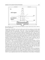
top of the atmosphere. The chosen UV photodiodes have a nominal FOV of ± 30º. This is the
solid angle contained between the normal to the dice and the imaginary line connecting the
sensing dice and the opaque border of the top of the caging, as shown in Figure 2. The
photodiodes are mounted in a circular pattern within a metallic box on the rover deck,
facing the sky. Each one is embedded within a magnet (to deflect the trajectories of in-falling
magnetic dust and protect the window from Martian dust deposition, mimicking the effect
of the magnet experiment of the MER rovers) [Kinch et al. 2006]. A scheme of this setup with
the nominal field of view of ± 30 º of a photodiode is shown in Figure 3. The whole REMS-
UV setup, in an anodized aluminium box of 55 mm x 68 mm x 16 mm with a D25 connector,
weights only 72 g. Photodiodes have the advantage of being small in size, light and robust
for operation under harsh conditions such as those expected for the MSL rover.
This sensor will deliver for the first time in-situ surface ultraviolet irradiance measurements
that will provide ground-truth to radiative transfer models and satellite reflectance
measurements as well as first order estimates of biological and chemical doses and UV
opacities. A solid understanding of the UV radiation behaviour of the Martian atmosphere is
important for photochemical models of the atmosphere [Rodrigo et al., 1990], for the
chemistry of the surface minerals [Holland 1978, Mukhin et al., 1996, Quin et al. 2001], has
biological implications and is paramount for the assessment of the possible habitability of
the Martian surface [Cockell et al. 2000, Patel et al. 2002, Patel et al. 2003, Patel et al. 2004a,
Patel et al. 2004B, Cordoba-Jabonero et al. 2003, Cordoba-Jabonero et al. 2005]. In addition,
satellite [Mateshvili et al. 2006, Montmessin et al. 2006] and Martian-ground based
measurements of UV radiation [Zorzano and Córdoba-Jabonero 2007], are important to
retrieve, through radiative transfer studies, [Zorzano et al. 2005], accurate information on
atmosphere aerosols, in particular aerosol size distribution, load, dust and cloud dynamics.
It is also relevant to estimate the ozone content which in turn serves as a proxy for Mars
atmospheric water vapour.
Previous space missions designed to explore the UV Martian conditions, such as the failed
Beagle lander mission, also considered a similar UV sensor concept based on photodiodes
[Patel et al. 2002]. However, because of their simplicity they also have certain limitations.
This study summarizes the evaluation of the response of this setup to representative
operation conditions.
Fig. 3. (Left) Front view of the photodiode and magnet. (Centre) Back view of the
photodiode with pins and magnet. (Right) Schematic representation of the nominal FOV of
the photodiode within REMS-UV box.
UV Photodiodes Response to
Non-Normal, Non-Colimated and Diffusive Sources of Irradiance
189
In this space application, the direct radiation source is the Sun. The incident UV radiation
comes both as a direct beam, with incident direction according to the solar zenithal angle at
the moment of observation, and as diffuse UV irradiance. This diffuse component is the
product of the scattering interaction between the incident solar radiation with the dust
aerosols and molecules of the thin Martian atmosphere [Zorzano et al. 2005, Zorzano and
Córdoba-Jabonero 2007, Zorzano et al. 2009]. The response of the photodiodes in this
operation environment shall be investigated.
In summary, we have three scenarios to consider. When the photon ray is within the
geometrical FOV, the direct beam is expected to be filtered. When the direct beam is
between the geometrical FOV and the critical angle FOV, secondary reflections against the
wall may allow extra photons to reach the dice avoiding the filter and thus inducing a
current leakage produced by an unfiltered contribution. Finally we shall consider the
response to the background diffuse irradiance, i.e. the radiation that has suffered scattering
with the atmosphere and reaches the sensor window from almost any direction. In this case
the sensor is excited by the diffuse irradiance contained within the solid angle of FOV,
which is a significant fraction of the sky diffused irradiance, and shall be filtered. The extra
diffuse radiation coming from rays with angles greater than this FOV, but still within the
critical angle FOV, can also excite the SiC dice through secondary reflections and, for some
photodetectors such as the one considered here, avoiding the filter action. The fraction of
diffuse radiation that gets to the dice not being filtered is proportional to the difference
between the nominal FOV and the critical angle FOV. If the downwelling diffuse irradiance
is uniform then this is a pure geometrical factor. There are second order corrections to this
due to the specific reflective, absorption and transmission characteristics of each filter that
will be also experimentally observed.
2. Characterization of the response under laboratory conditions
2.1 Spectral calibration of the response with a direct collimated beam of normal or
inclined incidence
The response of the sensor to a direct beam of collimated light can be calibrated under
controlled operation conditions. This has been done to characterize the spectral responsivity
of each photodiode and its dependence with angle of incidence. Its dependence with
temperature, the linearity of the response, the degradation with aging and thermal cycling
were also analyzed for this specific application in space instrumentation but the results of
these tests are beyond the scope of this chapter and shall not be discussed here.
A UV source, focusing optics, a monochromator, a calibrated beam splitter, a detector and a
multimeter have been used to calibrate spectrally the response of the photodiodes. One of
the photodiodes was sent to The National Physical Laboratory (NPL) (UK's National
Measurement Institute) for reference calibration and the results of this calibration setup
were referenced with this measurement. The spectral responsivity of each of the 6 chosen
photodiodes to a collimated direct beam at normal incidence and ambient temperature is
shown in Figure 4. The spectral response of the SiC sensing dice is the one labelled as ABC,
namely the one of the photodiode without filter. The rest of photodiodes have a filter and
the spectral responsivity is the result of the combined effect of this filter with the underlying
SiC responsivity.
The same study has been performed for inclined incidence. Figure 5 shows the measured
decay of responsivity (as percentage with respect to the one at normal incidence) for the
Photodiodes – Communications, Bio-Sensings, Measurements and High-Energy Physics
190
peak wavelength (the wavelength of maximal responsivity) for all chosen photodiodes
together with a 7
th
order polynomial fit (the r.m.s. error of the fit is 5,3
%)
. We observe in
this graph both the departure from cosine law and the response of the dices to radiation for
incident angles beyond the nominal FOV. This study has been performed for different polar
angles and the result is qualitatively similar (not shown), concluding that the response of the
photodiode to a direct collimated beam depends mostly on the azimuth angle. At the edge
of the nominal FOV (±30
o
) the response has decayed to a 40% with respect to the normal
(and not to 85% as would be expected by a pure cosine like response). Beyond this point
there is still significant signal (a 20% of the maximal). The difference between the slow decay
of ABC, A and E and the quick decay of B,C, and D is because the maximum of the spectral
response of the later ones shows a shift of about 20 nm towards lower wavelength ranges.
Furthermore, it is clearly observed in this graph that all the photodiodes show significant
responsivity beyond the nominal FOV of ± 30º.
Fig. 4. (Insert) View of the REMS UV box with 6 photodiodes. (Graph) Spectral responsivity,
calibrated at ambient temperature with a collimated beam at normal incidence.
2.2 Spectral characterization of the response of the non-filtered contribution with a
direct collimated beam at normal incidence
To evaluate qualitatively the spectral weight of the unfiltered contribution, a photodiode
was manipulated to separate, in the total current signal, the contribution from the filtered
signal and the unfiltered contribution. The same setup used for the spectral calibration of
flight model units was used here.
A C type photodiode was opened (by cutting the TO5 housing); an opaque element (a small
aluminium plate) was placed on top of the filter, blocking the passage of light rays through
this path. Photodiodes that have suffered this manipulation are here named “ob”. These
photodiodes deliver a current only when photons hit the SiC sensing dice avoiding the filter