Biosensors Emerging Materials and Applications Part 15 doc
Bạn đang xem bản rút gọn của tài liệu. Xem và tải ngay bản đầy đủ của tài liệu tại đây (1.85 MB, 40 trang )
A New Biosensor to enumerate Bacteria in Planktonic and Biofilm Lifestyle
551
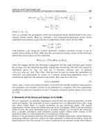
Fig. 2. BioTimer Assay correlation line. The typical BTA correlation line correlating the time
(t*) for color switching of BTA indicator and the log of number of bacteria initially present
in the samples (N
0
) is described by the linear equation t* = −a logN
0
+ b.
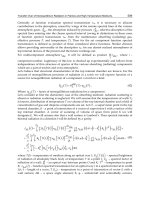
Moreover, the Eq (2) takes into account not only the t* of switching of different indicators,
but also the composition of different reagents through “a” parameter. As shown in Figure 3
the correlation lines for Lactobacillus rhamnosus performed using BT-PR reagent containing
glucose or lactose, as carbon source, differ only for “a” parameter involving the switching
time ( t* ) for the same number of bacteria (N
0
)
.
.
Fig. 3. Correlation lines of Lactobacillus acidophilus obtained using a BioTimer Assay Phenol
Red (BT-PR) specific reagent with 1% glucose (BT-PRglu) or 1% lactose (BT-PRlac).
Biosensors – Emerging Materials and Applications
552
Bacterial
species
BTA
reagent
Equation of
correlation line
r
Application Reference
Streptococcus
sobrinus
BT-RP -0.3056x + 8.2608 0.9997
Adhesion to dental
polymers
Berlutti et al.,
2003
Streptococcus
oralis
BT-RP -0.301x + 9.0615 0.9999
Adhesion to dental
polymers
Berlutti et al.,
2003
Lactobacillus
acidophilus
BT-RPglu -0.1857x + 7.9174 0.9903
Control of lyophilized
probiotic preparation
Valenti et al.,
personal
data
BT-RPlac -0.2773x + 7.4984 0.998
Control of lyophilized
probiotic preparation
Valenti et al.,
personal
data
Staphylococcus
aureus
BT-RP -0.5903x + 8.7219 0.9973
Adhesion to dental
polymers
Berlutti et al.,
2003
BT-RPMH -0.597x + 10.28 0.9990
Antibiotic
susceptibility of biofilm
Pantanella et
al., 2008
Staphylococcus
epidermidis
BT-RPMH -0.633x + 9.267 0.9980
Antibiotic
susceptibility of biofilm
Pantanella et
al., 2008
Enterococcus
f
aecalis
BT-RP -0.4767x + 10.022 0.9975
Laser disinfection of
dental root canals
Berlutti et al.,
personal
data; Telesca,
Master
Thesis, 2010
Escherichia coli
BT-RP -0.9678x + 10.347 0.9955
Adhesion to dental
polymers
Berlutti et al.,
2003
FBTA -0.8723+14.428 0.9970
Fecal contamination of
food
Berlutti et al.,
2008
Pseudomonas
aeruginosa
BT-RZ -0.4675x + 8.5841 0.9996
Adhesion to dental
polymers;
adhesion to SWCNT-
structured surfaces
Berlutti et al.,
2003;
Frioni et al.,
2010
Burkoldheria
cenocepacia
BT-RZ -0.415x + 9.018 0.9610 None
Berlutti et al.,
personal
data
Table 1. Correlation lines.
Likely, Eq.(2) takes into account also bacterial genera/species through “b” parameter. As
shown in Figure 4 the correlation lines for Staphylococcus aureus and Streptococcus sobrinus or
S. oralis performed using the same BTA reagent differ only for “b” parameter involving the
different metabolic activity specific for each bacterial genera/species.
Summarizing, in the BTA applications, the number of living planktonic bacteria in a sample
is determined inoculating the specific BTA reagent. The color switching of BTA indicator is
monitored and the time (t*) for color switching is recorded and used to determine the log N
0
through the specific correlation line.
A New Biosensor to enumerate Bacteria in Planktonic and Biofilm Lifestyle
553
Fig. 4. Correlation lines of Streptococcus sobrinus, Streptococcus oralis and Staphylococcus
aureus obtained using BT-PR reagent.
Similarly, it is possible to count bacteria in aggregated, adherent and biofilm lifestyle by
inoculating BTA reagents with sample containing aggregated bacteria or solid
supports/materials on which bacteria adhere or form biofilm (colonized material) without
sample manipulation. As the Eq. 2 describes the correlation between the time for color
switching of BTA indicators present in the original reagents and the CFUs (N
0
) of planktonic
bacteria, the number of bacteria in aggregated, adherent and biofilm lifestyle counted using
BTA can be defined as planktonic-equivalent CFUs (PE-CFUs).
However, it is possible to object that the metabolic rate of the same bacterium in different
lifestyle can be different and consequently the counts by BTA can be influenced by lifestyle.
In order to answer to this objection, S. sobrinus has been chosen as bacterial model because it
produces lactate as the principal end product of carbohydrate metabolism (Madigan, 2008;
Burne, 1998), which is easily detectable by high performance liquid chromatography system
(Berlutti, 2008; personal data). Planktonic and biofilm lifestyle S. sobrinus was cultured in
complete medium for 24 h at 37°C, in the absence or in the presence of glass beads,
respectively. S. sobrinus, indeed, colonizing the glass beads forms biofilm in 24 hours of
incubation. Both planktonic bacteria and colonized glass beads were used to inoculate BT-
PR reagents. The time for color switching of BT-PR reagents as well as the lactate
concentrations (c
lac
) at the moment of the color switching were recorded.
The values of lactate concentration c
lac
at the moment of color switching of BT-PR reagents
inoculated with different concentrations of planktonic N
0
were similar and corresponded to
a mean value of 770±33 mg/l (Table 2).
The values of c
lac
at the moment of color switching of BT-PR reagents inoculated with 1, 5, 10
colonized beads were similar and corresponded to a mean value of 760±45 mg/l (Table 2).
Therefore, the concentration of lactate needed for inducing color switching of the indicator
is independent from bacterial lifestyle. The sole difference observed among the samples was
the time required for color switching, the parameter pivotal for bacterial counts by BTA
(Berlutti et al., 2008 a; Valenti, personal data).
Biosensors – Emerging Materials and Applications
554
Lifestyle Inoculum c
lac
(mg/l) t* (hours)
Planktonic (log N
0
)
a
5 761±42 10.2
6 773±42 7.5
7 777±20 4.5
Biofilm (N
GB
)
1 805 ±48 4.5
5 710±52 2.7
10 740±45 1.5
Table 2. Lactate concentration (c
lac
) and switching time (t*) of BT-PR reagents inoculated
with Streptococcus sobrinus in planktonic and biofilm lifestyle. Legend:
a
planktonic inoculum
is prepared from broth cultures; biofilm inoculum is obtained utilizing colonized glass
beads (N
GB
).
Similarly to that demonstrated in counting planktonic bacteria by BTA, the time required for
BTA indicator switching is inversely related to the increasing of colonized glass bead (N
GB
)
number and consequently to the number of bacteria in biofilm (Table 2).
Therefore, the switching time t* is inversely proportional to the logarithm of the initial N
GB
,
according to the following equation
t* = −a
GB
logN
GB
+ b
GB
(3)
which is equivalent to the Eq. (2) describing the correlation line for bacteria in planktonic
lifestyle.
3. BioTimer Assay applications
It is important to again underline that the counts of bacteria in aggregated, adherent and
biofilm lifestyle, through BTA, do not require any manipulation of the samples, and this
characteristic represents an important advantage of BTA respect to other methods.
However, in the absence of a validated reference method, the number of bacteria in
aggregated, adherent and biofilm lifestyle carried out by BTA cannot be compared with
those obtained by other methods of bacterial enumeration in biofilm. This lack is a
disadvantage for all novel methods. Notwithstanding, BTA has been successfully applied to
enumerate bacteria in biofilm adherent on abiotic materials, on different foods and recently,
to detect the susceptibility of biofilm to antibiotics as well as the microbiological quality of
nano-particles to be in vivo administered.
3.1 BioTimer Assay to enumerate bacteria in adherent and biofilm lifestyle on abiotic
materials
The actual quantitative determination of bacteria in adherent and biofilm lifestyle on abiotic
materials is a concern for microbiologists. BTA has been successfully employed to estimate
bacterial population colonizing a variety of abiotic materials.
The first report concerned the evaluation of adhesion ability of different Gram-positive and
Gram-negative species on different adhesive poly(HEMA)-based hydrogels to be utilized in
dental restorative procedures (Berlutti et al., 2003). As matter of fact, the use of dental
polymers is a standardized practice in dental restorative procedures. However, bacteria
A New Biosensor to enumerate Bacteria in Planktonic and Biofilm Lifestyle
555
potentially causing oral pathologies may colonize these polymers. It is therefore of great
importance to evaluate both the susceptibility of the polymers to colonization by resident
and transient bacterial genera, and the importance of chemical factors triggering bacterial
adhesion. The study reported data of adhesion efficiency and biofilm formation of S.
sobrinus and Streptococcus oralis representing bacterial resident species, and Staphylococcus
aureus, Escherichia coli, and Pseudomonas aeruginosa considered transient bacteria in the oral
cavity. The dental polymers were prepared with 2-hydroxyethyl methacrylate (HEMA) and
different molar ratios of 2-acrylamido-2-methylpropane-sulfonic acid (AMPS) and/or 2-
methacryloyloxyethyl-tri-methyl-ammonium chloride (METAC) co-monomers.
In conditions mimicking those present in the oral cavity, all tested bacteria showed similar
adhesion percentages on the same dental polymer and different adhesion percentages on the
different dental polymers (Fig. 5). As matter of fact, the physico-chemical characteristics of
poly-HEMA based hydrogels are the major factors promoting bacterial adhesion. In
particular, the adhesion efficiency increased with increasing water content in the swollen
polymers and reached maximal values on cationic polymers. The highest adhesion
efficiency was recorded for the polymer p(HEMAco-METAC) (10:1) that showed also the
highest swelling ratio in double-distilled water.
BTA has been further employed in several microbiological studies in dentistry and, in
particular, to demonstrate the antibacterial efficiency of laser treatment of experimental
infections of dental root canals.
Fig. 5. Bacterial adhesion to different polymers. Adherent bacteria are expressed as
percentages of the cells initially present in the saliva-polymer mixtures. The polymers used
were: pH: p(HEMA); pHA:p(HEMA-co-AMPS) (10:1); pHM:p(HEMAco-METAC) (10:1);
pHAM-1:p(HEMA-co-AMPS-co-METAC) (10:1:1); pHAM-1.5:p(HEMA-co-AMPS-co-
METAC) (10:1:1.5); pHAM-2:p(HEMA-co-AMPS-co-METAC) (10:1:2). (Berlutti et al., 2003)
Biosensors – Emerging Materials and Applications
556
Fig. 6. Bactericidal activity of diode laser 808 nm treatment against Enterococcus faecalis CCM
2541 adherent on dental root canals. Dental roots were infected with Enterococcus faecalis
CCM 2541( 2.5 ± 0.7 x 10
6
CFUs). After 3 hours of incubation, dental root canals were treated
with 808 diode alone or in combination with NaOCl or betadine.
It is well known that dental root canals may be infected with different bacteria causing
endodontic as well as apical periodontitis and pulpitis. The treatment of canal and apical
periodontal infections consists in eradicating microbes or in reducing the microbial load and
preventing re-infection by orthograde root filling. The disinfectant treatment has a
remarkably high degree of success even if it cannot be excluded some fail (Mohammadi
&Abbott, 2009; Nair 2004). Enterococcus faecalis is associated with a significant number of
refractory endodontic infections (Vidana et al., 2010; Ricucci & Siqueira, 2010). Recently, a
different therapeutic approach for endodontic infections based on laser therapy has been
exploited (Schwarz et al, 2009; Romeo et al., 2003). BTA has been applied, using a
correlation line specific for E. faecalis, to evaluated the killing efficiency of the combined use
of diode 808nm laser and betadine or NaOCl disinfectants against E. faecalis adherent on
dental root canals after 3 hours of contact (Table 1) (Berlutti & Romeo, personal data).
Results have showed that the both disinfectants did not kill all adherent bacteria while the
combined use of disinfectants and diode 808nm laser significantly increased their
antibacterial activity, even if at different extent (Fig. 6).
Further experiments were carried out to evaluate the efficiency of treatments carried out
using diode 808nm and Er: YAG 2940nm laser against E. faecalis biofilm developed for 72
hours on dental root canals (Telesca V, European Master Degree On Oral Laser Applications
Thesis). The results, obtained counting bacterial population in biofilm by BTA, showed that
laser treatments significantly reduced bacterial number (Fig. 7).
3.2 BioTimer Assay to enumerate Escherichia coli in planktonic, adherent and biofilm
lifestyle on different foods and surfaces: applications in HACCP
Food safety is a global health goal. U.S. Food and Drug Administration (FDA) has
developed a comprehensive ‘Food Protection Plan’ in which food must be considered as a
A New Biosensor to enumerate Bacteria in Planktonic and Biofilm Lifestyle
557
Fig. 7. Bactericidal activity of 808 diode and Er: YAG laser treatment on Enterococcus faecalis
CCM 2541 biofilm developed on dental root canals. Dental roots were infected with
Enterococcus faecalis CCM 2541 (2.5 ± 0.7 x 10
6
CFUs). After 72 hours of incubation, dental
root canals were not treated (CTRL) or treated with 808 diode or Er: YAG laser. P values
≤0.05 were considered significant.
potential vehicle for intentional contamination (FDA, Food Protection Plan, 2007). Such
intentional contamination of food could result in human or animal illnesses and deaths, as
well as economic losses.
The European legislation through EC Regulation 852/2004 on the Hazard Analysis and
Critical Control Point (HACCP) application in primary and secondary food productions
indicates the systematic approach for food safety management. EC Regulation 2073/2005
followed by EC Regulation 1441/2007 identifies “ microbiological criteria for food and
foodstuffs” and indicated that “…foodstuffs should not contain microorganisms or their
toxins or metabolites in quantities that present an unacceptable risk for human health”.
In developed countries changes in the epidemiology of traditional infections have been
observed: in USA in 2008 the incidence of Salmonella serotype Typhimurium is decreased,
whereas the incidence of serotypes Newport, Mississippi, and Javiana is increased. In the same
year in European Economic Area/European Free Trade Association countries, the two most
common Salmonella serovars (S. enteritidis and S. typhimurium) representing 56 % and 22 %,
respectively, were found. Moreover, the increasing of incidence of re-emerging and
emerging pathogens like Escherichia coli O157, Listeria monocytogenes, Campylobacter jejuni,
Norovirus and Hepatitis A virus, responsible for majority of food-borne outbreaks was
observed (De Giusti et al., 2007; Velusamy et al. 2010; MMWR, 2008; ECDC. 2008).
Therefore, the food industry is strongly involved in real methods to detect the presence of
pathogenic microorganisms, as failure or delay in detecting bacterial pathogens may lead
to a dreadful effect.
Preparation and handling of safe food products requires the observance of hazard analysis and
critical control point (HACCP) principles including : 1- to carry out the hazard analysis; 2- to
determine the critical control points (CCPs); 3- to establish the critical limits; 4- to monitor the
procedures; 5- to carry out the corrective actions; 6- to verify the procedures, and 7- to establish
record-keeping and documentation procedures (EC Regulation 852/2004). In particular, this
Biosensors – Emerging Materials and Applications
558
Regulation reassesses the application of the HACCP procedure by extending it to the control
of primary production and reinforces the role of Good Manufacturing Practice.
The Commission Regulation on the Microbiological Criteria for Foodstuffs (EC Regulation
1441/2007 amending EC Regulation 2073/2005) identifies Escherichia coli as indicator of
good hygienical practice defining different limits of E. coli load in diverse foods and food
handling procedures. Therefore, E. coli plays a pivotal role in performing corrective hygienic
actions at CCPs to fit microbiological criteria of food safety as well as manufacturing,
handling and distribution processes. The EC Regulation 1441/2007 indicates also the
standard methods to count and identify E. coli (ISO 16649-2:2001). Conventional
microbiological analyses (ISO methods) such as bacterial culture, colony forming unit (CFU)
and other techniques as immunology-based and polymerase chain reaction-based methods
have been used to evaluate food safety. However, all these techniques provide results after
relatively long time spans (up to 72 hours) and many materials are needed. Moreover, ISO
methods analyse a small amount of food samples (up to 0.1 g) that may not be
representative of the actual bacterial contamination and they not guarantee reproducible
and real results except for bacteria in planktonic lifestyle.
As matter of fact, many bacterial pathogens are able to grow, survive and persist in foods as
well as to adhere both to catering surfaces and utensils also in biofilm lifestyle (Wilks et al.,
2005, 2006). Biofilm in foods shows high resistance to disinfectants or biocides (Byun et
al.,2007), thus causing food borne infections and diseases in humans (Gandhi, 2007; Oliver,
2005).
In foods, standardized enumeration of bacteria is based on CFUs count and on the most
probable number (MPN) method (EC Regulations 2073/2005 and 1441/2007). Even if MPN
could overcome the problem of counting bacteria in biofilm, it cannot be applied to count
bacteria on surfaces and, moreover, it is manual labour and time consuming. Therefore, the
development of microbiological methods allowing rapid and reliable detection of bacteria in
biofilm for evaluating bacterial contamination of food and surfaces is highly desirable.
For this purpose, BTA has been specifically modified for the detection of E. coli as biological
indicator of faecal contamination of food and surfaces. The modified BTA, named FoodBTA
(FBTA), utilizes the phenol red indicator, a reagent specific for E. coli, and its corresponding
correlation line (Table 1).
FBTA has been used for the evaluation of E. coli recovery in 122 food and surface samples.
FBTA results compared with those of reference method (CFU/g or CFU/cm
2
, respectively)
showed high overall agreement percentage (97.54%) as identical results were obtained in 119
out 122 samples and discordant results concerned only three samples (1 food, 2 surfaces).
Among the three discordant results, the food sample was positive using FBTA and negative
using reference method. It should be underlined that FBTA allows analysing a 10-fold greater
amount of food sample than reference method thus increasing the chance to detect E. coli
contamination. Moreover, FBTA counts a greater E. coli number in 8 out 9 positive food
samples than reference method. Concerning surface samples, the discrepancies could depend
on fact that samples were collected in nearby surfaces that may be differently contaminated.
The time required to achieve the results on E. coli contamination for all samples was 3-fold
shorter using FBTA than reference method (Fig. 8, panel A). The trend of promptness in the
results (Fig. 8, Panel B) clearly showed that FBTA may be considered very effective for
HACCP application, as corrective actions at CCPs can be quickly taken (Berlutti et al., 2008b).
Actually, using FBTA method, E. coli contamination can be detected in few hours and, in
particular, the time will be shorter in the presence of higher than lower E. coli contamination.
A New Biosensor to enumerate Bacteria in Planktonic and Biofilm Lifestyle
559
Fig. 8. Total time required to detect Escherichia coli contamination in all samples (Panel A)
and trend of promptness of the analyses by FBTA and Reference Method (RM) (Panel B)
(Berlutti et al., 2008b).
3.3 BioTimer Assay to detect the susceptibility of bacteria in planktonic and biofilm
lifestyle to antibiotics
Staphylococcus aureus and S. epidermidis biofilm represent great challenge for medicine as
they are involved in device- and specially catheter-related infections (Falagas et al., 2007).
Usually, antibiotic treatment of catheter-related infections is based on antibiotic
susceptibility tests performed on planktonic form of the clinical isolates instead on biofilm.
It is well known that microorganisms organized in biofilm exhibit higher levels of antibiotic
resistance than in planktonic form, so that a great part of therapeutic regimens based on
susceptibility of planktonic forms fails to eradicate biofilm infections (Carratalà, 2002;
Pascual et al., 1993). Therefore, it is imperative to set up a reliable method to detect antibiotic
susceptibility of clinical isolated bacteria in biofilm, rather in planktonic lifestyle. At now,
few methods are available to determine microbial antibiotic susceptibility of bacteria in
biofilm. The Calgary Biofilm Device is the most popular method (Ceri et al., 1999),
determining the minimal biofilm eradication concentration (MBEC) as the concentration of
antibiotic required killing 100% of bacteria in biofilm. Unfortunately, none of these methods
detects the actual number of bacteria in biofilm used as inoculum in MBEC tests. As
inoculum size influences the results of susceptibility tests (Egervarn et al., 2007), MBEC
values determined using the above mentioned methods, could be mistaken.
BTA has been applied to evaluate antibiotic susceptibility of Staphylococcus biofilm and for
the contemporaneous enumeration of viable bacteria after exposure to sub-inhibitory doses
Biosensors – Emerging Materials and Applications
560
of antibiotics (Pantanella et al., 2008). For these experiments, BT-PR Muller Hinton (BT-
PRMH) specific reagent has been set up to reliably determine antibiotic activity, and a
specific correlation line has been determined (Table 1). Moreover, a work flow of BTA
method to determine the minimal inhibitory concentration of a 24-hour-old Staphylococcus
biofilm has been presented (Fig. 9).
Fig. 9. Work flow of BioTimer Assay to determine the minimal inhibitory concentration of a
24-hour-old Staphylococcus biofilm (Pantanella et al., 2008).
Preliminary results obtained using BTA and reference antibiotic susceptibility test in
evaluating MICs of planktonic Staphylococcus agree at 100% thus demonstrating the BTA
reliability. Thereafter, BTA has been applied to study susceptibility of Staphylococcus biofilm
to four antibiotics chosen as prototypes of different mechanisms of action. In this set of
experiments, Staphylococcus biofilm has been developed on glass beads for 24, 48, and 72
hours. Colonized glass beads has been used as inoculum in antibiotic susceptibility assays in
BT-RPMH specific reagent (Table 1).
A New Biosensor to enumerate Bacteria in Planktonic and Biofilm Lifestyle
561
Antibiotics susceptibilities determined by BTA confirmed a greater resistance of biofilm
than of planktonic form according to the worldwide accepted literature (Lewis, 2001).
Unlikely to all antibiotic susceptibility tests, BTA is the first method allowing to know the
number of viable bacteria in the presence of sub-MBICs of antibiotics. This peculiar ability of
BTA method may have a great importance for clinicians in evaluating also the putative
therapeutic impact of sub-inhibitory doses of antibiotics against bacterial biofilm as they
may favor biofilm development (Mirani & Jamil,, 2010)
Moreover, the possibility to count viable bacteria in biofilm could also be employed to study
new anti-biofilm drugs. As matter of fact, the reported data show that antibiotics differently
kill bacteria in biofilm and that the killing is dependent on biofilm age (Donlan & Costerton,
2002). Sub-MBICs of gentamicin and ampicillin, for example, reduce the number of viable
Staphylococcus at higher extent in younger than older biofilm unlikely to sub-inhibitory
doses of ofloxacin and azithromycin (Pantanella et al., 2008). Therefore BTA could be useful
adopted in a wide range of microbiological laboratories to determine MBECs as well to
evaluate the anti-biofilm activity of new antibacterial drugs.
3.4 BioTimer Assay to detect the microbiological quality of nano-particles to be in
vivo administered
Infectious disease is one of the most important causes of mortality. Despite the great life
expectancy related to advanced health care, the increasing numbers of complicating health-
care infections remain a significant public health challenge. Biofilm lifestyle, more common
than planktonic one, plays a crucial role in human health despite the therapeutic use of
antibiotics (Brady et al., 2008; Bryers, 2008; Donlan & Costerton,, 2002). Moreover, biofilm-
mediated infections are very difficult to treat when biofilm develops on medical devices and
implanted biomaterials (Janatova,2000; Shunmugaperumal, 2010; Høiby et al., 2010).
Therefore, the possibility to counteract bacterial colonization of medical device and
biomaterial surfaces represents a crucial issue in human health. In the past few years
nanotechnology has broken into Medicine as tsunami involving in researchers with different
skills. Nano-structured materials have been recently proposed as pragmatic approach for
the development of new biomaterials able to counteract bacterial colonization and biofilm
development (Aslan et al., 2010). A fundamental prerequisite in studying bacterial adhesion
and biofilm formation on abiotic surfaces is the quantitative evaluation of the actual
bacterial number. The susceptibility of nano-structured medical devices and biomaterials to
microbial colonization and biofilm formation has not been thoroughly considered as well as
the sterility in process of manufacturing and storage of nano-structured medical devices.
The underestimation of the potential risk of contamination by adherent bacteria and/or
biofilm formation on nano-structured surfaces can lead to the unwanted onset of bacterial
infections likely to what happened in the early biomaterial era.
The ability of S. mutans to adhere and form biofilm on glass beads coated with single wall
carbon nano-tubes (SWCNTs-GBs) has been verified by atomic force microscopy (AFM)
(Figure 10).
The number of S. mutans adherent on SWCNTs (3 hours of incubation) and the number of
bacteria in biofilm (24 hours of incubation) has been detected by BTA. Results showed that
BTA was reliable to evaluate the number of S. mutans in adherent and biofilm lifestyle to
SWCNTs-GBs as well as to control the sterility of SWCNTs (Table 3).
Biosensors – Emerging Materials and Applications
562
Fig. 10. Atomic force microscopy of sterile (A) and colonized (B) glass beads coated with
single wall carbon nano-tubes.
Bacterial inoculum (N
0
)
Number of bacteria adherent
to SWCNTs
(3 h of incubation)
Number of bacteria adherent
in biofilm to SWCNTs
(24 h of incubation)
0 0 0
3.2*10
5
1.3±0.2*10
5
3.4±0.5*10
8
4*10
6
1.8±0.1*10
6
2.8±0.4*10
8
4.5*10
7
2.0±0.1*10
7
3.1±0.3*10
8
4.2*10
8
2.5±0.3*10
7
3.7±0.1*10
8
Table 3. Enumeration of Streptococcus mutans in adherent and biofilm lifestyle on SWCNTs-
GBs.
4. Conclusions and future perspectives
The quantitative microbiological risk assessment is an actual problem for analytical assays
and public health as well as for drug therapy to eradicate biofilm related infections.
A New Biosensor to enumerate Bacteria in Planktonic and Biofilm Lifestyle
563
Despite the efforts to discover novel microbiological protocols involving multidisciplinary
approaches, at now none validated method is available other than the CFU and MPN
protocols that are unreliable to quantitative evaluate bacteria in adherent and biofilm
lifestyle.
BTA utilizes original reagents specific for specific bacterial genera able to accelerate their
metabolism. In fact, BTA exploits the synthesis of different metabolites produced by
fermentative and non-fermentative bacteria evidenced by the switching of specific
indicators. Moreover, this novel quantitative microbiological assay inversely correlates the
time required for the switching of specific indicators with the number of bacteria present in
the samples at time 0. For this reason, even if BTA is a very sample microbiological method,
it requires a deep study to accurately define the composition of the reagents and the
indicators specific for the bacterial genera to be counted. Importantly, BTA does not require
any manipulation of the samples as well as it is not limited by the size and nature of the
samples.
On the basis of above reported data, even if BTA is not validated method, it should be
considered a useful tool in counting bacteria in planktonic, aggregated and biofilm lifestyle
present in fluid phase or adherent to abiotic or cell surfaces.
Therefore, BTA being a versatile method has been utilized to detect bacterial load on a
variety of samples.
In the study of the adhesion efficiency of bacteria to different biomaterials, BTA has been
successfully applied thus allowing a real control on new biomaterials to be in vivo applied.
In the food industry FBTA has been usefully applied to enumerate bacterial indicators of
good hygienically practice without any manipulation of samples. E. coli contamination has
been detected by BTA in significant shorter time than the reference methods thus allowing
to rapidly applying corrective majors at CCPs, to prevent food hazard and decrease
economic loss. Moreover, FBTA method has been also employed for the screening of food
samples at different steps of the food chain and for the determination of the safety of final
food products according to the recent Microbiological criteria for foodstuffs and to track
food products.
Concerning clinical application, the principal advantage of BTA is related to its employment
in the evaluation of antibiotic susceptibility of bacteria in biofilm lifestyle. At now BTA is the
first method that not only allows to determine the bactericidal concentrations of antibiotics
against biofilm, but also to count the number of bacteria resistant to antibiotic treatments.
This aspect of BTA performance could be helpful in order to evaluate the efficacy of
antibiotic treatment in eradicating biofilm.
Recently, BTA has been found to be reliable in quantitative evaluation of bacteria adherent
to nano-coated materials. This last BTA performance should be particularly relevant in the
microbiological risk assessment related to the present and increasing future use of nano-
materials to be in vivo applied.
Summarizing, BTA is an easy-to-perform and reliable biosensor which does not require a
sophisticated apparatus as well as a complex experimental procedure after drawing
correlation lines specific for each bacterial genus to be tested.
At now the main disadvantage of BTA is related to the lacking of a validated reference
method, which limits the possibility to compare its reliability, efficiency, and sensitivity with
reference methods, pivotal requisite for its validation and legal applications.
Biosensors – Emerging Materials and Applications
564
5. References
Aslan, S., Loebick, C.Z., Kang, S., Elimelech, M., Pfefferle, L.D., Van Tassel, P.R. (2010).
Antimicrobial biomaterials based on carbon nanotubes dispersed in poly(lactic-co-
glycolic acid). Nanoscale, Vol. 2, No. 9, pp. 1789–1794, ISSN 2040-3364, PMID
20680202
Berlutti, F., Pantanella, F., De Giusti, M., Tufi, D., Valenti, P., Boccia, A.(2008).
FoodBioTimerAssay: a new microbiological biosensor for detection of Escherichia
coli food contamination. Italian Journal of Public Health, Vol. 5, pp. 233-240, ISSN
1723-7815
Berlutti, F., Pantanella, F., Giona, M., Pagnanelli, F., Valenti, P. (2008) Indirect, easy-to-use
and reliable method for counting bacteria in biofilm. Proceedings of Biofilms III:
3rd International Conference, p. 103, 6 - 8 October, Munich, Germany. Available
from
Berlutti,
F., Rosso F., Bosso P., Giansanti ,F., Ajello, M., De Rosa, A., Farina, E., Antonimi,G.,
Valenti, P. (2003). Quantitative evaluation of bacteria adherent to polyelectrolyte
hema-based hydrogels. Journal of Biomedical Materials Research. Vol. 67 , No. 1, pp.
18-25, ISSN 0021-9304, PMID 14517857
Bestul, M.B., Vandenbussche, H.L. (2005). Antibiotic lock technique: review of the literature.
Pharmacotherapy. Vol. 25, No 2, pp.211-27, ISSN 0277-0008, PMID 15767236
Bochner, B.R., Savageau, M.A. (1977). Generalized indicator plate for genetic, metabolic, and
taxonomic studies with microorganisms. Appl Environ Microbiol., Vol. 33, No. 2, pp.
434-444, ISSN 0099-2240, PMID 322611
Boulos, L., Prevost, M., Barbeau, B., Coallier, J., Desjardins, R. (1999). LIVE/DEAD BacLight:
Application of a new rapid staining method for direct enumeration of viable and
total bacteria in drinking water. J Microbiol Meth., Vol. 37, No 1, pp. 77-86, ISSN
0167-7012, PMID 10395466
Brady, R.A., Leid J.G., Calhoun, J.H., Costerton, J.W., Shirtli, M. E. (2008). Osteomyelitis and
the role of biofilms in chronic infection. FEMS Immunol. Med. Microbiol., Vol. 52,
No. 1, pp. 13-22, ISSN 0928-8244, PMID 18081847
Bryers, J.D. (2008). Medical biofilms, Biotechnol. Bioeng., Vol. 100, No. 1, pp 1-18, ISSN 0006-
3592, PMID 18366134
Burne, R. A. (1998). Oral streptococci: products of their environment. J. Dent.Res., Vol. 77 No.
3, pp. 445–452, ISSN 0022-0345, PMID 9496917
Byun, M.W., Kim, J.H., Kim, D.H., Kim, H.J., Jo, C. (2007). Effects of irradiation and sodium
hypochlorite on the micro-organisms attached to a commercial food container. Food
Microbiology, Vol. 24, No. 5, pp. 544-548, ISSN 0740-0020, PMID 17367688
Carratalà, J. (2002). The antibiotic-lock technique for therapy of ‘highly needed’ infected
catheters. Clin Microbiol Infect., Vol. 8, No. 5, pp. 282–9, ISSN 1723-7815, PMID
12047405
Ceri, H., Olson, M.E., Stremick, C., Morck, D.W., Read, R.R., Buret, A.G. (1999). The Calgary
Biofilm Device: new technology for rapid determination of antibiotic
susceptibilities of bacterial biofilms. J Clin Microbiol., Vol. 37, No. 6, pp. 1771–6,
ISSN 0095-1137, PMID 10325322
Christensen, G.D., Simpson, W.A., Younger, J.J., Baddour, L.M., Barrett, F.F., Melton,bD.M.,
Beachey, E.H. (1985). Adherence of coagulasenegative staphylococci to plastic
tissue culture plates: A quantitative model for the adherence of staphylococci to
A New Biosensor to enumerate Bacteria in Planktonic and Biofilm Lifestyle
565
medical devices. J Clin Microbiol., Vol. 22, No. 6, pp. 996-1006, ISSN 0095-1137,
PMID 3905855
De Giusti, M. , De Medici, D., Tufi, D., Marzuillo, C., Boccia, A. (2007). Epidemiology of
emerging foodborne pathogens. Italian Journal of Public Health, Vol 4, No 1, pp. 24-
31, ISSN 1723-7815
Donlan, R.M., Costerton, J.W. (2002). Biofilms: survival mechanisms of clinically relevant
microorganisms. Clin. Microbiol. Rev., Vol. 15, pp. 167-193, ISSN 0893-8512, PMID
11932229
Dorobantu, L.S., Gray, M.R. (2010). Application of atomic force microscopy in bacterial
research. Scanning, Vol 32, No 2, pp. 74-96. ISSN 0161-0457, PMID 20695026
Egervarn, M., Lindmark, H., Roos, S., Huys, G., Lindgren, S. (2007). Effects of inoculum size
and incubation time on broth microdilution susceptibility testing of lactic acid
bacteria. Antimicrob Agents Chemother., Vol 51, No. 1, pp. 394-6, ISSN 0066-4804,
PMID 17060527
European Centre for Disease Prevention and Control (2010). ECDC, ISSN 1830-6160,
Available from
Directive (EC) No. 7/2006 of the European Parliament and of the Council of 15 February
2006 concerning the management of bathing water quality and repealing Directive
76/160/EEC Official Journal of the European Union 4.3.2006 EN L 64/37
Falagas, M.E., Fragoulis, K., Bliziotis, I.A., Chatzinikolaou, I. (2007). Rifampicin-
impregnated central venous catheters: a meta-analysis of randomized controlled
trials. J Antimicrob Chemother., Vol 59 No. 3 pp. 359-369, ISSN 0305-7453, PMID
17255143
FDA, Food Protection Plan. Department of health and human services. Maryland: U.S. Food
and Drug Administration; 2007. Available from www.fda.gov
Frioni ,A., Natalizi,T., Tendini, M., Fraveto, A., Pantanella, F., Berlutti, F., Pietropaoli, M.,
Passeri, D., Terranova, M. L., Rossi, M., Valenti, P. (2010). Biotimer Assay for
counting bacterial biofilm. Biophysics and Bioengineering Letters, Vol. 3, No. 2, pp. 1-
9, ISSN 2037-0199
Gandhi, M., Chikindas, M.L. (2007). Listeria: A foodborne pathogen that knows how to
survive. Int J Food Microbiol., Vol. 113, No. 1, pp.1-15, ISSN 0168-1605, PMID
17010463
Goel, A.S. and Richter-Dyn, N. (1974). Stochastic models in biology, Academic Press New
York, 1974. ISBN 0124192505
Gottenbos, B., van der Mei, H.C., Klatter, F., Nieuwenhuis, P., Busscher, H.J. (2002). In vitro
and in vivo antimicrobial activity of covalently coupled quaternary ammonium
silane coatings on silicone rubber. Biomaterials, Vol. 23, No. 6, pp. 1417-1423, ISSN
0142-9612, PMID 11829437
Høiby, N., Bjarnsholt, T., Givskov, M., Molin, S., Ciofu, O. (2010). Antibiotic resistance of
bacterial biofilms. Int J Antimicrob Agents., Vol.35, No. 4, pp. 322-32, ISSN 0924-8579,
PMID 20149602
Hope, C.K., Clements, D., Wilson ,M. (2002). Determining the spatial distribution of viable
and nonviable bacteria in hydrated microcosm dental plaque by viability profiling.
J Appl Microbiol., Vol. 93, No. 3, pp. 448-455, ISSN 1364-5072, PMID 12174043
International Organization for Standardization (ISO). Microbiology of food and animal
feeling stuffs. Horizontal method for the enumeration of β- glucuronidase-positive
Biosensors – Emerging Materials and Applications
566
Escherichia coli. Part.2: Colony-count technique at 44°C using 5-bromo-4-chloro-3-
indolyl β –D-glucuronide. ISO 16649-2:2001
Janatova, J. (2000). Activation and control of complement, inflammation, and infection
associated with the use of biomedical polymers. ASAIO J., Vol. 46, No. 6, pp. 53-62,
ISSN 1058-2916, PMID 11110295
John, T., Kopstein, A.B., John, O.C., Lai, C.I., Carey, R.B. (2001). In vitro adherence of
Staphylococcus epidermidis to silicone punctual plugs and collagen implants. J
Cataract Refract Surg., Vol. 27, No. 8, pp. 1298-1302, ISSN 0886-3350, PMID
11524204
Gfeller, K.Y., Nugaeva, N., and Martin H. (2005). Rapid Biosensor for Detection of
Antibiotic-Selective Growth of Escherichia coli. Applied and Environmental
Microbiology., Vol. 71, No. 5, pp. 2626–2631, ISSN 0099-2240, PMID 15870354
Lee, D.G., Park, S.J., Kim, S.J. (2007). Influence of pipe materials and VBNC cells on
culturable bacteria in a chlorinated drinking water model system. J Microbiol
Biotechnol., Vol. 17, No. 9, pp. 1558–62, ISSN 1017-7825, PMID 18062238
Lewis, K. (2001). Riddle of biofilm resistance. Antimicrob. Agents Chemother., Vol. 45, No. 4,
pp. 999–1007, ISSN 0066-4804, PMID 11257008
Madigan, M,, Martinko, J., Dunlap, P., Clark, D. (2008). Brock biology of microorganisms.
12th Pearson Ed. Ltd. UK. ISBN-10 0321536150
Merritt K., Gaind A., Anderson J.M. (1998). Detection of bacterial adherence on biomedical
polymers. J Biomed Mater Res., Vol. 39, No. 3, pp. 415-422, ISSN 0021-9304, PMID
9468050
Mirani, Z.A., Jamil, N. (2010). Effect of sub-lethal doses of vancomycin and oxacillin on
biofilm formation by vancomycin intermediate resistant Staphylococcus aureus. J
Basic Microbiol., Oct 21. [Epub ahead of print]) ISSN 0233-111X, PMID 20967790
Morbidity and Mortality Weekly Report. (June 2010). Summary of Notifiable Diseases —
United States, 2008. Vol. 57, No. 54, Available from
Mohammadi, Z., Abbott, P.V. (2009). Antimicrobial substantivity of root canal irrigants and
medicaments: a review. Aust Endod J., Vol. 35, No. 3, pp. 131-9, ISSN 1329-1947,
PMID 19961451
Nair, P.N. (2004). Pathogenesis of apical periodontitis and the causes of endodontic failures.
Crit Rev Oral Biol Med, Vol. 15, No. 6, pp. 348-81, ISSN 1329-1947, PMID 15574679
Oliver, S.P., Jayarao, B.M., Almeida, R.A (2005). Foodborne pathogens in milk and the dairy
farm environment: food safety and public health implications. Foodborne Pathog Dis,
Vol. 2, No. 2, pp. 115-29, ISSN 1535-3141, PMID 15992306
Pantanella, F., Valenti, P., Frioni, A., Natalizi, T., Coltella, L., Berlutti. F. (2008). BioTimer
Assay, a new method for counting Staphylococcus spp. in biofilm without sample
manipulation applied to evaluate antibiotic susceptibility of biofilm. Journal of
Microbiological Methods. J Microbiol Methods, Vol. 75, No. 3, pp. 478-84, ISSN 0167-
7012, PMID 18721833
Pascual, A, de Arellano, E.R., Martı´nez-Martı´nez L et al (1993). Effect of polyurethane
catheters and bacterial biofilms on the in vitro activity of antimicrobial agents
against Staphylococcus epidermidis.
J Hosp Infect, Vol.
24, No. 3, pp. 211–18, ISSN
0195-6701, PMID 8104211
A New Biosensor to enumerate Bacteria in Planktonic and Biofilm Lifestyle
567
Ramalho, R., Cunha, J., Teixera, P., Gibbs, P.A. (2001). Improved methods for the
enumeration of heterotrophic bacteria in bottled mineral waters. J Microbiol
Methods, Vol. 44, No. 2, pp. 97–103, ISSN 0167-7012, PMID 11165338
Regulation (EC) No. 1441/2007 modified of EC Regulation No. 2073/2005 of the European
Parliament and of the Council of 5 december 2007 on microbiological criteria of
foodstaffs, Official Journal of the European Union 7.12.2007 L322/12.
Regulation (EC) No. 178/2002 of the European Parliament and of the Council of 28 January
2002 on laying down the general principles and requirements of food law,
establishing the European Food Safety Authority and laying down procedures in
matters of food safety. Official Journal of the European Union 1.02.2002 L31/1.
Regulation (EC) No. 2073/2005 of the European Parliament and of the Council of 15
november 2005 on microbiological criteria of foodstaffs, Official Journal of the
European Union 22.12.2005 L338/1. Official Journal of the European Union
7.12.2007 L 322/12.
Regulation (EC) No. 852/2004 of the European Parliament and of the Council of 29 April
2004 on the hygiene of foodstuffs.Official Journal of the European Union 30.4.2004
L 139/1.
Ricucci, D., Siqueira, J.F. Jr (2010). Biofilms and apical periodontitis: study of prevalence and
association with clinical and histopathologic findings. J Endod, Vol. 36, No. 8, pp.
1277-88, ISSN 0099-2399, PMID 20647081
Romeo, U., Palaia, G., Pacifici, L., Ripari, F., Gambarini, G., Moroni, C., Tarsitani, G., Petti,.
S. (2003). Antimicrobial activity of Nd:YAG laser in endodontics. J Dent Res , Vol. 82
(Spec. Iss. B) 184, No 1378, ISSN 0022-0345
Rueckert, A., Ronimus, R.S., Morgan, H.W. (2005). Development of a rapid detection and
enumeration method for thermophilic bacilli in milk powders. J Microbiol Methods,
Vol. 60, No. 2, pp. 155–167, ISSN 0167-7012, PMID 15590090
Sandoe Jonathan, A. T., Wysome, J., Andrew, P., West, Heritage, J. and Wilcox, M. H
(2006). Measurement of ampicillin, vancomycin, linezolid and gentamicin activity
against enterococcal biofilms. Journal of Antimicrobial Chemotherapy, Vol. 57, No. 4,
pp. 767–770, ISSN 0305-7453, PMID 16464896
Schwarz, F,, Aoki, A., Sculean, A., Becker, J. (2009). The impact of laser application on
periodontal and peri-implant wound healing. Periodontol 2000, Vol. 51, pp. 79-108.
ISSN 0906-6713, PMID 19878471
Shunmugaperumal, T. (2010). Microbial colonization of medical devices and novel
preventive strategies. Recent Pat Drug Deliv Formul, Vol. 4, No. 2, pp. 153-73, ISSN
1872-2113, PMID 20236065
Telesca, V. (2010). Interaction of 810nm and 2940nm lasers with endodontic bacterial
pathogens. Thesis in European Master Degree On Oral Laser Applications
(EMDOLA), November 5, 2010, Tutors: Prof Umberto Romeo, Prof Francesca
Berlutti, Dr Gaspare Palaia.
Thorpe, T.C., Wilson, M.L., Turner, J.E., Di Guiseppi, J.L.,Willert, M., Mirrett, S., Reller, L.B.
(1990). BacT/Alert: An automated colorimetric microbial detection system. J Clin
Microbiol, Vol. 28, No. 7, pp. 1608-1612, ISSN 0095-1137, PMID 2116451
Vacheethasanee, K., Marchant, R.E. (2000). Surfactant polymers designed to suppress
bacteria (Staphylococcus epidermidis) adhesion on biomaterials. J Biomed Mater Res,
Vol. 50, No. 3, pp. 302-312, ISSN 0021-9304, PMID 10737871
Biosensors – Emerging Materials and Applications
568
Velusamy, V., Arshak, K , Korostynska, O., Oliwa, K , Adley, C. (2010). An overview of
foodborne pathogen detection: In the perspective of biosensors. Biotechnology
Advances, Vol. 28, No. 2, pp. 232–254, ISSN 0734-9750, PMID 20006978
Vidana, R., Sullivan, A., Billström, H., Ahlquist M., Lund B. (2010). Enterococcus faecalis
infection in root canals - host-derived or exogenous source? Lett Appl Microbiol, Vol.
52, No. 2, pp. 109-15, ISSN 0266-8254, PMID 21155997
Wilks, S.A., Michels, H., Keevil, C.W. (2006). Survival of Listeria monocytogenes Scott A on
metal surfaces: implications for cross-contamination. Int J Food Microbiol , Vol. 111,
No. 2, pp. 93-98, ISSN 0168-1605, PMID 16876278
Wilks, S.A., Michels, H., Keevil, C.W. (2005). The survival of Escherichia coli O157 on a range
of metal surfaces. Int J Food Microbiol, Vol. 105, No. 3, pp. 445-454, ISSN 0168-1605,
PMID 16253366
25
Indirect Amperometric Determination of
Selected Heavy Metals Based on Horseradish
Peroxidase Modified Electrodes
Philiswa N. Nomngongo
1
, J. Catherine Ngila
1,2
and Titus A. M. Msagati
2
1
School of Chemistry,
University of KwaZulu Natal
2
Department of Chemical Technology, University of Johannesburg
South Africa
1. Introduction
Due to the high toxicity of heavy metals, it is crucial to detect ultra low levels of the metals,
especially in drinking water. The common techniques include spectrometric techniques such
as inductively coupled plasma- atomic emission spectroscopy, ICP-AES (Bettinelli et al.
2000; Rahmi et al. 2007; Tuzen et al. 2008) as well as anodic stripping voltammetry (Brainina
et al. 2004). Even though ICP techniques have low detection limits (ranges from parts per
billion, ppb to parts per trillion, ppt (Berezhetskyy et al. 2008), however, they are unsuitable
for in-situ analysis, they are expensive, sophisticated and require skilled operators. For these
reasons, the development of alternative techniques such as electrochemical biosensor
techniques, offer alternative methods because they are sensitive, low cost and simple to
operate (Wang et al. 2009b).
Recent developments have shown the use of electrochemical biosensors as indirect methods
for detection of Cd
2+
, Cu
2+
, Cr
3+
, Zn
2+
, Ni
2+
and Pb
2+
using urease biosensor (Ilangovan et al.
2006; Tsai et al. 2003); Cd
2+
, Co
2+
, Zn
2+
, Ni
2+
and Pb
2+
using alkaline phosphatase
(Berezhetskyy et al. 2008); Cd
2+
, Cu
2+
, Zn
2+
and Pb
2+
by glucose oxidase (Ghica and Brett
2008); Hg
2+
using glucose oxidase invertase and mutarose (Mohammadi et al. 2005); Cu
2+
,
Cd
2+
, Mn
2+
and Fe
3+
using acetylcholinesterase (Stoytcheva 2002); and Cu
2+
, Cd
2+
, Zn
2+
and
Pb
2+
by nitrate reductase (Wang et al. 2009b).
Horseradish peroxidase (HRP) biosensor has so far only been reported for detection of
mercury (Han et al. 2001). This study sought to extend its application for detection of other
metals such as lead, cadmium and copper. We have chosen cadmium due to its similarities
with mercury with regards to toxicity as both metals, belong to the same group. In addition,
we have also chosen copper and lead because of their common occurrence in environmental
matrices (Pb from leaded petrol and Cu from wiring activities). Furthermore, copper is
reported to show interaction with biological systems (Cecconi et al. 2002; Uriu-Adams and
Keen 2005) and therefore interesting to see how it interacts with HRP enzyme.
The main aim of this present work is to investigate the inhibition of HRP enzyme by Cd, Pb
and Cu, a phenomenon that can be employed for their indirect determination. Kinetic
studies were done to determine the nature of enzyme inhibition (whether it is reversible or
irreversible and if reversible whether it is competitive or noncompetitive). The apparent
Biosensors – Emerging Materials and Applications
570
Michealis-Menten constant (
a
pp
M
K
) as well as maximum current (
max
I
) values in the absence
and the presence of metal inhibitor were investigated. The developed biosensor was applied
for determination of the Cd, Pb and Cu in tap water and landfill leachate sample.
2. Methodologies, results and discussion
2.1 Experimental reagents
All the chemicals used in this work were of analytical grade unless otherwise stated.
Horseradish peroxidase (E.C. 1.11.1.7, 169 Units mg
-1
powder, Sigma) aniline (99%),
hydrochloric acid (37%), N,N-dimethylformamide (DMF), disodium hydrogen phosphate
(dehydrated) and sodium dihydrogen phosphate (dehydrated) were all obtained from
Sigma-Aldrich (South Africa). Cadmium and copper stock solutions (1000 ppm) were
obtained from KIMIX Chemicals & Lab Supplies; and lead stock solution (1000 ppm) was
obtained from Saarchem-Holpro Analytic (PTY) Ltd. Working solutions of hydrogen
peroxide were prepared from 30% v/v stock solution obtained from Merck Chemical (PTY)
Ltd Phosphate buffer (PBS, 0.1 M, pH 7.0) was used as a supporting electrolyte as per
Songa et al. 2009.
3. Instrumentation
All electrochemical experiments were performed using BAS100W Electrochemical Analyzer
(Bioanalytical Systems, West Lafayette, IN, USA). A 15 mL electrochemical cell consisting of
Pt working electrode (A = 0.018 cm
2
), Pt wire auxiliary electrode and Ag/AgCl (saturated 3
M NaCl) reference electrode. Supporting electrolyte solutions was degassed with argon gas
before measurements performed at room temperature (20-25 °C). The PANI film was
characterized using both Perkin Elmer Spectra 100 FT-IR Spectrometer (attenuated total
reflectance, ATR) and UV-Vis Perkin Elmer Spectra spectrophotometer (PANI in DMF
solution in quartz cuvette). UV photolysis of the leachate water sample was carried out by
UV digester 705 equipped with a 500 W Hg lamp from Metrohm (Herisau, Switzerland).
Inductively coupled plasma optical emission spectroscopy (ICP-OES) analysis of Cd, Cu,
and Pb was performed using an Optima 5300 ICP-OES system (Perkin Elmer LLC, 761 Main
Avenue, Norwalk, USA) equipped with AS 93plus autosampler.
4. Preparation of polyaniline (PANI) film modified electrode
Aniline was distilled before use. The platinum working electrode was first polished
thoroughly with successive alumina slurries particle size of 1.0, 0.3 and 0.05 µm, and then
rinsed with distilled water after each polishing step followed by 10 min sonication with
ethanol and then water. The polyaniline (0.2 M aniline in 1.0 M HCl degassed in argon for 10
min) was electrochemically deposited on the platinum electrode (-200 mV to +1100 mV at 50
mVs
-1
for 20 cycles). The PANI- modified electrode was rinsed with water before use. The
modified electrode was used in subsequent biosensor fabrication.
5. Enzyme immobilization
The PANI film was reduced in PBS at a constant potential of –500 mV until the current
signal reached a steady state value. This was followed by the oxidation at +0.65 V for 20 min
Indirect Amperometric Determination
of Selected Heavy Metals Based on Horseradish PeroxidaseModified Electrodes
571
in the presence of HRP solution (50 µl of 2.0 mg ml
-1
in 1.0 ml fresh PBS). During the
oxidation process, the heme protein of HRP became electrostatically attached onto the PANI
film (Songa et.al. 2009; Mathebe et al. 2004) .The biosensor was stored in PBS at 4 °C when
not in use.
6. HRP Biosensor response to hydrogen peroxide
The response of the biosensor (Pt//PANI/HRP) to H
2
O
2
was studied at pH 7.0 in PBS.
Cyclic voltammetric (CV), differential pulse voltammetry (DPV) and amperometric
responses of the biosensor were recorded by adding small aliquots of 0.01-0.05 M H
2
O
2
.
7. Determination of Cd
2+
, Cu
2+
and Pb
2+
in model solutions
Amperometric measurements of HRP inhibition by cadmium, copper and lead were carried
out in a cell containing 2.0 ml of 0.1 M PBS (pH 7.02) and constant concentration H
2
O
2
(0.5
mM) with continuous stirring. The experiments were carried out at -0.20 V versus Ag/AgCl
(3 M NaCl) and allowing the steady-state current to be attained. An appropriate volume (µl)
of the inhibitor stock solution (10 ppm of each Cd
2+
, Cu
2+
and Pb
2+
) was then added using a
micropipette. After each experiment the enzyme electrode activity was regenerated by
rinsing the electrode with distilled water.
8. Analysis of heavy metals in tap water and landfill leachate samples
Water samples were collected as follows: Tap water was collected from the Laboratory Tap
at University of Western Cape, Bellville, Cape Town. Landfill leachate sample was collected
from the Marrianhill landfill (Ethekwini municipal solid waste deposit). The leachate water
sample was collected in polyethylene container and stored in the fridge at 4 °C.
Determination of heavy metals in tap water was achieved using standard addition method.
The pH of the tap water samples was first adjusted from 8.90 to 7.04 before the analysis was
carried out. The tap water sample (10 ml) was spiked with 0.1 ppm of each metal solution
(Cd
2+
, Cu
2+
and Pb
2+
) followed by amperometric analysis. For ICP-OES, the tap water
sample was analysed without the addition of metal standards.
Leachate water sample is rich with organics; therefore prior electrochemical analysis, the
organics were removed by passing the water sample through C-18 SPE column. The
cartridges were first conditioned with 5 mL methanol followed by 5 mL water. The C-18
column retained the organics and the water sample containing inorganics was collected. The
collected leachate water sample was spiked with 0.1 ppm of each metal solution (Cd
2+
, Cu
2+
and Pb
2+
) followed by Pt/PANI/HRP biosensor analysis.
For ICP-OES analysis, the leachate samples were filtered with 0.45 µm pore size filter
before they were subjected to UV digester. This procedure was done in order to destroy
all dissolved organic matter in the landfill leachate sample. A UV digester 705 equipped
with a 500 W Hg lamp from Metrohm was used. The quartz vessels were arranged
concentrically around the Hg lamp with a distance of 2.5 cm. Ten mL of leachate samples
were placed in quartz vessels and 100 μL H
2
O
2
was added to each sample. The solution
was irradiated with UV light for about 2 hour. The leachate water sample was then
analyzed by ICP-OES.
Biosensors – Emerging Materials and Applications
572
9. Results and discussion
9.1 Electrosynthesis of PANI film
Multiscan voltammetry of Pt/PANI electrode was performed (result not shown). The redox
peak currents increased with increasing scan rate while the peak potentials showed slight
increase in positive potential. These observations shows that the polymer is electroactive
and the peak currents are diffusion controlled (Mathebe et al. 2004). In order to calculate
surface concentration of the PANI film, Г*
PANI
, Brown-Anson equation (1) (Bard & Faulkner
2000) was used.
22
*
4
PAN
P
nF A
Iv
RT
Γ
=
(1)
where n is the number of electrons (n = 2) transferred, F is the Faraday constant (96584 C
mol
−1
), Γ*
PANI
is the surface concentration of the PANI film (mol cm
−2
), A is the surface area
of the electrode (0.0177 cm
2
), υ is the scan rate (V s
−1
), R is the gas constant (8.314 J mol K
−1
),
and T is the absolute temperature of the system (298 K). A graph of peak current versus scan
rate was obtained and the slope of the curve was used to calculate the surface concentration
of the PANI film. The surface concentration was found to be 7.8×10
-7
mol cm
-2
. The surface
concentration obtained in our study was comparable to that reported by Mathebe et al. 2004
(1.85×10
-7
mol cm
-2
).
The Randles-Sevcik equation (2) was used to calculate the diffusion coefficient of the
electrons within the polymer (Gau et al. 2005).
32 12 12
5
2.69 10
pe
inADCv=× (2)
where
p
i
is the peak current (A), n is the number of electrons appearing in half-reaction for
the redox couple, A is the area of the electrode (cm
2
), D is the diffusion coefficient (cm
2
/s),
C is the concentration (mol/cm
3
) and v is scan rate (V/s). Equation 2 was used to plot peak
current versus the square root of the scan rate and the slope of the linear regression was
used to estimate the diffusion coefficient of the electrons within the polymer (
e
D
) as
4.07×10
-8
cm
2
s
-1
.
10. Spectroscopic characterization of polyaniline
The absorption spectrum of PANI (dissolved in DMF) shows two characteristic absorption
peaks at 340 nm and 660 nm. The first absorption peak was assigned to π–π
*
transition of the
benzenoid rings and the second was attributed to the transition of benzenoid rings into
quinoid rings (Laska & Widlarz 2005; Kan et al. 2006). The results (UV-Vis characterization
of PANI) obtained in this study are in close agreement with the literature values (Laska
&Widlarz 2005; Kan et al. 2006; Singh et al. 2008; Mazeikiene et al. 2007; Kang, Neoh & Tan
1998).
The FTIR absorption band at 3325 cm
−1
was assigned to N-H stretching of the amine group
of polyaniline (spectrum not shown). The peaks at 1596 and 1493 cm
−1
which are
characteristics of, polyaniline, were most likely due to the C=C stretching of quinoid and
benzenoid groups, respectively (Lakshmi et al. 2009; Kim et al. 2001).
Indirect Amperometric Determination
of Selected Heavy Metals Based on Horseradish PeroxidaseModified Electrodes
573
11. Cyclic Voltammetric (CV) and Differential Pulse Voltammetric (DPV)
response of Pt/PANI/HRP biosensor to hydrogen peroxide
The electrochemical behaviour of Pt/PANI/HRP electrode in the absence and presence of
H
2
O
2
in PBS (0.1 M, pH 7.02) was studied using CV and DPV. Figure 1 shows CV (A) and
DPV(B) of Pt/PANI/HRP electrode in different concentrations of H
2
O
2
(0-6.9 mM) at scan
rate of 10 mV s
-1
and 20 mV s
-1
for CV and DPV, respectively. The value used in this study
for the optimum concentration of HRP was as per Ndangili et al. (2009) and Songa et al.
(2009). The effect of pH on HRP electrode response was investigated by CV in the pH ranges
from 5.5 to 8.5 in the presence of 1.0 mM H
2
O
2
. The HRP electrode response current
achieved a maximum value at pH 7.0. Therefore, in order to obtain maximum sensitivity, 0.1
M PBS solution of pH 7.0 was used throughout this study.
As expected, in the absence of H
2
O
2
, no significant current was observed. However
increasing the amount of H
2
O
2
showed increased cathodic peak current intensity due to
the reduction of H
2
O
2
. In order to confirm whether the change in current intensity
observed was due to the enzymatic catalytic reduction of H
2
O
2
, control experiments in the
absence of HRP were carried out. At both the bare electrode (Pt) and polymer modified
surface (Pt//PANI) no H
2
O
2
reduction current was observed at -200 mV. This is because,
the reduction reaction of H
2
O
2
at both electrodes in the absence of HRP, is very slow and
usually occurs at higher potentials. The difference in the observations made for the
control experiments (Pt and Pt/PANI) compared to that for Pt/PANI/HRP confirms that
the increase in the cathodic current was due to the direct electron transfer between the
HRP molecules and the electrode (Wang & Wang 2004). Moreover, PANI provides a
suitable platform for the immobilization of HRP on the platinum electrode surface and it
also mediates in electron transfer between HRP and the electrode (Gerard et al. 2002).
Thus the reduction peak is an indication of the electrocatalytic activity of the enzyme on
H
2
O
2
(Sun et al. 2004).
Figure 2 shows the possible mechanism of electric transduction between the platinum
electrode, PANI and HRP enzyme active site combined with electrocatalytic reduction
process of H
2
O
2
by HRP. It can be seen from figure 2 that H
2
O
2
is reduced by HRP to form
water and in turn HRP gets oxidized to form Compound I. The latter is converted to HRP
through the formation of intermediate (Compound II) via a two-electron reduction step
(Iwuoha et al. 1997). The reduction of Compound I is due to the direct electron transfer that
takes place between the PANI modified electrode and the enzyme (Liu & Ju 2002).
The relatively low potential value for H
2
O
2
reduction (-200 mV) in the presence of HRP,
ensures minimal risk of interfering reactions of other electroactive species in solution as well
as low background current and noise levels (Tong et al. 2007; Wang &Wang 2004)
.
12. Amperometric responses of Pt/PAN/HRP to H
2
O
2
Amperometric responses of Pt/PANI/HRP biosensor were investigated by consecutively
increasing the concentration of H
2
O
2
at a working potential of -200 mV. Figure 3 presents a
typical steady state current-time plots obtained with the fabricated biosensor upon
successive additions of 10 μL of 0.001 M H
2
O
2
into 2.0 mL PBS with the calibration plot as an
inset. It was observed that, upon the addition of H
2
O
2
into the PBS, the reduction current
rises sharply to reach the steady state value. In addition, the biosensor attained 95% steady
state current within 5 seconds after each addition of 10 µL 0.010 M H
2
O
2
. This observation
Biosensors – Emerging Materials and Applications
574
Fig. 1. Cyclic voltammograms and (B) Differential pulse voltammograms for the response of
the biosensors (Pt/PANI/HRP) to different concentrations of H
2
O
2
ranging from 0.5 to 6.9
mM made up in 0.1 M PBS (pH 7.02). CV experiments: scan rate, 10 mV/s; DPV
experimental conditions were: scan rate 20 mV s
-1
pulse width: 50 msec and pulse
amplitude: 20 mV. Arrow () indicate direction of potential scan
Indirect Amperometric Determination
of Selected Heavy Metals Based on Horseradish PeroxidaseModified Electrodes
575
Fig. 2. Mechanism of electric transduction between the platinum electrode, PANI and HRP
enzyme active site.
Fig. 3. Amperometric responses of Pt/PANI/HRP to successive additions of 10 µL 0.05 mM
of hydrogen peroxide (inset shows the calibration curve). Potential: −0.2 V; supporting
electrolyte: 0.1 M PBS (pH 7.02).