Adaptive Filtering Applications Part 7 docx
Bạn đang xem bản rút gọn của tài liệu. Xem và tải ngay bản đầy đủ của tài liệu tại đây (1.09 MB, 30 trang )
Adaptive Filtering by Non-Invasive VitalSignals Monitoring and Diseases Diagnosis
171
adaptive filtering of biosignals. The method which has to be applied depends on the case
under consideration and the availability of other sensors. For emergency, intensive care,
home care and long term monitoring and over all, where non-invasive measurement are
applied, the use of adaptive filter is of a great importance and in many cases is compulsory
to get the required results. It will also radically reduce the disturbances (alarm) for patient
and medical care stuff, reduce costs and enhance the medical systems.
8. References
Abdallah, O.; Piera Tarazona, A., Martínez Roca, T., Boutahir, H., Abo Alam, K. & Bolz, A.
(2006). Photoplethysmogram Signal Conditioning by Monitoring of Oxygen
Saturation and Diagnostic of Cardiovascular Diseases, 4th European Congress for
Medical and Biological Engineering, ISBN 978-3540892076, pp. (303-306), Antwerp,
September 2008,
Abdiel Foo Jong Yong & Sing Lim Chu. (2006). Pulse Transit Time based on Piezoelectric
Technique at the redial Artery. Journal of clinical monitoring and computing, (May 2006)
Vol. 20, Nr. 3, pp. 185-192
Abicht Jan-Michael. (2003) Computerunterstuetzte Analyse photoplethysmographischer
Signale, Dissertation zum Erwerb des Doktorgrades der Medizin an der Medizinischen
Fakultaet der Ludwig-Maximilians Universitaet zu München, October 2003, available
from:
Allen John. (2007) Photoplethysmography and its application in clinical physiological
Measurement, Physiological Measurement 28, , (February 2007), pp. R1–R39
Comtois, G.; Mendelson, Y., Ramuka, P. (2007). A Comparative Evaluation of Adaptive Noise
Cancellation Algorithms for Minimizing Motion Artifacts in a Forehead-Mounted
Wearable Pulse Oximeter, Conf proceeding IEEE Eng Medicine Biology Soc EMBS,
ISBN: 978-1-4244-0787-3, pp. 1528 – 1531, Lyon, August 2007
Fannin Christopher A. (1997). Design of an Analog Adaptive Piezoelectric Sensoriactuator,
Thesis submitted to the Faculty of the Virginia Polytechnic Institute and State University in
partial fulfillment of the requirements for the degree of MASTER OF SCIENCE, 1997
From : http://schoöar.lib.vt.edu/thesis/etd-8897-171952/unrestrictd/Cfannin.pdf
Garbey Marc Sun Nanfei, Merla Arcangelo, & Pavlidis Ioannis. (2007). Contact-Free
Measurement of Cardiac Pulse Based on the Analysis of Thermal Imagery, IEEE
Transactions On Biomedical Engineering, Vol. 54, No. 8, August 2007, pp. 1418-1426
Han D. K., Hong J. H., Shin J. Y. & Lee T. S. (2009). Accelerometer based motion noise analysis
of ECG signal, World Congress on Medical Physics and Biomedical Engineering, , IFMBE
Proceedings, Vol. 25/5, pp. 198-201, Munich, Germany, September 2009
Lee, Ju-Won and Lee, Gun-Ki (2005) Design of an Adaptive Filter with a Dynamic Structure for
ECG Signal Processing, International Journal of Control, Automation, and Systems, vol. 3,
no. 1, (March 2005), pp. 137-142,
Lisheng, X., David Z., & Kuanquan W. (2005). Wavelet-Based Cascaded Adaptive Filter for
Removing Baseline Drift in Pulse Waveforms, IEEE Transactions on Biomedical
Engineering, Vol. 52, No. 11, (November 2005), pp. 1973-1975, ISSN 0018-9294
Murugesan M. & Sukanesh R. (2009) Towards Detection of Brain Tumor in
Electroencephalogram Signals Using Support Vector Machines, International Journal of
Computer Theory and Engineering, Vol. 1, No. 5, (December, 2009), pp. 1793-8201
Murugesan, M. & Sukanesh, R. (2009). Automated Detection of Brain Tumor in EEG Signals
Using Artificial Neural Networks, Int. Conf. on Advances in Computing, Control, and
Telecommunication Technologies, pp. 284 – 288, Trivandrum, India, December 2009
Adaptive Filtering Applications
172
Oehler Martin Johannes (2009) Kapazitive Elektroden zur Messung bioelektrischer Signale,
Technischen Universitaet Carolo-Wilhelmina zu Braunschweig, Dissertation 2009
Available from: -
bs.de:8080/docportal/receiv/DocPortal_document_00031116
Ortolan, RL., Mori, RN. Pereira, RR., Cabral, CM., Pereira, JC. & Cliquet, AJ. (2003) Evaluation
of adaptive/nonadaptive filtering and wavelet transform techniques for noise
reduction in EMG mobile acquisition equipment, Neural Systems and Rehabilitation
Engineering, IEEE Transactions Vol. 11, No. 1, (March 2003) pp. 60 – 69, ISSN 1534-4320
Pandey Vinod, K. & Pandey Prem, C. (2007). Wavelet based cancellation of respiratory artifacts
in impedance cardiography, IEEE Intl. Conf. on Digital Signal Processing, Cardiff,
Wales, UK, July 2007
Philips Healthcare: ICG Impedance Cardiography, Non-invasive hemodynamic measure-
ments, />oducts/icg/
Prasad D.V. & Swarnalatha R. (2009) A New Method of Extraction of FECG from Abdominal
Signal, Int. Conf. On Biomedical Engineering, IFMBE Proceedings, Vol. 23, pp. 98–100,
Singapore, December 2008
Rasheed Tahir, Ho In Myung, Lee, Young-Koo, Lee Sungyoung, Lee Soo Yeol & Kim Tae-
Seong (2006). Constrained ICA Based Ballistocardiogram and Electro-Oculogram
Artifacts Removal from Visual Evoked Potential, EEG Signals Measured Inside MRI,
Lecture Notes in Computer Science, Vol. 4232, 2006, pp. 1088-1097,
Rik Vullings, Chris Peters, Massimo Mischi, Rob Sluijter, Guid Oei, & Jan Bergmans (2007)
Artifact reduction in maternal abdominal ECG recordings for fetal ECG estimation,
Proceedings of the 29th Annual International Conference of the IEEE EMBS, Lyon, France,
August 2007
Rosell, Javier., Cohen Kevin P. & Webster John G. (1995) Reduction of motion artifacts using a
two-frequency impedance plethysmograph and adaptive filtering, IEEE Transactions
On Biomedical, Engineering, Vol. 42, No. 10, (October 1995), pp.1044-148, ISSN 0018-
9294
Sudha S., Suresh G. R. & Sukanesh R. (2009) Speckle Noise Reduction in Ultrasound Images by
Wavelet Thresholding based on Weighted Variance, International Journal of Computer
Theory and Engineering, Vol. 1, No. 1, April 2009, pp. 1793-8201,
Rasheed T., Young-Koo L., Soo LY. and Kim TS. (2009). Attenuation of artifacts in EEG signals
measured inside an MRI scanner using constrained independent component analysis,
Physiol. Meas., Vol. 30, No. 4, April 2009, pp. 387–404
Volmer Achim, Orglmeister Reinhold & Feese Sebastian. (2010). Motion Artifact
Compensation for Photoplethysmographic Signals by Help of Adaptive Noise
Cancelation Motion, Automatisierungstechnik, Vol. 58, No. 5, May 2010, pp. 269-276
Xiaoxia Li, Gang Li, Ling Lin, Yuliang Liu, Yan Wang & Yunfeng Zhang. (2004). Application
of a Wavelet Adaptive Filter Based on Neural Network to Minimize Distortion of the
Pulsatile Spectrum, Advances in Neural Networks Lecture Notes in Computer Science, Vol.
3174, 2004, pp. 279-301, ISNN 2004
Xinsheng Yu, Don Dent & Colin Osborn. (1996). Classification of Ballistocardiography using
Wavelet Transform and Neural Networks, Annual International Conference of the IEEE
Engineering in Medicine and Biology Society, Vol. 3, October 1996, pp. 937 – 938, ISBN
07803-38111
Yan, YS., Poon CC., & Zhang, YT. (2005). Reduction of motion artifact in pulse oximetry by
smoothed pseudo Wigner–Ville distribution, Journal of NeuroEngineering and
Rehabilitation, 2 : 3, March 2005,
8
Noise Removal from EEG Signals
in Polisomnographic Records Applying
Adaptive Filters in Cascade
M. Agustina Garcés Correa and Eric Laciar Leber
Gabinete de Tecnología Médica, Facultad de Ingeniería, Universidad Nacional de San Juan
Argentina
1. Introduction
Polisomnography (PSG) is the standard technique used to study the sleep dynamic and to
identify sleep disorders. In order to obtain an integrated knowledge of different corporal
functions during sleep, a PSG study must perform the acquisition of several biological
signals during one or more nights in a sleep laboratory. The signals usually acquired in a
PSG study include the electroencephalogram (EEG), the electrocardiogram (ECG), the
electromiogram (EMG), the electro oculogram (EOG), the abdominal and thoracic
breathings, the blood pressure, the oxygen saturation, the oro-nasal airflow and others
biomedical records (Collop et. al., 2007).
Particularly, the EEG is usually analyzed by physicians in order to detect neural rhythms
during sleep. However, it is generally contaminated with different noise sources and mixed
with other biological signals. Their common artifacts sources are the power line interference
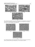
(50 or 60 Hz), the ECG and EOG signals. Figure 1 shows an example of real EEG ECG and
EOG signals recorded simultaneously in a PSG study. It can be seen that EEG signal is
contaminated by the QRS cardiac complexes which appear as spikes at the same time in
ECG record. Likewise, the low frequencies present in the contaminated EEG correspond to
the opening, closing or movements of the eyes recorded in EOG signal. These noise sources
increase the difficulty in analyzing the EEG and obtaining clinical information.
To correct, or remove the artifacts from the EEG signal, many techniques have been developed
in both, time and frequency domains (Delorme et. al., 2007; Sadasivan & Narayana, 1995).
More recently, component-based techniques, such as principal component analysis (PCA) and
independent component analysis (ICA); (Akhtar et. al., 2010; Astolfi et. al., 2010; Jung et. al.,
2000), have also been proposed to remove the ocular artifacts from the EEG. The use of Blind
Source Separation (BSS) (De Clercq et. al., 2005) and Parallel Factor Analysis (PFA) methods to
remove artifacts from the EEG have been used in this area too (Cichocki & Amari, 2002;
Makeig et. al., 2004). Wavelet Transform (WT) (Senthil Kumar et. al., 2009), WT combined with
ICA (Ghandeharion et. al., 2009) and Autoregressive Moving Average Exogenous (ARMAX)
(Hass et. al., 2003; Park et. al., 1998), have been applied too, to remove artifacts from EEG.
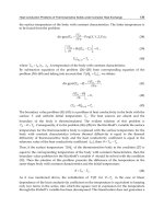
In this chapter, it is described a cascade of three adaptive filters based on a Least Mean
Squares (LMS) algorithm to remove the common noise components present in the EEG
signal recorded in polysomnographic studies.
Adaptive Filtering Applications
174
Adaptive filters method has been used, among other applications, in external
electroenterogram records (Mejia-García et. al., 2003) and in impedance cardiography
(Pandey et. al., 2005). Other applications of this filtering technique in biomedical signals
include, for example, removal of maternal ECG in fetal ECG records (Soria et. al., 1999)
detection of ventricular fibrillation and tachycardia (Tompkins, 1993), cancellation of heart
sound interference in tracheal sounds (Cortés, 2006), for pulse wave filter (Shen et. al., 2010),
for tumor motion prediction (Huang et. al., 2010), detection of single sweep event related
potential in EEG records (Decostre et. al., 2005), detection of SSVEP in EEG signals (Diez et.
al., 2011) and for motor imagery (Jeyabalan et. al., 2007).
In the particular case of artifacts removal in EEG records, He et. al. (2007) studied the accuracy
of adaptive filtering method quantitatively using simulated data and compared it with the
accuracy of the domain regression for filtering ocular artifacts from EEG records. Their results
show that the adaptive filtering method is more accurate in recovering the true EEG signals.
Kumar et. al. (2009) shows that adaptive filtering can be applied to remove ocular artifacts
from EEG with good results. Adaptive filters have been used to remove biological artifacts
from EEG by others authors (Chan et. al., 1998; Karjalainen et. al., 1999; Kong et. al., 2001).
In order to improve the signal to noise ratio of EEG signals in PSG studies, we had proposed
in a previous work a cascade of three adaptive filters based on a LMS algorithm (Garcés et.
al., 2007). The first filter in the cascade eliminates line interference, the second adaptive filter
removes the ECG complexes and the last one cancels EOG artifacts. Each stage uses a Finite
Impulse Response (FIR) filter, which adjusts its coefficients to produce an output similar to
the artifacts present in the EEG. In this chapter, we explain in detail the operation of the
cascade of adaptive filters including novel tests to determinate the optimal order of FIR filter
for each stage. Finally, we describe the results of the proposed filtering scheme in 18 real
EEG records acquired in PSG studies.
2. Materials
Eighteen PSG records belonging to sixteen subjects were selected from the MIT-BIH
Polysomnographic Database. All subjects are aged 44 +/- 12 years. This database contains
-0.05
0
0.05
Amplitud (u.a.)
-1.5
-1
-0.5
0
0.5
1
1.5
Amplitud (u.a.)
0 2 4 6 8 10
-0.5
0
0.5
Time (s)
Amplitud (u.a.)
EEG
ECG
EOG
Fig. 1. Some biological signals acquired in a PSG study a) EEG recording (corresponding to
Patient 41) corrupted with ECG and EOG artifacts, b) Real ECG signal, and c) Real EOG signal.
a)
b)
c)
Noise Removal from EEG Signals
in Polisomnographic Records Applying Adaptive Filters in Cascade
175
over 80 hours of four-, six-, and seven-channel PSG recordings. All of them contain EEG,
ECG and Blood Pressure (BP) signals, some of them have Nasal or Plethysmograph
Respiratory signals, five of them have O
2
Saturation signal, EOG and EMG signals. All the
subjects have ECG signals annotated beat-by-beat, and EEG and respiration signals annotated
by an expert with respect to sleep stages and apnea (Goldberger et. al., 2000). In this work were
used only the EEG, ECG and EOG signals, all of them were sampled at 250 Hz.
3. Common artifacts in EEG records
By artifacts it is understood all signals that appear in the EEG record which don't come from
the brain. The most common artifacts in the EEG signal appear during the acquisition due to
different causes, like as bad electrodes location, not clean hairy leather, electrodes
impedance, etc. There is also a finding of physiological artifacts, that is, bioelectrical signals
from other parts of the body (heart and muscle activity, eye blink and eyeball movement)
that are registered in the EEG (Sörnmo & Laguna, 2005).
The problem of those artifacts is that they can made a mistake in the analysis of a EEG
record, either in automatic method or in visual inspection by specialist (Wang et. al., 2008).
3.1 Power line interference
Biological records, especially EEG signals, are often contaminated with the 50 or 60 Hz line
frequency interference from wires, light fluorescents and other equipments which are
captured by the electrodes and acquisition system. The ignition of light of fluorescents
usually causes artificial spikes in the EEG. They are distributed in several channels of EEG
and can made a mistake in the analysis of the record (Sanei & Chambers, 2007)
3.2 Ocular artifacts
The human eye generates an electrical dipole caused by a positive cornea and negative
retina. Eye movements and blinks change the dipole causing an electrical signal known as
an EOG. The shape of the EOG waveform depends on factors such as the direction of eye
movements. A fraction of the EOG spreads across the scalp and it is superimposed on the
EEG (Vigon et. al., 2000).
Two kinds of ocular artifacts can be observed in EEG records, eye blinks and eye
movements. Eye blinks are represented by a low frequency signal (< 4 Hz) with high
amplitude. It is a symmetrical activity mainly located on the front electrodes (FP1, FP2) with
low propagation. Eye movements are also represented by a low frequency signal (< 4 Hz)
but with higher propagation, (Crespel et. al., 2006). In order for the EEG to be interpreted for
clinical use, those artifacts need to be removed or filtered from the EEG.
3.3 Cardiac artifacts
Cardiac activity may have pronounced effects on the electroencephalogram (EEG) because
of its relatively high electrical energy, especially upon the no-cephalic reference recordings
of EEG. The QRS complexes appear in the EEG signal like regular spikes (Sörnmo & Laguna,
2005). In figure 1 it can be observed the QRS complex present in a segment of EEG record.
The QRS amplitudes in the ECG are of the order of mV, but in the external EEG they have
been reduced. These artifacts in the EEG records could be clinically misleading.
Adaptive Filtering Applications
176
3.4 Other artifacts
The muscle disturbances are introduced in the EEG by involuntary muscle contractions of
the patient, thus generating an electromyogram (EMG) signal present in the EEG record.
The EMG and other biological artifacts have not been analyzing in the present work.
4. Methodology
Herein, we propose the use of adaptive filters to remove artifacts from EEG signal acquired
in PSG studies. Usually, biological signals (ECG, EOG and others) have overlaped spectra
with the EEG signal. For that, conventional filtering (band-pass, lower-pass or high-pass
filters) cannot be applied to eliminate or attenuate the artifacts without losing significant
frequency components of EEG signal.
Due to this reason, it is necessary to design specific filters to attenuate artifacts of EEG
signals in PSG studies. The adaptive interference cancellation scheme is a very efficient
method to solve the problem when signals and interferences have overlapping spectra.
Since the PSG recordings usually contain the ECG, EOG and EEG signals it is very
convenient to apply this method to filter this kind of records.
4.1 Adaptive filter
Adaptive filters are based on the optimization theory and they have the capability of
modifying their properties according to selected features of the signals being analyzed
(Haykin, 2005). Figure 2 illustrates the structure of an adaptive filter. There is a primary
signal d(n) and a secondary signal x(n). The linear filter H(z) produces an output y(n), which
is subtracted from d(n) to compute an error e(n).
The objective of an adaptive filter is to change (adapt) the coefficients of the linear filter, and
hence its frequency response, to generate a signal similar to the noise present in the signal to
be filtered. The adaptive process involves minimization of a cost function, which is used to
determine the filter coefficients. Initially, the adaptive filter adjusts its coefficients to
minimize the squared error between its output and a primary signal. In stationary
conditions, the filter should converge to the Wiener solution. Conversely, in non-stationary
circumstances, the coefficients will change with time, according to the signal variation, thus
converging to an optimum filter (Decostre
& Arslan, 2005).
Fig. 2. Structure of an adaptive filter.
In an adaptive filter, there are basically two processes:
a. A filtering process, in which an output signal is the response of a digital filter. Usually,
FIR filters are used in this process because they are linear, simple and stable.
Noise Removal from EEG Signals
in Polisomnographic Records Applying Adaptive Filters in Cascade
177
b. An adaptive process, in which the transfer function H(z) is adjusted according to an
optimizing algorithm. The adaptation is directed by the error signal between the
primary signal and the filter output. The most used optimizing criterion is the Least
Mean Square (LMS) algorithm.
The structure of the FIR can be represented as,
0
()
L
k
k
y
nwxnk
(1)
where L is the order of the filter, x(n) is the secondary input signal, w
k
are the filter
coefficients and y(n) is the filter output.
The error signal e(n) is defined as the difference between the primary signal d(n) and the
filter output y(n), that is,
en dn
y
n (2)
where,
0
L
k
k
en dn wxn k
(3)
The squared error is,
2
22
00
2
LL
kk
kk
e n d n dn wxn k wxn k
(4)
The squared error expectation for
N samples is given by
22
0
N
k
Ee n e n
(5)
2
1000
2
NLLL
kdx k lxx
nkkl
dn wr n wwr kl
(6)
where
r
dx
(n) and r
xx
(n) are, respectively, the cross-correlation function between the primary
and secondary input signals, and the autocorrelation function of the secondary input, that is
1
N
dx
n
r n dnxn k
(7)
1
N
xx
n
r n xnxn k
(8)
The objective of the adaptation process is to minimize the squared error, which describes a
performance surface. To get this goal there are different optimization techniques. In this
work, we used the method of steepest descent (Semmlow, 2004). With this, it is possible to
calculate the filter coefficient vector for each iteration k having information about the
previous coefficients and gradient, multiplied by a constant, that is,
Adaptive Filtering Applications
178
1
kk k
wn wn
(9)
where µ is a coefficient that controls the rate of adaptation.
The gradient is defined as,
2
k
k
en
wn
(10)
Substituting (10) in (9) leads to,
2
1
kk
k
en
wn wn
wn
(11)
Deriving with respect to w
k
and replacing leads to,
12
kk
k
en
wn wn en
wn
(12)
0
12
L
k
k
kk
k
dn wxn k
wn wn en
wn
(13)
Since d(n) and x(n) are independent with respect to w
k
, then,
12
kk
wn wn enxnk
(14)
Equation (14) is the final description of the algorithm to compute the filter coefficients as
function of the signal error
e(n) and the reference input signal x(n). The coefficient µ is a
constant that must be chosen for quick adaptation without losing stability. The filter is stable
if
µ satisfies the following condition, (Sanei & Chambers, 2007).
1
0
10. .
xx
LP
;
1
2
0
1
()
1
M
xx
n
Pxn
M
(15)
where
L is the filter order and P
xx
is the total power of the input signal.
4.2 Artifacts removal from EEG
As it is mentioned above, the adaptive interference cancellation is a very efficient method to
solve the problem when signals and interferences have overlap spectra.
The adaptive noise canceller scheme is arranged on the basic structure showed in Figure 2,
where the primary and secondary inputs are called as ”corrupted signal” and “reference
signal”, respectively.
In this scheme, it is assumed that the corrupted signal
d(n) is composed of the desired s(n)
and noise n
0
(n), which is additive and not correlated with s(n). Likewise, it is supposed that
the reference
x(n) is uncorrelated with s(n) and correlated with n
0
(n). The reference x(n)
feeds the filter to produce an output
y(n) that is a close estimate of n
0
(n) (Tompkins, 1993).
Noise Removal from EEG Signals
in Polisomnographic Records Applying Adaptive Filters in Cascade
179
To remove the main artifacts of the EEG signal, we propose a cascade of three adaptive
filters (see Figure 3). The input
d
1
(n) in the first stage is the EEG corrupted with artifacts
(EEG + line-frequency + ECG + EOG). The reference
x
1
(n) in the first stage is an artificial
sine function generated with 50 Hz (or 60 Hz, depends on line frequency). The output of
H
1
(z) is y
1
(n), which is an estimation of the line artifacts present in the EEG. This signal y
1
(n)
is subtracted from the corrupted
d
1
(n) to produce the error e
1
(n), which is the EEG without
line-interference. The
e
1
(n) error is forwarded as the corrupted input signal d
2
(n) to the
second stage. The reference input
x
2
(n) of the second stage can be either a real or artificial
ECG. The output of
H
2
(z) is y
2
(n), representing a good estimate of the ECG artifacts present
in the EEG record. Signal
y
2
(n) is subtracted from d
2
(n); its result produces error e
2
(n). Thus,
we have obtained the EEG without line and ECG artifacts. Then,
e
2
(n) enters into the third
stage as the signal
d
3
(n). The reference input x
3
(n) of filter H
3
(z) is also a real or artificial EOG
and its output is
y
3
(n), which is a replica of the EOG artifacts present in the EEG record.
Such
y
3
(n), subtracted from d
3
(n), gives error e
3
(n). It is the final output of the cascade filter,
that is, the clean EEG without artifacts.
The reference signals ECG and EOG and the corrupted EEG were acquired simultaneously
in polysomnographic studies. EEG, ECG and EOG records belonged to adult patients and
were downloaded from the MIT-BIH Polysomnographic Databas-Physiobank (Goldberger
et. al., 2000).
In section 4.3 there are present the tests that were carried out to determine the optimum
order
of H
1
(z), H
2
(z) and H
3
(z).
Fig. 3. Structure of adaptive filters cascade for artifacts removal on EEG signal acquired in
PSG studies.
4.3 Optimal order of FIR filters
To determine the optimum values of the orders L
1
, L
2
and L
3
of H
1
(z), H
2
(z) and H
3
(z) filters
the EEG signal were artificially contaminated with different coloured noises. The test to
Adaptive Filtering Applications
180
determinate the optimum values of the orders L
1
, L
2
and L
3
was done with a coefficient
convergence rates
μ fixed in 0.001. As soon as the optimum value of the L of each stage was
obtained the coefficient convergence rates
μ of each stage was recalculated with Eq. (15) to
assure an adequate adaptation. If
μ is too big, the filter becomes unstable, and if it is too
small, the adaptation may turn out too slow.
The tests were done using one stage of adaptive filter per time without using the cascade of
three filters.
4.3.1 Optimal estimation of order L
1
for filter H
1
(z).
The first stage filter attenuates the line frequency and was used to determinate the optimum
value
L
1
of H
1
(z). To determinate L
1
, the EEG was artificially contaminated with a sinusoidal
signal of 50 Hz which amplitude is adjusted in 30%, 50%, 80% and 100% of the Root Mean
Square (RMS) value of original EEG signal. Then, the filter order
L
1
was adjusted with
different values of 8, 16, 32, 64 and 128.
In order to study the filter performance, we estimated the Power Spectral Density (PSD) of
the original real EEG signal, the contaminated EEG and the different filtered versions of the
EEG signal. PSD was computed using the Burg method with a model order equal to 12.
Those graphics for one patient are presented in Figure 4 as an example.
Then, we estimated the normalized area below the frequency coherence function and the
maximum of temporal cross-correlation normalized function between the filtered EEG
signals and the contaminated EEG. If the signals are identical these parameters must be
equal to 1. This test was done for each patient.
Table 1 show the averaged values of two parameters for all EEG records of the database.
Contamination
of line
frequency
L
1
Coherence
Cross-
correlation
30%
8 0.9943 0.9760
16 0.9940 0.9727
32 0.9947 0.9657
64 0.9939 0.9497
128 0.9912 0.9062
50%
8 0.9936 0.9426
16 0.9932 0.9393
32 0.9938 0.9326
64 0.9930 0.9171
128 0.9902 0.8751
80%
8 0.9918 0.8739
16 0.9914 0.8706
32 0.9919 0.8643
64 0.9909 0.8500
128 0.9879 0.8111
100%
8 0.9903 0.8223
16 0.9898 0.8191
32 0.9901 0.8131
64 0.9890 0.7996
128 0.9859 0.7631
Table 1. Average values of the normalized parameters between filtered EEG signal and
contaminated EEG signal with line interference for different values of
L
1
.
Noise Removal from EEG Signals
in Polisomnographic Records Applying Adaptive Filters in Cascade
181
Figure 4 is an example of PSD graphics for a EEG recording (corresponding to Patient 48)
but all records of the database have a similar behaviour in the test. In this figure it could be
observed that as
L
1
increases, the attenuation of the 50 Hz interference is more significant.
However, if
L
1
is higher than 32, it can be seen than other frequencies of spectrum are
modified.
For this reason, there is a tradeoff between the 50 Hz interference attenuation and the
modification of the main frequency components of EEG signal.
In table 1 it can be observed that the best option between
L
1
=8, L
1
=16 and L
1
=32 is L
1
=16,
because it have the minimum area of coherence and similar values of maximum in cross-
correlation with
L
1
=32. Chosen this value of the order L
1
there is a loss of information of
original signal and there is not a modification in the rest of the spectrum.
It is concluded that the optimum value of
L
1
for the first filter is L
1
=16 (for a sampling
frequency of 250 Hz). For this order, the optimum value of the coefficient convergence rates
μ calculated with Eq. (15) must be positive and lower than 0.047
0 20 40 60 80 100 120
-70
-65
-60
-55
-50
-45
-40
Frequency (Hz)
Power/frequency (dB/Hz)
a)
0 20 40 50 60 80
-75
-70
-65
-60
-55
-50
-45
-40
Frequency (Hz)
Power/frequency (dB/Hz)
b)
Fig. 4. Power Spectral Density (PSD) of a EEG signal before and after the first adaptive filter
H
1
(z). a) In blue: PSD of original EEG, in red: PSD of EEG signal contaminated with an artificial
line interference. b) PSD of filtered EEG signal for different values of the order
L
1
. Red: original
EEG, Green:
L
1
=8, Orange: L
1
=16, Purple: L
1
=32, Light Blue: L
1
=64, Blue: L
1
=128.
Adaptive Filtering Applications
182
4.3.2 Optimal estimation of order L
2
for filter H
2
(z).
The second stage filter attenuates ECG artifacts (mainly QRS complexes) present in EEG
signal, and was used to determinate the optimum value of the order
L
2
of H
2
(z). To
determinate
L
2
, the EEG was artificially contaminated with a coloured noise, with a -3dB
bandwidth between 5 Hz and 40 Hz. This bandwidth was selected considering that QRS
complexes have almost their total energy in this frequency band (Thakor, 1984). Then, the
filter order
L
2
was adjusted with the different values of 16, 32, 64, 128, 256 and 512.
As a similar way to optimum value estimation of
L
1
, we estimated the PSD of the original
real EEG signal, the contaminated EEG and the different filtered versions of the EEG signal.
Figure 5 shows the PSD graphics for an EEG recording before and after the second adaptive
filter. In this figure it could be observed that the possible optimum values of
L
2
to filter the
cardiac frequencies between 5Hz and 40Hz are
L
2
=16, L
2
=32 or L
2
=64, because the rest of the
values of
L
2
modify the frequencies of the entire spectrum.
0 20 40 60 80 100 120
-66
-64
-62
-60
-58
-56
-54
-52
-50
-48
-46
Frequency (Hz)
Power/frequency (dB/Hz)
a)
0 20 40 60 80 100
-68
-66
-64
-62
-60
-58
-56
-54
-52
-50
-48
Frequency (Hz)
Power/frequency (dB/Hz)
b)
Fig. 5. Power Spectral Density (PSD) of a EEG signal before and after the second adaptive
filter
H
2
(z) . a) In blue: PSD of original EEG, in red: PSD of EEG signal contaminated with
coloured noise (5Hz to 40 Hz). b) PSD of filtered EEG for different values of the order
L
2
.
Red: original EEG, Green:
L
2
=16, Orange: L
2
=32, Purple: L
2
=64, Light Blue: L
2
=128, Blue:
L
2
=256, Black: L
2
=512.
Noise Removal from EEG Signals
in Polisomnographic Records Applying Adaptive Filters in Cascade
183
Table 2 shows the average of the normalized area below the frequency coherence function
and the maximum of temporal cross-correlation normalized function (between the filtered
EEG signals and the contaminated EEG) for all recordings analyzed and for different values
of
L
2
.
L
2
Coherence
Cross-
correlation
16 0.2588 0.5686
32 0.2596 0.5595
64 0.2927 0.5406
128 0.2641 0.5087
256 0.1756 0.4576
512 0.1579 0.3463
Table 2. Average values of the normalized parameters between filtered EEG signal and
contaminated EEG signal for different values of
L
2
.
In table 2 it can be observed that the best option between
L
2
=16, L
2
=32 or L
2
=64 is L
2
=32,
because it have the minimum value of the normalized area below the frequency coherence
function and the lower values of maximum of cross- correlation normalized function
without losing information and not modifying the spectrum of the original EEG signal.
It is concluded that the optimum value of
L
2
for second filter is L
2
=32. For this order, the
optimum value of the coefficient convergence rates
μ calculated with (15) must be positive
and lower than 0.02367.
4.3.3 Optimal estimation of order L
3
for filter H
3
(z).
As it is mentioned above, the third and last stage filter attenuates EOG artifacts present in
EEG. In this section, we determinate the optimum value of the order
L
3
of H
3
(z). To
determinate it, the EEG was artificially contaminated with a coloured noise with a -3dB
bandwidth between 0.5 Hz and 10 Hz. This bandwidth includes the main frequency
components of EOG artifacts. Then, we evaluated the filter performance with different
L3
values (4, 8, 16 and 32).
As a similar way to optimum value estimation of
L
1
and L
2
, we estimated the PSD of the
original real EEG signal, the contaminated EEG and the different filtered versions of the
EEG signal.
L
3
Coherence
Cross-
correlation
4 0.8773 0.8014
8 0.8586 0.7979
16 0.8579 0.7937
32 0.8584 0.7863
64 0.8586 0.7842
Table 3. Average values of the normalized parameters between filtered EEG signal and
contaminated EEG signal for different values of
L
3
.
Figures 6 and 7 show the PSD graphics for an EEG recording before and after the third
adaptive filter. It can be observed that all the values of the order
L
3
chosen have good result
Adaptive Filtering Applications
184
to filter the frequencies lower than 10 Hz (see Figure 6). No one introduce interferences in
other frequencies. But with values bigger than 256 it could be observed a distortion in high
frequencies and a loss of information of the original signal in low frequencies (see Figure 7).
The modification of the high frequencies and the losing of information in low frequencies
are shown in figure 7, where there have been filtered the contaminated EEG with values of
L
3
=256 and L
3
=512
Table 3 shows the averaged values of the normalized area below the frequency coherence
function and temporal cross-correlation normalized function (between the filtered EEG
signals and the contaminated EEG) for all recordings analyzed and for different values of
L
3
.
0 5 10 15 20 25 30
-62
-60
-58
-56
-54
-52
-50
-48
-46
-44
Frequency (Hz)
Power/frequency (dB/Hz)
a)
0 5 20 403010
-62
-60
-58
-56
-54
-52
-50
-48
Frequency (Hz)
Power/frequency (dB/Hz)
b)
Fig. 6. Power Spectral Density (PSD) of a EEG signal before and after the third adaptive filter
H
3
(z). a) In blue: PSD of original EEG, in red: PSD of EEG signal contaminated with coloured
noise (0.5 Hz to 10 Hz). b) PSD of EEG signal filtered for different values of the order
L
3
,
Red: original EEG, Green:
L
3
=4, Orange: L
3
=8, Purple: L
3
=16, Light Blue: L
3
=32,
In Table 3 it can be observed that the best option of the value of the order
L
3
for the third
filter is
L
3
=16, because it have the minimum value of the normalized area below the
frequency coherence function and the lower values of maximum of cross- correlation
Noise Removal from EEG Signals
in Polisomnographic Records Applying Adaptive Filters in Cascade
185
normalized function without losing information of original signal and not modifying the
spectrum of the original EEG. The results of the test using values of L
3
bigger than L
3=
256
have not been included in Table 3.
It is concluded that the optimum value of
L
3
for the third filter is L
3
=32. For this value, the
optimum value of the coefficient convergence rates μ calculated with (15) must be positive
and lower than 0.02367.
0 10 40 60 80 100 12020
-65
-45
Frequency (Hz)
Power/frequency (dB/Hz)
Fig. 7. Power Spectral Density of a EEG signal before and after the third adaptive filter H
3
(z).
In Red: PSD of the original EEG signal. In Green: PSD of the EEG signal contaminated with
coloured noise (0.5 Hz to 10 Hz). In Purple: PSD of the EEG filtered for order
L
3
=256. In
Blue: PSD of the EEG filtered for
L
3
=512. Note the modification in high frequencies and
losing of information in low frequencies.
5. Results
Eighteen real EEG records acquired in PSG studies were processed with the cascade of
adaptive filters. According to the previous tests, the values of the orders
L
1,
L
2
and L
3
were
adjusted as
L
1
= 16, L
2
= 32 and L
3
= 32.
As it was mentioned in section 2, only five subjects from the entire database have EOG
signals. So, the EEG signals of these five patients have been filtered with the entire cascade
shown in Figure 3. The others thirteen EEG (belonging to the rest of the patients) have not
been filtered with the last third stage.
The input
d
1
(n) in the first stage is the EEG corrupted with artifacts (EEG + line-frequency +
ECG + EOG). The reference
x
1
(n) in the first stage is an artificial sine function generated with
50 Hz with the same RMS of the EEG signal. The
e
1
(n), which is the EEG without line-
interference, is forwarded as the corrupted input signal
d
2
(n) to the second stage. The
reference input
x
2
(n) of the second stage is the real ECG. The error e
2
(n) is the EEG without
line and ECG artifacts and enters into the third stage as the signal
d
3
(n). The reference input
Adaptive Filtering Applications
186
x
3
(n) of filter H
3
(z) is a real EOG. The error e
3
(n) is the final output of the cascade filter, that
is, the clean EEG without artifacts.
In order to study the filter performance we estimated the normalized area below the
frequency coherence function and the maximum of temporal cross-correlation normalized
function between the filtered EEG signals of each stage and the original EEG for the entire
data base.
Table 4 shows the results obtained for each record of the database processed by the first
stage of the propose filter. In this table, it is presented the values of the normalized area of
frequency coherence function and the normalized maximum of temporal cross-correlation
between the contaminated signal
d
1
(n) and the error signal e
1
(n). Those values show that the
first stage attenuates the line interference.
Patient Coherence %
Cross-
correlation
1a 0.8690 0.6730
1b 0.8901 0.6349
2a 0.9833 0.4724
2b 0.9507 0.5417
3 0.9279 0.4044
4 0.9776 0.3615
14 0.9807 0.4698
16 0.9816 0.4452
32 0.9879 0.8309
37 0.9881 0.9293
41 0.9963 0.9857
45 0.9928 0.7017
48 0.9983 0.9413
59 0.9839 0.3970
60 0.9747 0.2807
61 0.9663 0.4281
66 0.9783 0.4213
67 0.9734 0.5504
average 0.9667 0.5816
Table 4. Normalized area of frequency coherence function and maximum of temporal cross -
correlation function between the signals
d
1
(n) and e
1
(n) of the first stage of proposed filter.
Figure 8 illustrates a temporal segment of 10s of the original EEG record (corresponding to
Patient 41) and its filtered version after the first stage of adaptive filter. In this figure it can
be observed that the 50 Hz power line component is significantly filtered.
Figure 9 shows the PSD function of the same original and filtered EEG signals shown in
Figure 8. The PSD of the filtered signal shows that the first stage attenuates the line-
frequency artifacts. The
H
1
(z) filter adapts the amplitude and the phase of the artificial
Noise Removal from EEG Signals
in Polisomnographic Records Applying Adaptive Filters in Cascade
187
sinusoidal signal x
1
(n) (50Hz) in order to have as output a replica, y
1
(n), of the line-
frequency artifacts present in the EEG.
After 50 Hz filtering, the EEG is forwarded to the second stage in order to remove ECG
artifacts (see Figure 3).
-0.1
0
0.1
0 2 4 6 8 10
-0.1
0
0.1
Time (s)
Amplitud (u.A)
Fig. 8. Example of a temporal segment of EEG filtered with stage 1 for patient 41. a) Red:
Original EEG contaminated with 50 Hz power line interference,
d
1
(n).b) Blue: EEG without
line interference,
e
1
(n).
0 10 20 30 40 50 60 70
-80
-60
-40
-20
0
20
Power Spectrum Magnitude (dB)
a)
0 10 20 30 40 50 60 70
-80
-60
-40
-20
0
20
Frequency (Hz)
Power Spectrum Magnitude (dB)
b)
Fig. 9. Example of first stage of the proposed filter. a) PSD of original EEG with artifacts. b)
PSD of first stage output
e
1
(n), where the 50 Hz component is attenuated.
Adaptive Filtering Applications
188
Table 5 shows the results obtained for each record of the database processed by the second
stage. In this table, it is presented the values of the normalized area of frequency coherence
function and the normalized maximum of temporal cross-correlation between the
contaminated signal
d
2
(n) and the error signal e
2
(n). Those values show that the second stage
attenuates QRS complexes artifacts introduced by ECG signal.
Patient Coherence
Cross-
correlation
1a 0.8528 0.7514
1b 0.8801 0.5180
2a 0.9709 0.9467
2b 0.9946 0.9845
3 0.9107 0.9460
4 0.9120 0.7910
14 0.9276 0.8768
16 0.9070 0.8757
32 0.8364 0.3333
37 0.8550 0.6725
41 0.8204 0.7826
45 0.7985 0.7981
48 0.9096 0.6893
59 0.9106 0.5431
60 0.8224 0.3027
61 0.8979 0.2482
66 0.8097 0.5319
67 0.8464 0.8209
avera
g
e 0.8342 0.6439
Table 5. Normalized area of frequency coherence function and maximum of temporal cross -
correlation function between the signals
d
2
(n) and e
2
(n) of the second stage of proposed
filter.
Figure 10 shows an example of 10s of EEG signal (corresponding to patient 41) processed by
the second filter. The contaminated signal
d
2
(n) is shown in red. It could be observed the
presence and morphology similarity of QRS complexes of the ECG (in green) in the EEG
record. The output signal
y
2
(n) of H
2
(z) is drawn in black colour, this signal is an estimation
of the ECG artifacts present in the EEG. The
H
2
(z) filter adapts the amplitude and the phase
of the reference signal
x
2
(n) (ECG signal) in order to have as output a replica of the artifacts
present in the EEG
After 50 Hz and ECG filtering, the EEG is forwarded to the third stage in order to remove
EOG artifacts.
Noise Removal from EEG Signals
in Polisomnographic Records Applying Adaptive Filters in Cascade
189
-0.05
0
0.05
-1
0
1
-0.04
0
0.06
0 2 4 6 8 10
-0.05
0
0.05
Time (s)
Amplitude (u.A.)
Fig. 10. Example of a temporal segment of EEG filtered with stage 2 for patient 41. In Red:
Contaminated EEG,
d
2
(n). In Green: ECG signal. In Black: output signal from H
2
(z), that is
y
2
(n). In Blue: EEG without ECG artifacts, e
2
(n).
Table 6 shows the results obtained for five records of the database processed by the third
stage. In this table, it is presented the values of the normalized area of frequency coherence
function and the normalized maximum of temporal cross-correlation between the
contaminated signal
d
3
(n) and the error signal e
3
(n), which is the final output of the
proposed filter.
As it has been mentioned before only five patients have been filtered with
the third stage, the rest of them do not have the reference signal
x
3
(n). Those values show
that this last stage attenuates artifacts introduced by the EOG.
Patient Coherence
Cross-
correlation
32 0.9985 0.9907
37 0.9912 0.7949
41 0.9859 0.6052
45 0.9990 0.9500
48 0.9527 0.7943
avera
g
e 0.9855 0.8270
Table 6. Normalized area of frequency coherence function and maximum of temporal cross -
correlation function between the signals
d
3
(n) and e
3
(n) of the third stage of proposed filter.*
Patient without available EOG signal.
Adaptive Filtering Applications
190
Figure 11 shows the same 10s of temporal EEG signal of patient 41. There it can be observed
all signals of third stage
. The contaminated signal d
3
(n) is drawn in red colour. It can be
observed the presence and morphology similarity of the EOG signal in the EEG record. The
output signal
y
3
(n) of H
2
(z) is in black colour in the figure, this signal is an estimation of the
EOG signal present in the EEG. The
H
3
(z) filter adapts the amplitude and the phase of the
reference signal
x
3
(n) (EOG signal) in order to have as output a replica of the EOG artifacts
present in the EEG.
-0.05
0
0.05
-0.2
0
0.2
-0.04
0
0.06
0 2 4 6 8 1
0
-0.05
0
0.05
Time (s)
Amplitude (u.A.)
Fig. 11. Example of temporal segment of EEG filtered with stage 3 for patient 41. In Red:
Contaminated EEG,
d
3
(n). In Green: EOG signal. In Black: output signal from H
3
(z), that is
y
3
(n). In Blue: EEG without EOG artifacts, e
3
(n).
Figure 12 show the PSD of the contaminated EEG of third stage,
d
3
(n), of the reference signal
x
3
(n),EOG, and of the filtered EEG signals illustrated in Fig. 11. Note that the low
frequencies of the EOG present in the contaminated EEG are attenuated in the filtered EEG
signal.
Figure 13 is shown temporal temporal segments of 10s of EEG. In this figure it could be
observed the attenuation of line frequency and biological artifacts without losing important
information of the EEG signal. Results show that the proposed adaptive filter cancels
correctly the line frequency interference and attenuate very well the biological artifacts
introduced by the ECG and the EOG.
6. Discussion and conclusion
In this chapter, a novel filtering method based on three adaptive filters in cascade has been
proposed to cancel common artifacts (line interference, ECG and EOG) present in EEG
signals recorded in PSG studies.
Noise Removal from EEG Signals
in Polisomnographic Records Applying Adaptive Filters in Cascade
191
0
0.2
0.4
0.6
0.8
1
x 10
-3
PSM (dB)
Power Spectrum Magnitude contaminated EEG
a)
0
0.5
1
x 10
-3
PSM (dB)
Power Spectrum Magnitude EOG
b)
0 0.5 1 1.5 2 2.5 3 3.5 4 4.5 5
0
0.5
1
x 10
-3
frequency (Hz)
PSM (dB)
Power Spectrum Magnitude filtered EEG
c)
Fig. 12. Example of third stage of the proposed filter a) PSD of the contaminated EEG, d
3
(n),
b) PSD of the reference signal
x
3
(n),EOG, c) PSD of the filtered EEG signal.
Adaptive Filtering Applications
192
-0.06
0
0.08
0 2 4 6 8 10
-0.06
0
0.08
Time (s)
Amplitude (u.A.)
Fig. 13. Example of temporal segments of contaminated EEG and EEG filtered with the
entire cascade for patient 41. In Red: Contaminated EEG,
d
1
(n), In Black: final filtered EEG
without line interference, ECG and EOG artifacts,
e
3
(n).
Other methods (like PCA, ICA, BSS or WT) have been described in the bibliography to
cancel these artifacts in the EEG signals. However, those methods have some restrictions.
For example, the properties of WT make it has an advantage in processing short-time
instantaneous signal, but it needs that the frequency range of the EEG signal was not
overlap with the bandwidth of noise sources and in this case the frequencies bands of the
ECG and EOG signal are overlap with the frequencies of the EEG. ICA is a developed
method for transforming an observed multidimensional vector into components that are
statistically as independent from each other as possible. This method needs that the
dimension of the signals were larger than that of original signals, and every original signal
must be non-Gaussian. With more observed signals ICA will get better filtering result,
which limits the application of this technique in few channels EEG recordings.
The main advantages of the proposed adaptive filtering method can be summarized as:
a.
The method does not have restrictions about the signal to be filtered.
b.
The implementation of adaptive filtering is very simple and fast and the results can be
obtained without complex calculations.
c.
The filter coefficients can be adapted to variations in heart frequency, abrupt changes in
the line frequency (caused, say, by ignition of electric devices) or modifications due to
eye movements.
d.
At each stage output, the error signals e
i
(n), EEG with one of the three attenuated
artifacts are present; such separation (by artifacts) may be useful in some applications
where such output might be enough.
e.
The filters have a linear phase response so no phase distortion is made. This is
particularly important for the analysis of neurological rhythms in EEG signals
Noise Removal from EEG Signals
in Polisomnographic Records Applying Adaptive Filters in Cascade
193
As soon as the optimal orders of the three filters were determinate, the method was tested in
18 real EEG records acquired in PSG studies. Figure 13 is a good example of an EEG record
corrupted by three types of artifacts and its corresponding filtered version. It can be seen
that all artifacts have been eliminated or attenuated, improving the quality of EEG record.
The remaining records analyzed in the work had obtained similar results and their filtered
EEGs don’t have large artifacts.
It has been concluded that proposed adaptive filtering scheme with the appropriate values
of order
L
i
, attenuate correctly ECG, EOG and line interference without removing significant
information embedded in EEG signals registered in PSG studies. Due to the fact that the
these studies usually have the ECG, EOG and EEG signals, the proposed cascade of
adaptive filters is very useful and appropriate for the analysis of PSG recordings in sleep
laboratories. The cascade could be used in others biomedical applications and in BCI
applications.
7. Acknowledgment
This work has been supported by grants from Consejo Nacional de Investigaciones
Científicas y Técnicas (CONICET) and Universidad Nacional de San Juan (UNSJ), both
Argentinian institutions.
8. References
Akhtar, M.T.; James, C.J & Mitsuhashi W. (2010). Modifying the Spatially-Constrained ICA
for Efficient Removal of Artifacts from EEG Data.
Proceedings of 4th International
Conference on Bioinformatics and Biomedical Engineering (iCBB
E), pp. 1-4. ISBN: 978-1-
4244-4713-8, Chengdu, China, June 18-20, 2010.
Astolfi, L.; Cincotti, F.; Mattia, D.; Babiloni, F.; Marciani, M.G.; De Vico Fallani, F.;
Mattiocco, M.; Miwakeichi, F.; Yamaguchi, Y.; Martinez, P.; Salinari, S.; Tocci, A.;
Bakardjian, H.; Vialatte, F.B.& Cichocki, A. (2006). Removal of ocular artifacts for
high resolution EEG studies: a simulation study,
Proceedings of 28th Annual
International Conference of the IEEE Engineering in Medicine and Biology Society
, pp.
976 – 979, ISBN 14244–0033–3, New York City, USA, Aug 30-Sept 3, 2006.
Chan, F.H.Y.; Qiu, W.; lam, F.K. & Poon, P.W.F. (1998). Evoke potential estimation using
modified time-sequence adaptive filter,
Medical & Biological Engineering &
Computing
, Vol. 36, No. 4, (July 1998) pp. 407-414
Cichocki, A. & Amari, S. (2002).
Adaptive Blind Signal and Image Processing, John Wiley &
Sons. ISBN 0-471-60791-6.
Collop, N.A.; Anderson, W.M.; Boehlecke, B.; Claman, D.; Goldberg, R. & Gottlieb, D.J.
(2007). Clinical guidelines for the use of unattended portable monitors in the
diagnosis of obstructive sleep apnea in adult patients. Portable Monitoring Task
Force of the American Academy of Sleep Medicine.
Journal Clinical Sleep Medicine.
Vol. 3, No. 7, (December 2007), pp.737-47.
Cortés, S.; Jane, R.; Torres, A.; Fiz, J.A. & Morera, J. (2006). Detection and Adaptive
Cancellation of Heart Sound Interference inTracheal Sounds,
Proceedings of the 28th
IEEE EMBS Annual International Conference,
pp. 2860-2863, ISBN 1-4244-0033-3, New
York City, USA, Aug 30-Sept 3, 2006.
Adaptive Filtering Applications
194
Crespel, A.; Gélisse, P.; Bureau, M. & Genton, P. (2005). Atlas of Electroencephalography (1
st
ed), Vol. 1 & 2, J. Libbey Eurotext, ISBN 2-7420-0600-1, Paris.
De Clercq, W.; Vergult, A.; Vanrumste, B.; Van Hees, J.; Palmini, A.; Van Paesschen, W.&
Van Huffel, S. (2005). A new muscle artifact removal technique to improve the
interpretation of the ictal scalp electroencephalogram,
Proceedings of the 27th Annual
Conference IEEE Engineering in Medicine and Biology
, pp. 944 - 947 ,ISBN: 0–7803–
8741–4, Shanghai, China, September 1-4, 2005.
Decostre, A. & Arslan, B. (2005). An Adaptive Filtering Approach to the Processing of Single
Sweep Event Related Potentials Data,
Proceedings 5th International. Workshop
Biosignal Interpretation
, pp. 1-3 , Tokyo, Japan, September 6-8, 2010.
Decostre, A. & Arslan, B. (2005). An Adaptive Filtering Approach to the Processing of Single
Sweep Event Related Potentials Data,
Proceeding 5th International. Workshop Biosignal
Interpretation
, pp. 1-3 ,Tokyo- Japan, September 2005
Delorme, A.; Sejnowski T. & Makeig, S. (2007). Enhanced detection of artifacts in EEG data
using higher order statistics and independent component analysis,
NeuroImage, Vol.
34, ( 2007), pp. 1443-1449, ISSN: 10538119.
Diez, P.; Garcés Correa, M.A. & Laciar, E. (2010). SSVEP Detection using Adaptive filters,
V
Congreso Latinoamericano de Ingeniería Biomédica (CLAIB2011)
, Habana, Cuba, May
16-21, 2011, In press.
Garcés Correa, A.; Laciar, E.; Patiño, H.D. & Valentinuzzi, M.E. (2007). Artifact removal
from EEG signals using adaptive filters in cascade,
Journal of Physics, Vol.90,
(September 2007), pp.1-10,
Ghandeharion H. & Ahmadi-Noubari, H. (2009). Detection and Removal of Ocular Artifacts
using Independent Component Analysis and Wavelets,
Proceedings of the 4th
International IEEE EMBS Conference on Neural Engineering,
pp.653-656, ISBN 978-1-
4244-2073-5, Antalya, Turkey, April 29 - May 2, 2009.
Goldberger, A.L.; Amaral, L.A.; Glass, N L.; Hausdorff, J.M.; Ivanov, P.C.; Mark, R.G.;
Mietus, J.E.; Moody, G.B.; Peng, C.K. & Stanley H.E. (2000). PhysioBank,
PhysioToolkit, andPhysioNet:
Components of a New Research Resource for Complex
Physiologic Signals, Circulation
, Vol. 101, No.23, pp. 215-220, June 2000
Hass, S.H.; Frei, M.G.; Osorio, I.; Pasik-Duncan, B. & Radel, J. (2003). EEG ocular artifact
removal through ARMAX model system identification using extended
least squares,
Comunication in formation and system, Vol. 3, No. 1, (June 2003), pp. 19-
40.
Haykin, S. (2005).
Neural Network (2
nd
), Pearson Prentice Hall, ISBN 81-7808-300-0, Delhi,
India.
He, P.; Kahle, M.; Wilson, G. & Russell, C. (2005). Removal of Ocular Artifacts from EEG: A
Comparison of Adaptive Filtering Method and Regression Method Using
Simulated Data,
Proceedings of the 2005 IEEE Engineering in Medicine and Biology 27th
Annual Conference
, pp. 1110-1113, ISBN 0-7803-8740-6, Shanghai, China, September
1-4, 2005
Huang, K.; Buzurovic, I.; Yu, Y. & Podder,T.K. (2010). A Comparative Study of a Novel AE-
nLMS Filter and Two Traditional Filters inPredicting Respiration Induced Motion
Noise Removal from EEG Signals
in Polisomnographic Records Applying Adaptive Filters in Cascade
195
of the Tumor, 2010 IEEE International Conference on Bioinformatics and Bioengineering,
pp.281-282 , ISBN 978-0-7695-4083-2, Philadelphia, May 31-Jun3, 2010.
Jeyabalan, V.; Samraj, A. & Chu-Kiong, L. (2007). Motor Imaginary Signal Classification
Using Adaptive Recursive Bandpass Filter and Adaptive Autoregressive Models
for Brain Machine Interface Designs
International Journal of Biological and Life
Sciences
, Vol. 3, No.2 ( March 2007), pp. 116-123. eISSN 2010-3832.
Jung, T. P.; Makeig, S.; Westerfield, M.; Townsend, J.; Courchesne, E. & Sejnowski, T. J.
(2000). Removal of eye activity artifacts from visual event-related potentials in
normal and clinical subjects.
Clinical Neurophysiology, Vol.111, No.10, (October
2000), pp. 1745-1758. ISSN 1388-2457.
Karjalainen, P.; Kaipio, J.; Koistinen, A. & Vauhkonen, M. (1999) Subspace regularization
method for the single trial estimation of evoked potentials,
IEEE Transactions on
Biomedical Engineering
, Vol. 46, No. 7,( July 1999), pp. 849-860, ISBN 0018–9294.
Kong, X. & Qiu, T. (2001) Latency change estimation for evoked potentials a comparison of
algorithms,
Medical & Biological Engineering & Computing, Vol. 39, No. 2, (2001), pp.
208-224.
Makeig S.; Debener S.; Onton J. & Delorme A. (2004).
A. Mining event-related brain dynamics.
Trends Cogn Sci
., pp.204-10., May 2004; Vol.8, No.5.
Mejia-Garcia, J. H.; Martinez-De-Juan, J. L.; Saiz, J.; Garcia-Casado, J. & Ponce, J.L. (2003)
Adaptive cancellation of the ECG interference in external electroenterogram,
Proceedings of the 25th Annual International Conference of the IEEE Engineering in
Medicine and Biology Society 2003,
Vol. 3, pp. 2639-2642, ISBN 0-7803-7789-3, Cancun,
Mexico September 17-21, 2003.
Pandey, V.K. & Pandey,P.C. (2005). Cancellation of Respiratory Artifact in Impedance
Cardiography,
Proceedings of the 2005 IEEE Engineering in Medicine and Biology 27th
Annual Conference,
pp. 5503-5506, ISBN 0-7803-8740-6, Shanghai, China, September
1-4, 2005
Park, H J.; Jeong, D U. & Park, K S. (2002).Automated Detection and Elimination of
Periodic ECG Artifacts in EEG Using the Energy Interval Histogram Method.
IEEE
transactions on Biomedical Engineering
. Vol. 49, No. 12, (December 2002), pp.1526-
1533, ISSN 0018-9294
Sadasivan, P.K & Narayana, D. (1995). Line interference cancellation from corrupted EEG
signals using Modified linear phase FIR digital filters,
Engineering in Medicine and
Biology Society, 1995 and 14th Conference of the Biomedical Engineering Society of India.
An International Meeting, Proceedings of the First Regional Conference,
pp. 3.35-3.36.
India, February 15-18, 1995.
Sanei, S. & Chambers, J. (2007).
EEG Signal Processing (1
st
ed.), Jhon Wiley & Sons, ISBN 13-
978-0-470-02581-9, England.
Semmlow, J.L. (2004).
Biosignal and Medical Image Processing, Marcel Dekker, ISBN 0-8247-
4803-4, United State of America.
Senthil Kumar, P.; Arumuganathan, R. & Vimal, C. (2009). An Adaptive method to remove
ocular artifacts from EEG signals using Wavelet Transform,
Journal of Applied
Sciences Research
, Vol. 5, No. 7, (2009 ), pp. 741-745, ISBN 741-745.