Adaptive Filtering Applications Part 6 pptx
Bạn đang xem bản rút gọn của tài liệu. Xem và tải ngay bản đầy đủ của tài liệu tại đây (2.83 MB, 30 trang )
Adaptive Filtering Applications
142
comparison with other modalities. Continuous wave near infrared (NIR) spectroscopy has
been applied to trans-abdominal fetal pulse oximetry (Ramanujam et al. 2000; Chance 2005;
Zourabian et al. 2000; Nioka et al. 2005; Vintzileos et al. 2005). The system consists of NIR
sources (halogen lamps) and a photomultiplier as detection unit (Ramanujam et al. 2000;
Chance 2005). The generated heat was justified by using cooling fans for the halogen lamps.
Recently, trans-abdominal oxygen saturation (S
p
O
2
) in animal (Nioka et al. 2005) and human
fetuses were successfully obtained in the laboratory (Vintzileos et al. 2005). However, the
proposed techniques require high power (a total of 80 W optical power) and a relatively
expensive detection unit (photo-multiplier).
In this project, we propose to design and develop a low-power optical FHR monitor. The
signal of interest is the photoplethysmogram (PPG), which is generated when a beam of
light is modulated by blood pulsations. PPG is a noninvasive technique for detecting blood
volume changes in living tissue by optical means consisting of a light emitting diode (LED)
and a photo-detector. One of the potential applications of the PPG technology is non-
invasive fetal heart rate detection through the maternal abdomen. In this application, the
light intensity is modulated by the mother as well as fetal blood circulation, producing a
combined signal which needs to be separated via digital signal processing (DSP) techniques.
The design of a fixed filter would not be adequate as the frequency spectrum of the noise
(maternal PPG) overlaps with the desired signal (fetal PPG). The adaptive filter will
automatically adjust its coefficients therefore achieve the high degree of noise rejection.
Such an approach - based on adaptive noise cancellation (ANC) - has been evaluated for
extraction of the fetal heart-rate using PPG signals from the maternal abdomen. A simple
optical model has been proposed in which the maternal and fetal blood pulsations result in
emulated signals where the lower SNR limit (fetal to maternal) is -25 dB (Zahedi & Beng,
2008). It is shown that the RLS algorithm is capable to extract the peaks of the fetal PPG from
these signals, corresponding to typical values of maternal and fetal tissues.
Subsequently, an optical fetal heart rate detection (OFHR) system has been designed and
developed using low-cost, low-power IR light (890 nm with optical power < 68 mW) and a
commercially available silicon photo-detector (Gan et al. 2009). Previous literature
(Ramanujam et al. 2000; Chance 2005; Zourabian et al. 2000; Nioka et al. 2005; Vintzileos et
al. 2005; Choe et al. 2003) shows that the Source-Detector separations depends on the type of
sources and the photo-detectors implemented in their studies. Since in our work the
developed instrument utilizes low optical power, the source-to-detector separation plays an
important role as it affects the detectivity of the photo-detector. This chapter discusses the
selection of S-D separation for the OFHR system based on the ANC limit and photo-
detector’s noise. The implementation of the ANC algorithm in OFHR system is also
discussed and the clinical trial results are also reported.
2. Materials and methods
2.1 Adaptive noise cancellation
Conventional digital signal processing techniques do exist to extract a desired biomedical
signal from a mixed signal which is usually contaminated by unwanted noises. Adaptive
filters are used for non-stationary signals where a sample-by-sample adaptation process is
required (Vaseghi, 2000; Widrow et al., 1975). Applications of adaptive filtering include
multi-channel noise reduction, radar or sonar signal processing, channel equalization for
cellular mobile phones, echo cancellation and low delay speech coding. This section
Application of Adaptive Noise Cancellation
in Transabdominal Fetal Heart Rate Detection Using Photoplethysmography
143
discussed the concept of the adaptive filtering, adaptive algorithm and the Recursive Least
Square (RLS) algorithm.
2.1.1 Concept of adaptive filtering
Adaptive filters consist of two distinct parts: a digital filter and the corresponding adaptive
algorithm, used to adjust the coefficients of the filter (Figure 1). In these algorithms, the error
signal e(n) defined as the difference between the output of the filter (y(n)) and a primary
input signal (d(n)), is minimized according to a least squares error criterion (Ifeachor &
Jervis, 2002).
Fig. 1. ANC system
The desired signal d(n) (Figure 1) is contaminated by an uncorrelated noise signal v
0
(n)
,
where n is the running time index. The result d(n) + v
0
(n) is the primary measurement signal
s(n). The reference input, v
1
(n) is only correlated with v
0
(n) and fed to an adaptive FIR filter.
The output of the FIR adaptive filter y(n) is subtracted from the primary input s(n) to
produce the error signal e(n):
0
() () () ()en dn v n yn
(1)
The adaptive filter uses e(n) to adjust its own impulse response to produce an output y(n) as
close a replica as possible to v
0
(n). Squaring and applying the expectation operator to both
sides of Equation 1:
2
22
00
() () () () 2 () () ()Ee n Ed n E v n
y
nEdnvn
y
n (2)
d(n) being uncorrelated with v
0
(n) and v
1
(n), E{d(n)(v
0
(n)-y(n))}= 0. Therefore Equation 2 can
be simplified:
2
22
0
E e (n) E d (n) E v n - y n (3)
An iterative procedure minimizes
2
Ee(n)
, which will occur when y(n) = v
0
(n) (ideal
situation) producing e(n) = d(n).
Adaptive Filtering Applications
144
2.1.2 Adaptive algorithm
In most adaptive systems, the digital filter in Figure 2 is realized using a transversal or finite
impulse response (FIR) structure. The FIR structure is the most widely used because of its
simplicity and stability.
A mth-order adaptive transversal filter is a linear time varying discrete-time system that can
be represented by:
1
1
0
() () ( )
m
i
i
y
nwnvni
(4)
where w
i
(n) is the adjustable weight and v
1
(n) and y(n) are the input and output of the filter.
The filter output is a time varying linear combination of the past input (Figure 2).
Fig. 2. Illustration of the configuration of an adaptive filter
Adaptive algorithm are used to adjust the coefficient of the digital filter (Figure 2) such that
the error signal e(n), is minimized according to the mean square error and least squares error
criterion (Ifeachor & Jervis, 2002). Common adaptation algorithms are least mean square
(LMS) and the RLS. The RLS algorithm minimizes the sum of the square of the error
whereas the LMS algorithm minimizes the mean square error. In terms of the computational
and storage requirements, the LMS algorithm is the most efficient and does not suffer from
the numerical instability problem (Ifeachor & Jervis, 2002). However, the recursive least
square (RLS) algorithm has superior convergence properties (Ifeachor & Jervis, 2002). It is
suitable for offline processing where computational requirement is not an issue.
2.1.3 Linear least-square error estimation
The principle of least-squares (LS) was introduced by the German mathematician Carl
Friedrich Gauss, who used it to determine the orbit of the asteroid ceres in 1821 by
formulating the estimation problem as an optimization problem (Manolakis et al., 2005).
The least-square approach provides a mechanism for designing fixed filters when the
properties of the signal source are known. More importantly, it provides a vehicle for adaptive
filter design that can operate in an environment of changing signal properties. The source
signal is modeled as the output of a linear discrete-time system with parameters which are
either known for the fixed algorithm or unknown in the adaptive case. Noise added to the
observations completes the signal description. The least-square algorithm is then required to
Application of Adaptive Noise Cancellation
in Transabdominal Fetal Heart Rate Detection Using Photoplethysmography
145
do the “best” filtering of the signal, employing as much of the priori signal and noise models
as is known. If these priori properties are unknown, then the LS algorithm is required to
identify the changed conditions and to adapt its parameters to the new signal environment.
The basic idea of the LS method is shown in Figure 3. An output signal, s(n) measured at the
discrete time, n in response to a set of input signal, v
1
(n). The input and output signals are
related by the simple regression model.
1
1
0
() () () ()
m
i
i
sn w nv n en
(5)
where e(n) is the measurement errors and w
i
(n) is the adjustable weight with mth order.
Fig. 3. An illustration of the basic idea of the LS method
The estimation error is defined as
1
1
0
() () () ()
m
i
i
en sn w nv n
1
()sn
T
wv (6)
where
v
1
= [v
1
(n), v
1
(n-1),…, v
1
(n-m-1)]
T
and w = [w
0
(n), w
1
(n),…, w
m-1
(n)]
T
. The filter weight,
w
i
(n) are determined by minimizing the sum of the squared errors
2
1
0
()
n
n
Een
(7)
that is, the energy of the signal.
To explore the relation between the filter coefficient,
w, and the error signal, e(n), Equation 6
can be written in matrix form for N samples measurement of the signals [s(0), s(1), , s(N–1)]
and signals [v
1
(n), v
1
(1),…, v
1
(N-1)] as
10 11 12 1 1
10 11 12 1 1
10 11 12 1 1
10 11 12 1 1
(0) (0) (0) (0) (0) (0)
(1) (1) (1) (1) (1) (1)
(2) (2) (2) (2) (2) (2)
(1) (1) (1) (1) (1) (1)
m
m
m
m
esvvv v
esvvv v
esvvv v
eN sN v N v N v N v N
0
1
2
1m
w
w
w
w
(8)
Adaptive Filtering Applications
146
or more compactly as
esVw (9)
where
e [e(0), e(1), , e(N–1)]
T
error data vector (N 1)
s [s(0), s(1), , s(N–1)]
T
primary data vector (N 1)
V [v
1
(0), v
1
(1), , v
1
(N-1)]
T
input data matrix (N m)
w [w
0
, w
1
, , w
m–1
]
T
weight vector (m 1) (10)
where
v
1
(n) [v
10
(0), v
11
(1),…, v
1m-1
(n)]. The energy of the error vector, that is the sum of
squared elements of the squared error vector, is given by the inner vector product as:
T
T
ee s Vw s Vw
T T TT TT
ss sVw V ws VwVw (11)
The gradient of the squared error function with respect to the filter coefficients is obtained
by differentiating Equation 11 with respect to
w as:
22
T
TTT
ee
sV wV V
w
(12)
The filter coefficients are obtained by setting the gradient of the squared error function of
Equation 12 to zero and yield:
TT
(V V)w V s (13)
or
T-1T
w(VV)Vs (14)
Note that the matrix
V
T
V is a time-averaged estimate of the autocorrelation matrix of the
input signal,
R
yy
and the vector V
T
s is a time-averaged estimate of the cross-correlation
vector of the input and the primary signals,
r
yx
2.1.4 Recursive least square algorithm
The RLS algorithm is based on the least-square method (Ifeachor & Jervis, 2002; Haykin,
2002). In recursive implementations of the method of least squares, the computation is
started with known initial conditions and use the information contained in new data
samples to update the old estimates. The RLS adaptive filters are designed so that the
updating of the coefficients is always achieved the minimization of the sum of the squared
errors. The RLS adaptive algorithm for updating the coefficients of the FIR filter is superior
to the LMS algorithm in convergence properties, eigen value sensitivity, and excess MSE.
The price paid for this improvement is additional computational complexity.
The computation of
w in Equation 14 requires time-consuming computation of the inverse
matrix. With the RLS algorithm the estimate of
w can be updated for each new set of data
Application of Adaptive Noise Cancellation
in Transabdominal Fetal Heart Rate Detection Using Photoplethysmography
147
acquired without repeatedly solving the time-consuming matrix inversion directly. A
suitable RLS algorithm can be obtained by exponential weighting the data to remove
gradually the effect of old data on
w and to allow the tracking of slowly varying signal
characteristic.
The derivation of the RLS algorithm can be found in the report (Gan, 2009) and the RLS
algorithm can be summarized as follows:
Input signals: v
1
(n) and d(n)
Initial values:
1
yy
n
Φ I
0
I
ww
For n = 1, 2, , compute
1.
Filter gain vector update :
1
1
1
11
fyy
T
fyy
nn
n
nn n
1
1
Φ v
k
v Φ v
(15)
2.
Error signal equation:
1
1
T
en dn n n wv (16)
3.
Filter coefficients adaptation:
1nn nenww k (17)
4.
Inverse correlation matrix update:
11
11
yy f yy f yy
nn nnn
T
1
ΦΦ kv Φ (18)
2.2 Photoplethysmography
Photoplethysmograph is an optoelectronic method for measuring and recording changes
in the volume of body parts such as finger and ear lobes caused by the changes in volume
of the arterial oxygenated blood, associated with cardiac contraction (Bronzino 2000). A
sample of few normal periodic PPG pulse waves is shown in Figure 4, where the steep rise
and dicrotic notch on the falling slope are clearly visible. When light travels through a
biological tissue (earlobe or finger), it is absorbed by different absorbing substances.
Primary absorbers are the skin pigmentation, bones and the arterial and venous blood.
The characteristics of the PPG pulse are influenced by arterial ageing and arterial disease
(Allen & Murray 2000).
The emitted light either red or infrared light emitting diode is detected by a photo-
detector. The time varying signals of the detected signal is called PPG. The PPG signal
contains AC and DC components: the AC component is mainly due to the arterial blood
pulsation and the DC component comes from the non-pulsating arterial blood, venous
blood and other tissues.
Adaptive Filtering Applications
148
Fig. 4. Typical PPG pulse wave signal acquired in our laboratory
The probes can be of two types, transmission or reflection. A transmission probe measures
the amount of light that passes through the tissue as in a finger clip probe. The photodiode
is located on the opposite side of the LED and the tissue is located between them. A
reflectance probe measures the amount of light reflected to the probe. However, the
detected light intensity of a reflectance probe is weaker than the transmission probe with the
same source to detector separation.
In this application, transmission probes are not suitable due to the very long optical path
that the light would have to travel to the photo-detector which is located opposite sides of
the maternal abdomen (Zahedi & Beng, 2008). The reflectance probe becomes the method of
choice where the photo-detector is placed on the same body surface (abdomen) making the
measurement of abdominal PPG signal possible (Zahedi & Beng, 2008).
2.3 Photo-detector noise
When designing an optical instrument, the photo-detector is an essential component.
Selection of an appropriate photo-detector resulted in better signal quality of the acquired
signals. The noise floor of the photo-detector will determine the maximum S-D separation
which is useful in the optical instruments.
Currently, the low noise photo-detector (from Edmund Optics Inc.) with noise equivalent
power as low as 1.8
10
-14
W/Hz
1/2
(0.051 cm
2
) (W57-522, Edmund Optics, Inc.) and 8.610
-14
W/Hz
1/2
(1.00 cm
2
) (W57-513, Edmund Optics, Inc.). Noise equivalent power is the incident
optical power required to produce a signal on the photo-detector that is equal to the noise
when the SNR is equal to one. These silicon photo-detectors are then utilized in the
following analysis.
Application of Adaptive Noise Cancellation
in Transabdominal Fetal Heart Rate Detection Using Photoplethysmography
149
Photo-detector area
(cm
2
)
R
(A/W)
R
sh
min
(M
)
Bandwidth
(Hz)
I
PN
(A)
0.051 0.62 600
100
8.29
10-14
1000
2.63
10-13
10000
8.29
10-13
100000
2.63
10-12
1.00 0.62 30
100
3.71
10-13
1000
1.17
10-12
10000
3.71
10-12
100000
10-11
Table 1. P
Noise
during photovoltaic operation at various bandwidths
The photo-detector can either operate in photovoltaic or photo-conductance condition.
Photovoltaic operation offered a low noise system compared to the photo-conductance
operation. Shot noise (due to the dark current) is the dominant noise component during
photo-conductance operation. Small photo-detector’s active area resulted in lower noise
level compared to the large photo-detector’s active area. Since strong scattering process for
the human tissue dispersed the light in random fashion (Bronzino, 2000), large photo-
detector’s active area increases the probability of detecting photons that exit from the
maternal layer. Therefore, photo-detector with 1 cm
2
area is proposed for the optical fetal
heart rate instrument. This value has thus been used in the rest of this work. Table 1 showed
the proposed silicon photo-detector’s noise, P
Noise
during photovoltaic operation at various
bandwidths. It shows that photo-detector’s noise increases with its bandwidth.
3. Results and discussions
This section discusses the determination of S-D separation based on the limit of ANC
operation. Results obtained in previous work (Zahedi & Beng, 2008) encouraged us to take
one step forward via practical implementation of the circuitry whereas digital synchronous
detection is utilized to further enhance the SNR. The design and development of the OFHR
system is described and results of the clinical trial are also reported.
3.1 Adaptive noise cancellation and the limit of the photo-detector
Since the adaptive noise canceling limit is -34.7 dB, the photo-detector used in the optical
fetal heart rate instrument must be able to detect fetal signal at this limit. By using Equation
19, the expected fetal optical power, P
F
at -34.7 dB is estimated and tabulated in Table 2.
10
10lo
g
34.7
F
Mam
P
dB
P
(19)
where P
F
is the estimated fetal optical power, P
M+am
is the optical power at photo-detector
using Monte Carlo simulation and -34.7 dB is the limit of the ANC operation. These values
were obtained through Monte-Carlo simulation using a three-layered tissue model
(maternal, amniotic, and fetal) (Zahedi & Beng, 2008). Optical properties (scattering and
Adaptive Filtering Applications
150
absorption coefficients) of the tissue model as well as respective thicknesses were obtained
from previous studies (Ramanujam et al. 2000; Gan, 2009), and simulation results were
based on the launching of two million photons with 1 mW optical power. The detailed
discussion of the Monte-Carlo simulation can be found in the previous report (Gan, 2009)
and will not be further discussed here.
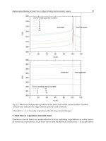
From Figure 5, when S-D separation larger than 4 cm (6 cm, 8 cm and 10 cm), the expected
optical power is below the photo-detector noise level. At 2 cm and 4 cm source to detector
separation, the expected fetal optical powers, 2293.99
10
-12
W/cm
2
and 5.9410
-12
W/cm
2
respectively, are higher than the photo-detector’s noise (1.17
10
-12
W/cm
2
) level. The photo-
detector is assumed to be operated at the photovoltaic condition with 1000 Hz bandwidth
and 1 cm
2
active area. Therefore, source to detector separation of 4 cm, which results in 70%
of optical power from fetal layer, is suitable to use with this low noise photo-detector. At 890
nm and 4 cm source-detector separation, the receiver sensitivity is optimized by considering
the limitation of the adaptive filter in FHR detection.
Source to detector separation
(cm)
Expected signal level,
P
M+am
(
10
-9
)
Expected P
F
signal level of
-34.7 dB
(
10
-12
)
2 6767.09 2293.99
4 17.53 5.94
6 0.31 0.11
8 0.37 0.13
10 0.09 0.03
Table 2. Expected P
F
signal level (-34.7 dB) at various source to detector separation
Fig. 5. Estimated P
F
(-34.7 dB) at 2.5 cm fetal depth
Application of Adaptive Noise Cancellation
in Transabdominal Fetal Heart Rate Detection Using Photoplethysmography
151
3.2 Implementation of ANC in transabdominal fetal Heart rate detection using PPG
In our work (Gan et al. 2009), a low-power optical technique is proposed based on the PPG
to non-invasively estimate the FHR. A beam of LED light (<68 mW) is shone to the maternal
abdomen and therefore modulated by the blood circulation of both mother and fetus
whereas maximum penetration is achieved at a wavelength of 890 nm. This mixed signal is
then processed by an adaptive filter with the maternal index finger PPG as reference input.
Figure 6 shows the optical fetal heart rate detection (OFHR) system block diagram whereas the
implementation by using National Instrument hardware and LabVIEW software are
illustrated in Figure 7 and Figure 8. In the OFHR system, the fetal probe (primary signal) is
attached to the maternal abdomen using a Velcro belt to hold the IR-LED and photo-detector,
separated by 4 cm. The reference probe is attached to the mother’s index finger as generally
practiced in pulse oximetry. As the selected IR-LED could only emit a maximum optical power
of 68 mW, the OFHR system operates with an optical power less than the limit of 87 mW
specified by the International Commission on Non-Ionizing Radiation Protection (ICNIRP)
(International Commission on Non-Ionizing Radiation Protection, 2000). In order to modulate
the IR-LED, the modulation signal is generated at a frequency of 725 Hz using software
subroutine through a counter port (NI-USB 9474) to the LED driver (Figure 6).
Fig. 6. OFHR system block diagram showing the hardware modules have been implemented
in LabVIEW.
The diffused reflected light from the maternal abdomen, detected by the low-noise photo-
detector, is denoted as I(M
1
, F), where M
1
and F denote the contribution to the signal from
the mother abdomen and fetus, respectively. A low-noise (6 nV/Hz
1/2
) transimpedance
amplifier is utilized to convert the detected current to a voltage level. The reference probe
(mother’s index finger) consists of an IR-LED and a solid-state photodiode with an
integrated preamplifier. The signal from this probe is denoted as I(M
2
), where M
2
refers to
the maternal contribution. Synchronous detection is not required at this channel as the
finger photoplethysmogram has a high signal to noise ratio (SNR).
Detected signals from both probes are simultaneously digitized with a 24-bit resolution data
acquisition card (NI-USB 9239, National Instruments, Inc.) at a rate of 5.5 kHz. The
Adaptive Filtering Applications
152
demodulation, digital filtering, and signal estimation are all performed in the digital
domain. Software implementation consists of generating a modulation signal, a
synchronous detection algorithm, down-sampling, high-pass filtering and ANC algorithm
(Zahedi & Beng, 2008). The entire algorithm and part of the instrument have been
implemented using Laboratory Virtual Instrumentation Engineering Workbench (LabVIEW
7.1, from National Instruments, Inc.). After pre-processing and applying the ANC algorithm
(Figure 9), the fetal signal and the fetal heart rate are displayed. The FHR is found by
estimating the prominent peak of the power spectral density using the Yule-Walker
autoregressive (AR) method (order of 20).
Figure 7 shows the laboratory prototype and the graphical user interface of the OFHR
system (in Figure 7, left) where the maternal index finger PPG (top), the abdominal PPG
(middle) and the estimated fetal PPG (bottom) are presented. There are three types of
Fig. 7. OFHR prototype
selectable displays (Figure 8) namely digital synchronous or lock-in amplifier (LIA), ANC
and heart rate trace. The purpose of the first two displays is to assist development and the
third one (Figure 8) indicates FHR values versus time (clinical application). The user can
either save the data to the personal computer for further analysis or just display it online.
Finally, a total of 24 data sets were acquired from six subjects at 37±2 gestational weeks from
the Universiti Kebangsaan Malaysia Medical Centre. This study was reviewed and
approved by the University Ethical Committee and written consent was obtained from all
patients who participated in this study after the procedure was clearly explained to them.
The process for subject recruitment and data acquisition are complied with the rules and
regulation as stated in Good Clinical Practice.
LED Driver
Fetal Probe Reference Probe
Application of Adaptive Noise Cancellation
in Transabdominal Fetal Heart Rate Detection Using Photoplethysmography
153
Fig. 8. Graphical User Interface of OFHR system. FHR trace menu
Fig. 9. ANC block diagram
In this study, all fetuses were singleton with gestation weeks from 30 week to 40 week.
Subjects with twin pregnancies, anterior placed placenta, obesity (BMI>30), gestational
diabetes mellitus (GDM) and hypertension were excluded from this study. In addition, all
fetuses in this study were found to be healthy by the obstetrician and born naturally
(vaginally) without any complication.
During the data acquisition, the fetal probe is fixed to a maternal abdomen and the reference
probe on her index finger in semi-Fowler position. The data analysis shows a correlation
coefficient of 0.97 (p-value < 0.001) between optical and ultrasound FHR with a maximum
error of 4%. Clinical results indicate that positioning the probe over the nearest fetal tissues
(not restricted to head or buttocks) improves signal quality and therefore detection accuracy.
4. Conclusions
A low power OFHR detection system has been designed and developed using low cost, very
low power (<68 mW) IR light and a commercially available silicon photo-detector. The
digital synchronous detection and adaptive filtering techniques have been successfully
Adaptive Filtering Applications
154
implemented using LabVIEW 7.1. By applying digital synchronous detection and adaptive
filtering techniques the FHR was determined with acceptable accuracy (maximum error of
4%) when compared to Doppler ultrasound. Attested by clinical results the probe
positioning influences the acquired signal’s quality and therefore affects the FHR results.
Locating the nearest fetal tissues (not restricted to head or buttocks) to the probe will help to
increase the signal quality and FHR determination accuracy.
The limitations of the optical technique are due to the presence of motion artifacts and
sensitivity to the probe placement. The presence of motion artifacts may cause loss of
correlation between the reference signal and the noise source (maternal PPG) in the mixed
signal recorded from the maternal abdomen. The performance of the adaptive filtering
scheme will suffer as a consequence, making the probe placement and stability an important
criterion. Besides that, finding a proper location is needed in order to get signals with good
SNR.
For the future development, by using an array of sensors to automatically select the channel
with the highest SNR will eliminate the positioning problem. The topology of the sources
and the photo-detector has to be determined. For the cost effective design, it is recommends
that more light sources are used instead of photo-detectors. A wearable system will make
the device more convenient for clinical applications in the near future. To ensure real-time
and low-power function, the whole system can be implemented using embedded processor.
The FHR will be wirelessly transmitted to another computing platform (PC or PDA) for
further analysis, storage and transmission (to the nursing entity at a clinic). The main
performance factors which will be considered are robustness, battery life, weight,
dimensions and ergonomy. It is thought that the using of the selected platform (ARM)
implementation will lead to a sufficiently low-cost bill-of material for the final product.
During development phase, EMC directives will be taken into account so that the system's
operation does not affect nor will be affected by other electronic devices. As a by-product of
the project and contribution to the scientific community, it is proposed that acquired data
during the project to be made available to a public data-base of biological signals
(www.physionet.org) maintained by MIT in the USA.
5. Acknowledgment
This work has been partially supported by research university grant UKM-AP-TKP-07-2009.
The authors would like to express their gratitude to Prof. Dr. M. A. J. M. Yassin and
Associate Prof. Dr. S. Ahmad for their assistance in collecting the clinical data, and the staff
at the Universiti Kebangsaan Malaysia Medical Centre, especially N. F. Mujamil for her
assistance in determining the fetal position through ultrasound scan.
6. References
Vaseghi, S.V. (2000). Advanced digital signal processing and noise reduction, Baffins Lane: John
Wiley & Sons Ltd
Widrow, B.; Glover Jr, J.R.; McCool, J.M.; Kaunitz, J.; Williams, C.S.; Hearn, R.H.; Zeidler,
J.R.; Eugene, D. & Goodlin, R.C. (1975). Adaptive noise cancelling: principles and
applications, Proceedings of the IEEE, Vol. 63, pp. 1692-1716
Ifeachor, E.C. & Jervis B.W. (2002). Digital signal processing: A practical approach, England:
Prentice Hall
Application of Adaptive Noise Cancellation
in Transabdominal Fetal Heart Rate Detection Using Photoplethysmography
155
Freeman, R.K.; Garite, T.J. & Nageotte, M.P. (2003). Fetal heart rate monitoring, Lippincott
William & Wilkins
Philip, J.S. (2002). Fetal distress. Current Obstetrics & Gynaecology, 12(1):5-21
Hershkovitz, R.; Sheiner, E. & Mazor, M. (2002). Ultrasound in obstetrics: a review of safety,
European Journal of Obtetrics & Gynecology and Reproductive Biology, Vol. 101, pp. 15-
18
Karlsson, B.; Berson, M.; Helgason, T.; Geirsson, R.T. & Pourcelot, L. (2000). Effects of fetal
and maternal breathing on the ultrasonic Doppler signal due to fetal heart
movement, European Journal of Ultrasound, pp. 47-52
Khandpur R. S. (2004). Biomedical Instrumentation: Technology and Applications, McGraw-Hill
Professional
Najafabadi, F. S.; Zahedi, E. and Mohd Ali, M.A. (2006). Fetal heart rate monitoring based on
independent component analysis, Computers in Biology and Medicine, Vol. 36, No. 3,
pp. 241-252
Ramanujam, N.; Vishnoi, G.; Hielscher, A.H.; Rode, M.E.; Forouzan, I. & Chance, B. (2000)
Photon migration through the fetal head in utero using continuous wave, near
infrared spectroscopy: clinical and experimental model studies, Journal of Biomedical
Optics, pp. 163-172
Chance, B. (2005). Transabdominal Examination Monitoring and Imaging of Tissue. U.S.
Patent 2005/0038344A1
Zourabian, A.; Chance, B.; Ramanujam, N.; Martha, R. & David A.B. (2000). Trans-
abdominal monitoring of fetal arterial blood oxygenation using pulse oximetry,
Journal of Biomedical Optics, Vol. 5, pp. 391-405
Nioka, S.; Izzetoglu, M.; Mawn, T.; Nijland, M.J.; Boas, D.A. & Chance, B. (2005). Fetal
transabdominal pulse oximeter studies using a hypoxic sheep model, The Journal of
Maternal-Fetal and Neonatal Medicine, Vol. 17, No. 6, pp. 393-399
Vintzileos, A.M.; Nioka, S. & Lake, M. (2005). Transabdominal fetal pulse oximetry using
near-infrared spectroscopy, American Journal of Obstetric & Gynaecology, Vol. 192,
pp. 129-133
Zahedi, E. & Beng, G.K. (2008). Applicability of adaptive noise cancellation to fetal heart rate
detection using photoplesthysmography. Computers in Biology and Medicine, Vol. 38,
No. 1, pp. 31-41
Choe, R.; Durduran, T.; Yu, G.; Nijland, M.J.M.; Chance, B.; Yodh, A.G. & Ramanujam, N.
(2003). Transabdominal near infrared oximetry of hypoxic stress in fetal sheep
brain in utero, Proceedings of the National Academy of Sciences, vol. 100, No. 22, pp.
12950-12954
Gan, K.B.; Zahedi, E. & Mohd. Ali, M.A. (2009). Trans-abdominal fetal heart rate detection
using NIR photopleythysmography: instrumentation and clinical results, IEEE
Transactions on Biomedical Engineering, Vol. 56, No. 8, pp. 2075-2082.
Manolakis, D.G.; Ingle, V.K. & Kogon, S.M. (2005). Statistical and adaptive signal processing.
Norwood:Artech House, Inc.
Haykin, S. (2002). Adaptive filter theory. Prentice Hall.
Gan K.B. (2009). Non-invasive fetal heart rate detection using near infrared and adaptive
filtering. Available online from: ()
Adaptive Filtering Applications
156
International Commission on Non-Ionizing Radiation Protection. (2000). ICNIRP statement
on light-emitting diodes (LEDs) and laser diodes: Implications for hazard
assessment, Health Phys., Vol. 78, No. 6, pp. 744–752
Bronzino, J.D. (2000). The biomedical engineering handbook: volume 1, Florida: CRC Press LLC
Allen, J. & Murray, A. (2000). Variability of photoplethysmography peripheral pulse
measurements at the ears, thumbs and toes, IEEE Proceeding Science and Technology,
Vol. 147, No. 6, pp. 403-407
7
Adaptive Filtering by Non-Invasive Vital
Signals Monitoring and Diseases Diagnosis
Omar Abdallah
1,2
and Armin Bolz
1
1
Institute for Biomedical Engineering, Karlsruhe Institute of Technology
2
Biomedatronik, Karlsruhe
Germany
1. Introduction
The reliability, reproducibility and accuracy of in-vivo measurements are of great
importance and have to be thoroughly studied and to a great extend achieved.
Reproducibility problems may result from the electronic components of the applied devices
and the variability of measured variables as well as noise sources. The inaccuracy is caused
by the approximation in the calculations or the used methods and by diverse sources of
errors resulting from the subject under considerations and its surroundings. In sensible
measurement like blood components, the positioning of the measuring sensor as well as the
variation in the applied pressure and the characteristics of contact area between sensor and
skin have a great effect on the accuracy and reproducibility of the measurements. The
ambient noise like high frequency and line frequency (50 or 60 Hz) noise can be filtered by
the detected biosignals like Photoplethysmogram (PPG) using the conventional analog or
digital filters without a great effort. The motion artifact of the subject caused by him as well
as by physical motion of body parts or by the surrounding has a varying frequency which
may have the same range of the signal frequency. It is difficult to filter noise from these
signals, and errors resulting from filtering can distort them. Usually physicians are misled
by these noisy signals and the analysis can go wrong. An adaptive filter is essential by bio-
signal and bio-image processing for noise cancellation without destroying or manipulating
the valuable detected information.
Biomedical signals such as photoplethysmogram (PPG) (Figure 1), electrocardiogram (ECG),
electroencephalogram (EEG), electromyogram (EMG) and impedance cardiogram (ICG) are
very important in the diagnosis of different pathological variations. By the detection of these
bio-signals as well as by the further derived parameters like oxygen saturation by pulse
oximeter, the motion artifact is a great challenge, which may lead to erroneous results or
even no results can be delivered [Lee].
The effectiveness of ECG monitors can be significantly impaired by motion artifact, which
can cause misdiagnoses, lead to inappropriate treatment decisions or trigger false alarms.
However, it is difficult to separate the noise from bio-signal due to its frequency spectrum
overlapping that of the ECG. A portable ECG recorder using accelerometer based on motion
artifact removal technique will be a great help for patients for tele-homecare or ambulatory
ECG monitoring.
Adaptive Filtering Applications
158
Fig. 1. Signal detection and processing by noninvasive diagnosis
A maternal electrocardiogram (mECG) and abdominal noise in abdominal maternal
recordings (especially by cardiotocography) can be orders of magnitude stronger than the
fetal electrocardiogram (fECG) signal. An adaptive filter using frequency-domain or time-
domain electrocardiogram features can be applied by the automatically extraction of a beat-
to-beat fECG from mECG using surface electrodes placed on the maternal abdomen [Rik]
[Prasad]. This will allow early diagnosis and monitoring treatment of certain fetal cardiac
disorders.
By non-contact ECG monitoring, cardiac activity and movements (that may be seen in part
in cardioballistogram CBG) may cause also disturbance to the detected signals, which can be
eliminated by applying an appropriate adaptive filter
High-quality EEG recording is crucial for diagnosis of different pathological variations. EEG
has biological artifacts and external artifacts. Biological artifacts can be EMG-, EOG-
(Electrooculograph) CBG or ECG-signal [Rasheed]. These artifacts appear as noise in the
recorded EEG signal individually or in a combined manner. These noise sources increase the
difficulty in analyzing the EEG and to obtaining clinical information. For this reason, it is
necessary to design specific filters to decrease such artifacts in EEG records. EEG quality in
the MR scanner is compromised by artifacts caused by interaction between the subject, EEG
electrode assemblies, and the scanner’s magnetic fields [Rasheed2009]. The three most
significant causes of EEG artifacts in the scanner are the large movements in the static field
like swallowing; the cardioballistogram and blood flow effects in the field associated with
the subject’s pulse; and the changing fields applied during fMRI image acquisition. Pulse
artifact is potentially a significant problem as it is normally large amplitude, widespread on
the scalp, and continuous. Using a cascade of adaptive filters based on a least mean squares
(LMS) algorithm can eliminate the undesired signals or interferences.
2. Photoplethysmogram
The photoplethysmogram (PPG) waveform comprises a pulsatile physiological waveform
superimposed on a slowly varying baseline with various lower frequency components. The
pulsatile one is attributed to cardiac synchronous changes in the blood volume with each
heart beat, and the second is attributed to respiration, sympathetic nervous system activity
and thermoregulation. Figure 2 shows a typical PPG signal without motion artifact. The
Adaptive Filtering by Non-Invasive VitalSignals Monitoring and Diseases Diagnosis
159
PPG technology has been used in a wide range of commercially available medical devices
for measuring oxygen saturation, blood pressure and arterial stiffness, cardiac output,
assessing autonomic function and detecting peripheral vascular diseases. Although the
origins of the components of the PPG signal are not fully understood, there is no doubt that
they can provide valuable information about the cardiovascular system and autonomic
nervous system. Hence, there is a great interest in the technique in recent years, driven by
the demand for low cost, very compact size, simple and portable technology for the primary
care and community based clinical settings, non-invasive technology without side effects or
risks as well as online monitoring capability and the advancement of computer-based pulse
wave analysis techniques and diagnosis [Allen, Abicht]. A computer aided analysis tool for
the hemodynamic diagnosis using PPG can be very helpful in clinical applications.
Automatic assessment of the reliability of reference heart rates from patient vital-signs
monitors incorporating both ECG and PPG based pulse measurements has been proposed
by Yu et al. They expressed reliability as a quality index for each reference heart rate. The
physiological waveforms were assessed using a support vector machine classifier and the
independent computation of heart rate made by an adaptive peak identification technique
that filtered out motion-induced noise [Allen].
Fig. 2. Photoplethysmogram PPG; top: detected raw signal, bottom: filtered signal
Also, due to demographic change, especially in the industrial countries, the personal health
care of old people is of great importance for prevention and rehabilitation. Continuous
monitoring of vital parameters is essential for that aim. By long term as well as by
emergency, a monitoring without interruption is crucial for the diagnosis of the case under
consideration. In many cases, a motion artifact caused by patient as well as by physical
motion of body parts or by the surrounding may have the same range of the signal
frequency. It is difficult to filter noise from these signals, and errors resulting from filtering
can distort them. Pulse oximeter for measuring oxygen saturation (S
P
O
2
) using more than
one PPG signal is a valuable device for monitoring patients in critical conditions. PPG and
the derived oxygen saturation are susceptible for motion artifact.
Pulse oximetry sensors use two Light Emitting Diodes (LEDs) which emit red and infrared
light that shine through a reasonably translucent part of the patient’s body. In pulse
Adaptive Filtering Applications
160
oximetry, it is called red light to the light band whose wavelength is comprised between
600-750 nm, while infrared light’s wavelength varies between 850 and 1000 nm. These two
wavelengths values are chosen because light absorption coefficient varies with the oxygen
concentration of in both the red and the infrared light. Figure 3 shows the two principles of
pulse oximetry: transmission and reflection pulse oximetry. By transmission Pulse oximetry,
the light sensitive photodetector (Photodiode PD), which acts as a receiver picking up the
light that passes through the measuring site, is opposite to the light emitter (light emitting
diode LED). By reflection pulse oximetry the PD and the LED`s lie at the same side of the
irradiated body portion.
Fig. 3. Operation of the pulse oximeter sensor, left: transmission pulse oximetry, right:
reflection pulse oximetry
Fig. 4. Light absorption by different tissue, at the top we see the plethysmogram generated
by the arterial pulsationen
Adaptive Filtering by Non-Invasive VitalSignals Monitoring and Diseases Diagnosis
161
Pulse oximeter works according to two physical principles: first, the presence of a pulse
wave generated by changes in blood volume (plethysmography) in the arteries and
capillaries (Figure 4) and second, the fact that oxyhemoglobin (O2Hb) and reduced
hemoglobin (Hb) have different absorption spectra (spectroscopy). Oxygenated hemoglobin
absorbs more infrared light and allows more red light to pass through. Deoxygenated (or
reduced) hemoglobin absorbs more red light and allows more infrared light to pass through.
3. Adaptive filtering of photoplethysmogram
We emphasize heir on the use of the adaptive filter by PPG, because of the importance of
this signal by detecting further parameters like pulse transit time (PPT); blood pressure
monitoring, Pulse rate variability and the application of it for the risk estimation and
diagnosis of cardiovascular diseases. Also the non-invasive calculation of concentration,
fractional oxygen saturation and further blood components like glucose may require also
the PPG signal analyzing. AC component of PPG signal caries important information for
diagnosis, but it may be affected by noise, which is sharing the same bandwidth. An
important application for the PPG is the calculation of oxygen saturation in emergency and
in intensive care, where the oxygen supplement of tissue has to be measured continuously.
The problem will be greater for example by detecting the PPG by low perfusion for the
monitoring of oxygen saturation, where a low signal to noise ratio is the result. An adaptive
filter will be the solution for this problem. Conventional filtering cannot be applied to
eliminate those types of artifacts because signal and artifacts have overlapping spectra. For
long term monitoring an adaptive filter is essential [Com 2007].
By pulse oximetry, Masimo adaptive filter is well known to the people working in this area.
The principle is easy and shortly described here: all detected samples of PPG`s by red and
infrared causing oxygen saturation below a certain value (e.g. 80%) are coming from venous
blood signals caused by motion artifact and has to be filtered. All signals causing saturation
higher than a threshold value (e.g. 90%) are the arterial signal. An intelligent algorithm is
designed according to this principle for the robust detection of oxygen saturation. By using
one PPG signal we cannot apply this algorithm. We used another algorithm by Filtering and
generation of reference noise depending on the detected signal.
Motion artefacts are one of the most important handicaps of photopletysmography and
pulse oximetry, as they suppose a big limitation and often become an insurmountable
obstacle on the utilization of this technology, since they are quite hard to cancel mainly due
to spectral characteristics of both, pulse signals and motion artifacts. In order to improve the
quality of Photoplethysmograms and pulse oximetry, some signal processing must be
implemented. Our research proposes, as viable solution, an Adaptive Filter in Noise
Cancellation configuration, working with a Least Mean Square Algorithm. At the end of the
system, we have carried out a reconstruction of the Photoplethysmogram and the signal that
we recover has a high enough quality for measuring fractional oxygen saturation of
hemoglobin in blood and for further diagnosis purposes.
An Adaptive Noise Cancellation (ANC) System has two inputs. This fact can be seen in the
Figure 5 presented below, more specifically in the diagram on the top. One is the Input
Signal, i.e., the signal corrupted by noise, coming from the sensor output, and the other one
is the Noise Reference, coming from the Synthesizer output. Both, the graphic of the Input
Signal and the generated plot of the Noise Reference appear in the Front Panel of the
corresponding LabVIEW program. Given that the Least Mean Square Algorithm provides
Adaptive Filtering Applications
162
adaptive filtering, the Noise Reference is adjusted to the real noise measured with the sensor
and, as a result, the output, Filtered Signal, naturally will be the filtered signal. In the
diagram below from the Figure 5 the main blocks of the Least Mean Square Algorithm
(LMS) implementation are presented. It is worth mentioning the fact that this algorithm is
recursive: the weights of the filter are calculated recursively to minimize the Mean Square
Error [Abdallah].
Fig. 5. Block diagram of the Adaptive Noise Cancellation (ANC)
4. Method and results by adaptive filtering of photoplethysmogram
Adaptive filters have been used to enable the measurement of photoplethysmogram PPG
under conditions, where movement of the body parts where the sensor is applied causes a
high noise to the signal. In this adaptive filter a noise reference and a signal reference are
used. We use the least mean square (LMS) method to extract the actual signal from the noisy
one.
For the first approximation to generate the reference signal a lowpass filter is used. Using
the resulting signal from this lowpass, an appropriate reference signal is generated. This
reference signal is in turn subtracted from the detected signal to generate a noise signal. The
generated noise signal is modified to synthesize the noise reference signal. The synthesized
reference noise is adjusted by the adaptive algorithm to the real one contained in the
measurement, and then subtracted from the detected noisy signal. The resulting signal is
modified to fulfill certain requirements (Figure 6)
The applied method discussed above can be used for the detection of a photopletysmogram
signal without the need for further signals of the same type or requiring a further sensor.
Figure 7 shows an example of the results obtained using this method. The algorithm was
tested for the calculation of oxygen saturation and accurate results are delivered under
artificial motion artifacts.
Adaptive Filtering by Non-Invasive VitalSignals Monitoring and Diseases Diagnosis
163
Fig. 6. Detected signal (top), generated reference signal (middle) and generated reference
noise for PPG filtering
Fig. 7. Schematic of the PPG filtering
Adaptive Filtering Applications
164
Each measurement from the applied PHM sensor contains seven signals of LEDs having
different wavelengths. Besides, a LED (which is off) acts as zero reference level. Since we
need two of them, first we have to separate them. Once these signals are presented
separately, we select the two of them that have been measured with the proper wavelengths
value for the calculation of oxygen saturation (LED having the wavelength 970 nm,
representing infrared light and a LED having the wavelength 660 nm, representing red
light). Then they are already adapted for being filtered by our system, which remove the
motion artifact from them. Finally, the filtered signals obtained after the program execution
can already be used to compute ratios regarding the SpO2, such as the so-called Ω ratio:
11
12
21
22
(,)
ln
(,)
(,)
ln
(,)
It
It
It
It
(1)
Where:
I(λ
1
,t
1
), I(λ
1
,t
2
),I(λ
2
,t
1
) and I(λ
2
,t
2
) are the light intensities measured at the instants t
1
, t
2
with
the wavelengths λ
1
,λ
2
respectively.
As results, examples of each step of the process described here are presented. First of all,
examples of the appearance of PHM measurements (and therefore, multiwavelength
measurements) are shown, both the whole measurement and a zoom of it (figure 8).
Fig. 8. Measurements of photoplethysmogram signals of PHM (right) and a zoom of it (left)
Next, the output given by the recovery of each signal is also presented. To demonstrate the
ability of the system presented here to make possible a precise enough computation of the
SpO2, we have calculated the value of the above-named Ω ratio for several measurements.
In order to make sure that the adaptive filter works well enough to get accurate SpO2
readings, the main goals are: first, to prove that the ratios obtained are included in an
acceptable range (bearing in mind that the values of this ratio allow us to estimate the
calibration that has to be applied later to the exact calculation of the SpO2). Next, it must be
proved that the values for the ratio when the signal is affected by motion artifacts keep quite
unchanging compared to those derived from the same signals without motion artifacts
[Figure 10]. The pulse amplitudes of the red and infrared signals are detected by the pulse
oximeter and measured to produce a certain ratio value, which is intrinsically related to the
functional oxygen saturation of (SpO2).
The signals shown in Figure 8 are measured by a Pulshemometer (PHM) sensor for the aim
of calculation of concentration and fractional oxygen saturation SaO2, which based on the
Adaptive Filtering by Non-Invasive VitalSignals Monitoring and Diseases Diagnosis
165
Principle of plethysmography (here volume change of arterial blood due to pulsation
generated from the heart) and optical spectroscopy. Also by our Project for the non-invasive
monitoring of glucose concentration in blood an adaptive filter for this aim is essential. For
in vivo measurement of blood components, the adaptive filter is necessary to get rid of the
noise and disturbances to the signal without any distortion of the detected useful signal that
may cause erroneous additive signals or that may reduce the information contents in the
detected signal. The Pulshemometer PHM sensor with a compact hardware circuit for
driving the LED’s and programmable digital potentiometer for adaptive programmable gain
amplification is shown in Figure 9.
Fig. 9. Pulshemometer PHM sensor for hemoglobin concentration and fractional and
fractional
Seven separated filtered PHM signals for and fractional oxygen saturation measurement
von PHM are shown in Figure 11 after normalization. For this sensor an adaptive filter is
essential for reliable and high accuracy results.
Wavelet transformation in combination with fuzzy and neuronal Networks (in some cases
cascaded) adaptive filtering is applied by different research groups. An energy ratio-based
method and a wavelet-based cascaded adaptive filter (CAF) can be applied for detecting and
removing baseline drift from pulse waveforms. This CAF outperforms traditional filters
both in removing baseline drift and in preserving the diagnostic information of pulse
waveforms [Lisheng]. Daubechies wavelet adaptive filter based on Adaptive Linear Neuron
networks is used to extract the signal of the pulse wave. Wavelet transform is a powerful
tool to disclose transient information in signals. The wavelet used is adaptive because the
parameters are variable, and the neural network based adaptive matched filtering has the
capability to “learn” and to become time-varying. So this filter estimates the deterministic
signal and removes the uncorrelated noises with the deterministic signal. This filter is found
to be very effective in detection of symptoms from pulsatile part of the entire optical signal
[Xiaoxia]. Fuzzy logic and Neuro-fuzzy can be used by adaptive filtering.
The method that has to be applied depends on the sensor applications and the case under
consideration, because intensive computation time, a high speed processor and a large saving
space may be needed, which may cause a delay time that disables an online monitoring. In
applications by multi-monitoring it will be possible to use other detected signals for the
purpose of filtering of a certain signal as will be discussed on the following section.