Ferroelectrics Material Aspects Part 15 ppt
Bạn đang xem bản rút gọn của tài liệu. Xem và tải ngay bản đầy đủ của tài liệu tại đây (7.16 MB, 35 trang )
Ferroelectrics – Material Aspects
480
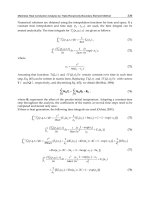
thin films with a tetragonal structure measured at 80 K (Yun et al., 2004). Recent theoretical
calculation showed a polarization of about 100 C/cm
2
in a rhombohedral structure as well
as about 150 C/cm
2
in a tetragonal structure. These values showed a good agreement with
the experimental ones (Ederer et al., 2006; Ricinschi et al., 2006).
1.3 General outline of chemical solution deposition
Chemical solution deposition (CSD) is one of the thin film fabrication methods, and it
includes spin-coating, drying and annealing processes. Precursor solution is deposited onto
a substrate by a spin-coating process. After the spin-coating process, a film dying process is
carried out to evaporate the solvent and decompose metal-organic compounds in the
precursor. An amorphous film is obtained at this stage. These processes are repeated several
times to obtain a desired film thickness. For the film crystallization, an annealing process is
carried out. It is usually carried out by a rapid thermal annealing (RTA) equipment to
crystallize and densify the film. Higher heating rate usually decomposes metal organic
compounds quickly and then desired oxide films with a higher density can be obtained
(Schwartz, 1997).
There are some advantages for CSD; (i) uniformity of the molecules in precursor solutions
and thin films, (ii) control of the film thickness by changing the solution concentration or the
coating speed, (iii) control of the composition ratio by mixing solutions, (iv) film fabrication
in ambient pressure, (v) synthesis of a non-equilibrium phase by the low-temperature
process. However, there are some disadvantages for this method; (i) possibility of cracks in a
film fabrication process, (ii) contamination which results in a difficulty of the manufacturing
process, (iii) films with low-coherency comparing with other thin film fabrication methods
such as pulsed laser deposition, chemical vapor deposition, and molecular beam epitaxy.
1.4 Precursor solutions for BiFeO
3
Precursor solutions for the CSD method are distinctly important. They consist of metal
organic compounds and solvent which determine process parameters such as drying and
annealing temperatures, film thickness per one spin-coating process, and coating affinity to
the substrates. In this chapter, BiFeO
3
thin films were prepared by CSD with precursor
solutions using 2-ethylhexanoate bismuth [Bi(OCO(CH)(C
2
H
5
)C
4
H
9
)
3
] and
trisacetylacetonato iron [Fe(C
5
H
7
O
2
)
3
] as metal organic materials, and toluene as a solvent.
2. Ferroelectric property of BiFeO
3
thin films prepared by CSD with
controlling Bi/Fe ratio in the precursor solution
In this section, we demonstrate the BiFeO
3
thin film growth with controlling Bi/Fe ratio of
the precursor solutions. Composition ratio affects the crystal growth and the electric
property of the films. We obtain both good crystallinity and ferroelectric polarization of 85
C/cm
2
with films using 10 mol% Bi-excess solution (Nakamura et al., 2007; Nakamura et
al., 2008).
2.1 Film preparation by CSD with controlling Bi/Fe ratio
BiFeO
3
thin films were deposited on a Pt (200 nm)/TiO
2
(40 nm)/SiO
2
(600 nm)/Si substrate
by CSD using precursor solutions of different Bi/Fe ratios: 10 mol% Fe-excess (10%Fe-ex.),
stoichiometric, 5 mol% Bi-excess (5%Bi-ex.), 10 mol% Bi-excess (10%Bi-ex.), and 20 mol% Bi-
BiFeO
3
Thin Films Prepared by Chemical Solution Deposition with
Approaches for Improvement of Ferroelectricity
481
excess (20%Bi-ex.). The precursor solution was spin-coated at 3000 rpm for 30 s and dried at
250°C for 5 min in air. These processes were repeated 20 times to obtain a film thickness of
250 nm. Then the films were annealed at 450 °C for 15 min in nitrogen atmosphere using the
RTA equipment. For electrical measurement, Pt top electrodes with a diameter of 190 m
and a thickness of 100 nm were formed on the films by rf sputtering at RT. We confirmed by
inductively coupled plasma (ICP) analysis that the composition ratios of BiFeO
3
thin films
were the same as the precursor solutions.
2.2 Crystal structure
Figure 1 shows
–2 scans of the XRD patterns of the BiFeO
3
thin films with different Bi/Fe
ratios. All the films show polycrystalline perovskite phase mainly. However, the 10%Fe-ex.
BiFeO
3
film has small amount of a Bi
2
Fe
4
O
9
phase and the 20%Bi-ex. film shows a Bi
2
O
3
phase. This indicates that excessive Fe or Bi compounds in the precursor solution tend to
form impurity phase. Comparing the peak intensities corresponding to the (010) and (110)
planes, the 10%Bi-ex. and 20%Bi-ex. films show higher diffraction intensities, indicating that
they are crystallized well. This result suggests that Bi compounds in the precursor solution
contribute the promotion of the film crystallization but that 5 mol% excess Bi is insufficient
for the crystallization of such films.
2.3 Surface texture and raman spectrum
Figures 2(a-e) show the atomic force microscope (AFM) images of BiFeO
3
films taken in a 20
× 20 m
2
area. All the BiFeO
3
thin films show a rosette structure, which consists of circular
regions with an uneven texture and outer regions with a flat surface. These structures were
also reported in PbZrO
3
thin films prepared by the sol–gel method (Alkoy et al., 2005). Table
I shows the percentages of circular regions, RMS roughnesses and total boundary lengths
surrounding the circular regions evaluated from Figs. 2(a-e). As can be seen in Table I, the
percentage of circular region and RMS roughness tend to increase with an increase in Bi/Fe
ratio. On the other hand, the total boundary length is ~200 m and seems to have no
systematic dependence.
Fig. 1. XRD
–2 patterns of BiFeO
3
thin films prepared using 10 mol%Fe-excess (10%Fe-
ex.), stoichiometric, 5 mol% Bi-excess (5%Bi-ex.), 10 mol% Bi-excess (10%Bi-ex.), and 20
mol% Bi-excess (20%Bi-ex.) precursor solutions.
Ferroelectrics – Material Aspects
482
Figure 3(a) shows an AFM image of the 10%Bi-ex. BiFeO
3
thin film with white circles
marking the measurement location of Raman spectroscopy. A laser with a 0.7 m spot size
and an excitation wavelength of 515 nm was applied to the film surface labelled ”Circular
region” and “Outer region” in Fig. 3(a). Figure 3(b) shows the Raman spectra measured at
RT in each measurement location shown in Fig. 3(a). The spectrum measured in BiFeO
3
ceramic is also shown as a reference. As shown in Fig. 3(b), the spectrum measured in the
circular region is almost similar to that of BiFeO
3
ceramic consisting of polycrystalline
grains. On the other hand, the spectrum measured in the outer region has a broad shape,
found frequently in amorphous materials, and is different from that of BiFeO
3
ceramic.
Fig. 2. 20 × 20 m
2
surface AFM images for the BiFeO
3
films of (a) 10%Fe-ex., (b)
stoichiometric, (c) 5%Bi-ex., (d) 10%Bi-ex., and (e) 20%Bi-ex Each film shows a rosette
structure, which consists of circular regions and outer regions.
Fig. 3. (a) AFM image of 10%Bi-ex. BiFeO
3
thin film with white circle marking the
measurement location of Raman spectroscopy. (b) Raman spectra measured at RT for each
location shown in (a). Spectrum of BiFeO
3
ceramic is also shown as a reference.
BiFeO
3
Thin Films Prepared by Chemical Solution Deposition with
Approaches for Improvement of Ferroelectricity
483
Sample
Circular region area
(%)
RMS roughness
(nm)
Boundary length
(mm)
10%Fe-ex. 46.0 3.6 238
Stoichiometric 48.1 5.1 225
5%Bi-ex. 41.1 5.6 196
10%Bi-ex. 85.9 4.8 165
20%Bi-ex. 53.7 6.8 208
Table 1. Circular region areas, RMS roughnesses, and boundary lengths for all samples.
The same results are also obtained in BiFeO
3
thin films prepared using the other precursor
solutions with different Bi/Fe ratios. These results indicate that the circular regions have a
BiFeO
3
crystalline phase, while the outer regions have an amorphous BiFeO
3
phase.
Moreover, it can be considered that each phase exists from the top to the bottom of the film
in a vertical direction because the excitation can sufficiently penetrate up to the bottom of
the film. Consequently, the BiFeO
3
thin films of 10%Bi-ex. and 20%Bi-ex. have more circular
regions and show good crystallinity, as shown in Fig. 1. This tendency is also observed in
the Pb(Zr,Ti)O
3
(PZT) thin film prepared by the sol–gel method. Excessive Pb compounds in
the precursor solution promote the formation of PZT and lead to show more circular regions
(Alkoy et al., 2005). From Figs. 1 and 2, however, the 20%Bi-ex. film shows a Bi
2
O
3
phase,
and the area of circular regions does not seem to increase so much compared with that in the
10%Bi-ex. film. This result suggests that excessive Bi compounds in the precursor solution
are more reactive, thereby they promote the formation of BiFeO
3
. However, the major
amount of Bi compounds tends to form Bi
2
O
3
as well as BiFeO
3
, therefore the circular region
does not seem to increase so much.
2.4 Ferroelectric property
Figure 4(a) shows the leakage current density versus electric field (J–E) property of the
BiFeO
3
thin films measured at RT. A comparatively larger leakage current is obtained in the
films that contain more Bi. Figure 4(b) shows P–E hysteresis loops of the BiFeO
3
thin film
measured at RT with a scanning frequency of 20 kHz. The 10%Bi-ex. BiFeO
3
thin film shows
more squareness in hysteresis loop than the other films. The remanent polarizations (P
r
) for
the maximum applied electric field of 1.2 MV/cm are 30, 38, 28, 85, and 53 C/cm
2
for the
films of 10%Fe-ex., stoichiometric, 5%Bi-ex., 10%Bi-ex., and 20% Bi-ex., respectively.
Fig. 4. (a) J–E characteristics of BiFeO
3
thin films measured at RT. (b) P–E hysteresis loops
measured at RT.
Ferroelectrics – Material Aspects
484
Fig. 5. Leakage current of BiFeO
3
thin films at 240 kV/cm as function of (a) amount of excess
Bi, (b) percentage of circular region area, (c) RMS roughness, and (d) boundary length.
Fig. 6. Remanent polarization of BiFeO
3
thin films as function of (a) amount of excess Bi and
(b) percentage of circular region area.
2.5 Relationship between surface texture and ferroelectricity
To investigate the influences of the surface texture and Bi/Fe ratio on the leakage current of
BiFeO
3
thin films, we consider the amount of excess Bi, percentage of circular region area,
BiFeO
3
Thin Films Prepared by Chemical Solution Deposition with
Approaches for Improvement of Ferroelectricity
485
RMS roughness, and boundary length at the surface between crystal and amorphous phases,
as shown in Table I. Figures 5(a-d) show the leakage current measured at 240 kV/cm versus
(a) amount of excess Bi, (b) circular region area, (c) RMS roughness, and (d) boundary
length. As shown in Figs. 5(a-c), leakage current tends to exponentially increase with an
increase in the amount of excess Bi, circular region area, and RMS roughness although some
scattering of the data is observed in Fig. 5(c). On the other hand, the length between the
circular regions and the outer regions does not seem to affect the leakage current as shown
in Fig. 5(d). These results suggest that the BiFeO
3
thin film prepared using the Bi excess
precursor solution tends to have more circular regions that have BiFeO
3
crystals and to have
a larger RMS roughness. From these leakage trends, leakage current mainly passes through
circular regions consisting of crystalline BiFeO
3
rather than through outer amorphous
region, and that current is increased by a rough surface. We further investigate the
influences of the surface texture and Bi/Fe ratio on the ferroelectric polarization of BiFeO
3
thin films. We plot the amount of excess Bi and percentage of circular region area that has
BiFeO
3
crystals, as shown in Fig. 3(b). Figures 6(a) and 6(b) show the remanent polarization
measured at RT versus (a) amount of excess Bi and (b) circular region area. The remanent
polarization increases with an increase in Bi ratio below the 10%Bi-ex. BiFeO
3
film.
However, the 20%Bi-ex. film decreases its remanent polarization because of the mixed phase
of BiFeO
3
and Bi
2
O
3
. As shown in Fig. 6(b), the remanent polarization linearly increases with
an increase in the percentage of the circular region area. From the extrapolated line in Fig.
6(b), fully crystallized BiFeO
3
thin films are expected to show 100 C/cm
2
. According to
leakage and polarization plots in Fig. 5 and Fig. 6, a 10 mol% Bi-excess solution gives BiFeO
3
thin films the best ferroelectric property with more circular regions.
3. Insertion effect of Bi-excess layer on BiFeO
3
thin films
In section 2, Bi-excess solution, or precursor solution with excessive Bi compounds,
promotes film crystallization, leading to a good ferroelectricity. In this section, we
demonstrate the insertion effect of Bi-excess layer to the stoichiometric BiFeO
3
thin films to
improve the crystal growth and ferroelectricity of the films (Nakamura et al., 2007;
Nakamura et al., 2008).
3.1 Insertion effect
Insertion effect, inserting Bi-excess BiFeO
3
layer to the film, is aiming to promote the crystal
growth of the film and to obtain a good ferroelectricity. There are some reports that
ferroelectric thin films prepared by CSD show a non-crystalline layer at the interface
between the thin film and the electrode. Such a layer is reported as an interfacial layer which
degrades the ferroelectric property of the film (Grossmann et al., 2002). These reports
suggest that the low crystallinity part is concentrated at the interface between the film and
the electrode. To improve the low crystallinity part, an insertion layer promoting crystal
growth will be effective.
In our BiFeO
3
thin films, a thin film with stoichiometric solution shows low crystallinity
with a small polarization, and a film with 10 mol% Bi-excess solution shows high
crystallinity and a large polarization. Thus an insertion layer with 10 mol% Bi-excess
solution is expected to be effective. To investigate the insertion effect of Bi-excess layers,
three types of thin films were prepared on Pt/TiO
2
/SiO
2
/Si substrates, as shown in Fig. 7:
stoichiometric BiFeO
3
thin film with Bi-excess top layer (Bi-T), bottom layer (Bi-B), and top
Ferroelectrics – Material Aspects
486
and bottom layer (Bi-TB). Then the films were annealed at 450 °C for 15 min in a nitrogen
atmosphere using the RTA process. For the electrical measurement, Pt top electrodes with a
diameter of 190 μm were formed by rf sputtering.
Fig. 7. Schematic models of BiFeO
3
thin films inserting Bi-excess top and bottom layer (Bi-
TB), top layer (Bi-T), and bottom layer (Bi-B).
3.2 Crystal structure
Figure 8 shows the θ–2θ scans of XRD patterns of the BiFeO
3
thin films with Bi-excess top
and bottom layer (Bi-TB), top layer (Bi-T), and bottom layer (Bi-B). These results show that
all the films exhibit mainly polycrystalline perovskite single phase without nonperovskite
phases such as Bi
2
Fe
4
O
9
and Bi
2
O
3
. Comparing peak intensities corresponding to (010) and
(110) planes, the crystallinity of Bi-TB is the best. Bi-T is the second best, followed by Bi-B.
This result indicates that the Bi-excess top layer improves the crystallization in the annealing
process. Moreover, this tendency suggests that the crystallization is produced from the
surface to the bottom using the RTA process. As for the difference between Bi-TB and Bi-T,
crystallinity of BiFeO
3
film can be enhanced by inserting the Bi-excess layer on the top
surface and the bottom.
Fig. 8. XRD
–2 patterns of Bi-TB, Bi-T, and Bi-B BiFeO
3
thin films.
BiFeO
3
Thin Films Prepared by Chemical Solution Deposition with
Approaches for Improvement of Ferroelectricity
487
3.3 Surface texture and raman spectrum
Figures 9(a-c) show the AFM images of BiFeO
3
films taken over a 10 × 10 μm
2
area. As can
be seen in Fig. 9(a), Bi-TB forms more grains than the others. On the other hand, Bi-T and Bi-
B form finer grains as well as larger grains, as shown in Figs. 9(b) and 9(c). In addition, Bi-T
seems to form larger grains than the film of Bi-B. The surface RMS roughness is estimated as
7.7, 6.7, and 5.0 nm for the films of Bi-TB, Bi-T, and Bi-B, respectively. The number of grains
and the surface roughness increase with increasing crystallinity, comparing Fig. 9 with Fig.
8. To investigate the difference between finer and larger grains, Raman spectroscopy was
carried out. A laser with a 0.7 μm spot size irradiated the points labelled A–C, which form
large grains, and D–F, which form fine grains, as shown in Figs. 9(a-c). Figures 9(d) and 9(e)
show Raman spectra measured at RT. These figures also include the spectrum measured in
BiFeO
3
ceramic, as a reference. As shown in Fig. 9(d), the spectra measured in the areas A–C
are almost the same as the spectrum of BiFeO
3
ceramic. On the other hand, the spectra
measured in the areas D–F are different from the spectrum of BiFeO
3
ceramic, as shown in
Fig. 9(e). These results indicate that the areas A–C have good BiFeO
3
crystals while the areas
D–F seem to be amorphized. Moreover, the area at which the BiFeO
3
crystal spectrum was
observed is the largest in Bi-TB. This result relates that Bi-TB crystallizes the best, comparing
Figs. 9 and 8.
Fig. 9. 10 × 10 μm
2
surface AFM images with markings of the typical locations of Raman
spectroscopy for the films of (a) Bi-TB, (b) Bi-T, and (c) Bi-B, respectively. Areas A–C form
large grains, while areas D–F form fine grains. (d) and (e) Raman spectra measured at RT for
the locations shown in Figs. 9(a)–9(c).
3.4 Ferroelectric property
Figure 10 shows the leakage current density versus electric field (J–E) of BiFeO
3
thin films
measured at (a) RT and (b) 80 K. When the electric field is lower than 300 kV/cm, the
Ferroelectrics – Material Aspects
488
leakage currents are almost unchanged among three types of films both at RT and 80 K. This
suggests that the amorphous phase of the surface limits the conduction in the case of lower
electric field, as mentioned in Abe et al. (Abe et al., 1993). On the other hand, when the
electric field is higher than 300 kV/cm at 80 K, the leakage current becomes large for the
film of Bi-TB. Therefore, it is suggested that the amorphous phase includes defects that limit
the carrier emission at the interface, and the leakage current increases at higher electric
fields in the Bi-TB film. In the case of Bi-T and Bi-B, the amorphous phase might suppress
the leakage current at high electric field. Figure 11 shows ferroelectric polarization versus
electric field (P–E) hysteresis loops of BiFeO
3
thin film at (a) RT and (b) 80 K, respectively. At
RT, the remanent polarizations (P
r
) for maximum applied electric field of 1.0 MV/cm are 55,
26, and 17 μC/cm
2
for the films of Bi-TB, Bi-T, and Bi-B, respectively. In addition, the
coercive field of Bi-TB is 385 kV/cm, which is the lowest in the three types of films. At 80 K,
the remanent polarizations for maximum applied electric field of 2.0 MV/cm are 65, 46, and
32 μC/cm
2
for the films of Bi-TB, Bi-T, and Bi-B, respectively. The remanent polarization of
Bi-TB is about twice that of the film prepared by stoichiometric solution (28 μC/cm
2
at RT,
and 38 μC/cm
2
at 80 K). These results show that BiFeO
3
thin film of Bi-TB gives the best
ferroelectric property among the three types of films, which is attributed to the good
crystallinity of the BiFeO
3
film, comparing Figs. 11 and 8.
Fig. 10. Leakage current characteristics of BiFeO
3
thin films measured at (a) RT and (b) 80 K.
Fig. 11. P–E hysteresis loops of BiFeO
3
thin films measured at (a) RT and (b) 80 K.
BiFeO
3
Thin Films Prepared by Chemical Solution Deposition with
Approaches for Improvement of Ferroelectricity
489
4. Improvement of ferroelectricity of BiFeO
3
thin films by postmetallization
annealing and electric field application
In this section, we describe the postmetallization annealing and electric field application by
using 10 mol% Bi-excess BiFeO
3
thin film which shows good ferroelectricity in section 2.
These are the ways to improve ferroelectricity of BiFeO
3
thin films. Postmetallizaton
annealing is the electrode annealing process to reduce the leakage current which has already
reported in several thin film materials such as BaTiO
3
, (Ba,Sr)TiO
3
, (Pb,Sr)TiO
3
, and PZT
after the deposition of top electrodes (Lee et al., 2004; Joo et al., 1997; Chung et al., 2001;
Thakoor, 1994). Electric field application is to apply a high electric field to reverse its
polarization reversal easily. It is typically carried out in bulk materials such as PZT (Kamel
et al., 2007). These two approaches are expected to be effective to improve ferroelectric
properties of BiFeO
3
thin films (Nakamura et al., 2009).
4.1 Film preparation methods
BiFeO
3
thin films were deposited on a Pt/TiO
2
/SiO
2
/Si substrate by CSD using 10mol % Bi-
excess precursor solution. Spin-coating and drying processes were the same as in chapter 2.
These processes were repeated 20 times to obtain a film thickness of 250 nm. Then, the films
were treated by the RTA process at 450 °C for 20 min in nitrogen atmosphere. For the
electrical measurement, Pt top electrodes were formed on the BiFeO
3
film by rf sputtering.
After the deposition of Pt top electrodes, the sample was divided into three pieces and
labelled as BFO, BFO-N, and BFO-O, respectively. Then the postmetallization annealing was
carried out for 5 min at 300 °C in nitrogen atmosphere for BFO-N, and oxygen atmosphere
for BFO-O by the RTA process. Finally, the following three films were obtained; BFO (as
prepared film without postmetallization annealing), BFO-N (the film with the annealing in
nitrogen atmosphere), and BFO-O (the film with the annealing in oxygen atmosphere).
4.2 Improvement of ferroelectric property of BiFeO
3
thin films by postmetallization
annealing
Figure 12(a) shows the
-2 patterns of the XRD of the BiFeO
3
thin films with and without
the postmetallization annealing. All the films consist mainly of polycrystalline perovskite
phase, but a Bi
2
O
3
phase is slightly observed. Evaluating the diffraction peak intensity of
each film, it does not change among three films. This result indicates that the crystallinity of
BiFeO
3
does not change by the postmetallization annealing. We note that the intensity of an
observed Bi
2
O
3
phase is much smaller than the BiFeO
3
phase and its intensity does not
change after the postmetallization annealing. Therefore, it is enough to evaluate the
dielectric property of BiFeO
3
and the annealing effect of the electrode. There may be a
possibility of a peak shift due to a strain relaxation between the BiFeO
3
film and the Pt
electrode by the postmetallization annealing, however, it is hard to observe the strain
relaxation from such a small 190 m diameter dot electrode because an incident X-ray beam
width is about 2 mm, making it difficult to analyze the crystalline property of the small area.
To confirm the strain relaxation, a 100 nm thick Pt film was deposited on the whole BiFeO
3
film surface and then the postmetallization annealing was carried out. Figure 12(b) shows
the XRD patterns near (010) peak before and after the Pt deposition and the
postmetallization annealing in nitrogen atmosphere. The peak intensity is decreased due to
the deposition of the Pt film, but the peak shift is not observed. This result suggests that a
clear strain relaxation does not occur near the interface between the BiFeO
3
film and the Pt
electrode after the postmetallization annealing.
Ferroelectrics – Material Aspects
490
Fig. 12. (a) XRD
-2 scans of as-prepared (BFO), N
2
annealed (BFO-N), and O
2
annealed
(BFO-O) BiFeO
3
thin films. (b) Expanded scans near the (010) peak before and after the Pt
deposition and the postmetallization annealing. A 100 nm thick of Pt film was deposited on
the whole surface of the BiFeO
3
film.
Figures 13(a) and 13(b) show the J-E characteristics of BiFeO
3
thin films measured at (a) 80 K
and at (b) RT. The leakage current is suppressed in both the BFO-N and BFO-O films at 80
K. This suppression is also observed at RT. Joo et al. reported that the leakage current is
suppressed by the postmetallization annealing in oxygen atmosphere due to the reduction
of oxygen vacancies in a film (Joo et al., 1997). Contrary to their result, our result suggests
that the postmetallization annealing improves the contact between the BiFeO
3
film and the
Pt electrode or the reduction of defects near the interface between the BiFeO
3
film and the Pt
electrode as suggested in Pt/PZT/Pt capacitors, rather than the compensation of oxygen
vacancies in the BiFeO
3
film.
Fig. 13. Current density-electric field (J-E) characteristics of BiFeO
3
thin films measured at (a)
80 K and at (b) RT.
Figures 14(a) and 14(b) show frequency dependences of the dielectric constant and the
dielectric loss tan measured at (a) 80 K and at (b) RT. The dielectric constant and the loss
tangent of BiFeO
3
thin films measured at 80 K are found to be 185 and 0.061, 172 and 0.045,
and 186 and 0.048 for the BFO, BFO-N, and BFO-O film with a measuring frequency of 1
MHz, respectively. In addition, the frequency variability of from 10
3
to 10
6
Hz is 20.5% BFO,
16.9% BFO-N, and 18.3% BFO-O. The reduction in the frequency variability and the
BiFeO
3
Thin Films Prepared by Chemical Solution Deposition with
Approaches for Improvement of Ferroelectricity
491
dielectric loss may be due to the reduction in the leakage current as shown in Fig. 13(a). The
same tendencies are also observed in the BiFeO
3
films measured at RT, as shown in Fig.
13(b). The frequency variability from 10
3
to 10
6
Hz is 30.6% BFO, 23.3% BFO-N, and 23.4%
BFO-O measured at RT. These results indicate that the frequency variability of and the
dielectric loss are successfully reduced by the postmetallization annealing.
Fig. 14. Dielectric constant-frequency (
-F) characteristics of BiFeO
3
thin films measured at
(a) 80 K and at (b) RT.
Figures 15(a) and 15(b) show P-E hysteresis loops of BiFeO
3
thin films measured at (a) 80 K
and at (b) RT with a scanning frequency of 20 kHz. The remanent polarizations (P
r
)
measured at 80 K under the maximum applied electric field of 1.2 MV/cm are 91, 87, and 89
C/cm
2
for the films of BFO, BFO-N, and BFO-O, respectively. In addition, the double
coercive field (2E
c
) is reduced at about 90 kV/cm in the nitrogen-annealed film. The slight
reduction of coercive field is also observed at RT, as shown in Fig. 15(b). This reduction of
the coercive field may be due to the improvement of the contact between the Pt electrode
and the BiFeO
3
film or the reduction in defects near the interface between BiFeO
3
film
and the Pt electrode, as mentioned above.
Fig. 15. P-E hysteresis loops measured at (a) 80 K and at (b) RT under 20 kHz triangular
scanning voltage of as-prepared (BFO), N
2
annealed (BFO-N), and O
2
annealed (BFO-O)
BiFeO
3
thin films.
Ferroelectrics – Material Aspects
492
4.3 Improvement of ferroelectric property of BiFeO
3
thin films by electric filed
application
To evaluate the effect of the electric field application, P-E hysteresis loops were measured at
80 K in a following order; the first measurement was carried out by the applied voltage from
5 to 70 V, which corresponds to 0.2 MV/cm to 2.8 MV/cm, and then, the second
measurement was carried out from 70 to 5 V with 1 kHz triangular wave. The temperature
was set at 80 K to reduce the thermal effect and to apply high electric field. This
measurement was carried out for the BFO-N film because it has the best insulation and
ferroelectric characteristics among three films. Figures 16(a-d) show hysteresis changes in
BFO-N film before (black lines) and after (red lines) applying the electric field of 0.4, 1.2, 2.0,
and 2.8 MV/cm corresponding 10, 30, 50, and 70 V, respectively at 80 K. After applying the
electric field of 2.8 MV/cm, which corresponds to 70 V, the shape of hysteresis loop is
dramatically changed in the second measurement. In the sequence from Figs. 16(a-d), the
remanent polarizations of the first measurement are 0.19, 29.9, 74.5, and 104 C/cm
2
under
the maximum field of 0.4, 1.2, 2.0, and 2.8 MV/cm, respectively. In addition, the remanent
polarizations of the second measurement are 17.4, 84.4, 97.6, and 106.5 C/cm
2
under the
maximum field of 0.4, 1.2, 2.0, and 2.8 MV/cm, respectively.
Fig. 16. P-E hysteresis of BiFeO
3
thin films measured at 80 K under the maximum field of (a)
0.4 MV/cm (10 V), (b) 1.2 MV/cm (30 V), (c) 2.0 MV/cm (50 V), and (d) 2.8 MV/cm (70 V),
respectively. Hysteresis loops were measured from 5 to 70 V the first measurement and then
measured from 70 to 5 V the second measurement.
Moreover, the leakage current is reduced at about 1 order of magnitude after the P-E
measurement, as shown in Fig. 17. Dependences of P
r
and E
c
on the applied electric field are
BiFeO
3
Thin Films Prepared by Chemical Solution Deposition with
Approaches for Improvement of Ferroelectricity
493
shown in Fig. 18. P
r
and E
c
obtained in the first measurement are gradually increased with
increase in the electric field, and then, the second measurement keeps large P
r
value even in
lower electric field. The third measurement from 5 to 70 V was carried out and values of P
r
and E
c
are almost the same as the second data, as shown in Fig. 18. Kohli et al. reported that
the hysteresis of the PZT thin film was changed by applying pulse electric field because of
the removal of 90° domain pinning (Kohli et al., 1998). Okamura et al. reported that the
wake-up phenomenon, which shows an increase in the remanent polarization by applying
switching pulses and removing the locked polarizations in the SrBi
2
Ta
2
O
9
film (Okamura et
al., 2000). The improvement of ferroelectricity is possibly due to the relaxation of pinned
domains or locked polarizations in the BiFeO
3
film by applying high electric field.
Fig. 17. Current density-electric field (J-E) characteristics of BiFeO
3
thin films measured at 80
K. The measurement was carried out before and after the P-E measurement shown in Fig. 5
(d).
Fig. 18. Applied field dependences of remanent polarization and coercive field at 80 K for
the first, the second, and the third measurement.
Ferroelectrics – Material Aspects
494
5. Conclusion
We describe BiFeO
3
thin films prepared by CSD with several approaches to improve its
ferroelectricity. Controlling Bi/Fe ratio in the precursor solution contributes the promotion
of the film crystallization and shows a large polarization of 85 C/cm
2
with 10 mol% Bi-
excess solution. Insertion of the 10 mol% Bi-excess layer to the stoichiometric BiFeO
3
films
also promotes the film crystallization, leading to the improvement of the ferroelectricity.
Ferroelectric property of films using 10 mol% Bi-excess solution can further improve by the
postmetallization annealing as well as the electric field application. These are the effective
methods to improve ferroelectricity of BiFeO
3
.
6. Acknowledgments
The authors thank Takaaki Nakamura, Hideo Fukumura, and Professor Hiroshi Harima of
Kyoto Institute of Technology for conducting Raman spectroscopy.
7. References
Roginskaya, Yu. E.; Venevtsev, Yu. N.; Fedulov, S.A. & Zhdanov, G.S. (1963). Soviet Physics
Crystallography, 8, 610
Palai, R.; Katiyar, R. S.; Schmid, H.; Tissot, P.; Clark, S. J.; Robertson, J.; Redfern, S. A. T.,
Catalan, G. & Scott, J. F. (2008).
phase and metal-insulator transition in
multiferroic BiFeO
3
. Physical Review B, 77, 014110-1-014110-11
Teague, J. R.; Gerson, R. & James, W. J. (1970). DIELECTRIC HYSTERESIS IN SINGLE
CRYSTAL BiFeO
3
. Solid State Communications, 8, 1073-1074
Lebeugle, D.; Colson, D.; Forget, A. & Viret, M. (2007). Very large spontaneous electric
polarization in BiFeO
3
single crystals at room temperature and its evolution under
cycling fields. Applied Physics Letters, 91, 022907-1-022907-3
Shvartsman, V. V.; Kleemann, W.; Haumont, R. & Kreisel, J. (2007). Large bulk polarization
and regular domain structure in ceramic BiFeO
3
. Applied Physics Letters, 90, 172115-
1- 172115-3
Wang, J.; Neaton, J. B.; Zheng, H.; Nagarajan, V.; Ogale, S. B.; Liu, B.; Viehland, D.;
Vaithyanathan, V.; Schlom, D. G.; Waghmare, U. V.; Spaldin, N. A.; Rabe, K. M.;
Wuttig, M. & Ramesh, R. (2003). Epitaxial BiFeO
3
Multiferroic Thin Film
Heterostructures. Science, 299, 1719-1722
Li, J.; Wang, J.; Wuttig, M.; Ramesh, R.; Wang, N.; Ruette, B.; Pyatakov, A. P.; Zvezdin, A. K.
& Viehland, D. (2004). Dramatically enhanced polarization in (001), (101), and (111)
BiFeO
3
thinfilms due to epitiaxial-induced transitions. Applied Physics Letters, 84,
5261-5263
Yun, K. Y.; Ricinschi, D.; Kanashima, T.; Noda, M. & Okuyama, M. (2004). Giant
Ferroelectric Polarization Beyond 150 C/cm
2
in BiFeO
3
Thin Film. Japanese Journal
of Applied Physics, 43, L647-L648
Ederer, C. & Spaldin, N. A. (2005). Effect of Epitaxial Strain on the Spontaneous Polarization
of Thin Film Ferroelectrics. Physical Review Letters, 95, 257601-1-257601-4
Ricinschi, D.; Yun, K. Y. & Okuyama, M. (2006). A mechanism for the 150 μC/cm
2
polarization of BiFeO3 films based on first-principles calculations and new
structural data. Journal of Physics: Condensed Matter, 18, L97-L105
BiFeO
3
Thin Films Prepared by Chemical Solution Deposition with
Approaches for Improvement of Ferroelectricity
495
Schwartz, W. R. (1997). Chemical Solution Deposition of Perovskite Thin Films. Chemistry of
Materials, 9, 2325-2340
Nakamura, Y.; Yun, K. Y.; Nakashima, S. & Okuyama, M. (2007). Sol-Gel Preparation and
Characterization of Multiferroic BiFeO
3
Thin Films with Various Bi/Fe Ratio.
Integrated Ferroelectrics, 95, 226-233
Nakamura, Y.; Nakashima, S. & Okuyama, M. (2008). Influences of Surface Texture and
Bi/Fe Ratio on Electric Properties of BiFeO
3
Thin Films Prepared by Chemical
Solution Deposition. Japanese Journal of Applied Physics, 47, 7250-7253
Nakamura, Y.; Nakashima, S.; Ricinschi, D. & Okuyama, M. (2007). The Effect of Bi-excess
Surface Layers on BiFeO
3
Thin Films Prepared by Chemical Solution Deposition.
2007 MRS Fall Proceedings, 1034-K11-10
Nakamura, Y.; Nakashima, S.; Ricinschi, D. & Okuyama, M. (2008). THE INSERTION
EFFECT OF Bi-EXCESS LAYERS ON STOICHIOMETRIC BiFeO
3
THIN FILMS
PREPARED BY CHEMICAL SOLUTION DEPOSITION. Functional Materials Letters,
1, 19–24
Nakamura, Y.; Nakashima, S. & Okuyama, M. (2009). Improvement of ferroelectric
properties of BiFeO
3
thin films by postmetallization annealing and electric field
application. Journal of Applied Physics, 105, 061616-1-061616-4
Alkoy, E. M.; Alkoy, S. & Shiosaki, T. (2005). Effects of Ce, Cr and Er Doping and Annealing
Conditions on the Microstructural Features and Electrical Properties of PbZrO
3
Thin Films Prepared by Sol–Gel Process. Japanese Journal of Applied Physics, 44,
6654-6660
Grossmann, M; Lohse, O; Bolten, D; Boettger, U.; Schneller, T. & Waser, R. (2002). The
interface screening model as origin of imprint in PbZr
x
Ti
1-x
O
3
thin films. I. Dopant,
illumination, and bias dependence. Journal of Applied Physics, 92, 2680-2687
Abe, K. & Komatsu, S. (1993). Dielectric Constant and Leakage Current of Epitaxially Grown
and Polycrystalline SrTiO
3
Thin Films. Japanese Journal of Applied Physics, 32, 4186-
4189
Lee, E. J. H.; Pontes, F. M.; Leite, E. R.; Longo, E.; Magnani, R.; Pizani, P. S. & J. A. Varela
(2004). Effects of post-annealing on the dielectric properties of Au/BaTiO3/Pt thin
film capacitors. Materials Letters, 58, 1715–1721
Joo, J. H.; Jeon, Y. C.; Seon, J. M.; Oh, K. Y.; Roh, J. S. & Kim, J. J. (1997). Effects of Post-
Annealing on the Conduction Properties of Pt/(Ba, Sr)TiO
3
/Pt Capacitors for
Dynamic Random Access Memory Applications. Japanese Journal of Applied Physics,
36, 4382-4385
Chung, H. J. ; Chung, S. J.; Kim, J. H. & Woo, S. I. (2001). The effect of post-annealing on the
electrical properties of (Pb,Sr)TiO
3
thin films prepared by liquid source misted
chemical deposition for ultra large-scale integration (ULSI) Dynamic random access
memory (DRAM) capacitor. Thin Solid Films, 394, 213-218.
Thakoor, S. (1994). Enhanced fatigue and retention in ferroelectric thin film memory
capacitors by post‐top‐electrode anneal treatment. Journal of Applied Physics, 75,
5409-5414
Kamel, T. M.; Kools, F. X. N. M. & With, G. (2007). Journal of the European Ceramic Society, 27,
2471–2479
Ferroelectrics – Material Aspects
496
Kohli, M.; Muralt, P. & Setter, N. (1998). Removal of 90° domain pinning in
(100) Pb(Zr
0.15
Ti
0.85
)O
3
thin films by pulsed operation. Applied Physics Letters, 72,
3217-3219
Okamura, S.; Takaoka, M.; Nishida, T. & Shiosaki, T. (2000). Increase in Switching Charge of
Ferroelectric SrBi
2
Ta
2
O
9
Thin Films with Polarization Reversal. Japanese Journal of
Applied Physics, 39, 5481-5484
23
Strontium Barium Niobate Thin Films for
Dielectric and Electro-Optic Applications
Mireille Cuniot-Ponsard
Laboratoire Charles Fabry de l’Institut d’Optique, C.N.R.S., Univ Paris Sud
France
1. Introduction
The existence of strontium barium niobate crystals (Sr
x
Ba
1-x
Nb
2
O
6
, noted SBN:100x) was first
reported in 1960 (Francombe, 1960) and first large SBN single crystals were grown by
Ballman and Brown over the range 0.25<x<0.75 (Ballman & Brown, 1966). In 1970, the Bell
Telephone Laboratories had published successive thorough investigations of the optical,
electrical, and structural properties of SBN crystals. The high values of the electro-optic and
pyroelectric coefficients oriented further work mainly towards holographic and pyroelectric
applications.
The development of SBN films started at the USSR Academy of Science in Novosibirsk
(Baginsky et al., 1978) using a RF sputtering technique. Different deposition techniques have
been then investigated: mainly sol-gel process, metal-organic chemical vapor deposition
(MOCVD), and pulsed laser deposition (PLD).
Thin SBN films are particularly attractive for their potential use as low voltage electro-optic
(e-o) waveguides. Electro-optic light modulation is a key function in light-wave
technologies, mostly realized by exploiting the linear e-o Pockels effect in ferroelectric bulk
crystals like lithium niobate (LN) for primary example. Optimizing the performance of an e-
o modulator involves minimizing the half-wave voltage-length product (V
L) and the drive
power (P). A considerable decrease in the required V
L and P values, by three orders of
magnitude, is expected from the replacement of bulk crystals by thin film waveguides about
1m thick. Beside LN and SBN, the ferroelectric materials which have been considered in
the literature in view of preparing electro-optic thin films are mainly BaTiO
3
(BT),
(Ba,Sr)TiO
3
(BST),
and (Pb,La)(Zr,Ti)O
3
(PLZT).
The implementation of Pockels e-o effect in thin film waveguides also opens up the path to
the realization of electrically-tunable photonic crystal (PC) devices. Through the engineering
of photonic band gaps, PC structures enable developing the functionality and reducing
radically the size of optical devices. Theoretically position and shape of a photonic band gap
can be electro-optically controlled. This e-o control considerably broadens the scope of PC
structures potential functionality. The future deployment of photonic technology largely
rests on the tunability of PC characteristics.
The Pockels electro-optic effect is the expression of the dielectric non linear properties in the
range of optical frequencies where ionic displacement is negligible and relative dielectric
permittivity is reduced to its electronic component. The interest for dielectric non linear
Ferroelectrics – Material Aspects
498
properties at lower frequencies, and particularly in the microwave region, has focused in the
literature on another material (BST). This chapter devotes a significant part to the excellent
dielectric non linear properties of SBN thin films. The great potential of SBN thin films is
linked to our ability to control preparation and characterization. Through the successive
sections, this chapter highlights some of the traps likely to deadlock research efforts.
2. Strontium barium niobate (SBN) crystals: main features
Strontium barium niobate (Sr
x
Ba
1-x
Nb
2
O
6
, noted SBN : 100x) crystallizes in the region 0.25
< x < 0.75 with the tetragonal tungsten bronze (TTB) structure represented in Figure 1
(Jamieson et al., 1968). The arrangement of NbO
6
octahedra in the form of five-members
rings provides three types of interstitial sites: trigonal sites are vacant, tetragonal (A1) and
pentagonal (A2) sites are partially occupied (5/6) by the divalent Sr and Ba atoms, and
partially vacant (1/6) for reasons of electroneutrality. In this structure NbO
6
octahedra are
not equivalent and two types must be distinguished. For both types the octahedral axes
are not perfectly perpendicular to the (a, b) plane but slightly tilted from the polar c-axis
(about 8°). Five formula units are necessary to form the unit cell depicted in the left part of
Figure 1. Cell dimensions decrease with increasing the Sr/Ba ratio due to the smaller
atomic radius of Sr from {a=b 12.48 Å, c 3.98 Å} when x25% to {a=b 12.43 Å, c 3.91
Å} when x75% at room temperature. This double variation in lattice parameters and
chemical composition modifies significantly the Curie temperature Tc of the ferroelectric
crystal: Tc decreases from about 220°C when x25% to about 60°C when x75% (Ballman
& Brown, 1967).
Above Tc the displacement of metallic atoms from their mean oxygen planes along the c-
axis becomes zero except for one of the two types of Nb atoms (80% of them), which are
distributed above and below oxygen planes with equal probability. The symmetry point
group of the crystal transforms from 4mm to42m, which is a non polar but also a non-
centrosymmetric class. Birefringence and second harmonic generation exist above Tc.
SBN is a disordered crystal since each interstitial site A1 or A2 may be either occupied or
vacant, and, if occupied, either by a Sr or a Ba atom. Local composition may change from
cell to cell. As a result SBN is a ferroelectric relaxor exhibiting a broad phase transition.
Fig. 1. View along the polar c-axis of the strontium barium niobate tetragonal tungsten
bronze structure (After Jamieson et al., 1968). Rings made of five NbO
6
octahedra form three
types of interstitial sites. The tetragonal (A1) and pentagonal (A2) positions are partially
occupied by Sr and Ba atoms (5 /6) and partially vacant (1/6).
Strontium Barium Niobate Thin Films for Dielectric and Electro-Optic Applications
499
SBN crystals are optically uniaxial negative (n
e
<n
o
) at room temperature. Compared to the
ordinary index n
o
in the (a, b) plane, the extraordinary index n
e
along the polar c-axis is
much more sensitive to both Sr content and temperature (Venturini et al., 1968). At =633
nm and room temperature, (n
o
, n
e
) vary from (2.314, 2.259) to (2.312, 2.299) when x varies
from 25 to 75%, respectively. The domain of transparency is 0.35m – 6 m.
Ferroelectric, dielectric and non linear optic properties of SBN crystals are very sensitive to
the Sr/Ba ratio consistently with the impact of this ratio on Curie temperature (Glass, 1969;
Lenzo, 1967). Increasing the Sr content reduces the interval between room and Curie
temperatures, thus inducing a drastic enhancement of the dielectric permittivity,
pyroelectric coefficient and non linear optic properties. Exceptionally large values of the
linear electro-optic coefficient have been obtained (r
33
=1340 pm/V at =633 nm) with a 75%
Sr content (Lenzo et al., 1967).
3. SBN thin films: preparation
3.1 Stoichiometry
The ternary phase diagram shown in Figure 2 (Carruthers & Grasso, 1970) indicates the
different phases that may crystallize when mixing the three basic oxides SrO, BaO, and
Nb
2
O
5
. The coloured field denotes the existence region of TTB SBN. The ternary solubility
was found to extend from about 4% excess (Ba+Sr) to about 1% excess Nb
2
O
5
.
Fig. 2. Ternary phase relationships in the room temperature isotherm of the system BaO-
SrO-Nb
2
O
5
, respectively denoted B-S-N in the chemical formula given along the axes. The
coloured field is the existence region of the tetragonal tungsten bronze SBN phase
(Carruthers & Grasso, 1970)
Stoichiometry of the deposit is a necessary but not sufficient condition for achieving single
phase SBN thin films. Each deposition technique possesses its specific conditions for SBN
stoichiometry, which must be established. In the case of the RF magnetron sputtering
technique, the mechanisms which control the target-film composition transfer have been
probed and their understanding exploited for stoichiometry control (Cuniot-Ponsard et al.,
2003a). An illustration is given in Figure 3: two deposition parameters, the R.F. power and
Ferroelectrics – Material Aspects
500
Fig. 3. (a) :Targets and sputtered films composition as determined from electron microprobe
analysis. Differences in film composition have been obtained by varying R.F. power and
oxygen percentage in the plasma. The other deposition parameters were fixed. (b) and (c) :
Ratio of Sr and Ba to Nb
2,
respectively, in films sputtered from a SBN:67 target as a function
of deposition rate when this deposition rate is exclusively varied by modifying R.F. power.
Oxygen percentage in the plasma is either zero (red symbols), or larger than 3% (3 to 15%)
(blue symbols).
oxygen percentage in the plasma, have been varied and the resulting film composition is
plotted in the ternary phase diagram. Targets and films composition has been determined
by electron microprobe analysis using a single crystal of known composition (SBN: 60) as a
standard. The film composition appears to be very sensitive to the varied parameters and
large deviations from stoichiometry are observed. The presence of oxygen in the plasma
changes the sign of the deviation: a lack of niobium becomes an excess niobium. This result
indicates that the sputtering yield of niobium from the target is lower than that of the two
other metallic atoms. On the other hand the Sr/Ba ratio is mainly determined by the target
composition and independent of deposition parameters, which suggests that divalent Sr and
Ba atoms have similar properties in terms of both sputtering yield and reactivity with
oxygen.
In both cases of non zero and zero oxygen percentages in the plasma, the opposite
deviations from the target stoichiometry depicted in Figures 3b and 3c unambiguously
decrease with increasing R.F. power. As might be expected, increasing R.F. power reduces
the difference between Nb and (Sr, Ba) sputtering yields but it may be hazardous for the
target integrity. Another way towards stoichiometry stands out in these figures, which
consists in adjusting the oxygen percentage between 0 and 3%. Then the deposition rate can
be chosen independently as low as desired. Stoichiometry is necessary to attempt to
crystallise a SBN single phase and it is not usually spontaneous. It is often inferred from the
observation of an X-ray diffraction spectrum consistent with that expected from SBN. The
next section discusses this point.
Strontium Barium Niobate Thin Films for Dielectric and Electro-Optic Applications
501
3.2 Crystallization: the X-ray diffraction traps
The implementation of the Pockels e-o effect in SBN films requires (001) oriented films.
Compared to the X-ray - 2 patterns of polycrystalline SBN (Fig. 4), a (001) oriented SBN
film is expected to display only the two peaks arrowed in the figure at 2 22.5° and 45.9°.
In Figure 4 the powder X-ray spectrum calculated from the atom distribution model of
Jamieson et al. is given for reference (Fig. 4a). As highlighted by vertical dashed lines (Fig.
4b-4c), an increase of the Sr content results in a slight shift of the SBN peaks towards higher
2 values due to the decrease in lattice constants. The difficulty of preparing a single SBN
phase increases with increasing the Sr/Ba ratio close to the limit where the SBN phase
becomes unstable (see Fig. 2). This explains the presence in the spectrum of the target SBN:
67 (Fig.4b) of parasitic peaks that issue from the phase SrNb
2
O
6
, noted SN in Fig. 2 (S for SrO
and N for Nb
2
O
5
).
20 22 24 26 28 30 32 34 46 48 50 52 54 56
2
Diffracted intensity
a: Calculated
SBN:75 powder
b: Experimental
Target SBN:67
SBN (002)
SBN (001)
SN
c: Experimental
Target SBN:48
Fig. 4. X-ray - 2 scan of a tungsten bronze SBN powder calculated from the atom
distribution model for SBN : 75 (a) and experimental X-ray - 2 scans measured from two
polycrystalline targets SBN: 67 (b) and SBN: 48 (c). Target SBN: 67 is not single phase.
In the ternary phase diagram shown in Figure 2 some phases (Sr
2
Nb
10
O
27
, BaNb
2
O
6
,
BaNb
6
O
16
, Ba
3
Nb
10
O
28
, respectively noted S
2
N
5
, BN, BN
3
and B
3
N
5
in the diagram), have a c-
cell parameter very close to that of the TTB SBN phase and consequently diffract their (001)
and (002) peaks in very close angular locations as illustrated in Figure 5. Experimental data
in the figure issue from the Joint Committee Powder Diffraction Standard - International
Centre for Diffraction Data. The other cell parameters of these parasitic phases are also close
to those (or to multiples) of the TTB SBN cell, so that the lattice-match and oriented (001)
growth are similarly probable. Consequently, and taking into account the likely occurrence
of a shift induced by epitaxial stress, the position of the (001) and (002) peaks in the X-ray
Ferroelectrics – Material Aspects
502
pattern of a (001) oriented film cannot be considered as a reliable signature of the TTB SBN
phase. On the other hand, the ratio of the (001) to the (002) peak intensities provides an
unambiguous means of distinguishing SBN from three of these four phases: S
2
N
5
, BN
3
and
B
3
N
5
. Phi-scan measurements are capable to rule out the remaining ambiguity.
22.2 22.5 22.8 45 46 47
0
10
20
30
40
50
60
70
80
90
100
110
Normalized diffracted intensity
of the (001) and (002) peaks
2
Calculated
SBN:75
JCPDS data
SBN:50
S
2
N
5
BN
BN
3
B
3
N
5
Fig. 5. Position and normalized diffracted intensity of the (001) and (002) peaks for the
phases S
2
N
5
, BN, BN
3
, B
3
N
5
, and SBN, from reported experimental data (Joint Committee
Powder Diffraction Standard – JCPDS - International Centre for Diffraction Data), and
calculated from the reported tetragonal tungsten bronze structure of SBN:75 (Jamieson et al.,
1968)
3.3 Epitaxial crystallization on MgO and Pt covered MgO substrates
Magnesium oxide is the most common substrate used to grow epitaxial SBN thin films.
Among the few substrate materials that can withstand temperatures higher than 700°C
without reacting with the film, the reasons for choosing MgO are a small lattice mismatch
(+1.2 to +1.6%, depending on Sr content, the positive sign implying a film tensile stress) and
a large index contrast with SBN (n = n
SBN
– n
MgO
0.5 at = 1.5 m). The oriented growth
(001) SBN // (001) MgO has been reported from various deposition techniques including
Metal Organic Chemical Vapor Deposition (MO-CVD) (Lu et al., 1994), Pulsed Laser
Deposition (PLD) (Schwyn Thöny et al., 1994), Plasma Enhanced-CVD (Zhu et al., 1995), sol-
gel synthesis (Sakamoto et al., 1996) and R.F. sputter deposition (Cuniot-Ponsard et al.,
2003a). Figure 6 shows the X-ray diffraction spectrum of a (001) oriented SBN thin film
prepared by RF magnetron sputtering of a ceramic target SBN: 60. The film was deposited
amorphous then crystallized by using rapid thermal annealing. Rocking curves of the (001)
and (002) peaks indicate a full width at half maximum of 0.65°.
When a bottom electrode is needed, a conductive crystalline substrate or a conductive
epitaxial coating must be used. Among the possible conductive materials to be deposited on
(001) MgO, platinum has been extensively studied for devices with ferroelectric, magneto-
resistive, or magneto-optic applications, due to its chemical stability at high temperatures.
Strontium Barium Niobate Thin Films for Dielectric and Electro-Optic Applications
503
20 25 30 35 40 45 50
6 8 10 12 14 20 22 24 26 28
b
Diffracted intensity
2 (°)
SBN :60 thin film
prepared on (001) MgO
SBN (002)
Ceram ic target
SBN:60
SBN (001)
a
c
Diffracted intensity
(°)
FWHM = 0.65 °
Fig. 6. (a) XRD - 2 scan of a polycrystalline SBN:60 target as reference (b) XRD - 2 scan
of a film prepared from the previous target on a (001) MgO substrate by using the RF
magnetron sputtering technique. (c) Rocking curves of the (001) and (002) peaks indicating a
full width at half maximum of 0.65°.
Whatever deposition technique the authors used to prepare Pt thin films onto (001) MgO,
the two orientations (111) and (001) were observed and found dominant at low and high
deposition temperatures, respectively. However the reported temperature range in which
the film orientation switches varies with the deposition technique: the lower the average
energy of the depositing species, the higher the substrate temperature necessary to yield a
dominant (001) Pt texture (Lairson et al., 1992; Narayan et al., 1994; McIntyre et al., 1995).
This suggests that the incident kinetic and subsequent thermal energies of the depositing
species are complementary regarding an energy threshold for the (001) Pt growth. The
involvement of kinetic phenomena in the orientation development of the films has also been
advanced. A decrease in the deposition rate has been reported (Ahn & Baik, 2002) to cause a
drastic lowering of the orientation switching temperature (500°C200°C). A similar
lowering (500°C350°C) can also be obtained with seeded substrates [Lairson et al., 1992).
The epitaxial orientation relationship is of the type [110] Pt // [110] MgO for both (001)Pt
and (111)Pt crystallites on (001)MgO, which means cube–on-cube orientation for (001) Pt.
Calculations have been carried out (MIntyre, 1997), which assign minima of the
Pt/(001)MgO interface energy to these two experimental in-plane alignments.
The (001) oriented growth of SBN onto Pt coated (001) MgO occurs exclusively on (001)
oriented Pt crystallites (Cuniot-Ponsard et al., 2006). Figure 7 shows the X-ray symmetric
patterns of two films simultaneously deposited and crystallized onto dominant (001) Pt and
(111) Pt coatings, respectively. These spectra demonstrate a strong correlation between a
(001) oriented SBN growth and the (001) orientation of underlying platinum. The integrated
intensities of (001) SBN and (002) Pt reflections are found proportional: both are reduced to
about 7% of their initial value from a spectrum (red) to the other (blue). Two other groups
have published results about SBN thin films grown onto Pt coated MgO substrates
[Sakamoto et al., 1996; Koo et al., 2000a]. Both prepared SBN by using a sol-gel process.