Báo cáo hóa học: " Abnormal joint torque patterns exhibited by chronic stroke subjects while walking with a prescribed physiological gait pattern" pdf
Bạn đang xem bản rút gọn của tài liệu. Xem và tải ngay bản đầy đủ của tài liệu tại đây (1.98 MB, 13 trang )
Journal of NeuroEngineering and
Rehabilitation
BioMed Central
Open Access
Research
Abnormal joint torque patterns exhibited by chronic stroke
subjects while walking with a prescribed physiological gait pattern
Nathan D Neckel*1,3, Natalie Blonien†1,2, Diane Nichols†1,2 and
Joseph Hidler†1,3
Address: 1Center for Applied Biomechanics and Rehabilitation Research (CABRR), National Rehabilitation Hospital, 102 Irving Street, NW,
Washington, DC 20010, USA, 2Physical Therapy Service, National Rehabilitation Hospital, 102 Irving Street, NW, Washington, DC 20010, USA
and 3Department of Biomedical Engineering, Catholic University, 620 Michigan Ave., NE, Washington, DC 20064, USA
Email: Nathan D Neckel* - ; Natalie Blonien - ;
Diane Nichols - ; Joseph Hidler -
* Corresponding author †Equal contributors
Published: 1 September 2008
Journal of NeuroEngineering and Rehabilitation 2008, 5:19
doi:10.1186/1743-0003-5-19
Received: 23 January 2008
Accepted: 1 September 2008
This article is available from: />© 2008 Neckel et al; licensee BioMed Central Ltd.
This is an Open Access article distributed under the terms of the Creative Commons Attribution License ( />which permits unrestricted use, distribution, and reproduction in any medium, provided the original work is properly cited.
Abstract
Background: It is well documented that individuals with chronic stroke often exhibit considerable gait
impairments that significantly impact their quality of life. While stroke subjects often walk asymmetrically,
we sought to investigate whether prescribing near normal physiological gait patterns with the use of the
Lokomat robotic gait-orthosis could help ameliorate asymmetries in gait, specifically, promote similar
ankle, knee, and hip joint torques in both lower extremities. We hypothesized that hemiparetic stroke
subjects would demonstrate significant differences in total joint torques in both the frontal and sagittal
planes compared to non-disabled subjects despite walking under normal gait kinematic trajectories.
Methods: A motion analysis system was used to track the kinematic patterns of the pelvis and legs of 10
chronic hemiparetic stroke subjects and 5 age matched controls as they walked in the Lokomat. The
subject's legs were attached to the Lokomat using instrumented shank and thigh cuffs while instrumented
footlifters were applied to the impaired foot of stroke subjects to aid with foot clearance during swing.
With minimal body-weight support, subjects walked at 2.5 km/hr on an instrumented treadmill capable of
measuring ground reaction forces. Through a custom inverse dynamics model, the ankle, knee, and hip
joint torques were calculated in both the frontal and sagittal planes. A single factor ANOVA was used to
investigate differences in joint torques between control, unimpaired, and impaired legs at various points in
the gait cycle.
Results: While the kinematic patterns of the stroke subjects were quite similar to those of the control
subjects, the kinetic patterns were very different. During stance phase, the unimpaired limb of stroke
subjects produced greater hip extension and knee flexion torques than the control group. At pre-swing,
stroke subjects inappropriately extended their impaired knee, while during swing they tended to abduct
their impaired leg, both being typical abnormal torque synergy patterns common to stroke gait.
Conclusion: Despite the Lokomat guiding stroke subjects through physiologically symmetric kinematic
gait patterns, abnormal asymmetric joint torque patterns are still generated. These differences from the
control group are characteristic of the hip hike and circumduction strategy employed by stroke subjects.
Page 1 of 13
(page number not for citation purposes)
Journal of NeuroEngineering and Rehabilitation 2008, 5:19
Background
Following stroke, individuals may experience weakness
[1-4], changes in passive joint properties [5], spasticity [68] and or altered muscle coordination [4,9-11]. In the
lower limbs, these impairments lead to walking deficits
such as decreased endurance [12], slower gait speed [13]
or an asymmetrical gait cycle [14]. Since asymmetric patterns are often equated to poor stability during gait which
increases the risk for falls [15], restoring gait symmetry is
often the goal of rehabilitative gait training. For example,
during body weight supported treadmill training, hemiparetic stroke subjects often produce a more symmetrical
gait pattern [16]. And it has been shown that symmetrical
gait patterns can be temporally induced in stroke subjects
after walking on a split belt treadmill with each belt running at a different speed [17].
An additional approach that may enable stroke subjects to
walk symmetrically is with the use of robotics. The Lokomat robotic orthosis is a device that guides a subject
through a symmetric physiological gait pattern as they
walk on a treadmill with or without body weight support
[18,19]. While gait training with the Lokomat has shown
the ability to improve the walking performance of acute
stroke subjects in clinical scales [20] and step length [21],
it is unclear whether symmetric kinematic training also
results in symmetric joint torques and muscle activation
patterns which underlie locomotion.
Unfortunately the joint torque patterns of stroke subjects
are poorly understood, most likely due to the practical difficulties associated with repeatedly testing stroke subjects
in the modern gait laboratory. Previous studies have
shown that stroke subjects exhibit greater knee flexion
during pre-swing [22] as well as greater peak ankle dorsiflexion torque and hip flexion torque during stance [23].
But these studies are based on no more than 5 non-consecutive steps, sometimes with the aid of a cane, or only
collecting data from one limb at time. To more accurately
quantify representative post-stroke kinetics, a large
number of steps equally collected from of a wider range of
impairment levels is required.
The goal of this study was to determine whether chronic,
hemiparetic stroke subjects that are guided through symmetric kinematic trajectories are capable of generating
symmetric joint torques and muscle activation patterns.
For this study, advanced instrumentation has been added
to the Lokomat that allows for the estimation of joint torques throughout the gait cycle while subjects walk in the
device [24]. A split belt instrumented treadmill was used
to capture the ground reaction force of each separate leg,
multi-degree of freedom load cells attached to the Lokomat leg cuffs and force sensors mounted to the foot lifters
measured the interaction forces between the subject and
/>
the Lokomat, and a motion capture system tracked the
location of each limb segment. Using this instrumentation, along with a custom inverse-dynamics algorithm
[24], the joint torques and muscle activation patterns
stroke subjects exhibit while moving through symmetric
kinematic patterns could be identified. Clinically, this
information is important for properly interpreting clinical
studies involving the Lokomat, and for increasing our
understanding of the capacity of hemiparetic stroke subjects to break out of stereotypical abnormal lower limb
motor behaviors that are often employed to compensate
for lower limb impairments.
Methods
Subjects
Ten chronic hemiplegic stroke subjects (age: 51–65, avg
56.5 yrs, SD 4.9) with mild to moderate lower limb
impairments (Fugl-Meyer lower limb scores 16–31 avg
21.1, SD 5.3) were tested along with five healthy subjects
with no known neurological impairments or gait disorders (age: 51–69, avg 58.8, SD 6.7). Stroke inclusion criteria included unilateral lesion of the cortex or subcortical
white matter with an onset greater than one year prior to
testing. Subjects were excluded from the study if they presented with severe osteoporosis, contracture limiting
range of motion, significant muscle tone, cardiac arrhythmia, or significant cognitive or communication impairment which could impede the understanding of the
purpose of procedures of the study (less than 24 on the
Mini Mental State Exam [25]). All experimental procedures were approved by the Institutional Review Boards of
Medstar Research Institute and the Catholic University of
America. Informed consent was obtained prior to each
test session.
Motor impairment was evaluated in the paretic lower
extremity using the Fugl-Meyer (FM) scale [26], which
ranges from 0 to 34 with the maximum score indicating
no observable deficits in function. In order to study hemiparetic stroke patients with mild to moderate impairment
levels, we targeted subjects having a FM score in the range
of 10–30.
Instrumentation
A Codamotion active marker system (Charnwood
Dynamics LTD, UK) was used to track the leg kinematics
of each subject in the same manner as Neckel and Hidler
[27]. Tracking kinematic patterns using a motion capture
system was necessary since subject's legs are not rigidly
coupled to the Lokomat and therefore do not move
through the same trajectory as the system's linkages [28].
Thus relying on the Lokomat potentiometers to measure
leg kinematics is highly inaccurate. Custom marker clusters were used such that the cuffs that fix the subject to the
Lokomat would not interfere with the placement of the 24
Page 2 of 13
(page number not for citation purposes)
Journal of NeuroEngineering and Rehabilitation 2008, 5:19
/>
active markers used. First, rigid plastic bases with foam
undersides were inserted under the Lokomat leg cuffs. The
motion tracking marker clusters were then fixed to rigid
plastic caps that fit firmly on top of both the base and
Lokomat leg cuff strap with Velcro straps. The Codamotion camera was placed approximately 2 meters in front of
the Lokomat. The marker positions were recorded at 100
Hz and exported to the software package Visual 3D (CMotion INC, Rockville MD) where a customized model of
each subject was created from anthropometric data. From
this model limb segment center of mass, segment acceleration, joint centers and limb angles were derived and
exported to the software package Matlab (Mathworks,
Natick MA) for further filtering and processing.
for each leg in the vertical, anterior-posterior, and mediallateral axes. Each of the six Lokomat cuff brackets that
couple the subject's leg to the device were instrumented
with 6 degrees of freedom loadcells (JR3 Inc, Woodland
CA) that measured the interaction forces and torques
applied to the subject's legs by the Lokomat. The Lokomat
is equipped with optional footstraps that lift the forefoot
up so that the toes can clear the ground during swing.
These footstraps were used on the affected leg of all stroke
subjects, where the tension in each strap was measured
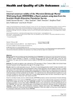
with uniaxial force sensors (MLP-50, Transducer Techniques, Temecula CA). A photograph of the loadcell setup
along with a schematic of the measured forces can be seen
in Figure 1.
An ADAL split-belt instrumented treadmill (TECHMACHINE, Andrézieux France; see Belli et al., 2001 for
detailed description [29]) was used below the Lokomat,
which allowed for ground reaction forces to be recorded
Electromyographic (EMG) recordings were collected from
the tibilias anterior, gastrocnemius, biceps femoris, vastus
medialis, rectus femoris, gluteus maximus, gluteus
medius, and adductor longus of both limbs in stroke sub-
Figure instrumentation
Setup of1
Setup of instrumentation. The photograph on the left shows the loadcells on the leg cuffs of the Lokomat which measure
the interactions between the subject and the device. The graphic on the right represents the recorded forces acting on a subject's right limb – ground reaction force, footstraps, and loadcells. Graphic adapted from Visual 3D (C-Motion INC, Rockville
MD).
Page 3 of 13
(page number not for citation purposes)
Journal of NeuroEngineering and Rehabilitation 2008, 5:19
jects and the left limb of four of the five control subjects
(one subject was improperly grounded and their EMG
data was not analyzed) using two Bagnoli-8 EMG system
(Delsys, Inc., Boston, MA). EMG data, along with the
forces and torques from the loadcells, were anti-alias filtered at 500 Hz prior to sampling at 1000 Hz using a 16bit data acquisition board (Measurement Computing,
PCI-DAS 6402, Middleboro, MA) and custom data acquisition software written in Matlab and stored for later analysis. Force plate data was further low-pass filtered using a
zero-delay fourth order Butterworth filter with a 25-Hz
cutoff frequency.
Protocol
The stroke subjects were first fitted with a harness so that
a portion of their body-weight could be supported while
control subjects did not wear the harness. Subjects were
led into the Lokomat and with the help of a physical therapist the device was adjusted so that the Lokomat hip and
knee centers lined up with those of the subject. After being
correctly aligned, the marker clusters were applied to the
subject's feet, shanks, and thighs. A neoprene band was
tightly wrapped around the subject's waist and individual
motion tracking markers were affixed to the boney landmarks of the pelvis.
After the subject was in the Lokomat, an experienced
physical therapist conducted a practice session for up to
2–3 minutes to allow the subject to acclimate to the
device. Stroke subjects began walking suspended above
the treadmill and the amount of body weight support provided by the accurate and constant Lokolift system [30]
was reduced until a minimum level that produced an
appropriate gait pattern was found. Inappropriate gait
patterns were judged by the physical therapists and
included such factors as impaired limb buckling during
stance, toe dragging through swing, and excessive trunk
movements that would not be analogous to a healthy gait
pattern. The levels of minimum body weight support
ranged from 11.5 to 25.6 percent of total body mass.
Following the acclimation period, the speed of the Lokomat was randomly adjusted to one of 4 different speeds
(1.5, 2.0, 2.5, and 3.0 km/hr), and after allowing the subject to acclimate to the new speed 30-seconds of data was
collected. The subject was told to try and match the kinematic pattern of the Lokomat to the best of their ability. It
should be noted that the Lokomat was run with 100%
guidance force under these trials, meaning the device was
in a pure position control mode rather than an impedance
mode. While the Lokomat has the ability to change the
amount of subject assistance, our goal was to determine
whether subjects assisted through physiological gait patterns produce symmetric, normal joint torques. For this,
position control mode was more appropriate than an
impedance mode. The remaining 3 speeds were tested in
/>
the same manner. Adequate rest breaks were taken
throughout the experiment to minimize fatigue. For the
purposes of this paper, only trials run at 2.5 km/hr are
reported.
Following all trials, a precision digitizing arm (MicroScribe MLX, Immersion, San Jose CA) was used to accurately locate the position of the Lokomat, load cells, and
foot lifter locations with respect to anatomical landmarks.
This information was necessary to determine the location
of the Lokomat forces acting on the subject's lower
extremities when computing the joint torques throughout
the gait cycle [24].
Data analysis
The vertical ground reaction forces were used to mark the
heel strike of each step, measured as the point were the
force exceeded 50 N. All experimental data (including that
calculated in Visual 3D) over the 30-second trials were
broken up into individual strides (from heel strike to heel
strike in the same leg), which were then resampled to the
same signal length. The subject kinematics calculated
from Visual 3D (limb segment center of mass location,
segment acceleration, joint center locations and limb segment locations) were combined with all the forces and
torques acting on the subject – the ground reaction forces
from the split-belt instrumented treadmill, as well as at
the Lokomat leg cuffs (location of the loadcells calculated
from the Lokomat potentiometers and digitized Lokomat
limb lengths) – into a custom inverse dynamics model
[24]. This model was then used to calculate joint torques
that the subjects were generating throughout the trial in
both the frontal and sagittal planes, as well as the torques
that the Lokomat were inducing on the subject. For each
subject, the data generated for all steps within a 30-second
trial was averaged for each limb.
Statistical analysis
A total of 5 kinematic and 5 kinetic measures of the profiles of the impaired, unimpaired, and control limbs (left
limb) were compared using a single factor ANOVA. The
kinematic measures were ankle, knee and hip range of
motion (ROM), maximum vertical pelvic displacement
from heelstrike, and the time in the gait cycle at which the
minimum pelvic displacement occurred. The kinetic
measures were maximum vertical ground reaction force,
maximum ankle dorsiflexion torque, magnitude of knee
extension torque at the midpoint of the initial swing
phase (68.5% gait cycle), the time at which the maximum
hip extension torque occurred, and the magnitude of the
hip adduction torque at mid swing (80% gait cycle). A
Bonferroni correction was used to reduce the risk of Type
I errors, so that with 10 measures tested, a α = 0.005 was
used for all comparisons.
Page 4 of 13
(page number not for citation purposes)
Journal of NeuroEngineering and Rehabilitation 2008, 5:19
The EMG activity from the selected muscle groups was
band-pass filtered (20–450 Hz), full-wave rectified, and
then smoothed using a 200-point RMS algorithm. For
each muscle recorded, the EMG traces were normalized to
that subject's highest value recorded across all trials to
allow for inter-subject comparison. The mean normalized
EMG trace for each subject was broken up into seven
phases of the gait cycle (initial loading 0–12%, midstance 12–30%, terminal-stance 30–50%, pre-swing 50–
62%, initial-swing 62–75%, mid-swing 75–87%, terminal-swing 87–100%) and each section integrated as in
Hidler and Wall [31].
Results
Kinematics
The mean ankle, knee and hip sagittal plane joint angles
for all three limbs tested (impaired, unimpaired, control)
are shown in Figure 2 with specific values found in Table
1. In general, there were only slight differences in the kinematic patterns exhibited between the control subjects
and the impaired and unimpaired limbs of the stroke subjects. At toe-off control subjects had a larger peak plantarflexion angle than either stroke ankle, but the impaired
ankle was slightly more plantarflexed throughout the rest
of the gait cycle. The knee angles were quite similar,
although the impaired knee tended to be slightly more
extended through the gait cycle, resulting in a peak flexion
angle through swing that was lower than either the unimpaired or control limb. The hip angles were similar as
well, with the impaired hip being more extended throughout the gait cycle, and the unimpaired hip being more
flexed, especially terminal swing and initial loading.
Figure 3 shows the mean vertical displacement of the pelvis center of gravity of the stroke and control groups from
heelstrike of the left leg (control) or unimpaired leg
(stroke) to single support on the left/unimpaired limb,
then to double limb support, and finishing with single
limbs support on the right/impaired limb. The pelvis of
stroke subjects consistently raised up higher during unimpaired limb support than during impaired limb support,
and the minimum pelvic height following unimpaired
limb support comes later in the gait cycle than the minimum pelvic height following normal single limb support.
The frontal plane angles were also derived and in general,
there was very little movement in the frontal plane, and
no differences between the three limbs tested.
Table 1 lists the average value, standard error of the mean,
and p-values for the 5 kinematic measures tested. There
were no significant kinematic differences between the
control limb and the unimpaired limb of stroke subjects,
no significant differences between the impaired limb and
control limb, and only 1 significant difference between
the impaired and unimpaired limb (ankle ROM).
/>
Kinetics
The mean vertical ground reaction forces (GRFs) throughout the gait cycle of the impaired, unimpaired, and control limbs are presented in Figure 4. For both the control
and stroke subjects, the vertical GRFs did not demonstrate
the classic double bump throughout stance. Since the
Lokomat is supported on a parallelogram that is supported by a large spring, the Lokomat maintains continuous upward lift to the subject through stance. While all
three traces follow similar paths for the 3 limbs, the
ground reaction force of the impaired limb tended to be
lower in magnitude than the unimpaired limb, which in
turn was less than the control. None of these differences
reached the significant level, presumably due to the large
variability in these measures.
The mean sagittal and frontal plane joint torques for the
ankle, knee, and hip for all three limbs as they progress
through the gait cycle is shown in Figure 5. Upon general
visual inspection, the sagittal ankle torques of the unimpaired and control limb follow very similar patterns,
whereas the sagittal ankle torque in the impaired limb of
stroke subject was quite different, with less dorsiflexion at
initial contact and continuous ankle extension during
swing. The diminished dorsiflexion results from the subject wearing the foot lifter, which reduces the need to flex
the ankle as it makes contact with the treadmill belt. Similarly, the continuous active ankle extension torque during swing results from the subject trying to extend their
ankle to a more neutral position. In the frontal plane,
stroke subjects exhibited larger eversion torques during
stance in both limbs. Neither of these torque profiles were
similar to the frontal plane torques in the control subjects,
where controls had a lower eversion torque during early to
mid stance and an inversion torque during late stance and
toe-off.
The knee torques generated in the sagittal plane in both
the impaired and unimpaired knees of the stroke subjects
follow similar patterns during stance, with lower extension torques than the controls in early stance. In midstance, stroke subjects tend to flex their knees to a greater
extent than controls in both limbs. From toe-off through
swing, the unimpaired limb behaved similar to the control limbs, but the impaired limb demonstrated a consistent, large extension torque at toe-off that is higher than
both the control and unimpaired limbs. In the frontal
plane, the unimpaired knee behaves similar to the control
knee but, with slightly less varus torque in early-stance.
The impaired limb is drastically different than the other
two torque profiles, where there were significant valgus
torques during mid to late stance as well as less valgus
through swing.
All 3 sagittal hip torques follow very similar patterns with
a few noteworthy differences. The maximum extension
Page 5 of 13
(page number not for citation purposes)
Journal of NeuroEngineering and Rehabilitation 2008, 5:19
/>
Figure 2
Joint kinematics
Joint kinematics. Mean sagittal joint angles of the ankle, knee, and hip through the gait cycle (from heelstrike to heelstrike).
Control – black, unimpaired – green, impaired – red. Shaded region represents 95% CI.
Page 6 of 13
(page number not for citation purposes)
Journal of NeuroEngineering and Rehabilitation 2008, 5:19
/>
Table 1: Mean kinematic measures.
Control
Ankle ROM
Knee ROM
Hip ROM
Time of Pelvis Min
Pelvis Max
Unimpaired
p vs Control
Impaired
p vs Control
p vs Unimpaired
28.53 (4.96)
57.21 (1.64)
44.47 (1.60)
3.79 (1.36)
0.89 (0.11)
28.98 (1.80)
57.07 (1.34)
48.38 (2.25)
4.16 (0.45)
1.23 (0.20)
0.918
0.952
0.273
0.750
0.260
17.75 (1.80)
53.18 (2.32)
41.84 (1.89)
7.97 (1.22)
0.76 (0.11)
0.025
0.273
0.387
0.056
0.476
<.001*
0.164
0.039
0.009
0.050
Standard error of the mean in parenthesis. * represents significant difference (p < .005).
torque was much greater in the unimpaired limb than the
impaired limb in early stance, peaking later in the gait
cycle that either of the other limbs. Surprisingly, the average maximum flexion torque was greater in the impaired
limb than the unimpaired limb or the control subjects at
the end of stance. In the frontal plane the impaired stroke
limb produced less abduction during stance and more
abduction during early to mid-swing than either the
unimpaired or control limbs.
Table 2 lists the average value, standard error of the mean,
and p-values for the 5 kinetic measures tested. There were
no significant differences between the control limb and
the unimpaired limb of stroke subjects, 3 significant differences between the impaired limb and control limb
(maximum ankle dorsiflexion, knee extension at initial
swing, and hip adduction at mid swing), and 3 significant
differences between the impaired and unimpaired limb
(maximum ankle dorsiflexion, knee extension at initial
swing, and hip adduction at mid swing).
Figure 3
Pelvic motion
Pelvic motion. Mean vertical displacement of the pelvis
center of gravity from heelstrike and single support of the
left/unimpaired limb to double limb support, and finishing
with single limb support on the right/impaired limb. Shaded
region represents 95% CI.
EMG
The mean integrated muscle activity of the eight muscle
groups for the impaired, unimpaired, and control limbs
over the seven phases of the gait cycle are shown in Figure
6. For the most part, the three groups behave quite similarly. In the gastrocnemius the impaired limb had slightly
higher activity during swing. In the tibilias anterior, mean
activity was 41% less in the impaired limb compared to
controls at terminal swing. During initial contact, the
biceps femoris on the impaired limb was slightly higher
and the vastus medialis was slightly lower than the other
two groups, but through the rest of gait these muscles were
very similar to unimpaired and control levels. There were
no notable differences between the gluteus medius activity of the three groups. The rectus femoris in the impaired
limb had a level of activity that was consistently higher
than the other two limbs (e.g. unimpaired and controls),
this mean activity was at least 2.6 times higher than the
control group from pre-swing through terminal swing.
This higher level of activity in the impaired limb was also
true in the adductor longus, but here the impaired values
were at least 53% higher than the unimpaired values dur-
Figure reaction force
Ground 4
Ground reaction force. Mean vertical ground reaction
force through the gait cycle. Control – black, unimpaired –
green, impaired – red. Shaded region represents 95% CI.
Page 7 of 13
(page number not for citation purposes)
Journal of NeuroEngineering and Rehabilitation 2008, 5:19
/>
Figure 5
Joint kinetics
Joint kinetics. Mean sagittal (top) and frontal (bottom) joint torques of the ankle, knee, and hip through the gait cycle. Control – black, unimpaired – green, impaired – red. Shaded region represents 95% CI.
ing terminal stance and pre-swing, and 2.4 times greater
than controls during pre-swing. All three groups produced
similar levels of gluteus maximus activity throughout the
gait cycle.
Lokomat torques
The mean sagittal and frontal torques induced by the
Lokomat at the subject's knee and hip throughout the gait
cycle are shown in Figure 7. The Lokomat consistently
induces low levels of torque in the frontal plane of all
three subject groups at the hip and knee, but there is notable variability in the sagittal plane.
At the knee, the control subjects experience low levels of
sagittal knee torque from the Lokomat throughout the
gait cycle. However, the unimpaired limb experiences a
greater extension torque at the knee from the Lokomat
during terminal swing and initial contact than either the
impaired or control limbs. The impaired limb experiences
a greater flexion torque at the knee from the Lokomat during late stance and pre-swing than the unimpaired and
control limbs.
At the hip, the Lokomat imparts an extension torque on
the control hip that starts at heelstrike, peaks during early
stance, and returns to zero by mid-stance. From mid to
late stance, the Lokomat produces a flexion torque on the
Table 2: Mean kinetic measures.
Control
GRF max
Ankle Flexion
Knee Extension
Initial Swing
Time of max
Hip Extension
Hip Adduction
Mid Swing
Unimpaired
p vs Control
Impaired
p vs Control
p vs Unimpaired
9.09 (0.36)
0.18 (0.03)
0.10 (0.03)
8.75 (0.49)
0.23 (0.04)
0.03 (0.03)
0.658
0.446
0.097
8.65 (0.41)
0.06 (0.02)
0.27 (0.02)
0.506
0.003*
0.001*
0.884
0.001*
<.001*
3.02 (1.15)
6.61 (0.87)
0.030
3.12 (1.31)
0.963
0.04
0.26 (0.07)
0.22 (0.06)
0.613
-0.07 (0.04)
0.001*
0.001*
Standard error of the mean in parenthesis. * represents significant difference (p < .005).
Page 8 of 13
(page number not for citation purposes)
Journal of NeuroEngineering and Rehabilitation 2008, 5:19
/>
Figure 6
Muscle activity
Muscle activity. Integrated mean normalized EMG values over the seven gait phases. Control – black, unimpaired – green,
impaired – red. Error bar represents standard error of the mean.
control subjects, followed by another slightly larger extension torque peak through swing. Both stroke limbs feel
this peak-valley-peak of Lokomat-induced torque at the
hip but it is shifted during early stance. Instead of an
extension torque being imparted on the hip by the Lokomat, the stroke limbs feel decreasing flexion torques.
Beyond mid stance, the impaired limb feels similar torques from the Lokomat as the control subjects, but the
unimpaired hip feels a much higher level of flexion at preswing and terminal swing. Statistical differences between
the induced Lokomat torques on the three limbs were not
found.
Discussion
The joint angles that both limbs of stroke subjects produce while walking in the Lokomat are similar to healthy
subjects, which is not surprising considering the Lokomat
guides subjects through a prescribed kinematic pattern.
Even at the ankle, which is not driven by the Lokomat,
subjects produce similar overall patterns in the sagittal
plane. Despite the kinematic patterns being similar, stroke
subjects still generated abnormal joint torque patterns,
particularly in the impaired limb. Many of these abnormal patterns are consistent with the clinical characteristics
of overground hemiparetic gait. Evidence of hip hiking,
stiff legged gait with impaired limb circumduction, and
asymmetric limb support times are still present in the
Lokomat, suggesting the inability of hemiparetic stroke
subjects to break out of stereotypical abnormal motor
behaviors even when the movement tasks are simplified.
The fact that there are no significant kinematic differences
between the stroke limbs and the control limbs can be
attributed to the fact that the Lokomat is guiding the limbs
of all subjects through pre-programmed trajectories. We
have previously shown that despite being firmly strapped
into the Lokomat, subjects retain the ability to move and
shift relative to the device [28]. Nevertheless, the angles
produced at the ankle, hip, and knee are quite similar,
even for the impaired limb of stroke subjects. The lone
kinematic difference is the ankle ROM between the
impaired limb and unimpaired limb. Since the Lokomat
does not drive the ankle, passive footstraps are often
applied to the impaired ankle, which assist dorsiflexion
during swing yet limit the extent of ankle plantarflexion.
It was expected that these footstraps would limit ROM for
the impaired limb compared to both the control and
unimpaired limbs. Surprisingly, differences were only significant when compared to the unimpaired limb and not
the control ankle ROM.
While there were no significant differences found for the
knee or hip kinematic patterns, the data presented in Figure 2 show some noteworthy characteristics. The differences between the knee impaired limb and both the
control or unimpaired limb can be explained in part by
Page 9 of 13
(page number not for citation purposes)
Journal of NeuroEngineering and Rehabilitation 2008, 5:19
/>
Figure 7
Robot induced torques
Robot induced torques. Mean torques induced at the joints by the Lokomat through the gait cycle. Control – black, unimpaired – green, impaired – red. Shaded region represents 95% CI. Vertical axis are scaled to match corresponding subject generated torques of figure 5.
stroke subjects trying to hyperextend the impaired knee
while standing. If stroke subjects comfortably stand with
the impaired limb in hyperextension, and are then set-up
in the Lokomat as such, the sagittal kinematic pattern of
the knee will be shifted slightly in extension. The average
difference between the unimpaired and impaired limbs
was 5 degrees, noticeable, but possibly clinically insignificant. The kinematic differences at the hip between the
unimpaired and impaired limb can be attributed to stroke
subjects attempting to get off of the impaired limb and
quickly back onto the unimpaired limb during normal
ambulation. In the Lokomat, this strategy manifests as a
lower range of motion of the impaired hip, and the flexion and extension peaks appear to be reached at an earlier
part of the gait cycle.
The pelvic vertical displacement of healthy subjects
traveled in the standard symmetric sinusoidal pattern,
while the pelvis of stroke subjects was higher during the
unimpaired single limb support phase than during the
impaired single limb support phase. This is consistent
with the common strategy of stroke subjects who hike the
hip up while on the unimpaired limb in order to have
enough clearance for the impaired limb to swing through.
One may believe that since the Lokomat imparts a physiological kinematic pattern that this hip hiking would be
unnecessary. However as described in the results section,
even though the stoke subject's kinematics are being
guided by the Lokomat, they still have a tendency to
extend their knee in pre-swing and also abduct their hip in
mid swing. Both of these stereotypical strategies result in
hip hiking as we saw in Figure 3. The Lokomat further
Page 10 of 13
(page number not for citation purposes)
Journal of NeuroEngineering and Rehabilitation 2008, 5:19
exacerbates this behavior since subjects cannot shift their
weight to the contralateral limb nor can they abduct their
leg to help with toe clearance. As a result, subjects tend to
hip hike for toe clearance. It is also worth mentioning that
the valleys of the pelvic traces that represent double limb
support are farther apart in the gait cycle between unimpaired limb single stance than impaired limb single stance
(not significantly different, p=.009), again, consistent
with the habit of spending more time on the unimpaired
limb.
As can be seen from the Table 2 there are no significant
kinetic differences between the unimpaired limb and the
control limb for the measures tested here. The vertical
ground reaction forces of all three limbs lack the characteristic double peak, which is typical in the Lokomat [24].
Even with a minimal amount of body weight support, the
vertical ground reaction forces of the stroke limbs were
lower, but not significantly different. Generally, stroke
subjects do not (or cannot) generate enough hip extension torque to carry the pelvis over the impaired limb
while it is in stance, and thus rush the gait cycle while on
the impaired limb and extend the gait cycle while on the
unimpaired limb. This behavior is still present during
Lokomat-assisted treadmill walking. The unimpaired hip
reaches its maximum extension later during early stance
than the controls or impaired limb; this may be due to
stroke subjects taking more time to vault the pelvis over
the unimpaired limb as the impaired limb goes through
swing. From Figure 3 it can be seen that the hip extension
torque during early stance of the impaired limb is lower
than that of the unimpaired limb. This can again be
explained by the stroke subject's tendencies to not fully
extend the impaired hip to pull the pelvis forward but to
quickly get off of the impaired limb during overground
ambulation.
From Figure 3 it can be seen that the control subjects reach
maximum hip abduction during mid-stance while the
unimpaired limbs reach maximum hip abduction during
late stance. If hip abduction torques are present to keep
the pelvis level during single limb support, this may help
explain the differences in hip abduction torques. If the
impaired limb is slow to enter swing, it could be rationalized that the maximum abduction torque of the unimpaired limb would occur later. It was also observed that
the unimpaired limb exhibits greater knee flexion during
early stance. This again may be to help vault the pelvis
over the unimpaired limb.
In the frontal plane, the stroke subjects lack any appropriate adduction torques of the impaired limb. Both the control and unimpaired limbs adduct the hip during swing
and early stance, presumably to keep the limb within the
workspace of the Lokomat and treadmill. But the
/>
impaired limb does the opposite and abducts or pushes
against the Lokomat through swing. This behavior is likely
a strategy to overcome a lack of knee flexion during swing
by circumducting the impaired limb outward to achieve
toe clearance through swing. While the Lokomat resists
much of this motion, subjects are able to over-power the
device slightly and achieve some out of plane movement.
This abnormal knee behavior of the impaired limb
presents itself as a significantly excessive extension torque
during initial swing. Again, this is part of the circumduction strategy of stroke subjects, where the knee is extended
or stiffened before the whole limb is swung out and
around during swing.
The impaired ankle exhibits little dorsiflexion and very
quickly transfers to plantarflexion during stance. This lack
of dorsiflexion may be due to the fact that the footlifter is
flexing the ankle for the stroke subject, and there is no
need for voluntary dorsiflexion of the impaired ankle.
Throughout stance of healthy over ground gait, the ankle
everts as a mechanism to prevent outward rolling of the
ankle. But in the Lokomat, we have found that healthy
subjects will invert during late stance and toe-off, presumably in response to the lack of lateral sway of the hips
above and the need to propel the limb straight forward
through swing. Both ankles of stroke subjects do not
behave in this manner in the Lokomat, instead producing
eversion torques during stance similar to healthy over
ground walking. The unimpaired limb will produce a
small inversion torque at toe-off, but from Figure 3 this
force appears to be smaller and occur later in the gait cycle
than in the control population.
The source of some of the abnormal torque behaviors of
stroke subjects during Lokomat assisted treadmill walking
can be traced back to the muscle activation patterns. The
lower tibilias anterior activity during early stance and
higher gastrocnemeous activity during swing can account
for the different sagittal torque profile of the impaired
ankle. Unfortunately the excessive impaired knee extension during toe off does not show up as a higher vastus
medialias activity, yet could result from other knee extensor muscles that were not recorded from. A trend that may
explain the low knee extension of both the impaired and
unimpaired limbs during mid stance is the higher biceps
femoris activity, possibly causing antagonistic activity and
thus reducing the net torque. The rectus femoris activity of
the impaired limbs of stroke subjects is higher throughout
the gait cycle than either the unimpaired or impaired
limbs but the impaired limb does not exhibit higher hip
flexion torques. However, the impaired hip does exhibit
lower extension torques, another instance of possible
antagonistic activity. Similar findings have been shown in
our previous work with stroke subjects performing isometric tasks [3]. And despite high aductor longus activity
Page 11 of 13
(page number not for citation purposes)
Journal of NeuroEngineering and Rehabilitation 2008, 5:19
/>
in the impaired limbs, stroke subjects were not able to
generate adequate hip adduction torques. Again, it is possible that other muscles that were not recorded from may
have provided some sort of antagonistic behavior. While
spasticity can contribute to abnormal torque patterns in
the limbs of stroke subjects [32], the subjects tested here
were pre-screened by a physical therapist and omitted
from the study if they had limited range of motion or significant muscle tone.
achieved, which has recently been shown in acute stroke
subjects following 4 weeks of Lokomat training [21]. The
purpose of this study was to simply determine whether
stroke subjects guided through a physiological gait pattern
would demonstrate symmetric, normal joint torques. Our
results indicate that despite symmetric, normal kinematics, the kinetic patterns of stroke subjects are consistent
with the stereotypical patterns often exhibited during
over-ground walking.
With the custom inverse dynamics model used, it was possible to calculate the forces that the Lokomat was applying
to the subject's hips and knees throughout the gait cycle.
In many cases it can be seen that the Lokomat was trying
to augment the torque patterns of the stroke subjects to
better match that of the controls. At the knee, the Lokomat
induced minimal forces on the control subjects, meaning
that the control subjects did a fine job of matching the
Lokomat. During late swing and early stance, the unimpaired limb exhibited more knee flexion than the controls
and thus the Lokomat produced extension torques about
the knee. Similarly, while the impaired knee was producing high extension torque during toe off, the Lokomat
tried to correct this behavior by applying a flexion torque.
The Lokomat did apply torques to the hips of the control
subjects but they are physiologically correct – extension
during early to mid stance, flexion from mid stance to toe
off and extension through swing. The unimpaired limb of
stroke subjects exhibited higher levels of extension and
less flexion than the controls through stance and thus the
Lokomat was constantly producing a flexion torque on
the unimpaired hip through stance. During mid swing the
unimpaired limb behaved similar to the control limbs
and the Lokomat torques applied to each limb were also
similar. Yet at the very end of swing, the Lokomat firmly
flexed the hip, but this is not in response to a voluntary
extension on the part of the unimpaired limb, for the subjects' hip torques were minimal at this time. It seems as if
the stroke subjects simply relaxed and allowed the Lokomat to guide their leg into heel contact. The impaired hip
did not produce any torques that are significantly different from controls, but the Lokomat torques applied to the
impaired limb are only similar to those applied to the
control limb from late stance through early swing. During
mid stance the Lokomat produced minimal torques about
the hip and yet the impaired limb extended slightly less
than the control limb. And as in the unimpaired limb, the
impaired limb expressed very low voluntary hip torques,
so the Lokomat supplied most of the required hip flexion
during late swing.
Conclusion
It should be noted that the results of this study do not
indicate individuals following stroke cannot be trained to
walk symmetrically in the Lokomat since only one test session was run. Over time, gains in symmetry may be
4.
As would be expected with Lokomat assisted walking,
there were very few kinematic differences between the
impaired, unimpaired, and control limbs as subjects
walked in the device. However, the torques that stroke
subjects produce while moving through these patterns are
both asymmetrical and quite different from age matched
controls. Based on our findings, it appears that hemiparetic stroke subjects cannot break out of stereotypical
abnormal motor behaviors in their lower extremities even
when the complexity of the task is reduced. Future studies
will investigate whether long-term symmetric training
with devices such as the Lokomat leads to more symmetric
motor patterns and ultimately improved walking ability
in hemiparetic stroke subjects.
Competing interests
The authors declare that they have no competing interests.
Authors' contributions
NN designed the experiment, carried out the experiments,
collected and analyzed the data, and drafted the manuscript. NB prepared subjects and assisted with the experiments. DN prepared subjects and assisted with the
experiments. JH designed the experiment, developed the
instrumentation and data collection software, and helped
draft the manuscript. All authors read, edited, and
approved the final manuscript.
Acknowledgements
We would like to extend our sincere thanks to the subjects who participated in the study, as well as the students who helped in the data collection.
This work was funded by the Whitaker Foundation (Arlington, VA; PI: J.
Hidler).
References
1.
2.
3.
5.
Bourbonnais D, Noven S Vanden: Weakness in patients with
hemiparesis. Am J Occup Ther 1989, 43(5):313-9.
Duncan PW, Badke MB: Stroke Rehabilitation: The Recovery of Motor
Control Chicago: Year Book Medical Pub; 1987.
Neckel N, Pelliccio M, Nichols D, Hidler J: Quantification of functional weakness and abnormal synergy patterns in the lower
limb of individuals with chronic stroke. J Neuroeng Rehabil. 2006,
3:17.
Hidler JM, Carroll M, Federovich EH: Strength and coordination
in the paretic leg of individuals following acute stroke. IEEE
Trans Neural Syst Rehabil Eng 2007, 15(4):526-34.
Chung SG, Van Rey E, Bai Z, Roth EJ, Zhang LQ: Biomechanic
changes in passive properties of hemiplegic ankles with spastic hypertonia. Arch Phys Med Rehabil 2004, 85(10):1638-46.
Page 12 of 13
(page number not for citation purposes)
Journal of NeuroEngineering and Rehabilitation 2008, 5:19
6.
7.
8.
9.
10.
11.
12.
13.
14.
15.
16.
17.
18.
19.
20.
21.
22.
23.
24.
25.
26.
27.
28.
29.
30.
Corcos DM, Gottlieb GL, Penn RD, Myklebust B, Agarwal GC:
Movement deficits caused by hyperexcitable stretch reflexes
in spastic humans. Brain 1986, 109(Pt 5):1043-58.
Bobath B: Adult Hemiplegia: Evaluation and Treatment London: William
Heinnemann; 1978.
Black I, Nichols D, Pelliccio M, Hidler J: Quantification of reflex
activity in stroke survivors during an imposed multi-joint leg
extension movement. Exp Brain Res 2007, 183(2):271-81.
Knuttson E, Richards C: Different types of disturbed motor control in gait of hemiparetic patients. Brain 1979, 102:405-430.
Knutsson E, Martensson A: Dynamic motor capacity in spastic
paresis and its relation to prime mover dysfunction, spastic
reflexes and antagonist co-activation. Scand J Rehabil Med 1980,
12(3):93-106.
Chae J, Yu DT: Neuromuscular electrical stimulation for
motor restoration in hemiparesis. Top Stroke Rehabil 2002,
8(4):24-39.
Dean CM, Richards CL, Malouin F: Walking speed over 10
metres overestimates locomotor capacity after stroke. Clin
Rehabil 2001, 15:415-421.
Von Schroeder HP, Coutts RD, Lyden PD, Billings E: Gait parameters following stroke: a practical assessment. J Rehabil Res Dev
1995, 32:25-31.
Olney SJ, Griffin MP, Monga TN, McBride ID: Work and power in
gait of stroke patients. Arch Phys Med Rehabil 1991, 72:309-314.
Titianova EB, Tarkka IM: Asymmetry in walking performance
and postural sway in patients with chronic unilateral cerebral infarction. J Rehabil Res Dev 1995, 32(3):236-44.
Hassid E, Rose D, Commisarow J, Guttry M, Dobkin BH: Improved
gait symmetry in hemiparetic stroke patients induced during body weight supported treadmill stepping. J Neurol Rehabil
1997, 11(1):21-26.
Reisman DS, Wityk R, Silver K, Bastian AJ: Locomotor adaptation
on a split-belt treadmill can improve walking symmetry
post-stroke. Brain 2007, 130(Pt 7):1861-72.
Colombo G, Joerg M, Schreier R, Dietz V: Treadmill training of
paraplegic patients using a robotic orthosis. J Rehabil Res Dev
2000, 37(6):693-700.
Hidler J, Nichols D, Pelliccio M, Brady K: Advances in the understanding and treatment of stroke impairment using robotic
devices. Top Stroke Rehabil 2005, 12(2):22-35.
Mayr A, Kofler M, Quirbach E, Matzak H, Frohlich K, Saltuari L: Prospective, blinded, randomized crossover study of gait rehabilitation in stroke patients using the Lokomat gait orthosis.
Neurorehabil Neural Repair 2007, 21(4):307-14.
Husemann B, Müller F, Krewer C, Heller S, Koenig E: Effects of
locomotion training with assistance of a robot-driven gait
orthosis in hemiparetic patients after stroke: a randomized
controlled pilot study. Stroke 2007, 38(2):349-54.
Chen G, Patten C: Joint moment work during the stance-toswing transition in hemiparetic subjects. J Biomech 2008,
41(4):877-83.
Kerrigan DC, Karvosky ME, Riley PO: Spastic paretic stiff-legged
gait: joint kinetics. Am J Phys Med Rehabil 2001, 80(4):244-9.
Hidler J, Neckel N: Inverse-dynamics based assessment of gait
using a robotic orthosis. Conf Proc IEEE Eng Med Biol Soc 2006,
1:185-8.
Folstein MF, Folstein SE, McHugh PR: "Mini-mental state". A
practical method for grading the cognitive state of patients
for the clinician. J Psychiatr Res 1975, 12(3):189-98.
Fugl-Meyer AR, Jääskö L, Leyman I, Olsson S, Steglind S: The poststroke hemiplegic patient. 1. a method for evaluation of
physical performance. Scand J Rehabil Med 1975, 7(1):13-31.
Neckel ND, Hidler JM: Method for Motion Tracking Inside the
Lokomat Robotic Orthosis. Proceedings of the American Society of
Biomechanics Annual Conference: 6–9 Sept 2006; Blacksburg. Meeting
chair Stefan Dumas .
Neckel N, Wisman W, Hidler J: Limb alignment and kinematics
inside a Lokomat robotic orthosis. Conf Proc IEEE Eng Med Biol
Soc 2006, 1:2698-701.
Belli A, Bui P, Berger A, Geyssant A, Lacour JR: A treadmill ergometer for three-dimensional ground reaction forces measurement during walking. J Biomech 2001, 34(1):105-12.
Frey M, Colombo G, Vaglio M, Bucher R, Jörg M, Riener R: A novel
mechatronic body weight support system. IEEE Trans Neural
Syst Rehabil Eng 2006, 14(3):311-21.
/>
31.
32.
Hidler JM, Wall AE: Alterations in muscle activation patterns
during robotic-assisted walking. Clin Biomech (Bristol, Avon) 2005,
20(2):184-93.
Mirbagheri MM, Alibiglou L, Thajchayapong M, Rymer WZ: Muscle
and reflex changes with varying joint angle in hemiparetic
stroke. J Neuroeng Rehabil 2008, 27(5):6.
Publish with Bio Med Central and every
scientist can read your work free of charge
"BioMed Central will be the most significant development for
disseminating the results of biomedical researc h in our lifetime."
Sir Paul Nurse, Cancer Research UK
Your research papers will be:
available free of charge to the entire biomedical community
peer reviewed and published immediately upon acceptance
cited in PubMed and archived on PubMed Central
yours — you keep the copyright
BioMedcentral
Submit your manuscript here:
/>
Page 13 of 13
(page number not for citation purposes)