Báo cáo hóa học: "Short-term locomotor adaptation to a robotic ankle exoskeleton does not alter soleus Hoffmann reflex amplitude" doc
Bạn đang xem bản rút gọn của tài liệu. Xem và tải ngay bản đầy đủ của tài liệu tại đây (662.67 KB, 8 trang )
RESEA R C H Open Access
Short-term locomotor adaptation to a robotic
ankle exoskeleton does not alter soleus
Hoffmann reflex amplitude
Pei-Chun Kao
1*
, Cara L Lewis
2
, Daniel P Ferris
1
Abstract
Background: To improve design of robotic lower limb exoskeletons for gait rehabilitation, it is critical to identify
neural mechanisms that govern locomotor adaptation to robotic assistance. Previously, we demonstrated soleus
muscle recruitment decreased by ~35% when walking with a pneumatically-powered ankle exoskeleton providing
plantar flexor torque under soleus proportional myoelectric control. Since a substantial portion of soleus activation
during walking results from the stretch reflex, increased reflex inhibition is one potential mechanism for reducing
soleus recruitment when walking with exoskeleton assistance. This is clinically relev ant because many
neurologically impaired populations have hyperactive stretch reflexes and training to reduce the reflexes could lead
to substantial improvements in their motor ability. The purpose of this study was to quantify soleus Hoffmann (H-)
reflex responses during powered versus unpowered walking.
Methods: We tested soleus H-reflex responses in neurologically intact subjects (n=8) that had trained walking with
the soleus controlled robotic ankle exoskeleton. Soleus H-reflex was tested at the mid and late stance while
subjects walked with the exoskeleton on the treadmill at 1.25 m/s, first without power (first unpowered), then with
power (powered), and finally without power again (second unpowe red). We also collected joint kinematics and
electromyography.
Results: When the robotic plantar flexor torque was provided, subjects walked with lower soleus
electromyographic (EMG) activation (27-48%) and had concomitant reductions in H-reflex amplitude (12-24%)
compared to the first unpowered condition. The H-reflex amplitude in proportion to the background soleus EMG
during powered walking was not significantly different from the two unpowe red conditions.
Conclusion: These findings suggest that the nervous system does not inhibit the soleus H-reflex in response to
short-term adaption to exoskeleton assistance. Future studies should determine if the findings also apply to long-
term adaption to the exoskeleton.
Background
Many research groups are developing robotic lower limb
exoskeletons to assist in locomotion training after neu-
rological injury [1-6]. The exoskeletons are intended to
reduce manual effort from therapists and improve reha-
bilitation outcomes. Though reducing manual effort
from therapists is clearly being achieved by current
devices, results for improving rehabilitation outcomes
are still equivocal. Studies have demonstrated that the
choice of computer control algorithms for robotic g ait
devices can affect the process of motor learning to
robotic assistance [2,7-11]. However, there is no clear
theory on how different control algorithms specifical ly
alter mechanisms or aspects of neural control [12,13].
To design better robotic gait devices that can enhance
therapy, it is critical to identify neural mechanisms that
govern locomotor adaptation to robotic assistance.
In recent studies from our laboratory, we examined
how healthy young subjects adapted to a robotic ankle
exoskeleton during walking [14,15]. The exoskeleton
provided plantar flexor torqu e under proportional myo-
electric control of soleus electromyographic (EMG)
* Correspondence:
1
School of Kinesiology, University of Michigan, Ann Arbor, Michigan 48109-
2214, USA
Kao et al. Journal of NeuroEngineering and Rehabilitation 2010, 7:33
/>JNER
JOURNAL OF NEUROENGINEERING
AND REHABILITATION
© 2010 Kao et al; licensee BioMed Central Ltd. This is an Open Access article distributed under the terms of the Creative Commons
Attribution License ( which permits unrestricted use, distr ibution, and reproduction in
any medium, provided the original work is properly cited.
activation. We have focused on the ankle joint because
it produces a majority of the positive mechanical work
during stance in h uman walking [16] and insuffic ient
plantar flexor torque generat ion has been shown to be a
major factor limiting mobility after neurological injuries
[17-19]. When the robotic assistance was first intro-
duced, subjects walked on the ball of their foot during
stance due to the increased plantar flexion torque. After
two thirty-minute training sessions three days apart,
subjects had reduced soleus muscle activation by ~35%
and walked smoothly with the exoskeleton mechanical
assistance. A large portion of soleus muscle activation is
a direct result of proprioceptive feedback, including th e
stretch reflex response [20-27]. Thus, the nervous sys-
tem could inhibit reflex activation during walking with
the exoskeleton as a mechanism for reducing soleus
recruitment.
Increased stretch reflex inhibition with robotic exoske-
leton training would be particularly relevant to gait
rehabilitation for individuals after neurological injuries.
Individuals who had stroke, spi nal cord injury, cerebral
palsy, and traumatic brain injury often demonstrate
abnormally high stretch reflexes that substa ntially affect
their movement capabilities [28-34]. A number of
research groups have been investigating training meth-
ods to inhibit reflexes and their results demonstrated
that reflex responses can be manipulated both in patient
populations [28,35-37] and neurologically intact subjects
[38-42]. Chen et al (2006) concluded that conditioning
of reflex responses in a rat model can improve func-
tional locomotion after spinal cord injury [37]. If a
robotic exoskeleton could be used to induce an altera-
tion of reflex responses during human walking, it would
have considerable potential as an aid for gait rehabilita-
tion in addition to reducing manual assistance from the
therapists. The added m echanical torque provided by
the robotic exoskeleton may enhance motor adaptation
as subjects would need to tune their muscle activations
correctly by normalizing the exaggerated reflexes.
The purpose of this study was to quantify soleus reflex
responses in neurologically intact subjects trained to
walk with the robotic ankle exoskeleton. By identi fying
how devices modify musculoskeletal and neural systems
with use in neurologically intact subjects, researchers
and clinicians have a much better chance of determining
which patient populations might benefit from practice
with the robotic devices. We used the Hoffmann (H-)
reflex, an electrical analogue of the stretch reflex, to
examine soleus reflex responses during walking both
with the exoskeleton powered and with the exoskeleton
unpowered. The H-reflex is elicited by stimulating the
afferent nerve (Ia sensory) directly and bypassing the
muscle spindle. H-reflex measurements have been
extensively used to study how the stretch reflex is
modulated centrally [43-45]. The H-reflex is highly task-
dependent and is modulated frequently both within a
gait cycle and during different motor behaviors
[43,44,46-49]. A reduction in H-reflex amplitude has
been associated with mastering new motor tasks such as
balancing during standing [39,40], perturbed cycling
[38], and backward walking tasks [41,50]. In a pilot
study, a single subject that had trained with the ankle
exoskeleton for several years demonstrated a much
lower H-reflex amplitude in proportion to the back-
ground EMG during powered walking compared to dur-
ing unpowered walking [51]. Based on that finding, we
hypothesized that subjects would have lower H-reflex
magnitudes when normalized to background soleus
activity during adapted p owered walking than during
unpowered walking. In this study, we tested eight sub-
jects who had trained to walk with the robotic ankle
exoskeleton for two training sessions. A previous study
demonstrated that healthy subjects reached steady-state
dynamics of powered walking within the two thirty-min-
ute training sessions [14]. This adaptation period might
be enough to elicit a change neurologically because
further biomechanical modifications wou ld be relatively
small and/or require much longer training periods.
Methods
Subjects
Eight healthy, neurologically intact subjects (4 male,
4 female, age 23.6 ± 7.3 years, height 174.2 ± 11.4 cm,
mass 70.6 ± 15.3 kg, mean ± SD) gave written informed
consent and participated in the study. The University of
Michigan Medical School Institutional Review Board
approved the protocol, and the study conformed to the
standards set by the Declaration of Helsinki.
Experimental design and protocol
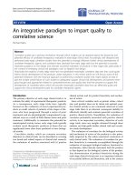
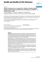
We constructed a custom-made orthosis (Figure 1) for the
left lower limb of each subject. The exoskeleton consisted
of a carbon fiber shank section and a polypropylene foot
section. A metal hinge betwee n the sections allowed free
sagittal plane rotation of the ankle joint. Two artificial
pneumatic muscles attached to the exoskeleton provided
substantial plantar flexor torque. During powered walking,
the peak plantar flexor torque provided by the ankle exos-
keleton was ~47% of the total ankle joint mo ment at
push-off [15]. Details of the design and performance of the
exoskeleton are documented elsewhere [52-54]. We imple-
mented proportional myoelectric control (i.e., amplitude
and timing) of the artificial muscles through desktop com-
puter and real-time control board (dSPA CE Inc.). A cus-
tom real-time computer controller regulated air pressure
in the artificial plantar flexor muscles proportional to the
processed soleus electromyographic signals (EMG) via a
pressure regulator. The EMG signal from t he soleus was
Kao et al. Journal of NeuroEngineering and Rehabilitation 2010, 7:33
/>Page 2 of 8
high-pass filtered with a second-order Butterworth filter
(20-Hz cutoff frequency) to remove movement artifact,
full wave rectified, and low-pass filtered with a second-
order Butterworth filter (10-Hz cutoff frequency) to
smooth the signal. Adjustable gains scaled the control sig-
nals and a threshold cutoff eliminated background noise.
Soleus H-reflex was tested while subjects walked with
the exoskeleton on the treadmill at 1.25 m/s, first with-
out power (first unpowered), t hen with power (pow-
ered), and finally without power again (second
unpowered). Before the testing of soleus H-reflex, sub-
jects had completed two 30-minute treadmill training
sessions for walking with the powered ankle exoskeleton
controlled by soleus EMG [14,15]. In addition, on the
day of soleus H-reflex testing, subjects were given time
(i.e., 5 minutes for unpowered conditions and 15 min-
utes for the powered condition) to re-familiarize them-
selves to walk with the exoskeleton prior to the nerve
stimulations. The same protocol of soleus H-reflex testing
repeated in the second unpowered condition was for mon-
itoring the influence of multiple stimuli on the H-reflex
amplitudes (e.g., homosynaptic depression) [55].
Data acquisition and analysis
We collected ankle kinematics, artificial muscle force,
electromyography (EMG) and ground reaction forces
while subjects walked on a custom-constructed force-
measuring split-belt treadmill. The three-dimensional
kinematic data were collected by using 8-camera video
system (120 Hz, Motion Analysis Corporation, Santa
Rosa,CA).Artificialmuscleforcedatawerecollected
with force transdu cers (1200 Hz, Omega Engineering)
mounted on the bracket of orthosis. We plac ed bipolar
surface electrodes on the left shan k to record EMGs
(1200 Hz, Konigsberg Instruments Inc.) from tibialis
anterior (TA), soleus (SOL), medial gastrocnemius
(MG), lateral gastrocnemius (LG).
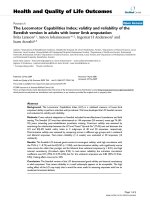
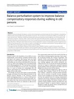
Soleus H-reflex measurements
We elicited the soleus H-reflex by stimulating (DS7AH
constant current stimulator, Digitimer Ltd.) the tibial
nerve with a cathode placed in the popliteal fossa and
an anode (7-cm diameter) on the patella (Figure 2). The
electrical stimulus was a 1-mi llisecond monophasic
square pulse. We located the optimal site of tibial nerve
stimulation using the criterion that a larger M-wave
amplit ude could be elicit ed at the same low intensity of
stimulus. Before the walking trials, we measured the
peak-to-peak amplitudes of M and H waves from sur-
face electrodes (2000 Hz) across different stimulation
intensities to gather a standing H- reflex and M-wave
recruitment curve.
For the walking trials, we tested the soleus H-reflex in
the 3 conditions (first unpowered, powered and second
unpowered). We used a footswitch (B&L engineering) to
detect heel strikes in real time an d estimated the dura-
tion of a gait cycle from at least 90 strides in each con-
dition. We divided the gait cycle into 16 equal epochs
(10 epochs in the stance). The majority of powered
assistance occurred at the middle to late stance, and this
was the time period of the largest reductions in the
soleus muscle activation [14,15]. Because a large number
of stimuli can inhibit H-reflex responses and be uncom-
fortable for subjects, we evoked soleus H-reflexes for
only three epochs: two during mid-stance (epoch 5 and
6) and one during late stance (epoch 8).We used a cus-
tom-written program and a real-time control board
(dSPA CE Inc.) to control the timing of electrical stimuli
and to measure the resulting M-wave and H-wave peak-
to-peak amplitudes (2000 Hz). We randomly dispersed
the stimuli to each of the 3 epochs. The program sent a
stimulus at least every 4 seconds.
ThesizeoftheM-waveasapercentageofthemaxi-
mal M-wave (i.e., M
max
, maximal evoked muscle
response) has been used regularly to control constant
effective stimulus intensity to the afferent nerve
[43,47,49,56]. While walking, the relative movement
between stimulating electrode and the nerve may change
M
max
over a stride [49]. To account for changes in
M
max
, we first collected M
max
data (3 M
max
measure-
ments) of each epoch by delivering a larger stimulus
Figure 1 Subjects wore a custom fit orthosis on their left lower
limb. The orthosis was hinged at the ankle to allow free sagittal
plane rotation. Soleus EMG activation was recorded and processed
to be used to control air pressure in the artificial pneumatic muscles
proportionally. As air pressure increased, the artificial muscles started
to develop tension and become shortened, allowing the powered
exoskeleton to provide plantar flexor torque controlled by soleus
muscle activation.
Kao et al. Journal of NeuroEngineering and Rehabilitation 2010, 7:33
/>Page 3 of 8
than the one evoked M
max
during quiet standing (at
least 1.2 times of stimulation intensity for evoking M
max
during quiet standing).
The effective stimulus intensity used for the H-reflex
measurements was the intensity to evoke a corresponding
M-wave that is 25% of M
max
for that epoch. The program
monitored the peak-to-peak amplitude of the M-wave
produced by the stimulus, and calculated the ratio of the
M-wave amplitude to the M
max
of that epoch. We only
accepted H-reflex measurements where the M-wave was
25 ± 10% of the corresponding M
max
. To ensure constant
stimulus intensity over the gait cycle, we manually
adjusted the intensity of subsequent stimuli if the ratio
was not within the range of 25 ± 10%. We collected
10 measurements of H-reflex where the corresponding
M-wave was 25 ± 10% of M
max
in each epoch.
For background soleus EMG amplitudes, we calculated
the mean of rectified averaged soleus EMG of each time
epoch. We normalized the H-reflex amplitudes and
mean EMG measurements to the M
max
for that time
epoch. This procedure corrected for changes in H-reflex
and background EMG values due to movement of t he
muscle fibers relative to the recording electrodes [49].
Since the H-reflex amplitude depends on the back-
ground level of motor activity [56], we calculated the
ratio o f H-reflex amplitude to its corresponding back-
ground EMG amplitude. Thus, the variables we derived
were H-wave amplitude (H/M
max
), background EMG
amplitude (EMG/M
max
), and the ratio of H-wave and
background EMG (H/EMG). To reduce the inter-subject
variabi lity, we then normalized the H-re flex, mean EMG
amplitudes and the ratio between H-reflex and
Figure 2 Soleus H-reflexes were evoked at epoch 5, 6, and 8 (circled). We stimulated the tibial nerve with a cathode placed in the popliteal
fossa and an anode on the patella. The effective stimulus intensity used for the H-reflex measurements was the intensity to evoke a
corresponding M-wave that is 25% of M
max
for that epoch. We only accepted the measurements of H-waves where their preceding M-waves
were 25 ± 10% of the corresponding M
max
.
Kao et al. Journal of NeuroEngineering and Rehabilitation 2010, 7:33
/>Page 4 of 8
background EMG in each condition to the values of the
first unpowered condition.
Statistics
We performed Friedman tests to test for differences in
normalized H-reflex amplitudes, soleus EMG amplitudes
and the ratio between H-reflex and background EMG at
the three epochs among the three conditions (first
unpowered, powered, and second unpowered). For the
small sample size, we chose the nonparamet ric methods
because the validity of this approach does not depend
crucially on normality assumption. We set the signifi-
cance level at p < 0.05. If a main effect (i.e., condition)
was detected, we used Wilcoxon signed ranks tests to
discriminate differences between the powered condition
and each of the two unpowered conditions (i.e., powered
vs. first unpowered, powered vs. second unpowered)
with Bonferroni’s correction (adjusted a = 0.025). All
statistical analyses were performed in SPSS statistics
version 17.0 (SPSS Inc., Chicago, Illinois).
Results
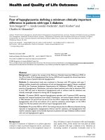
When the robotic plantar flexor torque was provided,
subjects walked with decreased soleus EMG and differ-
ent ankle joint kinematics at late stance (Figure 3).
Compared to the unpowered condition, subjects had
similar ankle joint angle profiles during initial to middle
stance but the ankle angle profiles deviated from the
unpowered ankle angle profiles at epoch 7 (Figure 3A).
In addition, the soleus activation was significantly lower
in the powered condition for epochs 5 (0.60 ± 0.17;
Friedman test, p = 0.002; both Wilcoxon signed ranks
tests, p < 0.025), epoch 6 (0.52 ± 0.21; Friedman test,
p = 0.002; b oth Wilcoxon signed ranks tests, p <0.025)
and epoch 7 (0.65 ± 0.22; Friedman test, p = 0.018; both
Wilcoxon signed ranks tests, p < 0.025) but not for
epoch 8 (0.73 ± 0.22, Friedman test, p =0.18)andthe
rest of the epochs in stance compared to the two
unpowered conditions (Figure 3B, Figure 4B). The
soleus EMG amplitudes as well as H-wave amplitudes in
the first unpowered condition were equal to 1.0 (100%)
for the three epochs because we normalized the data in
each condition to the first unpowered condition.
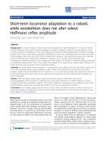
The reduction in soleus EMG activation was much
more than the reduction in H-wave amplitude during
powered walking. Subjects had significantly lower H-
wave amplitudes at epoch 5 (0.76 ± 0. 13; Friedman test,
p = 0.021; b oth Wilcoxon signed ranks tests, p <0.025)
but not at epoch 6 (0.80 ± 0.22, Friedman test, p =
0.066) and epoch 8 (0.88 ± 0.46, Friedman test, p =
0.867) during powered walking (Figure 4A). Compared
to the 27-48% of decrease in soleus EMG activation, H-
wave amplitudes were only lowered by 12-24% in the
powered condition. Thus, the ratio of H-wave amplitude
and background soleus EMG amplitude during powered
walking (epoch 5: 1.33 ± 0.26, epoch 6: 1.62 ± 0.60,
epoch 8: 1.11 ± 0.67) were not significantly different
from the two unpowered conditions (Figure 4C). A con-
dition effect was detected in the epoch 5 (Friedman test,
p = 0.028) but not in the epoch 6 (Friedman test, p =
0.066) and epoch 8 (Friedman test, p =0.651).For
further comparisons at epoch 5, the ratio of H-wave and
soleus EMG in the powered condition was significantly
different from the ratio in the first unpowered condition
(Wilco xon signed ranks test, p = 0.012) but not the sec-
ond unpowered condition (Wilcoxon signed ranks test,
p = 0.109).
Discussions
The confirmation of re-adaptation to the robotic ankle
exoskeleton was essential before performing soleus H-
reflex tests. Our previous studies [9,14] have shown that
subjects reached steady state o f powered walking much
faster at the second training session (~6 minutes) than
the first session (~25 minutes). For this study, 15 min-
utes of re-familiarization period in the third session was
sufficient to ensure the adaptation. In another published
Figure 3 Ankle joint angle profile (A) and normalized soleus
EMG (B). Data are the average of all subjects. (A) Ankle joint angle
profiles are shown for unpowered (black) and powered condition
(red). The error bars represent ± 1 standard deviation. Positive
values indicate ankle plantar flexion. (B) Normalized soleus EMG of
each time epoch was shown for the first unpowered (black),
powered (red), and second unpowered (grey). Epoch 5, 6, and 8
(circled) were the points in time when we performed the H-reflex
measurements.
Kao et al. Journal of NeuroEngineering and Rehabilitation 2010, 7:33
/>Page 5 of 8
study, we documented the results when using catch
trials (i.e., turning off the exoskeleton assistance unex-
pectedly) [57] to assess the presence of negative afteref-
fects, a benchmark of motor adaptation [58].
Our findings do not support the hypothesis that the
normalized amplitude of soleus H-reflex is reduced
when training with a robotic ankle exoskeleto n under
soleus proportional myoelectric control. With short
term training, our subjects reduced soleus background
EMG by ~35% and had less concomitant reductions i n
H-reflex amplitude by ~20% during steady-state pow-
ered walking. As a result, subjects demonstrated slightly
higher H-reflex amplitude relative to their background
muscle activity compared to unpowered walking.
The amplitude of the soleus H-reflex depends on presy-
naptic modulation of Ia afferents (e.g., increased
presynaptic inhibition) as well as overall excitability of the
motoneuron pool (e.g., a decrease in the voluntary drive of
soleus muscle). The unaltered H-reflex modulation in this
study indicates that stretch reflex inhibition (i.e., increased
presynapt ic inh ibition of Ia afferents) is likely not one of
the mechanisms for reducing soleus EMG when adapting
to robotic assistance with short term training. Instead, our
results suggest that mechanisms for this short-term adap-
tation to the robotic assista nce could be decreased excit-
ability of the soleus motoneuron pool, r esulting from
increased inhib ition of the motor neurons or a reduction
in supra-spinal drive [59].
Adaptation to the robotic exoskelet on assistance dur-
ing walking may occur in two phases, a quick adaptation
that occurs in the first few hours or days and a much
longer adaptation that continues for weeks [60-62]. The
two adaptation phases may have been reflected by the
difference between our current study results on newly
trained subjects and the pilot study on a long-term
trained subject [51]. When initially walking w ith the
robotic ankle exoske leton, subjects’ gait patterns were
greatly disturbed by the additional ankle mechanical tor-
que provided [14]. Decreased motor output of soleus
motor neurons du e to increased post-synaptic inhibition
or a reduction in supra-spinal excitation [63] would be
strategies to quickly reduce significant amount of so leus
EMG without altering the excitability of reflex pathway.
With longer term training, modulation of spi nal reflex
pathways by supra-spinal centers (i.e., increased pre-
synaptic inhibition of Ia afferents) could contribute to
soleus EMG reduction without need for constant
supraspinal inhibition. The different sensorimotor cali-
bration after long term training may result from
repeated motor adaptation to the robotic assistance [61].
During the initial learning of a motor task, increased
attention may also enhance the reflex responses. Pre-
vious studies have shown greater H-reflex responses
during the initial training o n a novel locomotion task
such as obstacle avoidance during walking [64] and
backward walking [41]. In our study, the subjects had
trained with the robotic-assisted walking for two thirty-
minute sessions and had a 15-minute period of practice
with powered walking by the time of H-reflex testing.
From subjects’ comments after data collection, it seemed
that a certain amount of attention or concentr ation was
necessary to walk smoothly with the augmented
mechanical plantar flexor torque provided by the exos-
keleton at the third session. This may have contributed
to the enhanced H-reflex amplitude relative to the back-
ground EMG in the powered walking in our study.
Conclusions
Our findings suggest that the nervous system does not
inhibit the soleus H-reflex in response to short-term
(A)
1.5
*
Normalized
H-wave
amplitude
1
0
0.5
1.5
1
0.5
(B)
Normalized
Soleus EMG
amplitude
**
2
(C)
Normalized
0
1
0
Normalized
ratio of
H-wave and EMG
(H/EMG)
Epoch 5
Epoch 6
Epoch 8
Epoch 5
Epoch 6
Epoch 8
First unpowered Powered Second unpowered
Figure 4 Normalized H-wave amplitude (A), normalized soleus
EMG amplitude (B), and normalized ratio of H-wave amplitude
to background EMG (C). Amplitudes of H-wave and soleus
rectified EMG were first normalized to the peak-to-peak amplitude
of M
max
of that time epoch. To reduce the inter-subject variability,
we then normalized the amplitudes in each condition to the values
of the first unpowered condition. Thus, the normalized data in the
first unpowered condition were 1.0 (100%) for the three epochs.
Kao et al. Journal of NeuroEngineering and Rehabilitation 2010, 7:33
/>Page 6 of 8
adaption to exoskeleton assistance as a mechanism for
reducing soleus muscle recruitment. Likely mechan-
isms for the decrease in soleus EMG include spinal or
supraspinal post-synaptic inhibition of the soleus
motor neurons. Previous results that found H-reflex
inhibition in a subject with long term exoskeleton
training experience [51] suggest that the neural
mechanisms involved in the adaptation to the exoske-
leton may change with extended practice. It is
unknown how much time or how many repetitions are
needed to transition from adapted motor patterns (i.e.,
motor adaptation) to well learned motor behaviors
(i.e., motor learning) [58]. Results from our previous
studies suggest that it is faster to achieve steady state
performance biomechanically than neurologically
[9,14]. Future studies should examine other potential
neural mechanisms both in short-term and long-term
adaptation to the exoskeleton as considerable evidence
suggests that robotic exoskeletons and orthoses have
strong potential for improving mobility in patients
with neurological impairments [10-13].
Acknowledgements
The authors thank Evelyn Anaka, Danielle Sandella, Catherine Kinnaird and
members of the Human Neuromechanics Laboratory for assistance in
collecting data. We also thank Anne Manier for help with fabricating the
orthosis. Supported by NIH R21 NS062119 (DPF) and F32 HD055010 (CLL).
Author details
1
School of Kinesiology, University of Michigan, Ann Arbor, Michigan 48109-
2214, USA.
2
College of Health & Rehabilitation Sciences: Sargent College,
Boston University, Boston, Massachusetts 02215, USA.
Authors’ contributions
PCK recruited subjects, managed data collections, completed data analysis
and drafted the manuscript. CLL developed a custom-written program to
control the timing of electrical stimuli, assisted with data analysis and
helped edit the manuscript. DPF conceived of the study, provided guidance
on experimental design, and helped draft and edit the manuscript. All
authors read and approved the final manuscript.
Competing interests
The authors declare that they have no competing interests.
Received: 18 January 2010 Accepted: 26 July 2010
Published: 26 July 2010
References
1. Werner C, Von Frankenberg S, Treig T, Konrad M, Hesse S: Treadmill
training with partial body weight support and an electromechanical gait
trainer for restoration of gait in subacute stroke patients: a randomized
crossover study. Stroke 2002, 33:2895-2901.
2. Sawicki GS, Domingo A, Ferris DP: The effects of powered ankle-foot
orthoses on joint kinematics and muscle activation during walking in
individuals with incomplete spinal cord injury. Journal of Neuroengineering
and Rehabilitation 2006, 3:3.
3. Wirz M, Colombo G, Dietz V: Long term effects of locomotor training in
spinal humans. Journal of Neurology Neurosurgery and Psychiatry 2001,
71:93-96.
4. Colombo G, Wirz M, Dietz V: Driven gait orthosis for improvement of
locomotor training in paraplegic patients. Spinal Cord 2001, 39:252-255.
5. Banala SK, Kim SH, Agrawal SK, Scholz JP: Robot Assisted Gait Training
With Active Leg Exoskeleton (ALEX). 10th IEEE International Conference on
Rehabilitation Robotics; Jun 13-15 Noordwijk, NETHERLANDS 2007, 2-8.
6. Emken JL, Harkema SJ, Beres-Jones JA, Ferreira CK, Reinkensmeyer DJ:
Feasibility of manual teach-and-replay and continuous impedance
shaping for robotic locomotor training following spinal cord injury. Ieee
Transactions on Biomedical Engineering 2008, 55:322-334.
7. Emken JL, Benitez R, Reinkensmeyer DJ: Human-robot cooperative
movement training: Learning a novel sensory motor transformation
during walking with robotic assistance-as-needed. Journal of
Neuroengineering and Rehabilitation 2007, 4:8.
8. Aoyagi D, Ichinose WE, Harkema SJ, Reinkensmeyer DJ, Bobrow JE: A robot
and control algorithm that can synchronously assist in naturalistic
motion during body-weight-supported gait training following neurologic
injury. 10th IEEE International Conference on Rehabilitation Robotics; Jun 13-
15 Noordwijk, NETHERLANDS 2007, 387-400.
9. Cain SM, Gordon KE, Ferris DP: Locomotor adaptation to a powered
ankle-foot orthosis depends on control method. Journal of
Neuroengineering and Rehabilitation 2007, 4:48.
10. Edgerton VR, Roy RR: Robotic training and spinal cord plasticity. Brain
Research Bulletin 2009, 78:4-12.
11. Reinkensmeyer DJ, Patton JL: Can Robots Help the Learning of Skilled
Actions? Exercise and Sport Sciences Reviews 2009, 37:43-51.
12. Huang VS, Krakauer JW: Robotic neurorehabilitation: a computational
motor learning perspective. Journal of Neuroengineering and Rehabilitation
2009, 6:5.
13. Marchal-Crespo L, Reinkensmeyer DJ: Review of control strategies for
robotic movement training after neurologic injury. Journal of
Neuroengineering and Rehabilitation 2009, 6:20.
14. Gordon KE, Ferris DP: Learning to walk with a robotic ankle exoskeleton.
Journal of Biomechanics 2007, 40:2636-2644.
15. Kao PC, Lewis CL, Ferris DP:
Invariant ankle moment patterns when
walking with and without a robotic ankle exoskeleton. Journal of
Biomechanics 2010, 43:203-209.
16. Kuo AD, Donelan JM, Ruina A: Energetic consequences of walking like an
inverted pendulum: step-to-step transitions. Exercise and Sport Sciences
Reviews 2005, 33:88-97.
17. Nadeau S, Arsenault AB, Gravel D, Bourbonnais D: Analysis of the clinical
factors determining natural and maximal gait speeds in adults with a
stroke. Am J Phys Med Rehabil 1999, 78:123-130.
18. Kim CM, Eng JJ: The relationship of lower-extremity muscle torque to
locomotor performance in people with stroke. Physical Therapy 2003,
83:49-57.
19. Chen G, Patten C: Joint moment work during the stance-to-swing
transition in hemiparetic subjects. Journal of Biomechanics 2008,
41:877-883.
20. Yang JF, Stein RB, James KB: Contribution of peripheral afferents to the
activation of the soleus muscle during walking in humans. Experimental
Brain Research 1991, 87:679-687.
21. Sinkjaer T, Andersen JB, Larsen B: Soleus stretch reflex modulation during
gait in humans. Journal of Neurophysiology 1996, 76:1112-1120.
22. Nielsen JB, Sinkjaer T: Afferent feedback in the control of human gait.
Journal of Electromyography and Kinesiology 2002, 12:213-217.
23. Mazzaro N, Grey MJ, Sinkjaer T: Contribution of afferent feedback to the
soleus muscle activity during human locomotion. Journal of
Neurophysiology 2005, 93:167-177.
24. Mazzaro N, Grey MJ, Sinkjaer T, Andersen JB, Pareyson D, Schieppati M:
Lack of on-going adaptations in the soleus muscle activity during
walking in patients affected by large-fiber neuropathy. Journal of
Neurophysiology 2005, 93:3075-3085.
25. Rossignol S, Dubuc RJ, Gossard JP: Dynamic sensorimotor interactions in
locomotion. Physiological Reviews 2006, 86:89-154.
26. af Klint R, Nielsen JB, Cole J, Sinkjaer T, Grey MJ: Within-step modulation of
leg muscle activity by afferent feedback in human walking. Journal of
Physiology-London 2008, 586:4643-4648.
27. af Klint R, Nielsen JB, Sinkjaer T, Grey MJ: Sudden Drop in Ground Support
Produces Force-Related Unload Response in Human Overground
Walking. Journal of Neurophysiology 2009, 101:1705-1712.
28. Phadke CP, Wu SS, Thompson FJ, Behrman AL: Comparison of soleus H-
reflex modulation after incomplete spinal cord injury in 2 walking
Kao et al. Journal of NeuroEngineering and Rehabilitation 2010, 7:33
/>Page 7 of 8
environments: Treadmill with body weight support and overground.
Archives Of Physical Medicine And Rehabilitation 2007, 88:1606-1613.
29. Yang JF, Fung J, Edamura M, Blunt R, Stein RB, Barbeau H: H-reflex
modulation during walking in spastic paretic subjects. Canadian Journal
of Neurological Sciences 1991, 18:443-452.
30. Dietz V: Spinal cord lesion: effects of and perspectives for treatment.
Neural Plast 2001, 8:83-90.
31. Dietz V: Proprioception and locomotor disorders. Nature Reviews
Neuroscience 2002, 3:781-790.
32. Knikou M, Angeli CA, Ferreira CK, Harkema SJ: Soleus H-reflex modulation
during body weight support treadmill walking in spinal cord intact and
injured subjects. Experimental Brain Research 2009, 193:397-407.
33. Yelnik A, Albert T, Bonan I, Laffont I: A clinical guide to assess the role of
lower limb extensor overactivity in hemiplegic gait disorders. Stroke
1999, 30:580-585.
34. Knikou M, Angeli CA, Ferreira CK, Harkema SJ: Soleus H-reflex gain,
threshold, and amplitude as function of body posture and load in spinal
cord intact and injured subjects. International Journal of Neuroscience 2009,
119:2056-2073.
35. Trimble MH, Kukulka CG, Behrman AL: The effect of treadmill gait training
on low-frequency depression of the soleus H-reflex: comparison of a
spinal cord injured man to normal subjects. Neuroscience Letters 1998,
246:186-188.
36. Trimble MH, Behrman AL, Flynn SM, Thigpen MT, Thompson FJ: Acute
effects of locomotor training on overground walking speed and H-reflex
modulation in individuals with incomplete spinal cord injury. Journal of
Spinal Cord Medicine 2001, 24:74-80.
37. Chen Y, Chen XY, Jakeman LB, Chen L, Stokes BT, Wolpaw JR: Operant
conditioning of H-reflex can correct a locomotor abnormality after
spinal cord injury in rats. Journal of Neuroscience 2006, 26:12537-12543.
38. Mazzocchio R, Kitago T, Liuzzi G, Wolpaw JR, Cohen LG: Plastic changes in
the human H-reflex pathway at rest following skillful cycling training.
Clinical Neurophysiology 2006, 117:1682-1691.
39. Mynark RG, Koceja DM: Down training of the elderly soleus H reflex with
the use of a spinally induced balance perturbation. Journal of Applied
Physiology 2002, 93:127-133.
40. Trimble MH, Koceja DM: Modulation of the triceps surae H-reflex with
training. International Journal of Neuroscience 1994, 76:293-303.
41. Schneider C, Capaday C: Progressive adaptation of the soleus H-reflex
with daily training at walking backward. Journal of Neurophysiology 2003,
89:648-656.
42. Thompson AK, Chen XY, Wolpaw JR:
Acquisition of a Simple Motor Skill:
Task-Dependent Adaptation Plus Long-Term Change in the Human
Soleus H-Reflex. Journal of Neuroscience 2009, 29:5784-5792.
43. Ferris DP, Aagaard P, Simonsen EB, Farley CT, Dyhre-Poulsen P: Soleus H-
reflex gain in humans walking and running under simulated reduced
gravity. Journal of Physiology (London) 2001, 530:167-180.
44. Schneider C, Lavoie BA, Capaday C: On the origin of the soleus H-reflex
modulation pattern during human walking and its task-dependent
differences. J Neurophysiol 2000, 83:2881-2890.
45. Sinkjaer T: Muscle, reflex and central components in the control of the
ankle joint in healthy and spastic man. Acta Neurol Scand Suppl 1997,
170:1-28.
46. Stein RB, Capaday C: The modulation of human reflexes during functional
motor tasks. Trends in Neurosciences 1988, 11:328-332.
47. Capaday C, Stein RB: Amplitude modulation of the soleus H-reflex in the
human during walking and standing. Journal of Neuroscience 1986,
6:1308-1313.
48. Dyhre-Poulsen P, Simonsen EB, Voigt M: Dynamic control of muscle
stiffness and H reflex modulation during hopping and jumping in man.
Journal of Physiology (London) 1991, 437:287-304.
49. Simonsen EB, Dyhre-Poulsen P: Amplitude of the human soleus H reflex
during walking and running. Journal of Physiology-London 1999,
515:929-939.
50. Ung RV, Imbeault MA, Ethier C, Brizzi L, Capaday C: On the potential role
of the corticospinal tract in the control and progressive adaptation of
the soleus h-reflex during backward walking. Journal of Neurophysiology
2005, 94:1133-1142.
51. Ferris DP, Kinnaird CR: Robotic lower limb orthoses for gait rehabilitation
after incomplete spinal cord injury. Proceedings of the 2008 Annual
Meeting of the American Spinal Injury Association, June 19-22 San Diego, CA
2008.
52. Ferris DP, Czerniecki JM, Hannaford B: An ankle-foot orthosis powered by
artificial pneumatic muscles. Journal of Applied Biomechanics 2005,
21:189-197.
53. Ferris DP, Gordon KE, Sawicki GS, Peethambaran A: An improved powered
ankle-foot orthosis using proportional myoelectric control. Gait and
Posture 2006, 23:425-428.
54. Gordon KE, Sawicki GS, Ferris DP: Mechanical performance of artificial
pneumatic muscles to power an ankle-foot orthosis. Journal of
Biomechanics 2006, 39:1832-1841.
55. Knikou M: The H-reflex as a probe: Pathways and pitfalls. Journal of
Neuroscience Methods 2008, 171:1-12.
56. Capaday C: Neurophysiological methods for studies of the motor system
in freely moving human subjects. J Neurosci Methods 1997, 74:201-218.
57. Kao PC, Lewis CL, Ferris DP: Joint kinetic response during unexpectedly
reduced plantar flexor torque provided by a robotic ankle exoskeleton
during walking. Journal of Biomechanics 2010, 43:1401-1407.
58. Reisman DS, Bastian AJ, Morton SM: Neurophysiologic and rehabilitation
insights from the split-belt and other locomotor adaptation paradigms.
Physical Therapy 2010, 90:187-195.
59. Wolpaw JR: The complex structure of a simple memory. Trends in
Neurosciences 1997, 20:588-594.
60. Wolpaw JR, Okeefe JA: Adaptive Plasticity in the Primate Spinal Stretch
Reflex - Evidence for a 2-Phase Process. Journal of Neuroscience 1984,
4:2718-2724.
61. Bastian AJ: Understanding sensorimotor adaptation and learning for
rehabilitation. Current Opinion in Neurology 2008, 21:628-633.
62. Luft AR, Buitrago MM: Stages of motor skill learning. Molecular
Neurobiology 2005, 32:205-216.
63. Shefchyk SJ, Jordan LM: Excitatory and Inhibitory Postsynaptic Potentials
in Alpha-Motoneurons Produced During Fictive Locomotion by
Stimulation of the Mesencephalic Locomotor Region. Journal of
Neurophysiology 1985, 53:1345-1355.
64. Hess F, van Hedel HJA, Dietz V: Obstacle avoidance during human
walking: H-reflex modulation during motor learning. Experimental Brain
Research 2003, 151:82-89.
doi:10.1186/1743-0003-7-33
Cite this article as: Kao et al.: Short-term locomotor adaptation to a
robotic ankle exoskeleton does not alter soleus Hoffmann reflex
amplitude. Journal of NeuroEngineering and Rehabilitation 2010 7:33.
Submit your next manuscript to BioMed Central
and take full advantage of:
• Convenient online submission
• Thorough peer review
• No space constraints or color figure charges
• Immediate publication on acceptance
• Inclusion in PubMed, CAS, Scopus and Google Scholar
• Research which is freely available for redistribution
Submit your manuscript at
www.biomedcentral.com/submit
Kao et al. Journal of NeuroEngineering and Rehabilitation 2010, 7:33
/>Page 8 of 8