Báo cáo sinh học: "Use of recombinant lentivirus pseudotyped with vesicular stomatitis virus glycoprotein G for efficient generation of human anti-cancer chimeric T cells by transduction of human peripheral blood lymphocytes in vitro" pot
Bạn đang xem bản rút gọn của tài liệu. Xem và tải ngay bản đầy đủ của tài liệu tại đây (580.46 KB, 10 trang )
BioMed Central
Page 1 of 10
(page number not for citation purposes)
Virology Journal
Open Access
Research
Use of recombinant lentivirus pseudotyped with vesicular
stomatitis virus glycoprotein G for efficient generation of human
anti-cancer chimeric T cells by transduction of human peripheral
blood lymphocytes in vitro
Anthony Simmons*
†1,3
, Robert P Whitehead
2
, Andrey A Kolokoltsov
3
and
Robert A Davey
†3
Address:
1
Department of Pediatrics, University of Texas Medical Branch, Galveston, Texas, USA,
2
Department of Internal Medicine, University of
Texas Medical Branch, Galveston, Texas, USA and
3
Department of Microbiology & Immunology, University of Texas Medical Branch, Galveston,
Texas, USA
Email: Anthony Simmons* - ; Robert P Whitehead - ; Andrey A Kolokoltsov - ;
Robert A Davey -
* Corresponding author †Equal contributors
Abstract
Background: Genetic redirection of lymphocytes that have been genetically engineered to
recognize antigens other than those originally programmed in their germlines is a potentially
powerful tool for immunotherapy of cancers and potentially also of persistent viral infections. The
basis for this procedure is that both cancers and some viruses have developed strikingly similar
mechanisms of evading attacks by host immune mechanisms. To redirect human peripheral blood
lymphocytes (PBLs) with a chimeric T cell receptor (chTCR) so that they recognize a new target
requires a high degree of transfection efficiency, a process that is regarded as technically
demanding.
Results: Infection with a retroviral vector carrying a chTCR cassette was shown to transduce
100% of rapidly dividing murine T cells but typically, only ~10% of PBLs could be infected with the
same vector. In contrast with other retroviruses, lentiviruses integrate their genomes into non-
dividing cells. To increase host cell range, vesicular stomatitis virus G protein was pseudotyped with
a lentivirus vector, which resulted in ~100% PBL transduction efficiency. Signaling of PBLs bearing
chimeric receptors was shown by specific proliferation on exposure to cells expressing cognate
ligand. Further, T-bodies against CEA showed a startling abilty to cause regression of maligant colon
tumors in a nude mouse model of human cancer.
Conclusion: A lentivirus/VSV pseudotyped virus, which does not require replicating cells for
integration of its genome, efficiently transduced a high proportion of human PBLs with chTCRs
against CEA. PBLs transduced by infection with a lentivirus/VSV pseudotyped vector were able to
proliferate specifically in vitro on exposure to CEA-expressing cells and further they had a startling
therapeutic effect in a mouse model of human colon cancer.
Published: 28 February 2006
Virology Journal2006, 3:8 doi:10.1186/1743-422X-3-8
Received: 10 January 2006
Accepted: 28 February 2006
This article is available from: />© 2006Simmons et al; licensee BioMed Central Ltd.
This is an Open Access article distributed under the terms of the Creative Commons Attribution License ( />),
which permits unrestricted use, distribution, and reproduction in any medium, provided the original work is properly cited.
Virology Journal 2006, 3:8 />Page 2 of 10
(page number not for citation purposes)
Background
It has become increasingly apparent that the scope of
immunization and immunotherapy is applicable not only
to infectious agents but also to tumors. Persistent viruses
and tumors escape immune surveillance by a variety of
common mechanisms, one of the more prominent being
down-regulation of class I major histocompatibility mol-
ecules thereby preventing recognition by cytotoxic T cells.
Antibodies on the other hand retain their ability to recog-
nize native antigens but in case of tumors their potential
effects are compromised by their failure to penetrate into
neoplastic tissue.
In the current work, the term T-bodies is used to describe
T lymphocytes whose targets were redirected using viral
vectors engineered to convey to lymphocytes chTCR cas-
settes, based on a single chain antibody variable frag-
ments for antigen recognition [1-6]. Specifically, the T-
bodies described recognize an antigen that is expressed
selectively by cells in growing tumors.
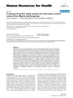
Simplified scheme of T cell activation by A) the T cell receptor and the co-stimulatory receptor, CD28 and B) a chimeric receptor in which co-stimulation and antigen specific activation are provided by a single molecule that is not dependent on MHC-1-associated peptide presentationFigure 1
Simplified scheme of T cell activation by A) the T cell receptor and the co-stimulatory receptor, CD28 and B) a chimeric
receptor in which co-stimulation and antigen specific activation are provided by a single molecule that is not dependent on
MHC-1-associated peptide presentation. *LCK is a lipid raft-associated protein that phosphorylates ITAMs of the TCR zeta
chain, allowing them to bind the zeta associated protein kinase ZAP-70. LCK then also activates ZAP-70 by phosphorylation,
continuing the T cell signaling cascade. [44]. While it is generally accepted that LCK is activated when CD4 or CD8 co-recep-
tors are cross-linked with the TCR and MHC complex, it is also known that some LCK associates with CD8 in the absence of
MHC [45]. It is possible that LCK activation is a function of the surrounding membrane environment rather than the clustering
of co-receptors with T cell receptor and MHC [46].
A. ANTIGEN PRESENTING CELL B. TUMOR CELL
MHC/Peptide CD80/86 Native antigen
Down-stream signaling events e.g.
IL-2 gene transcription and associated cell proliferation
Interferon gamma gene transcription,
Cytotoxicity
V
H
]
G
F
P
C
D
2
8
V
H
]
G
F
P
C
D
2
8
Ż
Extracellula
r
Cytoplasm
J
HGH
Ti
Extracellular
Cytoplasm
Membrane
CD3
ZAP-70
Zeta-chain associated
protein (ZAP-70) binds to
ITAMs that are
phosphorylated (Ź). Binding
of ZAP-70 to ] is a crucial
step i
nT
-
cell activation
CD28
LCK*
LCK*
ZAP-70
Ż
V
L
V
L
Membrane
Virology Journal 2006, 3:8 />Page 3 of 10
(page number not for citation purposes)
Vectors based mainly the on the oncoretrovirus Murine
Leukemia Virus (MLV) backbone have been tried by sev-
eral groups [1-6] to transduce lymphocytes with genes
encoding chTCRs to combat experimental tumors in mice
with, in practical terms, limited therapeutic success. The
major problems encountered, including in our own pre-
liminary experiments, were inability to grow MLV vectors
to a sufficiently high titer to infect and therefore transduce
a high proportion of PBLs and the dependence on
oncoretroviruses on dividing cells in order for integration
of their genomes into host cell DNA. Eshhar et al. [7]
showed that the efficiency of transduction could be
increased to 35–70% by pseudotyping the retrovirus with
the envelope protein of another retrovirus, Gibbon ape
leukemia virus. In contrast with oncoretroviruses, the
genus lentiviruses have the ability to transduce non-divid-
ing cells. In addition to murine killer cells, we describe sta-
ble transduction of a very high proportion of human PBLs
with a lentivirus/vesicular stomatitis virus pseudotype.
It was shown previously [8] that tissue tropism of lentivi-
ruses can be broadened by constructing pseudotyped viral
particles comprising a lentiviral genome enveloped by the
surface glycoprotein from vesicular stomatitis virus (VSV-
G). VSV, a rhabdovirus, has been commonly used for
pseudotyping retroviruses because it is highly stable and
confers an exceptionally wide host range, because of the
binding of VSV-G to a cell surface lipid. We therefore
chose to test the ability of VSV pseudotypted lentivirus
containing a chimeric T cell receptor for PBL transduction
and compare its efficiency with a well known retrovirus
for conveying the same chimeric receptor gene. A models
of colon cancer in athymic mice was chosen to explore the
efficacy of human T-bodies created by lentiviral vectors
against a human tumor. The significance of colon cancer
is undisputed. Colorectal cancer is the third most com-
mon malignant neoplasm in the world [9] and the second
leading cause of cancer deaths (irrespective of gender) in
the United States [10,11].
Chemotherapy is commonly used in the treatment both
of colon. An alternative approach to chemotherapy that is
receiving much attention is adoptive immunotherapy
with immune cells that have been manipulated ex vivo.
Despite its promise, effective responses to adoptive
immunotherapy have been documented against only a
restricted number of tumor types and this approach to
cancer therapy has been further restricted by toxicity asso-
ciated with the need for exogenous administration of
interleukin-2. A prominent reason for failure of both vac-
cination and adoptive immunotherapy is the common
ability of tumors to down-regulate molecules of the major
histocompatibility complex, a property also shared with
many persistent viruses.
Though it has been established that human tumors may
express multiple antigens that can be recognized by cyto-
toxic T lymphocytes. [12], tumors are not normally
attacked by the host's immune system. Binding of antigen
to the TCR complex results in a cascade of events com-
mencing with phosphorylation of critical domains of the
receptor by membrane-associated Src family of protein
tyrosine kinases (PTKs) such as LCK and Fyn, which leads
to a cascade of downstream kinase activities that ulti-
mately cause Ca
++
fluxing, new gene transcription and/or
cell cycle progression (figure 1). A key role in signal trans-
duction is played by domains of the CD3 ζ (zeta) subunit
known as Immunoreceptor Tyrosine-based Activation
Motifs (ITAMs). When ITAMs are phosphorylated by LYK
or Fyn they can bind SH2 domains of other kinases, espe-
cially the zeta associated protein, ZAP-70. The ensuing
events lead ultimately to transcription of genes, including
interleukin-2, that promote cell proliferation.
Conventional wisdom recognizes that activation of naïve
T cells requires two signals [13], the primary one being
interaction between MHC/peptide and the TCR and a sec-
ond, co-stimulatory signal, transmitted by interaction
between CD28 on the lymphocyte and CD80 (B7-1)/86
(B7-2), generally present on professional antigen present-
ing cells [14]. Like many T cell receptors, signal transduc-
tion by CD28 appears to involve phosphorylation of
tyrosine residues in its cytoplasmic domain. The cytoplas-
mic tail of CD28 is very short (~40 amino acids) and has
only four tyrosines, only one of which (tyrosine 188 of the
mouse sequence) appears to be essential for co-stimula-
tion as assessed by expression of CD69 and production of
interleukin-2 (IL-2). [15].
To feasibly and safely cause regression of carcinomas, the
targets recognized by the chTCR is of paramount impor-
tance because it must be tumor selective or expressed at
very low levels in normal tissues to avoid significant col-
lateral damage by the T-body. CEA was selected for the
tumor-selective target in the reported studies because CEA
is expressed selectively on most colorectal and several
other cancer cell types. As a result it has been a popular
target for a variety of immunotherapeutic trials. CEA rep-
resents a family of molecules that is involved in regulation
of cellular differentiation during embryogenesis.
Although it is expressed at very low levels by normal adult
tissues, CEA is present in often high levels in cancers of the
colon, pancreas, breast, thyroid, lung, ovaries, and stom-
ach.
In clinical trials, colon cancer patients have been vacci-
nated with CEA peptides, CEA-pulsed dendritic cells or
viral vectors containing CEA and co-stimulatory mole-
cules. These strategies occasionally have been shown to
engender antigen specific T cell responses and occasion-
Virology Journal 2006, 3:8 />Page 4 of 10
(page number not for citation purposes)
ally partial tumor regression in vaccine recipients, which
indicates that it may be possible to overcome the potential
problems of immune evasion. However, only those
patients whose disease is limited to a few specific sites
have benefited from this approach to date and moreover
the benefits have been short-lived [16-20].
Regression of CEA
+
colon cancers caused by systemic
administration of T-bodies in a scid mouse model of
human colon carcinoma was demonstrated previously by
Haynes et al [1-3]. Here we show that T-bodies (o2f the
structure illustrated in figure 2) constructed from human
PBLs by transduction with lentiviral/VSV pseudotypes and
directed at CEA have considerable promise for develop-
ment as therapies for cancer.
Results
Redirection of human PBLs and murine MD45 cells with
chTCRs by stable transduction
High efficiency stable transduction of lymphocytes is gen-
erally regarded as technically demanding. Transfection
typically has an unacceptably low efficiency and retroviral
transduction is a widely preferred option. This option
however requires production of high titer viruses, which
are typically packaged in a derivative of NIH3T3 or 293
cells [21].
Direct transfection of PBLs was tried with limited success
(<10%) using the calcium phosphate precipitation tech-
nique and other contemporary methods (Lipofectamine,
Invitrogen; FuGENE 6 and X-tremeGENE, Roche). The
focus was then changed to retroviral vectors for introduc-
tion of the chTCR cassette into the human genome. Retro-
viruses have the unique advantage of integration into all
host cell genomes and infect a broad range of cell-types.
The caveat to the preceding statement is that most retrovi-
ruses require rapidly dividing cells to achieve integration
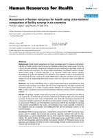
Assembly of a chTCR cassetteFigure 2
Assembly of a chTCR cassette. Antibody V
L
(red) and V
H
(yellow) were linked by a flexible 15 amino acid hinge to allow correct
scFv refolding and reconstitution the antigen binding site. Two thirds of the hinge sequence was attached using specifically
designed PCR primers, allowing their splicing to create a scFv. To avoid creation of a sub-optimal (Gly
4
Ser)
2
hinge, alternative
glycine codons were used. To the C terminus of the scFv, extracellular and transmembrane portions of either mouse or human
CD28 (etm28) are covalently attached to aid interaction between the chTCR and target and enable fluidity of the chTCR
within the cell membrane. The remainder of the chTCR is as described.
Function:
Antigen recognition trans- co- activation tracking
membrane stimulation
Hinge
V
L
V
H
V
L
V
H
etm28 CD28 TCR
]
GFP
(
Gly
4
Ser)
3
V
L
Hinge V
H
etm28 CD28 TCR
]
GFP
MD45 murine T cell line transduced by infection with a MLV-based retroviral vector (retro-X) containing a GFP- chTCR cassette, demonstrating high efficiency transduction with retro-X and a continuous growing cell population, judged here by expression of GFPFigure 3
MD45 murine T cell line transduced by infection with a MLV-
based retroviral vector (retro-X) containing a GFP- chTCR
cassette, demonstrating high efficiency transduction with
retro-X and a continuous growing cell population, judged
here by expression of GFP.
Virology Journal 2006, 3:8 />Page 5 of 10
(page number not for citation purposes)
but lentiviruses have the capability to integrate into the
genomes of non-replicating cells.
Whilst high transduction efficiencies could be achieved
for a rapidly growing mouse MD45 cell line (Figure 3)
using a commercially available MLV-based system (Retro-
X, Invitrogen), the maximum viral titer produced by PT67
packaging cells was 10
6
transforming units (TU)/ml when
titrated in 293 cells. This titer was adequate for infecting
all MD45 cells but in contrast, 10
6
TU/ml was not suffi-
cient to efficiently transduce human peripheral blood
mononuclear cells that were stimulated to proliferate with
anti-CD3 and anti-CD28. Typically, only ~10% of cells
(not shown) became infected as judged by expression of
GFP, a component of the receptor construct. A variety of
techniques were tried to increase the viral titer (including
overnight incubation of packaging cells at 32°C prior to
virus harvest) without additional success. [22].
Consequently, we tried the use of a vesicular stomatitis
virus (VSV) G protein pseudotyped lentivirus vector to
improve both the viral titer and transduction efficiency of
target cells. Using this approach, viral titers of 10
7
TU/ml
(in 293 cells) or greater were obtained, which enabled 5–
10 TU/cell to be used for infection of 10
6
PBLs. With this
approach it was possible to achieve stable transduction of
near 100% of anti-CD3/anti-CD28 stimulated PBLs (Fig-
ure 4).
In Vitro detection of CEA T-body proliferation
To demonstrate antigen-specific stimulation of human
CEA T bodies generated in the laboratory, a standard [
3
H]-
thymidine uptake proliferation assay was used (figure 5).
PBLs tranduced with the chTCR proliferated vigorously on
exposure to irradiated CEA positive (SW403) but not CEA
negative (COLO 320 HSR) colon carcinoma cells. Further,
little stimulation of control cells tranduced with a GFP
cassette alone or untransduced PBLs was observed.
Finally, the T-bodies proliferated vigorously on cross-link-
ing of their natural TCRs with anti-CD3 plus anti-CD28.
Thymidine uptake in the presence of cell culture medium
alone was considered background. The results were inter-
preted as specific redirected signaling of peripheral blood
lymphocytes by the chTCR.
Regression of tumors in an experimental model of human
colon carcinoma
Tumor growth was first visible 3 weeks after injection of
cells and tumors grew to a diameter of ~1.5 cm 4 weeks
after they first became visible (figure 6). 10
7
chimeric or
control T cells were adoptively transferred via tail veins to
tumor cell recipients three weeks after tumor cells. Regres-
sion was dramatic (Figure 6 and 7) with CEA-specific T-
bodies alone, which are expected to attack only cells at the
tumor rim. Experimental colon cancers recurred after
treatment with CEA T-bodies and all mice we dead by day
100.
Discussion
The main thrust of the current work was to show that high
efficiency stable transduction of human PBLs is a feasible
prospect for generating anti-cancer chimeric T cells for use
in cancer immunotherapy. The target selected in the cur-
rent work was CEA, a tumor-selective antigen
Several possible mechanisms for tumor escape from
immune surveillance have been demonstrated in model
systems. Mechanisms of immune evasion include presen-
tation of a tumor antigen by tumor cells without the nec-
essary co-stimulatory signal [23,24], suppression or
anergy of tumor-infiltrating T-cells [25-29], inability of a
tumor antigen to induce high avidity T-cells. [30,31] and
possibly most significant and general of all, tumor cells
commonly down-regulate expression of either class I or
class II major histocompatibility complex (MHC) mole-
cules needed for presentation of antigens to lymphocytes
CEA-specific human T-bodiesFigure 4
CEA-specific human T-bodies. (A) Anti-CD3/CD28 stimulated human PBLs transduced by infection with a VSV/lentivirus
pseudotype virus carrying a CEA-specific chTCR (phase); (B) same field viewed by fluorescence for GFP; (C) overlay of A and
B, demonstrating the presence of the GFP containing chTCR cassette in all cells.
A B C
Virology Journal 2006, 3:8 />Page 6 of 10
(page number not for citation purposes)
[16,32-34]. Adoptive immunotherapy with autologous
chimeric T lymphocytes that recognize a tumor antigen
has enormous therapeutic potential to produce regression
of tumors in humans with advanced cancers. A useful
anti-cancer T-body may be defined as an autologous lym-
phocyte whose natural target has been redirected to a
tumor specific antigen by introduction of a chTCR. The
approach that seems most promising in animal models is
the use of a single chain antibody variable fragment (scFv)
against the tumor coupled to T cell receptor signaling
domains for activation effector functions. A great advan-
tage of this approach is that stimulation of a cell with a
scFv-based receptor does not depend on expression of
major histocompatibility molecules by the target cells.
Prior reported studies, using a similar model in scid mice
[3] to the nude mouse model described here, demon-
strated tumor inhibition when T-bodies were adminis-
tered on one day after tumor cells.
CEA-specific proliferation of human lymphocytes measured by uptake of [
3
H]-thymidineFigure 5
CEA-specific proliferation of human lymphocytes measured by uptake of [
3
H]-thymidine. T-bodies (labeled T-body in the fig-
ure) were produced by infection of 10
6
PBLs with 10
7
TU/ml of a virus comprising a VSV envelope and a lentiviral genome con-
taining the CEA-CD28-CD3zeta-GFP cassette. T-bodies (■) responded to irradiated CEA+ SW 403 stimulator cells but the
CEA- COLO 320 control cells ( ) did not. PBLs transduced with a GFP cassette alone did not respond. All cells proliferated
vigorously in response to anti-CD3/CD28 (ᮀ) and no CEA-CD28- CD3ζ-GFP T-bodies responded to medium alone.
Effectors and stimulators
COLO 320 + SW403
CONTROL + SW403
CONTROL + ANTI-CD3
COLO 320 + SW403
T-BODY + SW403
T-BODY + ANTI-CD3
COLO 320 + MEDIUM
T-BODY + MEDIUM
GFP
+
PBLs + MEDIUM
0 2 4 6 8 10 12 14 16
COLO 320 + SW403
T-BODY + GFP
+
PBLs
GFP
+
PBLs + ANTI-CD3
Virology Journal 2006, 3:8 />Page 7 of 10
(page number not for citation purposes)
In the current work, chTCR cassettes combining intracel-
lular CD28 and CD3 sequences were constructed and
inserted into lentivirus/VSV pseudotypic vectors which
were subsequently used for transduction of PBLs.
A weakness of many prior attempts has been failure to
take into account the need for T cells to receive a second,
co-stimulatory, signal from accessory molecules on anti-
gen presenting cells such as CD80 and CD86. However,
many T bodies have been investigated that supply only a
single activation signal, generally from ITAMs derived
from the CD3 ζ (zeta) chain component of the T cell
receptor. Addition of the part of the small intracellular
domain of the co-stimulatory receptor molecule CD28
has been shown to improve the responses of T-bodies in
vitro and in vivo. [2]. Recently, Hombach et al. [6] exam-
ined the requirement for stimulation of CD28 in chTCR
by CD80/86 and found that proliferation, cytokine secre-
tion and cytolysis were differentially modulated by recep-
tor cross-linking. These authors found that cytolysis in
particular did not require an interaction between CD28
and CD80/86. The implications of these findings are that,
while tumor cell lysis by chimeric T cells is independent of
CD28, IL-2 secretion will be lacking under these circum-
stances. A lack of IL-2 has the obvious consequence of
impaired Th1 cellular responses, for which IL-2 is a potent
stimulator. Thus there may be deficient recruitment of
natural killer and other key effector cells. Pinthus et al.
[35] used a strategy of preconditioning the bone marrows
of immunodeficient mice to accept redirected effector
lymphocytes, by total body irradiation or low doses
cyclphosphamide. This had the effect of stimulating secre-
tion of SDF-1, a powerful mediator of chemotaxis for
CXCR-4 expressing killer cells, improving the homing effi-
ciency of chimeric PBLs to bone marrow and enabling
artificially induced bone metastases from prostate cancer
to be treated successfully after intravenous administration
of T-bodies. It remains to be shown whether similar strat-
egies will be required to prepare other metastatic sites for
retention of adoptively transferred T bodies.
Unlike the signal transduction events that follow ligand
binding by the natural TCR, which involve clustering of
CD28 (and perhaps other) molecules capable of provid-
ing co-stimulation into the same vicinity, the chimeric
TCRs generated here provide stimulation and co-stimula-
tion in an antigen-dependent manner from the same mol-
ecule. Haynes et al. [2] showed previously that co-
stimulation provided superior efficacy over CD3-zeta
alone for stimulating chimeric T-cells.
It is possible that chTCR may be recruited to lipid rafts and
this may provide an explanation for their ability to trans-
mit a signal to the host cell, given the known association
of LCK with rafts. Understanding the molecular processes
involved in activation of T cells via a chTCR is important
because host T cells may fail to respond to stimuli [36], at
least in part, due to abnormal expression of signal trans-
duction molecules [37,38], which may create a barrier to
use of chTCR that depends on proximal components of
the T cell signaling cascade. Fitzer-Atlas et al. [39] demon-
strated that a scFv-PTK chTCR could bypass proximal TCR
transduction steps and directly stimulate T cell effector
mechanisms, indicating that inclusion of distal members
of the TCR stimulation cascade offers alternative
approaches to the receptor structure described here.
Panels A and B: Established tumor (V = 146 mm
3
) 28 days after subcutaneous injection of COLO320 and SW403 cells respec-tively into left flanks of NU/J Foxn1nu mice (H2
d
)Figure 6
Panels A and B: Established tumor (V = 146 mm
3
) 28 days after subcutaneous injection of COLO320 and SW403 cells respec-
tively into left flanks of NU/J Foxn1nu mice (H2
d
). For advanced tumors it was possible to measure W and L using a ruler. A cal-
iper was used for regressing tumors (e.g. panel C, 30 days after intravenous CEA T body treatment; pre- and post -treatment.
RTV C vs. B = 0.43).
B
C
A
Virology Journal 2006, 3:8 />Page 8 of 10
(page number not for citation purposes)
Conclusion
We conclude that pseudotyped virus comprising a lentivi-
rus, which does not require replicating host cells for inte-
gration of its genome, together with an envelope
containing vesicular stomatitis virus glycoprotein G, is
superior to an oncoretrovirus carrying the same transgene
for efficient transduction of human PBLs. Using this
approach we were able to efficiently redirect PBLs with
chTCRs against a human tumor selective target. PBLs
expressing CEA-specific chimeric receptors proliferated
specifically in vitro on exposure to CEA-expressing cells.
CEA specific T bodies had startling therapeutic effects in a
mouse model of human colon cancer. Thus, this vector
has potential for redirection of human PBLs to chimeric
anti-tumor T cells, forming basis for immunotherapy of
many different human cancers
Methods
Mouse cells and human lymphocytes
MD45 cells, a murine cell NK-like T cell line, were
obtained from Zelig Eshhar (Weizmann Institute of Sci-
ence, Rehovot, Israel). PBLs were separated from 100 ml
samples of whole blood by centrifugation (800 Xg)
through Ficoll-Plaque Plus (Amersham Biosciences).
Banded PBLs were washed twice in phosphate buffered
saline and resuspended in RPMI1640 (Gibco, NY) at a
concentration of 10
7
cells/ml. All cells were propagated in
a 5%CO
2
atmosphere using RPMI1640 (Gibco, NY) sup-
plemented with10% fetal bovine serum, 2 mM L-
glutamine, antibiotics (penicillin/streptomycin), 10 mM
HEPES Buffer, 10 mM and sodium bicarbonate.
Generation of chTCRs against CEA
The chTCR against CEA was generated from:
An anti-human CEA single chain antibody which was pro-
vided by Hinrich Abken (Cologne, Germany).
CD28 sequences that were PCR cloned in one section
from human cDNA prepared by reverse transcription of
splenocyte DNA using published primers [40].
Generation of lentiviral vectors
The expression cassettes described above were inserted
into a derivative of pLENTI6 (Invitrogen, CA) that drives
transgene expression under control of a CMV promoter.
Lentivirus was then produced by calcium phosphate-
mediated transient transfection of the lentiviral expres-
sion construct together with pLP1 (encodes the HIV gag-
pol structural proteins), pLP2 (encodes HIV rev) and
pVSV-G (encodes VSV G protein) into 293FT cells. Trans-
fection efficiency of cells was measured by detection of
GFP expression in the 293FT cells, 2 days post-transfec-
tion, and typically exceeded 95%. At the same time culture
supernatants containing lentivirus were harvested and fil-
tered through a 0.45 µm filter to remove cell debris. Virus
was concentrated when required by centrifugation for 3 h
at 25,000 × g. Pellets were then resuspended in DMEM
and used immediately or frozen in aliquots at -80°C.
In vitro cell proliferation assay
To test the ability of T-bodies constructed in the way
described to generate a signal from the chimeric receptor,
the ability of CEA-specific cells to proliferate in vitro when
stimulated by soluble recombinant CEA (Protein Sciences
Corp, Meriden, CT) was examined by measuring uptake of
3
[H]-thymidine. Serial dilutions of each sample were
tested in triplicate, starting with 10
6
cells/100 µl incubated
in Aim-V serum free medium (Invitrogen) in wells of 96-
well round-bottom microtiter plates. Cells were stimu-
lated with 10 µg/ml recombinant CEA and unstimulated
control cells were also included in the assay. Uptake of
3
The effect of immunotherapy on the mean volumes (+SEM) of CEA positive and CEA negative human tumors that were established in nude (NU/J Foxn1nu) mice (see figure 6)Figure 7
The effect of immunotherapy on the mean volumes (+SEM)
of CEA positive and CEA negative human tumors that were
established in nude (NU/J Foxn1nu) mice (see figure 6).
Groups of five mice were injected once with 10
7
tumor cells
(day 0). On d28 well-established tumors (e.g. see figure 6)
were treated by a single intravenous injection of 10
7
normal
PBLs ( ) or 10
7
PBLs transduced using a retroviral vector
containing either a CEA-specific scFv-CD28-zeta-GFP chi-
meric receptor ( ) or a GFP gene alone ( ). The CEA-
specific T-bodies caused unexpectedly startling regression
colon tumors 4 weeks after systemic administration of T-
bodies (** p < 0.01). Control cells had little effect. The effect
was specific for CEA as CEA- (COLO 320) cells were not
affected by any cells transferred. By day 58, all mice had died
from the tumor (cross) except those treated with the CEA-
specific T-body. Despite 100% survival of T-body treated
mice on day 58, all groups (treated and untreated) had suc-
cumbed to the tumor by 100 days.
0
20
40
60
80
100
120
140
160
180
200
SW403 - d28
(pre-treatment)
COLO 320 -
d28 (pre-
treatment)
SW403 d58 COLO 320 d58
mean tumor volume (mm
3
)
ᅤ
ᅤ
ᅤ
ᅤ
ᅤ
5/5 5/5 5/5 5/5 5/5
**
**
Virology Journal 2006, 3:8 />Page 9 of 10
(page number not for citation purposes)
[H] was used as a standard measure of proliferation [41].
During the final 16 hours of culture, the cells were pulsed
by adding 1 µCi
3
[H]-thymidine to each well. Cells were
harvested and the uptake of isotope was measured stand-
ard using a Wallac 1205 Beta plate liquid scintillation sys-
tem (Wallac Inc., Gaithersburg, MD).
Induction of human tumors in athymic BALB/c mice
All animal experiments were done in compliance with the
Animal Welfare Act (P.L. 89–544, as amended by P.L. 91–
579, P.L. 94–279, and P.L. 99–108), The Guide for Care
and Use of Laboratory Animals (NIH Publication No. 93-
23, 1985 or succeeding revised editions), and the PHS
Policy of Humane Care and Use of Laboratory Animals.
To simulate a human colon cancer in vivo, groups of five
athymic (NU/J Foxn1nu) 'nude' mice (H2
d
) were injected
subcutaneously (day 0) with either: 10
7
CEA+ (SW403;
ATCC CCL-230) colon cancer cell or 10
7
CEA- (COLO
320; ATCC CCL 220.1) colon cancer cells.
With both cell-types, tumor growth was first visible 3
weeks after injection of cells and tumors grew to a diame-
ter of ~1.5 cm in 4 weeks (e.g. figure 6).
Therapy of established tumors with lentiviral transduced T
cells
10
7
chimeric or control T cells were adoptively transferred
via the tail veins to tumor cell recipients three weeks after
tumor cells. Prior reported studies using a model of colon
cancer in scid mice [3] demonstrated tumor inhibition
when T-bodies were administered one day after tumor
cells were administered. To assess the impact of therapy
on advanced tumors, a conventional approach was used
for calculating tumor volumes [42] which involves meas-
uring tumor widths in two perpendicular planes and cal-
culating their volume using the following formula for
ellipsoid tumors. [43]:
V = W
2
× L × 0.52, where
V = volume, W = the largest tumor diameter in centimeters
and L = the smallest tumor diameter.
Before and 4 weeks after therapy the individual relative
tumor volumes (RTV) were used as an objective measure
of efficacy and calculated as follows:
RTV = V2/V1 where V2 is the volume in cubic millimeters
4 weeks after a single intravenous injection of T-bodies
and V1 is the volume at before T-body administration.
Competing interests
The author(s) declare that they have no competing inter-
ests.
Authors' contributions
AS and RPW conceived of this project and coordinated all
experiments described. RAD and AK were responsible for
advice and assistance with making the lentivirus-VSV
pseudotyped viruses.
Acknowledgements
This work was supported by a gift from the Gillson Longenbaugh Founda-
tion (Houston, TX). The authors thank Hinrich Abken for the gift of a plas-
mid containing a cloned ScFv against CEA and Drs. Abken and Zelig Eshhar
for invaluable advice during the course of this project.
References
1. Haynes NM, Snook MB, Trapani JA, Cerruti L, Jane SM, Smyth MJ,
Darcy PK: Redirecting mouse CTL against colon carcinoma:
superior signaling efficacy of single-chain variable domain
chimeras containing TCR-zeta vs Fc epsilon RI-gamma. J
Immunol 2001, 166:182-187.
2. Haynes NM, Trapani JA, Teng MW, Jackson JT, Cerruti L, Jane SM,
Kershaw MH, Smyth MJ, Darcy PK: Rejection of syngeneic colon
carcinoma by CTLs expressing single-chain antibody recep-
tors codelivering CD28 costimulation. J Immunol 2002,
169:5780-5786.
3. Haynes NM, Trapani JA, Teng MW, Jackson JT, Cerruti L, Jane SM,
Kershaw MH, Smyth MJ, Darcy PK: Single-chain antigen recogni-
tion receptors that costimulate potent rejection of estab-
lished experimental tumors. Blood 2002, 100:3155-3163.
4. Bach N, Waks T, Eshhar Z: Specific lysis of tumor cells by a nat-
ural-killer-like cell line transfected with chimeric receptor
genes. Tumor Targeting 1995, 1:203-209.
5. Pinthus JH, Waks T, Kaufman-Francis K, Schindler DG, Harmelin A,
Kanety H, Ramon J, Eshhar Z: Immuno-gene therapy of estab-
lished prostate tumors using chimeric receptor-redirected
human lymphocytes. Cancer Res 2003, 63:2470-2476.
6. Hombach A, Wieczarkowiecz A, Marquardt T, Heuser C, Usai L, Pohl
C, Seliger B, Abken H: Tumor-specific T cell activation by
recombinant immunoreceptors: CD3 zeta signaling and
CD28 costimulation are simultaneously required for effi-
cient IL-2 secretion and can be integrated into one combined
CD28/CD3 zeta signaling receptor molecule. J Immunol 2001,
167:6123-6131.
7. Eshhar Z, Waks T, Bendavid A, Schindler DG: Functional expres-
sion of chimeric receptor genes in human T cells. J Immunol
Methods 2001, 248:67-76.
8. Gallardo HF, Tan C, Ory D, Sadelain M: Recombinant retrovi-
ruses pseudotyped with the vesicular stomatitis virus G glyc-
oprotein mediate both stable gene transfer and
pseudotransduction in human peripheral blood lym-
phocytes. Blood 1997, 90:952-957.
9. Shike M, Winawer SJ, Greenwald PH, Bloch A, Hill MJ, Swaroop SV:
Primary prevention of colorectal cancer. The WHO Collab-
orating Centre for the Prevention of Colorectal Cancer. Bull
World Health Organ 1990, 68:377-385.
10. American Cancer Society.: Cancer Facts and Figures 2004.
In American Cancer Society Atlanta, GA; 2004.
11. Jemal A, Murray T, Samuels A, Ghafoor A, Ward E, Thun MJ: Cancer
statistics, 2003. CA Cancer J Clin 2003, 53:5-26.
12. Renkvist N, Castelli C, Robbins PF, Parmiani G: A listing of human
tumor antigens recognized by T cells. Cancer Immunol Immu-
nother 2001, 50:3-15.
13. Bretscher P: The two-signal model of lymphocyte activation
twenty-one years later. Immunol Today 1992, 13:74-76.
14. June CH, Bluestone JA, Nadler LM, Thompson CB: The B7 and
CD28 receptor families. Immunol Today 1994, 15:321-331.
15. Sadra A, Cinek T, Arellano JL, Shi J, Truitt KE, Imboden JB: Identifi-
cation of tyrosine phosphorylation sites in the CD28 cyto-
plasmic domain and their role in the costimulation of Jurkat
T cells. J Immunol 1999, 162:1966-1973.
16. Rosenberg SA, Yang JC, Schwartzentruber DJ, Hwu P, Marincola FM,
Topalian SL, Restifo NP, Dudley ME, Schwarz SL, Spiess PJ, Wunder-
lich JR, Parkhurst MR, Kawakami Y, Seipp CA, Einhorn JH, White DE:
Immunologic and therapeutic evaluation of a synthetic pep-
Publish with BioMed Central and every
scientist can read your work free of charge
"BioMed Central will be the most significant development for
disseminating the results of biomedical research in our lifetime."
Sir Paul Nurse, Cancer Research UK
Your research papers will be:
available free of charge to the entire biomedical community
peer reviewed and published immediately upon acceptance
cited in PubMed and archived on PubMed Central
yours — you keep the copyright
Submit your manuscript here:
/>BioMedcentral
Virology Journal 2006, 3:8 />Page 10 of 10
(page number not for citation purposes)
tide vaccine for the treatment of patients with metastatic
melanoma. Nat Med 1998, 4:321-327.
17. Marchand M, van Baren N, Weynants P, Brichard V, Dreno B, Tessier
MH, Rankin E, Parmiani G, Arienti F, Humblet Y, Bourlond A, Vanwi-
jck R, Lienard D, Beauduin M, Dietrich PY, Russo V, Kerger J, Masucci
G, Jager E, De Greve J, Atzpodien J, Brasseur F, Coulie PG, van der
BP, Boon T: Tumor regressions observed in patients with
metastatic melanoma treated with an antigenic peptide
encoded by gene MAGE-3 and presented by HLA-A1. Int J
Cancer 1999, 80:219-230.
18. Nestle FO, Alijagic S, Gilliet M, Sun Y, Grabbe S, Dummer R, Burg G,
Schadendorf D: Vaccination of melanoma patients with pep-
tide- or tumor lysate-pulsed dendritic cells. Nat Med 1998,
4:328-332.
19. Fong L, Hou Y, Rivas A, Benike C, Yuen A, Fisher GA, Davis MM, Eng-
leman EG: Altered peptide ligand vaccination with Flt3 ligand
expanded dendritic cells for tumor immunotherapy. Proc Natl
Acad Sci U S A 2001, 98:8809-8814.
20. von Mehren M, Arlen P, Gulley J, Rogatko A, Cooper HS, Meropol NJ,
Alpaugh RK, Davey M, McLaughlin S, Beard MT, Tsang KY, Schlom J,
Weiner LM: The influence of granulocyte macrophage colony-
stimulating factor and prior chemotherapy on the immuno-
logical response to a vaccine (ALVAC-CEA B7.1) in patients
with metastatic carcinoma. Clin Cancer Res 2001, 7:1181-1191.
21. Pear WS, Nolan GP, Scott ML, Baltimore D: Production of high-
titer helper-free retroviruses by transient transfection. Proc
Natl Acad Sci U S A 1993, 90:8392-8396.
22. Lee SG, Kim S, Robbins PD, Kim BG: Optimization of environ-
mental factors for the production and handling of recom-
binant retrovirus. Appl Microbiol Biotechnol 1996, 45:477-483.
23. Wick M, Dubey P, Koeppen H, Siegel CT, Fields PE, Chen L, Blue-
stone JA, Schreiber H: Antigenic cancer cells grow progres-
sively in immune hosts without evidence for T cell
exhaustion or systemic anergy. J Exp Med 1997, 186:229-238.
24. Allison J, Stephens LA, Kay TW, Kurts C, Heath WR, Miller JF, Krum-
mel MF: The threshold for autoimmune T cell killing is influ-
enced by B7-1. Eur J Immunol 1998, 28:949-960.
25. Correa MR, Ochoa AC, Ghosh P, Mizoguchi H, Harvey L, Longo DL:
Sequential development of structural and functional altera-
tions in T cells from tumor-bearing mice. J Immunol 1997,
158:5292-5296.
26. Zea AH, Curti BD, Longo DL, Alvord WG, Strobl SL, Mizoguchi H,
Creekmore SP, O'Shea JJ, Powers GC, Urba WJ, .: Alterations in T
cell receptor and signal transduction molecules in
melanoma patients. Clin Cancer Res 1995, 1:1327-1335.
27. Mizoguchi H, O'Shea JJ, Longo DL, Loeffler CM, McVicar DW, Ochoa
AC: Alterations in signal transduction molecules in T lym-
phocytes from tumor-bearing mice. Science 1992,
258:1795-1798.
28. Horiguchi S, Petersson M, Nakazawa T, Kanda M, Zea AH, Ochoa
AC, Kiessling R: Primary chemically induced tumors induce
profound immunosuppression concomitant with apoptosis
and alterations in signal transduction in T cells and NK cells.
Cancer Res 1999, 59:2950-2956.
29. Friberg M, Jennings R, Alsarraj M, Dessureault S, Cantor A, Exter-
mann M, Mellor AL, Munn DH, Antonia SJ: Indoleamine 2,3-diox-
ygenase contributes to tumor cell evasion of T cell-mediated
rejection. Int J Cancer 2002, 101:151-155.
30. Alexander-Miller MA, Leggatt GR, Berzofsky JA: Selective expan-
sion of high- or low-avidity cytotoxic T lymphocytes and effi-
cacy for adoptive immunotherapy. Proc Natl Acad Sci U S A 1996,
93:4102-4107.
31. Targoni OS, Lehmann PV: Endogenous myelin basic protein
inactivates the high avidity T cell repertoire. J Exp Med 1998,
187:2055-2063.
32. Jager E, Ringhoffer M, Altmannsberger M, Arand M, Karbach J, Jager
D, Oesch F, Knuth A: Immunoselection in vivo: independent
loss of MHC class I and melanocyte differentiation antigen
expression in metastatic melanoma. Int J Cancer 1997,
71:142-147.
33. Cabrera T, Collado A, Fernandez MA, Ferron A, Sancho J, Ruiz-
Cabello F, Garrido F: High frequency of altered HLA class I phe-
notypes in invasive colorectal carcinomas. Tissue Antigens 1998,
52:114-123.
34. Cabrera T, Angustias FM, Sierra A, Garrido A, Herruzo A, Escobedo
A, Fabra A, Garrido F: High frequency of altered HLA class I
phenotypes in invasive breast carcinomas. Hum Immunol 1996,
50:127-134.
35. Pinthus JH, Waks T, Malina V, Kaufman-Francis K, Harmelin A, Aizen-
berg I, Kanety H, Ramon J, Eshhar Z: Adoptive immunotherapy of
prostate cancer bone lesions using redirected effector lym-
phocytes. J Clin Invest 2004, 114:1774-1781.
36. Letourneur F, Klausner RD: T-cell and basophil activation
through the cytoplasmic tail of T-cell-receptor zeta family
proteins. Proc Natl Acad Sci U S A 1991, 88:8905-8909.
37. Pawelec G, Zeuthen J, Kiessling R: Escape from host-antitumor
immunity. Crit Rev Oncog 1997, 8:111-141.
38. Yun CO, Nolan KF, Beecham EJ, Reisfeld RA, Junghans RP: Target-
ing of T lymphocytes to melanoma cells through chimeric
anti-GD3 immunoglobulin T-cell receptors. Neoplasia 2000,
2:449-459.
39. Fitzer-Attas CJ, Schindler DG, Waks T, Eshhar Z: Harnessing Syk
family tyrosine kinases as signaling domains for chimeric sin-
gle chain of the variable domain receptors: optimal design
for T cell activation. J Immunol 1998, 160:145-154.
40. Deshpande M, Venuprasad K, Parab PB, Saha B, Mitra D: A novel
CD28 mRNA variant and simultaneous presence of various
CD28 mRNA isoforms in human T lymphocytes. Hum Immu-
nol 2002, 63:20-23.
41. Steinman RM, Inaba K: Stimulation of the primary mixed leuko-
cyte reaction. Crit Rev Immunol 1985, 5:331-348.
42. Bras-Goncalves RA, Rosty C, Laurent-Puig P, Soulie P, Dutrillaux B,
Poupon MF: Sensitivity to CPT-11 of xenografted human
colorectal cancers as a function of microsatellite instability
and p53 status. Br J Cancer 2000, 82:913-923.
43. Reijerkerk A, Meijers JC, Havik SR, Bouma BN, Voest EE, Gebbink
MF: Tumor growth and metastasis are not affected in
thrombin-activatable fibrinolysis inhibitor-deficient mice. J
Thromb Haemost 2004, 2:769-779.
44. Janeway C, Travers P, Walport M, Capra JD: Immunobiology: The
Immune System in Health and Diseases. In Current Biology Pub-
lications New York, NY; 1999:157, 174-176, 238, 430-431
45. Bachmann MF, Gallimore A, Linkert S, Cerundolo V, Lanzavecchia A,
Kopf M, Viola A: Developmental regulation of Lck targeting to
the CD8 coreceptor controls signaling in naive and memory
T cells. J Exp Med 1999, 189:1521-1530.
46. Ilangumaran S, Rottapel R: Regulation of cytokine receptor sign-
aling by SOCS1. Immunol Rev 2003, 192:196-211.