Báo cáo hóa học: " Myocardium-derived conditioned medium improves left ventricular function in rodent acute myocardial infarction" pot
Bạn đang xem bản rút gọn của tài liệu. Xem và tải ngay bản đầy đủ của tài liệu tại đây (5.09 MB, 18 trang )
RESEARCH Open Access
Myocardium-derived conditioned medium
improves left ventricular function in rodent
acute myocardial infarction
Steve Leu
1,2†
, Ying-Hsien Kao
3
, Cheuk-Kwan Sun
4†
, Yu-Chun Lin
1,2
, Tzu-Hsien Tsai
1
, Li-Teh Chang
5
, Sarah Chua
1
,
Kuo-Ho Yeh
1
, Chiung-Jen Wu
1
, Morgan Fu
1*
, Hon-Kan Yip
1,2*
Abstract
Background: We investigated whether myocardium-de rived conditioned medium (MDCM) is effect ive in
preserving left ventricular (LV) function in a rat acute myocardial infarction (AMI) model.
Methods: Adult male Sprague-Dawley (SD) rats (n = 36) randomized to receive either left coronary artery ligation
(AMI induction) or thoracotomy only (sham procedure) were grouped as follows (n = 6 per group): Group I, II, and
III were sham-controls treated by fresh medium, normal rat MDCM, and infarct-related MDCM, respectively. Group
IV, V, and VI were AMI rats treated by fresh medium, normal MDCM, and infarct-related MDCM, respectively. Either
75 μL MDCM or fresh medium was administered into infarct myocardium, followed by intravenous injection (3 mL)
at postoperative 1, 12, and 24 h.
Results: In vitro studies showed higher phosphorylated MMP-2 and MMP-9, but lower a-smooth muscle actin and
collagen expressions in neonatal cardiac fibroblasts treated with MDCM compared with those in the cardiac
fibroblasts treated with fresh medium (all p < 0.05). Sirius-red staining showed larger collagen deposition area in LV
myocardium in Group IV than in other groups (all p < 0.05). Stromal cell-derived factor-1a and CXCR4 protein
expressions were higher in Group VI than in other groups (all p < 0.05). The number of von Willebrand factor- and
BrdU-positive cells and small vessels in LV myocardium as well as 90-day LV ejection fractio n were higher, whereas
oxidative stress was lower in Group VI than in Group IV and Group V (all p < 0.05).
Conclusion: MDCM therapy reduced cardiac fibrosis and oxidative stress, enhanced angiogenesis, and preserved
90-day LV function in a rat AMI model.
Background
Although transplantation of a v ariety of s tem cells has
been reported to be benefici al in improving infarct- and
ischemia-related LV dysfunction [1-5], the underlying
mechanisms are still poorly understood [3-5]. It has
been proposed that implanted mese nchymal stem cells
(MSCs) differentiated into functional cardiomyocytes to
replace the lost myocardium, thereby improving heart
function [6]. However, accumulating evidence has
shown that only a few implanted stem cells subsequently
express myogenic cell-like phenotype in ischemic zone
[3-5,7]. Direct cellular participation, therefore, seems an
unlikely explanation for the improvement in LV func-
tion after cell therapy. In contrast, growing data
[4,5,8-11] support that angiogenesis, trophic and para-
crine (i.e. cytokine and chemokine) effects, as well as
stem cell homing appear to be possible mechanisms
underlying the improved heart function following stem
cell treatment.
Matrix metalloproteinases (MMPs) participate in redu-
cing cardiac remodeling through regulating the degrada-
tion of extracellular matrix (ECM) and fibrosis after
acute myocardial infarction (AMI) [12,13]. Cardiac
fibroblasts (CFBs), which constitute 60-70% of cells in
the human heart, have distinctive properties of secreting
* Correspondence: ;
† Contributed equally
1
Division of Cardiology, Department of Internal Medicine, Chang Gung
Memorial Hospital - Kaohsiung Medical Center, Chang Gung University
College of Medicine, Kaohsiung, Taiwan
Full list of author information is available at the end of the article
Leu et al. Journal of Translational Medicine 2011, 9:11
/>© 2011 Leu et al; licensee BioMed Central Ltd. This is an Open Access article distributed under the terms of the Creative Commons
Attribution License (http://creativecommons.o rg/licenses/by/2.0), which permits unrestricted use, distribution, and reproduction in
any medium, provided the original work is properly cited.
cytokines and chemokines in response to various stimuli
such as ischemia or mechanical stress to the heart [12].
In addition, CFBs have been reported to have the ability
of secreting MMPs i n response to the stimulation from
implanted mesenchymal stem cells in ischemia area
[13]. Furthermore, abundant data from both clinical
observational and experimental studies have revealed
that ischemic preconditioning can salvage myocardium
in the settings of ischemia-reperfusion inju ry and AMI
[14-17]. Additionally, enhancement of neovascularization
and collateral circulation in ischemic area, which has
been observed in AM I patients with ischemic precondi-
tioning [18,19], has also been reported to contribute to
better prognostic outcome [19,20]. These finding s
[14-20] raise the hypothesis that ischemic precondition-
ing may participate in enhancing the secretion of che-
mokines/cytokines which are essential for angiogenesis/
neovascularization.
In the present study, therefore, we first prepared myo-
cardial infarct-related myocardium-derived conditioned
medium (MDCM) to mimic the setting of ischemic pre-
conditioning. We further tested the hypothesis that the
conditioned medium from in vitro culturing of different
cellular components of the heart including cardiomyo-
cytes, endothelial cells, and CFBs may contain SDF-1a
and vascular e ndothelial growth factor (VEGF), two key
angiogenesis-related mediators, and other cytokines. The
therapeutic impact of the conditioned medium on cardiac
remodeling, heart function, cardiac fibrosis, and angiogen-
esis was also investigated in vivo in a rat AMI model.
Methods
Ethics
All experimental animal procedures were approved by
the Institute of Animal Care and Use Committee at our
hospital and performed in accordance with the Gui de
for the Care and Use of Laboratory Animals (NIH publi-
cation No. 85-23, National Academy Press, Washington,
DC, USA, revised 1996).
Animals, Protocol and Procedure
Experimental procedures were performed in pathogen-
free, adult male Sprague-Dawley (SD) rats, weighing
275-300 g (Charles River Technology, BioL ASCO Tai-
wan Co., Ltd., Taiwan). The detailed procedure was
based on our previous report [4]. Briefly, SD rats were
anesthetized by intraperitoneal injections of chloral
hydrate (35 mg/kg). The rat was placed in a supine posi-
tion on a warming pad at 37°C after being shaved on
the chest and then intubated with positive-pressure ven-
tilation (180 mL/min) with room air using a Small Ani-
mal Ventilator (SAR-830/A, CWE, Inc., USA). Under
sterile conditions, the heart was exposed via a left thora-
cotomy at the level of 5
th
intercostal space.
Sham-operated control rats (n = 18) that only received
thoracotomy without left coronary artery ligation
(LCAL) were further divided into three groups (n = 6
per group): Group I [Sham controls with 75 μloffresh
medium (DMEM plus 10% of fetal bovine serum)]
infused into LV anterior wall at six different si tes);
Gro up II [Sham controls with 75 μlofnormalratmyo-
cardium-derived conditioned medium (MDCM) injected
into LV anterior wall]; Group III (Sham controls
with 75 μl of infarct-related MDCM injected into LV
anterior wall).
AMI induction (n = 18) was performed through left
coronary artery ligation (LCAL) 2 mm below the left
atrium with a 7-0 prolene suture. Regional myocardial
ischemia was confirmed through the observation of a
rapid discoloration over the anterior surface of the LV
together with the development of akinesia and dilatation
over the at-risk area. These rats were further assigned
into three groups (n = 6 per group): Group IV (AMI
induction plus 75 μl of fresh medium injected into LV
anterior wall at six different sites); Group V (AMI
induction plus 75 μl of normal rat MD CM injected into
LV anterior wall), and Group VI (AMI induction plus
75 μl of infarct-related MDCM injected into LV anterior
wall). Both fresh and conditioned media were injected
into the ischemic area of LV wall 30 minutes after AMI
induction. Three milliliters of either MDCM or fresh
medium was intravenously administered at postoperative
1, 12, and 24 h for individual Group of rats (Figure 1B).
To determine the impact of conditioned medium ther-
apy on collagen deposition in infarct area using Sirius
red staining, sixteen additional adult male SD rats hav-
ing received the same procedure and treatment as
Groups I, IV, V, and VI (n = 4 in each group) we re also
included in this study.
Preparation of Conditioned Media for Infusion
Twelve extra SD rats, including six normal rats and six
rats 72 h after LCAL were utilized for media preparation
(Figure 1A). Each rat was euthanized by an overdose of
intraperitoneal sodium pentobarbital and the heart was
then removed immediately after opening the chest wall
and attached to the perfusion pump. All procedures and
the ingredients of the perfusion solutions were in accor-
dance with previously reported protocols [21]. Briefly,
the adult male SD rats (~350 g) were euthanized by an
intraperitoneal injection of sodium pentobarbital
(100 mg/kg). Cell component of myocardium was iso-
latedbyamodifiedmethodofMitraandMorad.The
heart was removed and perfused retrogradely at 37°C
for 5 minutes with Ca
2+
-free Tyrode solution containing
(in mM) 137 NaCl, 5 KCl, 1 MgCl
2
,10D-glucose,and
10 NaHEPES (HEPES neutralized to pH 7.4 with
NaOH). This was followed by recirculation of the same
Leu et al. Journal of Translational Medicine 2011, 9:11
/>Page 2 of 18
solution containing (U/ml) 300 collagenase (type I) and
1 protease (type XIV) for 10 minutes and then perfusion
with enzyme-free Tyrode solution containing 0.2 mM
CaCl
2
for a further 5 minutes to stop enzymatic diges-
tion. The ventricles were cut radially, and the cells were
dispersed at room temperature for experiments within 8
h of isolation. The myocardium components of each rat,
which included cardiomyocytes, endothelial cells, and
CFBs, were collectively isolated and cultured in DMEM
culture medium [in 50 mL of 150 cm
2
flask(1.0×10
6
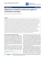
Figure 1 Detailed protocol and procedure. Schematic illustration of the detailed protocol on preparative procedure of conditioned media and
treatment courses as well as in vitro and ex vivo molecular-cellular studies.
Leu et al. Journal of Translational Medicine 2011, 9:11
/>Page 3 of 18
cells per mL culture medium )]. The sup ernatants were
collected at 36 h after cell culture and then stored at -
20°C for future use. These supernatants were defined
as 1) Normal (without AMI) MDCM and 2) Infarct-
related MDCM.
Definition of Conditioned Medium
The culture media utilized in the current study were
categorized into (1) Fresh medium (G1); (2) Normal
MDCM derived from cardiac cellular components of
normal rat hearts (G2); (3) Infarct-related MDCM
derived from cardiac cellular components of infarcted
hearts (G3). To investigate the concentration-dependent
impact, two concentrations (i.e. 10% and 20%) of G2 and
G3 media were adopted in the current study. The 10%
G2 medium was prepared by mixing 10% of G2 with 90%
of G1, while the 20% G2 medium was prepared by mixing
20% of G2 with 80% of G1. Similarly, the 10% and 20%
G3mediawerepreparedbymixing10%and20%ofG3
with 90% and 80% of G1, respectively.
Functional Assessment by Echocardiography
Transthoracic echocardiography was performed in each
group prior to and on day 90 after AMI induction with
the anesthetized rats in a supine position by an animal
cardiologist blinded to the design of the experiment
using a commercially available echocardiographic system
(UF-750XT) e quipped with a 8-MHz linear-array trans-
ducer for animals (FUKUDA Denshi Co. Hongo, Bun-
kyo-Ku, Tokyo, Japan). M-mode tracings of LV were
obtained with the heart being imaged in 2-dimensional
mode in short-axis at the level of the papillary muscle.
Left ventricular internal dimensions [end-systolic dia-
meter (ESD) and end-diastolic diameter (EDD)] were
measured according to the American Society of Echo-
cardiography leading-edge method using at least three
consecutives cardiac cycles. The LV ejection fraction
(LVEF) was calculated as follows: LVEF (%) =
[(LVEDD
3
-LVEDS
3
)/LVEDD
3
] × 100
Preparation of Neonatal Cardiac Fibroblasts and Grouping
(Figure 1)
Three-day-old newborn SD rats were euthanized by an
overdose of intraperitoneal sodium pentobarbital. The
hearts were removed after opening the chest wall and
cut into pieces, followed b y further lyses in enzymatic
digestive solution [50 mL PBS buffer containing 0.07 g
collagenase IV (Sigma), 14 mg protease XIV ( Sigma)
and 0.09 g glucose]. Finally, the CFBs were collected
and co-cultured with conditioned media.
The harvested CFBs (Figure 1A) were then divided
into three groups according to the culture medium in
whichtheywereincubated:Group1(5.0×10
5
CFBs
cultured in fresh medium for 48 h), Group 2 (5.0 × 10
5
CFBs co-cultured with 10% and 20% of normal MDCM
for 48 h, respectively), and Group 3 (5.0 × 10
5
CFBs co-
cultured with 10% and 20% of infarct-related MDCM
for 48 h, respectively).
Cellular Proliferation Test
To evaluate whether MDCM treatment promotes cellu-
lar proliferation in the infarct area, 5-bromodeoxyuri-
dine (BrdU) was intravenously given in Groups I, IV,
and VI animals on days 3, 5, 7, 9, and 12 after acute
AMI induction for labeling the proliferating cells.
Specimen Collection
Rats in each group were euthanized on day 90 after
AMI induction, and heart in each rat was rapidly
removed and immersed in cold saline. For immunohis-
tofluorescence (IHF) study, the heart tissue was rin sed
with PBS, embedded in OCT compound (Tissue-Tek,
Sakura, Netherlands) and snap-frozen in liquid nitrogen
before being stored at -80°C. For immunohistochemical
(IHC) staining, heart tissue was fixed in 4% formalde-
hyde and embedded in paraffin.
IHC Staining
Cardiac cross-sections were collected in the sixteen
additional rats in Groups I, IV, V, and IV (n = 4 per
group). To analyze the extent of collagen synthesis and
deposition, three cardiac paraffin sections (6 μm) at
3 mm intervals were stained with picro-Sirius red (1%
Sirius red in saturated picric acid solution) for one hour
at room temperature using standard methods. The sec-
tions were then washed twice with 0.5% acetic acid.
After dehydration in 100% ethanol thrice, the sections
were cleaned with xylene and mounted in a r esinous
medium. Ten low power fields (×10) of each section
were used to identify Sirius red-positive area on each
section. Image-pro plus 6.1 software (Media Cybernetics,
Inc., Bethesda, MD, USA) was used to calculate the total
cross-sectional area of left ventricle and the total area of
Sirius red-positive staining. The mean area of collagen
deposition (A) was obtained by summation of Sirius
red-positive areas on each section divided by the total
numbers of sectio ns. In addition, the mean cross-sec-
tional area (B) of left ventricle was obtained by dividing
the sum of all cross sectional areas with the total num-
ber of sectio ns examined. Finally, the percentage change
in area of collagen deposition was obtained by dividing
(A) with (B), followed by multiplication by 100%.
IHC of blood vessels was performed by incubating the
tissue sections with an anti-a-SMA (1:400) primary anti-
body at room tem perature for 1 h, followed by washing
with PBS thrice. Ten minutes after the addition of the
anti-mouse-HRP conjugated secondary antibody, the tis-
sue sections were washed with PBS thrice again. The
Leu et al. Journal of Translational Medicine 2011, 9:11
/>Page 4 of 18
3,3’ diaminobenzidine (DAB) (0.7 gm/tablet) (Sigma)
was then added, followed by washing with PBS thrice
after one minute. Finally, hematoxylin was added as a
counter-stain for nuclei, followed by washing twice with
PBS after one minute. Three sections of LV myocardium
were analyzed in each rat. For quantification, three ran-
domly selected HPFs (×100) were analyzed in each sec-
tion. The mean number per HPF for each animal
was then det ermined by summation of all numbers
divided by 9.
Western Blot Analysis for Connexin (Cx)43, CXCR4,
Stromal Cell-Derived Factor (SDF)-1a, and Oxidative
Stress Reaction in LV Myocardium
Equal a mounts (10-30 mg) of protein extracts from
remote viable LV myocardium were loaded and sepa-
rated by SDS-PAGE using 8-10% acrylamide gradients.
Following electrophoresis, the separated proteins were
transferred electrophoretically to a polyvinylidene
difluoride (PVDF) membrane (Amersham Biosciences).
Nonspecific proteins were blocked by incubating the
membrane in blocking buffer (5% nonfat dry milk in
T-TBS co ntaining 0.05% Tween 20) overnight. The
membranes were incubated with the indicated primary
antibodies (Cx43, 1:1000, Chemicon ; CXCR4, 1:1000,
Abcam; SDF-1, 1:1000, Cell Signaling; Actin, 1:10000,
Chemicon) for 1 h at room temperature for Cx43 and
CXCR4 and overnight at 4°C for SDF-1, respectively.
Horseradish peroxidase-conjugated anti-mouse immu-
noglobulin IgG (1:2000 , Amersham Biosciences) wa s
applied as the second antibody for Cx43 for 1 h at
room temperature; Horseradish peroxidase-conjugated
anti-rabbi t imm unoglobulin IgG (1:2000, Cell Signaling )
wasappliedasthesecondaryantibodyfor1hfor
CXCR4 and 45 minutes for SDF-1 at room temperature.
The washing procedure was repeated eight times
within 1 h.
The Oxyblot Oxidized Protein Detection Kit was pur-
chased from Chemicon (S7150). The oxyblot procedure
was performed according to our recent study [5].
The procedure of 2,4-dinitrophenylhydrazine (DNPH)
derivatization was carried out on 6 μgofproteinfor15
minutes according to manufacturer’s instructions. One-
dimensional electrophoresis was carried out on 12%
SDS/polyacrylamide gel after DNPH derivatization. Pro-
teins were t ransferred to nitrocellulose membranes
which were then incubated in the primary antibody
solution (anti-DNP 1: 150) for 2 h, followed by incuba-
tion with second antibody solution (1:300) for 1 h at
room temperature. The washing procedure was repeated
eight times within 40 minutes.
Immunoreactive bands were visualized by enhanced
chemiluminescence (ECL; Amersham Biosciences)
which was then exposed to Biomax L film (Kodak). For
quantification, ECL signals were digitized using Labwork
soft ware (UVP). For oxyblot protein analysis, a standard
control was loaded on each gel.
Real-Time Quantitative PCR Analysis
Real-time polymerase chain reaction (RT-PCR) was con-
ducted using LightCycler TaqMan Master (Roche,
Germany) in a single capillary tube according to the
manufacturer’s guidelines for individual component con-
centrations as we previously reported [5]. Forward and
reverse primers were each designed based on individual
exons of the target gene sequence to avoid amplifying
genomic DNA.
During PCR, the probe was hybridized to its comple-
mentary single-strand DNA sequence within the PCR
target. As amplification occurred, the probe was
degraded due to the exonuclease activity of Taq DNA
polymerase, thereby separating the quencher from
reporter dye during extension. During the entire amplifi-
cation cycle, light emission increased exponentially.
A positive result was determined by identifying the
threshold cycle value at which reporter dye emission
appeared above background.
Zymography Analysis Amplification
For zymography, supernatants from cultured neonatal
cardiac fibroblasts (CFBs) (Group 1, 10% and 20% of
Groups 2 and 3) w ere collected and centrifuged (500 g,
5 min) to remove cells and debris. Protein extract was
electrophoresed in 8% SDS-PAGE containing 0.1%
gelatin. After migration and washing, gels wer e incu-
bated (16 h, 37°C) in activation buffer (50 mM Tris-
base at pH 7.5, 5 mM CaCl
2
, 0.02% Na N
3
,and1μM
ZnCl
2
). Gels were stained with Coomassie staining
solution (0.5% Coomassie, 50% MeOH, 10% acetic acid,
and 40% H
2
O) for 90 minutes, followed by destaining
(0.5% Coomassie, 50% MeOH, 10% acetic acid, and
40% H
2
O). Quantification of Western blot and zymo-
graphy was performed with densitometry (TotalLab
v1.10, Nonlinear Dynamics; Durham, NC, http://www.
nonlinear.com).
Statistical Analysis
Data were expressed as mean val ues (mean ± SD). The
significance of differences between two groups was eval-
uated with t-tes t. The significance of differences among
the g roups was evaluated using analysis of variance fol-
lowe d by Bonferroni multiple-comparison post hoc test.
Statistical analyses were performed using SAS statistical
software for Windows version 8.2 (SAS institute, Cary,
NC). A probability value <0.05 was considered statisti-
cally significant.
Leu et al. Journal of Translational Medicine 2011, 9:11
/>Page 5 of 18
Results
Impact of Conditioned Medium on Cardiac Fibroblast
Gene Expressions
The mRNA e xpression of a -smooth muscle actin
(a-SMA)(Figure2A)inculturedCFBswasnotably
higher in Group 1 (CFBs cultured in fresh medium)
than in Group 2 (CFBs co-cultured with normal
MDCM) and Group 3 (CFBs co-cultured with infarct-
related MDCM), and notably higher in Group 2 than in
Group 3. On the other hand, the mRNA expressions of
both collagen type I a-1 (Figure 2B) and collagen type I
a-2 (Figure 2C) in cultured CFBs were similar between
Group 1 and Group 2, whereas their expressions were
notably suppressed in Group 3 compared with those in
Group 1 and 2.
The mRNA expression of major activator membrane
type 1-matrix metalloproteinase (MT1-MMP) (Figure 2D)
in cultured CFBs was notably higher in Group 3 than in
Group 1 and 2, and was significantly higher in Group 2
than in Group 1. In addition, the mRNA expressions of
MMP-2 (Figure 2E) and MMP-9 (Figure 2F) in cultured
CFBs were notab ly higher in Group 3 than in Group 1
and 2, and were remarkably higher in Group 2 than in
Group 1. In contrast, the mRNA expression of tissue
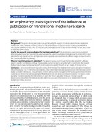
Figure 2 Impact of conditioned medium on cardiac fibroblast gene expressions. Effects of fresh medium (G1), 10% and 20% concentration
of normal rat myocardium-derived conditioned medium (MDCM) (G2) and 10% and 20% of myocardial infarct-related MDCM (G3) on gene
expressions of neonatal cardiac fibroblasts (n = 6 in each group). (A) mRNA expression of a-smooth muscle actin (SMA). G1 vs. G2 (10% & 20%)
vs. G3 (10% & 20%), p < 0.01. Symbols (*, †, ‡, §, ¶) indicate significance (at 0.05 level) (by Bonferroni multiple comparison post hoc test). (B) &
(C) mRNA expressions of both collagen type I a-1 (B) and collagen type I a-2 (C). *p < 0.02 between the indicated groups. (D) mRNA expression
of major activator membrane type 1-matrix metalloproteinase (MT1-MMP). *p < 0.01 between the indicated groups. (E) & (F) mRNA expressions
of matrix metalloproteinase (MMP)-2 and MMP-9. *p < 0.01 between the indicated groups. (G) mRNA expression of tissue inhibitor of
metalloproteinase-2 (TIMP-2). *p < 0.02 between the indicated groups. (H) mRNA expressions of vascular endothelial growth factor (VEGF). *p <
0.01 between the indicated groups. (I) mRNA expressions of vascular endothelial growth factor (VEGF). *p < 0.001 between the indicated groups.
Leu et al. Journal of Translational Medicine 2011, 9:11
/>Page 6 of 18
inhibitor of metalloproteinase-2 (TIMP-2) (Figure 2G) in
cultured CFBs was notably lower in Group 3 than in
Group 1 and 2, and was markedly lower in Group 2 than
in Group 1.
The mRNA expression of VEGF (Figure 2H) in cul-
tured CFBs was re markably increased in Group 3 than
in Group 1 and 2, and was significantly increased in
Group 2 than in Group 1. Furthermore, the mRNA
expression of SDF-1a (Figure 2I) in cultured CFBs was
similar between Group 1 and Group 2, whereas it was
notably increased in Group 3 than in the other groups.
Impact of Conditioned Medium on Protein Expressions of
Collagen Type I a-1 and a-SMA
Western blot analysis demonstrated that the protein
expression of collagen type I a-1 (Figure 3, left panel)
in cultured CFBs was remarkably lower in Group 3 than
in Group 1 and Group 2, and was sig nificantly lower in
Group 2 than in Group 1. Moreover, the a-SMA pro-
tein expression (Figure 3, right panel) in cultured CFBs
was significantly suppressed in Group 3 than in the
other two groups, but it did not differ between Group 1
and Group 2.
Comparison of the Expressions of Gelatinolytic Activity of
MMP-2 and MMP-9 in Supernatant of Cultured Neonatal
Cardiac Fibroblasts
The expressions of both pro-MMP-2 (pro-peptide) and
active MMP-2 (cleaved) (Figure 4, left panel) were sub-
stantially increased in Group 3 compared with those in
the other two groups, and were notably increased in
Group 2 than in Group 1. Similarly, the expressions of
both pro-MMP-9 (pro-peptide) and active MMP-9
(cleaved) showed consistent c hanges among the three
groups (Figure 4, left panel).
Increased Concentration of Interleukin (IL)-10,
Transforming Growth Factor (TGF)-b, VEGF, SDF-1a and
Basic Fibroblast Growth Factor (bFGF) in Infarct-related
Conditioned Medium
To determine the trophic effects of t he conditioned
media, the concentrations of five most common and
important chemokines (i.e. IL-10, TGF-b, VEGF, SDF-1a,
and bFGF) were measured by ELISA (Figure 5, A-D).
The concentration of IL-10 in normal MDCM was too
low to be detected. The concentration o f TGF-b in
serum [i.e. fetal bovine serum (FBS)] of fresh medium
was not measured because of its originally high concen-
tration. As compared with normal MDCM, the concen-
tration of TGF-b was remarkably higher in infarct-related
MDCM. The concentration of VEGF did not differ
between fresh medium and normal MDCM, whereas it
was significantly higher in infarct-related MDCM com-
pared with both fresh medium and normal MDCM. The
concentrations of SDF-1a and bFGF were notably higher
in normal MDCM and infarct-related MDCM than in
fresh medium, and significantly higher in infarct-related
MDCM than in normal MDCM.
Increased mRNA Expression of IL-10, TGF-b, VEGF, SDF-1a,
and bFGF in 36-hour cultured myocardium components
To determine whether the trophic effects of chemokines
in conditioned medium were derived from cultured cellu-
lar components, the mRNA expressions of IL-10, TGF-b,
VEGF, SDF-1a, and bFGF (Figure 5, E-I) were measured
in this study. The mRNA expressions of IL-10 and TGF-
b, two indicators of anti-inflammation, were remarkably
higher in infarct-related cultured cellular com p o ne n t s
than in normal cultured cellular components. Besides,
the mRNA expressions of VEGF, SDF-1a,andbFGF,
three pro-angiogenic indexes, were substantially higher
in infarct-related cultured cellular components than in
normal cultured cellular components.
Impact of Conditioned Medium Treatment on 90-Day Left
Ventricular Function and Fractional Shortening
The initial left ventricular ejection fraction (LVEF), frac-
tional shortening (FS), LVEDD and LVESD we re similar
among the six groups (Table 1). Besides, there was also
no significant difference between the 90-day LVEF and
FS among Group I, II and III. However, the 90-day
LVEF and FS were remarkably lower, whereas the
LVEDD and LVESD were notably higher in Group IV,
V, and VI than in Gro up I, II, and III. Furthermore, the
90-day LVEF and FS were significantly lower in Group
IVthaninGroupVandVI,andnotablylowerin
Group V than in Group VI. Moreover, the 90-day
LVEDD and LVESD were significantly higher in Group
IV than in Group V and Group VI, and notably higher
in Group V than in Group VI. These findings imply that
conditioned media, especially those derived from the
infarcted heart, was effective in preserving LV function
and inhibiting LV remodeling after AMI.
Impact of Conditioned Medium Treatment on Regulating
mRNA Expressions of SDF-1a, VEGF, Endothelial Nitric
Oxide Synthase (eNOS), Bcl-2, Bax, and Caspase-3 in LV
Myocardium
The impact of conditioned medium treatment on 90-day
left ventricular function and fractional shortening is
shown in Table 1. Real-time PCR analyses showed
remarkably lower mRNA expressions of SDF-1a, VEGF,
eNOS and Bcl-2 in Group IV than in other groups
(Figure 6). Conversely, the mRNA expressions of Bax and
caspase 3 were notably higher in Group IV than in other
groups. These findings suggest that conditioned medium
therapy up-regulated chemokines for angiogenesis and
suppressed cellular apoptosis in LV myocardium.
Leu et al. Journal of Translational Medicine 2011, 9:11
/>Page 7 of 18
Impact of Conditioned Medium Treatment on Oxidative
Stress
Western blotting revealed that although the mitochondrial
oxidative stress in LV myocardium did not differ among
Group I, II, and III on day 90 after AMI induction, it was
significantly higher in G roup IV than in other groups
and was notably higher in Group V than in Group VI
(Figure7).Theresults,therefore, showed an increase in
oxidative stress after AMI that was significantly suppressed
by MDCM, especially infarct-related MDCM.
Impact of Conditioned Medium Treatment on Enhancing
Protein Expressions of Cx43, CXCR4, and SDF-1a
Cx43proteinexpressioninLVmyocardiumonday90
after AMI in duction was similar among Group I, II, and
III, and was also similar between Group IV and Group
V (Figure 8, left panel). On the other hand, the expres-
sion was markedly higher in Group I, II, and III than in
Group IV, V, and VI, and notably higher in Group VI
than in Group IV and V. The results, therefo re, demon-
strated a notable suppression in Cx43 expression after
Figure 3 Impact of conditioned medium on protein expressions of collagen type I a-1 and a-SMA. (Left Panel) Protein e xpression of
collagen type I a-1 (COL1A1) in cultured cardiac fibroblasts (CFBs) (n = 6 per group). *p = 0.002 between the indicated groups. Protein
expression of COL1A1 in cultured CFBs. *p = 0.01 between the indicated groups. (Right Panel) Protein expression of a-smooth muscle actin
(a-SMA) in cultured CFBs (n = 6 per group). G1 vs. 10% G2 vs. 10% G3, p = 0.031. G1 vs. 20% G2 vs. 20% G3, p = 0.003.
Leu et al. Journal of Translational Medicine 2011, 9:11
/>Page 8 of 18
AMIinduction.Theexpression,however,wassignifi-
cantly restored after administration of infarct-related
MDCM.
CXCR4 protein expression in LV myocardium on day
90 after AMI induction did not differ among Group I,
II, and III was also similar between Group IV and V
(Figure 8, middle panel). However, the expression was
significantly higher in Group IV, V, and VI than in
Group I, II, and III, and was significantly higher in
Group VI than in Group IV and V.
In addition, there was also no significant difference in
SDF-1a protein expression in LV myocardium among
Group I, II and III and among Group IV, V and VI on
day 90 after AMI (Figure 8, right panel). However, the
expression was significantly higher in Group IV, V, and
VI than in Group I, II and III.
Impact of Conditioned Medium on Number of von
Willebrand Factor (vWF)-Positive Cells
Immunofluorescent staining identified remarkably
higher number of vWF-positive cells, a marker of
endothelial cells, in Group VI than in other groups (Fig-
ure 9). The number was also sign ificantly higher in
GroupI,II,andIIIthaninGroupIVandV,andalso
notably higher in Group V than in Group IV. However,
it showed no difference among Group I, II, and III.
These findings indicate that treatment with infarct-
related MDCM had a positive impact on angiogenesis.
Impact of Conditioned Medium on Cellular Proliferation
in Infarct Area of Left Ventricle
To determine whether conditioned medium treatment
enhanced cellular proliferation in LV infarct area,
Figure 4 Gelatinolytic activity of MMP-2 and MMP-9 in supernatant of cultured neonatal cardiac fibroblasts. Expressions of supernatant
gelatinolytic activity of MMP-2 and MMP-9 in fresh medium versus different conditioned media (n = 6 in each group). (Left Panel) Pro-MMP-2
and MMP-2 (cleaved). (1) G1 vs. 10% G2 vs. 10% G3, p < 0.0001 (* vs. ‡ or † vs. ¶, p < 0.001). (2) G1 vs. 20% G2 vs. 20% G3, p < 0.0001 (§ vs.
** or # vs. ##, p < 0.001). (Right Panel) Pro-MMP-9 and MMP-9 (cleaved). (1) G1 vs. 10% G2 vs. 10% G3, p < 0.0001 (* vs. ‡ or † vs. ¶, p < 0.001).
(2) G1 vs. 20% G2 vs. 20% G3, p < 0.0001 (§ vs. ** or # vs. ##, p < 0.001).
Leu et al. Journal of Translational Medicine 2011, 9:11
/>Page 9 of 18
intra-venous injection of BrdU was given to Group I,
IV,andVI.Theresultsdemonstratedthatbyday90
after AMI induction, the cellular uptake of BrdU, an
index of cellul ar proliferation, was remarkably elevated
in Group VI compared with that in other groups
(Figure 10). It was also significantly higher in Group
IV than in Group I.
Impact of Conditioned Medium on Reducing Collagen
Expression
To investigate whether conditioned medium treatment
reduced collagen expression in infarct area of LV myo-
cardium, Sirius red staining was performed for Group I,
IV, V, and VI in the current study. The collagen deposi-
tion area was substantially higher in Group IV than in
Figure 5 ELISA analysis on conditioned medium and mRNA expression profile of cultured cellular components. Comparison of ELISA
findings of supernatant concentrations of transforming growth factor (TGF)-b, VEGF, stromal cell-derived factor (SDF)-1a, and basic fibroblast
growth factor (bFGF) between normal MDCM and infarct-related MDCM after 36 h cell culture (n = 6 per group). (A) TGF-b, * vs. †, p < 0.001;
(B) VEGF, *p < 0.0001 between the indicated groups; (C) SDF-1a, *p < 0.05 between the indicated groups; (D) bFGF, *p < 0.03 between the
indicated groups. Comparisons of mRNA expressions of IL-10, TGF-b, VEGF, SDF-1a, and bFGF in normal cultured cardiac cell components and
infarct-related cultured cell components after 36 h cell culture (n = 6 per group). (E) IL-10, * vs. †, p < 0.0001; (F) TGF-b, * vs. †, p = 0.0001;
(G) VEGF, * vs. †, p = 0.0017; (H) SDF-1a, * vs. †, p < 0.0001; (I) bFGF, * vs. †, p < 0.0001.
Table 1 Echocardiographic Findings Prior to and on Day 90 after AMI
Variables Group I (n = 6) Group II (n = 6) Group III (n = 6) Group IV (n = 6) Group V (n = 6) Group VI (n = 6) P‡ value
LVEF (%)* 81.5 ± 2.07 80.8 ± 1.39 79.7 ± 1.48 80.8 ± 1.44 81.7 ± 3.18 80.2 ± 1.75 0.512
FS (%)* 42.4 ± 1.95 43.7 ± 2.46 42.9 ± 1.26 43.2 ± 1.82 44.2 ± 1.88 43.6 ± 1.61 0.648
LVEDD (cm)* 0.60 ± 0.01 0.61 ± 0.01 0.62 ± 0.02 0.60 ± 0.02 0.59 ± 0.03 0.60 ± 0.01 0.871
LVESD (cm)* 0.33 ± 0.02 0.32 ± 0.01 0.34 ± 0.01 0.32 ± 0.02 0.31 ± 0.03 0.33 ± 0.01 0.794
LVEF (%)† 79.8
a
± 1.46 79.3
a
± 2.44 79.2
a
± 2.07 63.4
b
± 1.71 69.8
c
± 2.03 74.8
d
± 2.87 <0.0001
FS (%)† 43.0
a
± 1.21 43.2
a
± 1.75 43.1
a
± 0.85 30.9
b
± 0.50 35.2
c
± 2.19 38.7
d
± 1.21 <0.0001
LVEDD (cm)† 0.61 ± 0.01
a
0.60 ± 0.01
a
0.59 ± 0.02
a
1.0 ± 0.01
b
0.77 ± 0.02
c
0.69 ± 0.03
d
<0.0001
LVESD (cm)† 0.34 ± 0.01
a
0.31 ± 0.02
a
0.33 ± 0.02
a
0.66 ± 0.02
b
0.49 ± 0.02
c
0.40 ± 0.02
d
<0.0001
Data expressed as means ± SD.
AMI = acute myocardial infarction; LVEF = left ventricular ejection fraction; FS = fractional shortening; LVEDD = left ventricular end-diastolic dimension; LVESD =
left ventricular systolic dimension.
*Transthoracic echocardiography performed at day 0 prior to AMI induction.
†Transthoracic echocardiograp hy performed on day 90 after AMI induction.
Group I = sham control treated by fresh medium;
Group II = sham control treated by normal heart myocardium-derived conditioned medium (MDCM);
Group III = sham control treated by infarcted-related MDCM;
Group IV = AMI induction treated by fresh medium;
Group V = AMI induction treated by normal heart MDCM;
Group VI = AMI induction treated by infarcted-related MDCM.
‡One-way ANOVA on the arcsine transformed data was use d to improve the normality for statistical analysis. Letters (
a, b, c, d
) indicate significance (at 0.05 level)
by Bonferroni multiple comparison post hoc test (
a
versus
b, c, d
,
b
versus
c, d
,
c
versus
d
, all p values <0.05).
Leu et al. Journal of Translational Medicine 2011, 9:11
/>Page 10 of 18
Figure 6 Impact of conditioned medium treatment on mRNA expression of angiogenic and apoptotic factors in left ventricular
myocardium. Real-time PCR showing significantly lower mRNA expressions of (A)SDF-1a, (B)VEGF, (C) endothelial nitric oxide synthase (eNOS),
and (D) Bcl-2 in LV myocardium in Group IV (AMI treated by fresh medium) than in other groups (p < 0.03) (n = 6 in each group). Note also
remarkably higher gene expressions of (E) Bax and (F) caspase-3 in Group IV than in other groups (p < 0.01).
Figure 7 Impa ct of conditioned medium treatment on oxidative stress . Western blotting of oxidative index in left ventricular (LV)
myocardium of Group I to VI on day 90 after AMI induction (left), with quantification results of each group (n = 6) shown (right). *p < 0.003
between the indicated groups.
Leu et al. Journal of Translational Medicine 2011, 9:11
/>Page 11 of 18
other groups. It was also rem arkably higher in Group V
than in Group VI and I, and was significantly higher in
GroupVIthaninGroupI(Figure11).Thesefindings
suggest that treatment with infarct-related MDCM sig-
nificantly inhibited collagen deposition in infarct zone of
LV myocardium.
Impact of Conditioned Medium on Angiogenesis
IHC staining for Group I, IV, V and VI demonstrated
notably higher number of small vessels positively stained
for a-SMA in Group VI than in other groups (Figure 12).
The number was also remarkably higher in Group I than
in Group IV and V, and was notably increased in Group V
than in Group IV. These findings indicate that treatment
with infarct-related MDCM significantly enhanced
neovascularization.
Discussion
The present study, which investigated the potential
impact of MDCM on heart function and LV remodeling
in a rat AMI model, provided several valuable implica-
tions. First, the gene expression of collagen and the pro-
tein expressions of both the collagen and a-SMA of
CFBs were significantly s uppressed after co-culturing
with infarct-related MDCM. Second, gelatinolytic analy-
sis demonstrated notably increased MMP-2 and MMP-9
activities in CFBs after co-culturing with infarct-related
MDCM. Third, ELISA finding showed remarkably
higher VEGF, SDF-1a, bFGF, and TGF-b levels in
infarct-related MDCM compa red with those in normal
MDCM. Fourth, fibrosis and oxidative stress in LV myo-
cardium were markedly attenuated, whereas CXCR4 and
SDF-1a protein expressi ons as we ll as ang iogen esis/vas-
culogenesis were substantially increased after treatment
with infarct-related MDCM on day 90 after AMI.
Importantly, both LVEF and FS were notably preserved
and LV remodeling was remarkably suppressed follow-
ing infarct-related MDCM administration.
Conditioned Medium Treatment Improved LV Function
after AMI
Although stem cell therapy appears to be an attractive
and promising option in treatment of ischemic organ
dysfunction [1-6,8-10], the principal mechanism is still
poorly defined [3-5,8,9]. Growing evidence suggests that
the reparation, regeneration, and improvement in
ischemic organ dysfunction after stem cell therapy is
mainly due to its cytokine/paracrine [3-5,10,11,13]
effects and angiogenesis [3-5,8,9] rather than the results
of differentiation of transplanted cells per sec into parti-
cular cell phenotype. Indeed, studies have reveale d that
MSC-derived conditioned medium significantly contri-
butes to the positive impacts of cell therapy [13,22].
Interestingly, while the conditioned medium derived
from MCSs has been well reported to preserve the
function of other ischemia-related organ disorders
[13,22,23], the therapeutic benefit of MDCM in ische-
mia-related LV dysfunction has not been reported. The
novel finding in the present study is that infarct-related
MDCM notably preserved heart function and markedly
Figure 8 Impact of conditioned medium treatment on protein expressions of Cx43, CXCR4, and SDF-1a. Western blot of LV myocardium
(n = 6 in each group). (Left) Protein expression of connexin43 (Cx43). *p < 0.0001 between the indicated groups. (Middle) Protein expression of
CXCR4. *p < 0.001 between the indicated groups. (Right) Protein expression of SDF-1a. *p < 0.001 between the indicated groups.
Leu et al. Journal of Translational Medicine 2011, 9:11
/>Page 12 of 18
Figure 9 Impact of conditioned medium on number of von Willebrand factor (vWF)-positive cells. Immunofluorescent staining (400×) for
von Willebrand factor (vWF)-positive cells in LV myocardium in sham-operated controls and infarcted animals (n = 6 in each group). *p < 0.001
between the indicated groups. Scale bars in right lower corner represent 50 μm.
Leu et al. Journal of Translational Medicine 2011, 9:11
/>Page 13 of 18
attenuated LV remodeling after AMI. Furthermore,
although it is less effective compared with infarct-related
MDCM, normal MDCM treatment still significantly
improved heart function after AMI. Therefore, our find-
ings, in addition to strengthening those of previous stu-
dies [13,22,23], further highlight the therapeutic
potential of conditioned medium derived from myocar-
dial components of ischemic heart, a mimicked ischemic
preconditioning, in the treatment of ischemic hea rt
disease.
Interestingly, previous clinical observational studies
[3,21] have shown that patients with ischemic precondi-
tioning experience less myocardial damage, better pre-
servation of LV function, and more favorable clinical
outcome after AMI compared with those without. Con-
sistently, numerous animal model studies [22,23] have
also establish ed a therapeutic benefit of preconditioning
in preventing myocardial damage from ischemia-
reperfusion injury. Although the precise mechanisms of
preconditioning against myocardial damage from AMI
attack or ischemia-reperfusion injury are t ill not fully
understood, this phenomenon may at least partly
account for the positive therap eutic impact of treatment
with infarct-related MDCM on LV function in the cur-
rent study.
Possible Mechanisms Underlying MSC-Derived
Conditioned Medium Therapy in Improving Heart
Function
The paracrine mediators secreted by MSCs have
been identified to be chemokines and cytokines in
both the cultured medium and MSC-implanted area
[11,13,22,24]. The chemokines, which consist mainly of
SDF-1a, VEGF, and HGF, are called trophic factors that
have been reported to contribute to the mobilization of
endothelial progenitor cell/MSC into circulation and
homing to ischemic area for angiogenesis/vasculogenesis
and regeneration, thereby improving ischemia-related
organ dysfunction [24-27]. In addition, cytokines includ-
ing M MP-2, MMP-9, and TIMP, which are well known
Figure 10 Impact of conditioned medium on cellula r proliferation in infarct area of left ventri cle. Immunohistochemical (IHC) staining
(400×) for the distribution of proliferative cells in infarction area of LV myocardium (n = 6 in each group). *p < 0.0001 between the indicated
groups. Scale bars in right lower corner represent 50 μm.
Leu et al. Journal of Translational Medicine 2011, 9:11
/>Page 14 of 18
Figure 11 Impact of conditioned medium on collagen expression. Sirius red staining for collagen deposition in LV myocardium (n = 4). *p
< 0.001 between the indicated groups.
Figure 12 Impact of conditioned medium on angiogenesis. The number of arterioles in infarct LV myocardium (n = 6). Quantification (right
panel) of small vessels (diameters ≤15 mm) (yellow arrows) on 90 day following AMI induction (200 ×). *p < 0.0001 between the indicated
groups. Scale bars in right lower corner represent 50 μm.
Leu et al. Journal of Translational Medicine 2011, 9:11
/>Page 15 of 18
regulators of extra-cellular matrix (ECM) formation
[13,28], have also been shown to modulate CFB activity
and play an essential role in regulating LV remodeling
[13]. Indeed, previous studies have already demonstrated
the importance of trophic mediators in this process
[11,13,22,24-28].
Improvement of Heart Function after AMI from Findings
of Current Study–Paracrine Effects and Angiogenesis
One important finding in the current study is that ELISA
showed a remarkably higher level of VEGF, a common
index of angiogenesis, and SDF-1a, a well-known trophic
chemokine, in infarct-related MDCM compared with nor-
mal MDCM and fresh medium. Moreover, real-time PCR
showed that the mRNA expressions of VEGF and SDF-1a
in both cultured CFBs and infarcted LV myocardium were
significantly higher using normal MDCM compared with
fresh medium. The expressions of these mediators, inter-
estingly, were further enhanced when infarct-related
MDCM was used. Furthermore, Western blot analysis
demonstrated a notable increase in CXCR4 protein
expression, a marker of endothelial progenitor cells, in
infarcted LV myocardium when normal MDCM was used
instead of fresh medium. It was further upregulated after
administration of infarct-related MDCM. Moreover, the
protein expression of SDF-1a, a chemokine for EPC mobi-
lization, was also elevated in infarcted LV after administra-
tion of infarct-related MDCM compared with infusion of
fresh medium. Finally, real-time PCR and Immunofluores-
cent staining of infarcted LV myocardium showed that the
expression of eNOS, an indicator of endothelial function,
and the number of vWF-positive cells, a marker of
endothelial cells, were significantly higher when normal
MDCM was applied and further elevated when infarct-
related MDCM was given as compared with fresh med-
ium. The results, in addition to strengthening those of
previous studies [24-27], are consistent with other findings
in this study including an increase in the positivity of
a-SMA staining (i.e. an indicator of angiogenesis/vasculo-
genesis) and cellular proliferation in infarcted LV myocar-
dium. Taken together, our findings could, at least in part,
account for the preservation of cardiac function in the set-
ting of AMI after MDCM treatment.
Interestingly, rece nt studies have shown that gene
therapy using over-expressions of VEGF and SDF-1
genes significantly improves ischemia-related LV dys-
function in experimental studies [29,30]. Similarly,
results of the current study using myocardial infarction-
induced enhancement of paracrine secretions for treat-
ment of AMI, in addition to being c omparable to those
of the recent studies, further clarify the roles of chemo-
kine/cytokine and the mechanisms underlying the
improvement in heart function after AMI.
Inhibition of LV Remodelling–Crucial Role of MMPs
The principal finding in the present study is that both con-
ditioned media enhanced the mRNA expressions of
MMP-2, MMP-9, and TM1-MMP in cultured CFBs. In
contrast, TIMP-2 mRNA expression in cultured CFBs, an
indicator of the trend of developing cardiac fibrosis,
was markedly suppressed by both conditioned media.
Additionally, the gelatinolytic activities of MMP-2 and
MMP-9 in supernatant of cultured CFBs were remarkably
upregulated by both conditioned media. On the other
hand, a-SMA expression and collagen secretion by cul-
tured CFBs were remarkably suppressed by conditioned
media. Furthermore, Sirius-red staining showed that the
fibrosis in LV infarct area was significantly reduced by
normal MDCM and furt her suppressed by infarct-related
MDCM as compared with fresh medium. Our findings,
therefore, in addition to reinforcing the results of previous
studies [13], may partially explain the attenuation of post-
AMI LV remodeling after MDCM treatment.
Impact of Conditioned Medium on Oxidative Stress,
Cellular Apoptosis, and Cx43 expression
The mRNA expressions of Bax and caspase-3, indexes of
apoptosis, were notably reduced in infarcted LV myocar-
dium after treatment with either conditioned medium
compared with fresh medium. On the other h and, the
expressions of Bcl-2 and eNOS, two indicators of anti-
apoptosis, were significantly elevated in infarcted LV
myocardium following administration of the two types of
conditioned media compared to fresh medium treatment.
Besides, Western blot demonstrated re markably reduced
oxidative stress in infarcted LV myocardium after adminis-
tration of normal MDCM compared to treatment with
fresh medium. It was further suppressed in infarct-related
medium. The link between increased oxidative stress and
cellular apoptosis has been established in ischemic condi-
tion [4,5,31]. Furthermore, an association between an
increase in both cellular apoptosis and oxidative stress and
a decreased Cx43 expression in ischemic myocardium,
which plays a key role in electrical coupling between cardi-
omyocytes [32,33], has been demonstrated in our previous
studies [4,5]. The notable reduction in protein expression
of Cx43 in infarcted LV myocardium and its restoration
after administration of infarct-related MDCM further sup-
port our findings of less LV remodeling and better LV
function in animals receiving infarct-related MDCM com-
pared with the other treatment groups.
Study Limitations
This study has limitations. First, the harvested cellular ele-
ments from ex vivo digestion contain various cellular com-
ponents including cardiomyocytes and CFBs that together
constitute 90% of cells in myocardium and also endothelial
Leu et al. Journal of Translational Medicine 2011, 9:11
/>Page 16 of 18
cells that make up less than 10 % of the cell population.
Therefore, although both chemokines and cytokines were
identified in MDCM, this study cannot specifically identify
their exact sources. Second, since a variety of complex
cytokine-mediated interactions after my ocardial injury
have been suggested [34], other mediators that may parti-
cipate in the process of post-AMI LV remodeling can-
not be identified without a detailed p roteomic screening
study for MDCM. Third, since the heart has b een sug-
gested to contain endogenous cardiac stem cells [35],
their precise involvement in tissue regeneration and
repair after MDCM treatment remains unknown.
Finally, although studies have previously reported that
myocardium-derived medium can induce the differen-
tiation of bone marrow mesenchymal stem cells [36],
thecurrentstudydidnotevaluatetheimpactofMDCM
on the differentiation of the stem cells to provide infor-
mation to address this issue.
In conclusion, although the exact mechanisms underly-
ing the positive therapeutic potential of MDCM treatment
in suppressing LV remodeling and preserving LV function
after AMI remain uncertain, our demonstration of further
enhancement of the therapeutic effect using infarct-related
conditioned medium suggests that an interplay of cyto-
kines, a reduction in oxidative stress, an en hanced stem
cell homing effect and angi ogenesis appear to be the key
elements contr ibuting to the improvement in heart func-
tion after infarction. Thesefindingsalsosupportthe
proposal that the positive impact of MSC therapy on
ischemia-rel ated heart dysfunction is due to i ts paracrine
effects instead of differentiation of implanted MSCs into
specific cell phenotype in the ischemic area.
Acknowledgements
This study was supported by a program grant from Chang Gung Memorial
Hospital, Chang Gung University (grant no. CMRPG 880291).
Author details
1
Division of Cardiology, Department of Internal Medicine, Chang Gung
Memorial Hospital - Kaohsiung Medical Center, Chang Gung University
College of Medicine, Kaohsiung, Taiwan.
2
Center for Translational Research in
Biomedical Sciences, Chang Gung Memorial Hospital - Kaohsiung Medical
Center, Chang Gung University College of Medicine, Kaohsiung, Taiwan.
3
Department of Medical Research, E-DA Hospital, I-Shou University,
Kaohsiung, Taiwan.
4
Division of General Surgery, Department of Surgery,
Chang Gung Memorial Hospital - Kaohsiung Medical Center, Chang Gung
University College of Medicine, Kaohsiung, Taiwan.
5
Basic Science, Nursing
Department, Meiho University, Pingtung, Taiwan.
Authors’ contributions
All authors have read and approved the final manuscript. SL, YHK, YCL, and
CKS designed the experiment, drafted and performed animal experiments.
LTC, THT, SC, KHY, and CJW were responsible for the laboratory assay and
troubleshooting. MF and HKY participated in refinement of experiment
protocol and coordination and helped in drafting the manuscript.
Author’s information
Cheuk-Kwan Sun contributed equally as the first author to this work. Morgan
Fu contributed equally compared with the corresponding author to this
work.
Competing interests
The authors declare that they have no competing interests.
Received: 17 September 2010 Accepted: 18 January 2011
Published: 18 January 2011
References
1. Strauer BE, Brehm M, Zeus T, Kostering M, Hernandez A, Sorg RV, Kogler G,
Wernet P: Repair of infarcted myocardium by autologous intracoronary
mononuclear bone marrow cell transplantation in humans. Circulation
2002, 106:1913-1918.
2. Wang JS, Sh um-Tim D, Chedrawy E, Chiu RC: Thecoronarydeliveryof
marrow str omal cells for myocardial regeneration: pathophysiologic
and therapeutic implications. J Thorac Cardiovasc Surg 2001,
122:699-705.
3. Dai W, Hale SL, Martin BJ, Kuang JQ, Dow JS, Wold LE, Kloner RA:
Allogeneic mesenchymal stem cell transplantation in postinfarcted rat
myocardium: short- and long-term effects. Circulation 2005, 112:214-223.
4. Yip HK, Chang LT, Wu CJ, Sheu JJ, Youssef AA, Pei SN, Lee FY, Sun CK:
Autologous bone marrow-derived mononuclear cell therapy prevents
the damage of viable myocardium and improves rat heart function
following acute anterior myocardial infarction. Circ J 2008, 72:1336-1345.
5. Sun CK, Chang LT, Sheu JJ, Chiang CH, Lee FY, Wu CJ, Chua S, Fu M,
Yip HK: Bone marrow-derived mononuclear cell therapy alleviates left
ventricular remodeling and improves heart function in rat-dilated
cardiomyopathy. Crit Care Med 2009, 37:1197-1205.
6. Makino S, Fukuda K, Miyoshi S, Konishi F, Kodama H, Pan J, Sano M,
Takahashi T, Hori S, Abe H, et al: Cardiomyocytes can be generated from
marrow stromal cells in vitro. J Clin Invest 1999, 103:697-705.
7. Toma C, Pittenger MF, Cahill KS, Byrne BJ, Kessler PD: Human
mesenchymal stem cells differentiate to a cardiomyocyte phenotype in
the adult murine heart. Circulation 2002, 105:93-98.
8. Tse HF, Kwong YL, Chan JK, Lo G, Ho CL, Lau CP: Angiogenesis in
ischaemic myocardium by intramyocardial autologous bone marrow
mononuclear cell implantation. Lancet 2003, 361:47-49.
9. Davani S, Marandin A, Mersin N, Royer B, Kantelip B, Herve P, Etievent JP,
Kantelip JP: Mesenchymal progenitor cells differentiate into an
endothelial phenotype, enhance vascular density, and improve heart
function in a rat cellular cardiomyoplasty model. Circulation 2003,
108(Suppl 1):II253-258.
10. Mangi AA, Noiseux N, Kong D, He H, Rezvani M, Ingwall JS, Dzau VJ:
Mesenchymal stem cells modified with Akt prevent remodeling and
restore performance of infarcted hearts. Nat Med 2003, 9:1195-1201.
11. Zhang M, Mal N, Kiedrowski M, Chacko M, Askari AT, Popovic ZB, Koc ON,
Penn MS: SDF-1 expression by mesenchymal stem cells results in trophic
support of cardiac myocytes after myocardial infarction. Faseb J 2007,
21:3197-3207.
12. Porter KE, Turner NA: Cardiac fibroblasts: at the heart of myocardial
remodeling. Pharmacol Ther 2009, 123:255-278.
13. Mias C, Lairez O, Trouche E, Roncalli J, Calise D, Seguelas MH, Ordener C,
Piercecchi-Marti MD, Auge N, Salvayre AN, et al: Mesenchymal stem cells
promote matrix metalloproteinase secretion by cardiac fibroblasts and
reduce cardiac ventricular fibrosis after myocardial infarction. Stem Cells
2009, 27:2734-2743.
14. Ottani F, Galvani M, Ferrini D, Sorbello F, Limonetti P, Pantoli D, Rusticali F:
Prodromal angina limits infarct size. A role for ischemic preconditioning.
Circulation 1995, 91:291-297.
15.
Noda T, Minatoguchi S, Fujii K, Hori M, Ito T, Kanmatsuse K, Matsuzaki M,
Miura T, Nonogi H, Tada M, et al: Evidence for the delayed effect in
human ischemic preconditioning: prospective multicenter study for
preconditioning in acute myocardial infarction. J Am Coll Cardiol 1999,
34:1966-1974.
16. Divald A, Kivity S, Wang P, Hochhauser E, Roberts B, Teichberg S,
Gomes AV, Powell SR: Myocardial ischemic preconditioning preserves
postischemic function of the 26S proteasome through diminished
oxidative damage to 19S regulatory particle subunits. Circ Res 2010,
106:1829-1838.
17. Cheng Y, Zhu P, Yang J, Liu X, Dong S, Wang X, Chun B, Zhuang J,
Zhang C: Ischaemic preconditioning-regulated miR-21 protects heart
against ischaemia/reperfusion injury via anti-apoptosis through its target
PDCD4. Cardiovasc Res 2010, 87:431-439.
Leu et al. Journal of Translational Medicine 2011, 9:11
/>Page 17 of 18
18. Cribier A, Korsatz L, Koning R, Rath P, Gamra H, Stix G, Merchant S, Chan C,
Letac B: Improved myocardial ischemic response and enhanced
collateral circulation with long repetitive coronary occlusion during
angioplasty: a prospective study. J Am Coll Cardiol 1992, 20:578-586.
19. Kosuge M, Kimura K, Kojima S, Sakamoto T, Ishihara M, Asada Y, Tei C,
Miyazaki S, Sonoda M, Tsuchihashi K, et al: Effects of preinfarction angina
pectoris on infarct size and in-hospital mortality after coronary
intervention for acute myocardial infarction. Am J Cardiol 2003,
92:840-843.
20. Anzai T, Yoshikawa T, Asakura Y, Abe S, Akaishi M, Mitamura H, Handa S,
Ogawa S: Preinfarction angina as a major predictor of left ventricular
function and long-term prognosis after a first Q wave myocardial
infarction. J Am Coll Cardiol 1995, 26:319-327.
21. Cao CM, Yan WY, Liu J, Kam KW, Zhan SZ, Sham JS, Wong TM: Attenuation
of mitochondrial, but not cytosolic, Ca2+ overload reduces myocardial
injury induced by ischemia and reperfusion. Acta Pharmacol Sin 2006,
27:911-918.
22. Caplan AI, Dennis JE: Mesenchymal stem cells as trophic mediators. J Cell
Biochem 2006, 98:1076-1084.
23. Togel F, Weiss K, Yang Y, Hu Z, Zhang P, Westenfelder C: Vasculotropic,
paracrine actions of infused mesenchymal stem cells are important to
the recovery from acute kidney injury. Am J Physiol Renal Physiol 2007,
292:F1626-1635.
24. Sadat S, Gehmert S, Song YH, Yen Y, Bai X, Gaiser S, Klein H, Alt E: The
cardioprotective effect of mesenchymal stem cells is mediated by IGF-I
and VEGF. Biochem Biophys Res Commun 2007, 363:674-679.
25. Takahashi T, Kalka C, Masuda H, Chen D, Silver M, Kearney M, Magner M,
Isner JM, Asahara T: Ischemia- and cytokine-induced mobilization of bone
marrow-derived endothelial progenitor cells for neovascularization. Nat
Med 1999, 5:434-438.
26. Schober A, Karshovska E, Zernecke A, Weber C: SDF-1alpha-mediated
tissue repair by stem cells: a promising tool in cardiovascular medicine?
Trends Cardiovasc Med 2006, 16:103-108.
27. Kocher AA, Schuster MD, Bonaros N, Lietz K, Xiang G, Martens TP,
Kurlansky PA, Sondermeijer H, Witkowski P, Boyle A, et al: Myocardial
homing and neovascularization by human bone marrow angioblasts is
regulated by IL-8/Gro CXC chemokines. J Mol Cell Cardiol 2006,
40:455-464.
28. Huang PH, Chen YH, Wang CH, Chen JS, Tsai HY, Lin FY, Lo WY, Wu TC,
Sata M, Chen JW, Lin SJ: Matrix metalloproteinase-9 is essential for
ischemia-induced neovascularization by modulating bone marrow-
derived endothelial progenitor cells. Arterioscler Thromb Vasc Biol 2009,
29:1179-1184.
29. Elmadbouh I, Haider H, Jiang S, Idris NM, Lu G, Ashraf M: Ex vivo delivered
stromal cell-derived factor-1alpha promotes stem cell homing and
induces angiomyogenesis in the infarcted myocardium. J Mol Cell Cardiol
2007, 42:792-803.
30. Das H, George JC, Joseph M, Das M, Abdulhameed N, Blitz A, Khan M,
Sakthivel R, Mao HQ, Hoit BD, et al: Stem cell therapy with overexpressed
VEGF and PDGF genes improves cardiac function in a rat infarct model.
PLoS One 2009, 4:e7325.
31. Garrido AM, Griendling KK: NADPH oxidases and angiotensin II receptor
signaling. Mol Cell Endocrinol 2009, 302:148-158.
32. Jalife J, Morley GE, Vaidya D: Connexins and impulse propagation in the
mouse heart. J Cardiovasc Electrophysiol 1999, 10:1649-1663.
33. Vozzi C, Dupont E, Coppen SR, Yeh HI, Severs NJ: Chamber-related
differences in connexin expression in the human heart. J Mol Cell Cardiol
1999, 31:991-1003.
34. LaFramboise WA, Scalise D, Stoodley P, Graner SR, Guthrie RD,
Magovern JA, Becich MJ: Cardiac fibroblasts influence cardiomyocyte
phenotype in vitro. Am J Physiol Cell Physiol 2007, 292:C1799-1808.
35. Mazhari R, Hare JM: Mechanisms of action of mesenchymal stem cells in
cardiac repair: potential influences on the cardiac stem cell niche. Nat
Clin Pract Cardiovasc Med 2007, 4(Suppl 1):S21-26.
36. Xie XJ, Wang JA, Cao J, Zhang X: Differentiation of bone marrow
mesenchymal stem cells induced by myocardial medium under hypoxic
conditions. Acta Pharmacol Sin 2006, 27:1153-1158.
doi:10.1186/1479-5876-9-11
Cite this article as: Leu et al.: Myocardium-derived conditioned medium
improves left ventricular function in rodent acute myocardial infarction.
Journal of Translational Medicine 2011 9:11.
Submit your next manuscript to BioMed Central
and take full advantage of:
• Convenient online submission
• Thorough peer review
• No space constraints or color figure charges
• Immediate publication on acceptance
• Inclusion in PubMed, CAS, Scopus and Google Scholar
• Research which is freely available for redistribution
Submit your manuscript at
www.biomedcentral.com/submit
Leu et al. Journal of Translational Medicine 2011, 9:11
/>Page 18 of 18