- Trang chủ >>
- Khoa Học Tự Nhiên >>
- Vật lý
nanocrystalline apatite-based biomaterials, 2009, p.95
Bạn đang xem bản rút gọn của tài liệu. Xem và tải ngay bản đầy đủ của tài liệu tại đây (1.26 MB, 95 trang )
NANOCRYSTALLINE
APATITE-BASED BIOMATERIALS
No part of this digital document may be reproduced, stored in a retrieval system or transmitted in any form or
by any means. The publisher has taken reasonable care in the preparation of this digital document, but makes no
expressed or implied warranty of any kind and assumes no responsibility for any errors or omissions. No
liability is assumed for incidental or consequential damages in connection with or arising out of information
contained herein. This digital document is sold with the clear understanding that the publisher is not engaged in
rendering legal, medical or any other professional services.
NANOCRYSTALLINE
APATITE-BASED BIOMATERIALS
D. EICHERT
C. DROUET
H. SFIHIA
C. REY
AND
C. COMBES
Nova Science Publishers, Inc.
New York
Copyright © 2009 by Nova Science Publishers, Inc.
All rights reserved. No part of this book may be reproduced, stored in a retrieval system or
transmitted in any form or by any means: electronic, electrostatic, magnetic, tape, mechanical
photocopying, recording or otherwise without the written permission of the Publisher.
For permission to use material from this book please contact us:
Telephone 631-231-7269; Fax 631-231-8175
Web Site:
NOTICE TO THE READER
The Publisher has taken reasonable care in the preparation of this book, but makes no expressed
or implied warranty of any kind and assumes no responsibility for any errors or omissions. No
liability is assumed for incidental or consequential damages in connection with or arising out of
information contained in this book. The Publisher shall not be liable for any special,
consequential, or exemplary damages resulting, in whole or in part, from the readers’ use of, or
reliance upon, this material.
Independent verification should be sought for any data, advice or recommendations contained
in this book. In addition, no responsibility is assumed by the publisher for any injury and/or
damage to persons or property arising from any methods, products, instructions, ideas or
otherwise contained in this publication.
This publication is designed to provide accurate and authoritative information with regard to the
subject matter covered herein. It is sold with the clear understanding that the Publisher is not
engaged in rendering legal or any other professional services. If legal or any other expert
assistance is required, the services of a competent person should be sought. FROM A
DECLARATION OF PARTICIPANTS JOINTLY ADOPTED BY A COMMITTEE OF THE
AMERICAN BAR ASSOCIATION AND A COMMITTEE OF PUBLISHERS.
LIBRARY OF CONGRESS CATALOGING-IN-PUBLICATION DATA
Nanocrystalline apatite-based biomaterials / D. Eichert ... [et al.].
p. ; cm.
Includes index.
ISBN 978-1-60741-212-0 (E-Book)
1. Bone substitutes. 2. Nanocrystals. 3. Apatite. I. Eichert, D.
[DNLM: 1. Bone Substitutes--metabolism. 2. Apatites--chemistry. 3. Biomimetic Materials-chemistry. WE 200 N186 2009]
RD755.6.N36 2009
610.28--dc22
2008042003
Published by Nova Science Publishers, Inc. + New York
CONTENTS
Preface
vii
Chapter 1
Introduction
1
Chapter 2
Early Works and the Way Bone Mineral was
Conceived
3
Chapter 3
Synthesis of Nanocrystalline Apatites
9
Chapter 4
Characterization of Apatites
13
Chapter 5
A Model for Nanocrystalline Apatites
29
Chapter 6
Physico-Chemical Properties of Nanocrystalline
Apatites
33
Processing of Nanocrystalline Apatite-Based
Biomaterials
45
Biological Properties of Nanocrystalline Apatites
59
Chapter 7
Chapter 8
Conclusion
63
References
65
Index
77
PREFACE
The improvement of the biological activity and performance of bone
substitute materials is one of the main concerns of orthopaedic and dental surgery
specialists. Biomimetic nanocrystalline apatites exhibit enhanced and tunable
reactivity as well as original surface properties related to their composition and
mode of formation. Synthetic nanocrystalline apatites analogous to bone mineral
can be easily prepared in aqueous media and one of their most interesting
characteristics is the existence of a hydrated surface layer containing labile ionic
species. Ion exchange and macromolecule adsorption processes can easily and
rapidly take place due to strong interactions with the surrounding fluids. The ion
mobility in the hydrated layer allows direct crystal-crystal or crystal-substrate
bonding. The fine characterization of these very reactive nanocrystals is essential
and can be accomplished with different tools including chemical analysis and
spectroscopic techniques such as FTIR, Raman and solid state NMR. The
reactivity of the hydrated layer of apatite nanocrystals offers material scientists
and medical engineers extensive possibilities for the design of biomaterials with
improved bioactivity using unconventional processing. Indeed apatitic
biomaterials can be processed at low temperature which preserves their surface
reactivity and biological properties. They can also be associated in various ways
with active molecules and/or ions. Several examples of use and processing of
nanocrystalline apatites involved in the preparation of tissue-engineered
biomaterials, cements, ceramics, composites and coatings on metal prostheses are
presented.
Chapter 1
INTRODUCTION
Osteoarticular pathologies are, at all ages, the first cause of handicap and raise
concern in public healthcare. Articular aging, traumatology, child growth defects,
bone tumor treatment, osteoporosis and related bone failures, constitute points for
which research efforts are necessary. Bone diseases and induced defects are also
of prime importance in maxillo-facial surgery and odontology especially for aging
populations. The shortcomings of bone auto- or allografts which in addition
involve secondary operations, risks of disease transmission as well as
immunological rejection and morbidity justified the development of synthetic
bone graft materials.
In the past decades, the effort to adapt the first biomaterials taken from other
technical domains (e.g. alumina, carbon, titanium carbide and nitride, plaster of
Paris), or to design new materials better suited to biological applications, led to
significant advances. However in the opinion of many researchers the ultimate
achievement would be the perfect imitation of biological tissues and more
importantly the improvement of biological repair and maintenance processes.
Ideally, a substitute material should mimic the living tissue’s mechanical,
chemical, biological and functional properties; however the design of a complex
structure such as bone is still impossible to achieve without the aid of Nature
which masters, using sophisticated chemical properties and processes, this high
performance mineral-protein composite.
Poorly crystalline apatites (PCA) are the major inorganic constituent of
mineralized tissues in vertebrates. The imitation of bone mineral has inspired the
research and development of calcium phosphate (CaP) based biomaterials. The
most famous and most widely used CaP compound is stoichiometric
hydroxyapatite processed as dense or porous ceramics, coatings and composites.
2
D. Eichert, C. Drouet, H. Sfihia et al.
However, the design of biomaterials has evolved and today the function of a
biomaterial is not restricted to physical substitution but the novel biomaterial
should actively participate in the process of bone regeneration implying reactivity
of the component(s). In this view we will show in this chapter how nonstoichiometric nanocrystalline apatite-based biomaterials can fulfil two of the
main challenges: mimicking bone mineral crystal structure and composition and
exhibiting a controlled reactivity regarding interactions with components of
biological fluids (ions, proteins).
This chapter reports part of our “bioinspired” research based on the idea that
the development and the processing of bioactive biomaterials for bone substitution
or regeneration applications can take advantage of a thorough knowledge and
understanding of the structure and properties of the tissue to be substituted. Due to
the complex structure and heterogeneity of biological systems such as bone,
synthetic apatites are generally used to throw light on the surface reactivity of
bone mineral. However the characterization of nanocrystalline apatite analogous
to bone mineral is difficult due to its relative instability and poor crystallinity.
Even though the results are complex, recent fine investigations on synthetic PCA
revealing their original surface reactivity are useful both in biomaterials or
biomineralization.
The present chapter gathers the main results of our most recent investigations
on nanocrystalline apatite composition, structure and properties with the aim of
better understanding bone mineral properties and achieving better design of new
bioactive nanocrystalline apatite based biomaterials or improving the biological
performance of existing bone substitutes. This chapter which alternates between
results on synthetic apatites and their significance for biological apatites is
organized in seven sub-sections: 1) a description of bone composition and
structure and the way it was perceived through the 20th century are presented first,
2) synthetic routes of nanocrystalline apatites are discussed with an emphasis on
biomimetic nanocrystalline apatite preparation and maturation at room
temperature and at physiological pH as developed in our research group, 3) the
complementary characterization techniques that we use to investigate the global
and local fine structure and composition of nanocrystalline apatites are reported 4)
a model for apatite nanocrystals is put forward which can explain the properties of
synthetic and biological apatite nanocrystals, 5) the physico-chemical properties
of biomimetic apatites and their involvement in the biological behavior of PCA
based materials are presented and discussed, 6) examples of nanocrystalline
apatite based biomaterial showing some potential with regards to nanocrystalline
apatite surface reactivity are described, 7) finally, the biological properties of
apatites are presented.
Chapter 2
EARLY WORKS AND THE WAY BONE MINERAL
WAS CONCEIVED
The studies on bone mineral composition and structure were rather puzzling
for the first investigators. Before the development of structural analysis by X-ray
diffraction (XRD), until the beginning of the 20th century, chemical composition
was one of the major characterization tools used to identify biominerals. Other
identification methods, for example based on the use of the optical properties of
crystals could not be applied to biological apatites due to the small size of their
crystals. Chemical analyses revealed the diversity of phosphate-containing
biominerals. Three major components are always present: calcium, phosphate and
carbonate, and were readily identified but they showed variable contents
depending on the species, the individuals, their location in the body or the age and
the type of mineralized tissue considered, in contrast with the other major
biomineral, calcium carbonate, showing a rather constant composition. It was then
accepted, in accordance with the hypothesis of Haüy concerning the composition
of calcium phosphate-carbonate minerals, that several phases co-existed in hard
tissues of vertebrates: essentially tricalcium phosphate and calcium carbonate
[McConnel 1973].
The first structural identifications of biominerals using X-ray diffraction were
obtained by de Jong in 1926 [de Jong 1926]. He established that calcium
phosphate biominerals of vertebrates corresponded to an apatite structure and
since that time bone mineral has been frequently identified as hydroxyapatite
(HA):
Ca10 (PO4)6 (OH)2
4
D. Eichert, C. Drouet, H. Sfihia et al.
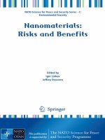
which was later considered to crystallize in the hexagonal system (space group
P63/m), see on figure 1, and then shown to be monoclinic, at room temperature,
when stoichiometric [Posner 1958, Elliott 1973]. The XRD data revealed very
broad bands indicating low cristallinity. However, the identification of a poorly
crystalline apatite structure did not explain the existence of significant amounts of
carbonate in all mineralized biological tissues. Thus it was considered that bone
mineral was a mixture of poorly crystalline apatite and amorphous calcium
carbonate or that carbonate ions were at least in part adsorbed on the apatite
crystal surface [Elliott 1994].
OH
O
Ca
P
Figure 1. Projection on the (001) plane of hydroxyapatite structure. The two Ca2+ triangles
lining the "tunnels" of the structure are located at z ẳ and ắ. OH- ions are slightly under or
above the triangles [Cazalbou 2004] - Reproduced by permission of The Royal Society of
Chemistry.
It was only in the sixties that studies on synthetic carbonated apatites
established that all carbonate ions could in fact be located within the apatite
structure [Legeros 1968, Labarthe 1973]. But this has been the object of much
controversy. Detailed studies indicated that carbonate ions could be located in the
two anionic sites of the apatite structure: in PO43- sites (type B carbonated apatite)
and OH- sites (type A carbonated apatite). Bone apatites were believed to
correspond essentially to type B carbonated apatite whereas enamel contained
both type A and B carbonate. The fraction of type A carbonate in dental enamel
was evaluated at about 10% of the total carbonate content using infrared
Early Works and the Way Bone Mineral was Conceived
5
spectroscopy [Elliott 1985]. The use of carbonated apatite as a model for
biological calcifications of hard tissues of vertebrates is nowadays accepted. The
variability of the composition of apatite minerals and their mode of formation
however needed further investigation.
From a kinetic point of view the direct formation of apatite crystals in
calcified tissues was considered as unlikely, regarding the slow growth rate of
apatite crystals and the existence in body fluids of crystal growth inhibitors such
as magnesium and carbonate ions [Campbell 1991]. The improvement of
crystallinity of bone apatites upon aging, revived for a while the theory of an
amorphous phase considered as a necessary precursor in the formation of
biological apatites [Termine 1966]. This hypothesis based on studies dealing with
the formation and stability of amorphous calcium phosphates and its progressive
conversion into apatite, compared to bone mineral evolution upon aging, was thus
considered as quite consistent for a decade. In the eighties however it was
demonstrated that bone mineral was composed of apatite nanocrystals with no or a
non-detectable amorphous phase [Grynpas 1984]. The peculiar shape of bone
crystals (platelets) showing non-equivalent a and b directions perpendicular to the
c-axis of the hexagonal structure (figure 1), however led to the hypothesis that
apatite formation involved another precursor phase, very close to apatite and
indiscernible considering the very poor crystallization state of the mineral:
triclinic OctaCalcium Phosphate (OCP) [Brown 1987]:
Ca8 (PO4)4 (HPO4)2, 5H2O
The OCP structure has been shown to consist in the association of an apatitelike layer and a hydrated layer [Mathew 1988]. Precipitating as platelet-shaped
crystals, this phase exhibits a high crystal growth rate and hydrolyzes readily in
aqueous media into apatite, forming interlayered compounds with hydroxyapatite
and preserving the original platelet shape of the crystals. This model does not
however explain all the variability of apatite compositions and particularly the
very similar role played by carbonate and HPO42- ions in bone mineral and
synthetic analogues [Neuman 1956].
The chemical composition of biological apatite has been the object of several
approximations frequently based on the composition of model minerals or
synthetic analogues. A general chemical formula proposed by Winand for HPO42-containing apatite was [Winand 1961]:
Ca10-x (PO4)6-x (HPO4)x (OH)2-x with 0 ≤ x ≤ 2
6
D. Eichert, C. Drouet, H. Sfihia et al.
and by Labarthe et al. for carbonate-containing apatites [Labarthe 1973]:
Ca10-x (PO4)6-x (CO3)x (OH)2-x with 0 ≤ x ≤ 2.
These formula establish a similar behavior for bivalent ion substitution of trivalent
phosphates: the creation of a cationic vacancy and an anionic vacancy in
monovalent sites. These chemical formulas are consistent with the limit
composition observed (x=2) and the decrease of the OH- content when the amount
of carbonate and/or HPO42- in the apatite increases. Other chemical formulas have
been proposed. The most general one [Rey 2006]:
Ca10-x+u (PO4)6-x-y (HPO42- or CO32-)x+y (OH)2-x+2u-y with 0 ≤ x ≤ 2 and 0 ≤ 2u +y ≤ x
is however of little relevance for biological apatites which are best approximated
by the simple combination of the two previous formulas taking into account the
possible existence of type A carbonates:
Ca10-x (PO4)6-x (HPO4 or CO3)x (OH or ½ CO3)2-x with 0 ≤ x ≤ 2
The compilation of different cortical bone analyses suggests a relatively
homogeneous composition [Legros 1987]:
Ca8.3 (PO4)4.3 (HPO4 or CO3)1.7 (OH or ½ CO3)0.3
characterized by a very high vacancy content close to the maximum (x=1.7). The
carbonate content varies with age: it is very low in embryonic bone, and can
represent up to 80% of the bivalent ions in the bone mineral of old vertebrate
animals. The OH- content of bone is very low at any age and OH- ions can barely
be detected [Rey 1995, Pasteris 2004]. The composition of tooth enamel crystals
reveals a radically different chemical composition:
Ca9.4 (PO4)5.4 (HPO4 or CO3)0.6 (OH or ½ CO3)1.4
showing a much lower vacancy content, unveiling the unique adaptability of
apatites to their biological functions [Cazalbou 2004].
Other minor substitutions are found in biological apatites involving for
example trivalent cations (e.g. rare earth elements, actinides) or monovalent
cations (especially Na+) for Ca2+, tetravalent ions replacing PO43-, and bivalent
ions replacing OH-. Several charge compensation mechanisms have been
Early Works and the Way Bone Mineral was Conceived
7
proposed. Although the ability of the apatite structure to fix many elements has
several consequences regarding intoxications with mineral ions and diseases, such
possibilities seem to have a minor influence on the chemical formula of apatites in
calcified tissues due to the low amounts of these foreign ions [Iyengar 1999].
The present data underline the strong heterogeneity of bone mineral and
apatitic biomineralizations. The global compositions do not reflect strong local
variations between osteons and within osteons, and probably between the crystals
themselves [Paschalis 1996]. In addition several properties of bone mineral such
as ion exchange suggest the existence of surface modifications and possibly
surface compositions different from the bulk at the level of a nanocrystal. The
heterogeneities of the mineral are among its chief characteristics, mainly related to
bone remodeling processes and formation conditions. Parameters susceptible to
evaluate these characteristics would be of great utility.
The replacement and healing of damaged hard tissues have always been a
concern for human beings as shown by the examination of mummies. It is
however only very recently that calcium phosphates have been used for bone
substitution and repair [Jarcho 1979]. The first to be used were stoichiometric
hydroxyapatite (HA) and β-tricalcium phosphate (β-TCP) which are stable CaP at
high temperature and can be easily sintered into ceramics. They are still the major
industrial CaP biomaterials. β-TCP was shown to be bioabsorbable and replaced
by bone whereas HA constituted non-degradable materials. β-TCP is mainly used
as a bioceramic whereas HA is also being processed for other biomaterials uses
such as the coating of metallic prostheses where it was found to considerably
improve bone repair as an "osteoconductive" material or composite ceramicpolymer materials showing strong mechanical analogies with bone tissues and
excellent bone bonding abilities [de Groot 1987, Bonfield 1988]. Biphasic
Calcium Phosphates (BCP), associating these two high-temperature CaP allow a
controlled resorption rate and have been reported to offer superior biological
properties [Daculsi 2003, Legeros 2002]. They are progressively replacing β-TCP
ceramics in Europe. A new technological step was made with the development of
CaP cements [Brown 1986]. These materials are able to set and harden in a living
body and most can be injected. Despite their poor mechanical properties they
offer a number of advantages and are increasingly used for several applications.
More recently biomimetic coatings involving low temperature nanocrystalline
CaP have been proposed - some have been claimed to exhibit osteoinductive
properties [Habibovic 2006].
Chapter 3
SYNTHESIS OF NANOCRYSTALLINE APATITES
As mentioned above, synthetic nanocrystalline apatites are of undeniable
interest in the preparation of apatite-based bioceramics for bone substitution,
repair or augmentation applications. However, synthetic apatites are also prepared
and studied to better understand the formation of biological apatites and some of
their properties [Legeros 1994]. Since the eighties, several synthetic routes have
emerged and in the near future they could challenge the high-energy conventional
processes involving high temperatures. Among the major advantages of these
emerging unconventional processes are the use of low temperature (from room
temperature (RT) to about 400°C), the flexibility and range of chemical
compositions, and the physical, chemical and biological properties of these CaP.
Synthetic apatites can be prepared by several methods (precipitation under
conditions of constant or changing composition, hydrolysis, solid/solid reaction at
high temperature, hydrothermal methods) the type of which determines the
amount and kind of substitution in the apatite. In this section we will focus on the
synthesis of calcium-deficient apatites in solution systems.
Several processes (precipitation by double decomposition, sol-gel method,
hydrolysis) involving various media (aqueous, hydro-alcoholic or organic
solutions) leading to calcium phosphate apatites have been reported. Sol-gel
processes still raise some problems: the long time needed for the preparation of
the sol, and the presence of other calcium phosphate phases depending on aging
time and temperature [Liu 2002]. Calcium-deficient or substituted apatites can
also be prepared by hydrolysis of amorphous calcium phosphate, dicalcium
phosphate dihydrate, octacalcium phosphate, or α and β tricalcium phosphate for
example. Hydrolysis of these calcium phosphate phases to yield apatite can
proceed through a dissolution-reprecipitation mechanism depending on the pH,
10
D. Eichert, C. Drouet, H. Sfihia et al.
the temperature and the presence of other ions. The latter can act as inhibitors
(magnesium, pyrophosphate ions) or promotors (fluoride ions) of hydrolysis of
dicalcium phosphate dihydrate (DCPD: CaHPO4 2H2O) and OCP for example
[Legeros 1994].
Two major parameters determine the crystallinity and the calcium deficiency
of the apatite obtained by precipitation methods: temperature (ambient
temperature to 100°C) and pH (basic). When precipitated from solutions at
temperatures between 80°C and 100°C, the higher the initial pH, the lower the
calcium deficiency. Precipitation at temperatures under 80°C leads to less and less
crystallized apatites, and the synthesis of poorly crystalline apatites analogous to
bone mineral can be easily achieved at ambient temperature and physiological pH
according to the method reported in the next sub-section.
Interestingly, precipitation using a hydro-alcoholic medium with a dielectric
constant lower than that of water, provides control of hydrogenphosphate and
carbonate ion content in apatite analogous to bone mineral [Zahidi 1985,
Rodrigues 1998, Dabbarh 2000]. In addition, several studies reported the
influence of the drying process and temperature on the composition, structure and
degree of crystallinity of apatitic calcium phosphates [Dabbarh 2000, Lebugle
1986].
3.A. POORLY CRYSTALLINE APATITE (PCA) SYNTHESIS
The results presented in this chapter are related to poorly crystalline apatites
synthesized at ambient temperature and physiological pH by double
decomposition between a phosphate and carbonate solution (for example, 40g of
(NH4)2HPO4, 20g of NaHCO3 and concentrated ammonia solution 1 ml in 500 ml
of deionized water) and a calcium solution (fr example, 17.7g of Ca(NO3)2 4H2O
in 250 ml of deionized water) as previously published [Rey 1989]. The calcium
solution is rapidly poured into the phosphate and carbonate solution at room
temperature (20°C) and stirred only for a few minutes. For investigations on
freshly-precipitated nanocrystalline apatites, the precipitate is then very quickly
filtered under vacuum and washed with deionized water (2 liters). Then the gel is
freeze-dried and finally stored in a freezer to prevent further maturation of the
PCA nanocrystals. This method leads to a carbonated poorly crystalline apatite
analogous to bone mineral [Rey 1995]. Non-carbonated poorly crystalline apatite
can be prepared by this method with a carbonate-free phosphate solution. Other
recipes involving cationic and anionic solutions with slightly different Ca/P ratio
and no concentrated ammonia solution also leads to poorly crystalline apatite. In
Synthesis of Nanocrystalline Apatites
11
all cases, the large excess of phosphate (and bicarbonate) ions in the solution
provides pH buffering at pH = 7.4.
3.B. POORLY CRYSTALLINE APATITE (PCA) MATURATION
To study the physical-chemical properties of nanocrystalline apatites, the
apatite can be left to mature after precipitation at room temperature in the mother
solution without stirring and in a stoppered vial to minimize the release and
uptake of CO2 at physiological pH. This evolution in solution (maturation) is an
important process that can help us understand the evolution of the composition,
structure and properties of biological and synthetic biomimetic apatites after
different aging times (corresponding to young and old bones for example).
After maturation during variable periods of time (from an hour to several
months), the precipitates were filtered under vacuum and washed with deionized
water. In the case of subsequent ion exchange experiments, part of the gel is then
freeze-dried (reference sample) and the other part is used for ion exchange
treatments.
The study of maturation properties of PCA is presented in section 6.A.
3.C. IONIC EXCHANGE (DIRECT AND INVERSE) ON POORLY
CRYSTALLINE APATITE (PCA)
Two kinds of ion exchange experiments can be performed on PCA: anionic
exchange (and the study of the reversibility of exchange between carbonate and
hydrogenophosphate) ions, and cationic exchange (and the study of the
reversibility of exchange between calcium and other cations such as strontium or
magnesium ions as reported in this chapter). Also, such ion exchange experiments
can be carried out on immature gels or on apatite samples matured for various
durations.
The ion exchange is performed by exposing either the gel or the mature
sample of nanocrystalline carbonated apatite (CA) to an "exchange" solution
containing the target ion (HCO3- or HPO42- for anionic hydrogenphosphate ⇔
carbonate exchange, Mg2+ or Sr2+ for cationic exchange with Ca2+) at varying
concentrations (for example 1 M for 10 minutes). The starting salts used for the
preparation of the "exchange" solution can be NaHCO3, (NH4)2HPO4, Mg(NO3)2
and Sr(NO3)2. Part of the "exchanged" samples are then filtered, washed with
12
D. Eichert, C. Drouet, H. Sfihia et al.
deionized water and freeze-dried. The other part is re-suspended for 10 minutes in
the "inverse exchange" solution, containing the initial ion at a concentration of 1
M. In all cases, the samples are finally filtered, washed with deionized water and
freeze-dried. In the text the notation "CA/X" represents a carbonated apatite
exchanged with the ion X, and "CA/X/Y" represents the same sample after
inverse exchange with the ion Y. For example, "CA/Sr" refers to a carbonated
apatite for which part of the calcium ions has been exchanged with strontium ions,
and "CA/Sr/Ca" refers to the same sample after inverse exchange of Sr2+ by Ca2+.
The studies of ionic exchange properties of PCA are presented in section 6.B.
Chapter 4
CHARACTERIZATION OF APATITES
4.A. CHEMICAL ANALYSIS
Depending on the precipitation conditions, on the maturation time and/or on
ion exchange treatments, the composition of calcium-deficient apatites can vary
significantly. The determination of calcium, total phosphate and carbonate ions
can be easily performed in different ways whereas the direct evaluation of
hydrogenphosphate ions which are one of the most important markers of PCA and
bone mineral crystals has not yet been possible (only indirect measurements are
available). All the chemical analysis methods used for apatite characterization are
based on the dissolution of apatite in acidic solution before the analysis (calcium
and orthophosphate ions determination) or during the analysis (carbonate ions).
Calcium concentration can be determined by complexometry with EDTA and
the phosphorus concentration by UV-visible spectrophotometry of the phosphovanado-molybdenum complex. The Ca/P atomic ratio of apatites can be calculated
from the result of these two analyses. The relative uncertainty on calcium and
phosphorus concentrations has been evaluated at 0.5 %.
The titration of HPO42- ions is more complex because the two inorganic
3-
2-
orthophosphate ions, PO4 and HPO4 , encountered in apatites are in rapid
equilibrium in solution. So the dissolution of apatite in acidic solution and the
determination of total phosphorus content by colorimetry cannot distinguish
2-
3-
2-
HPO4 and PO4 . Therefore, the only way to determine the HPO4 content is to
condense the ions into pyrophosphates according to equation 1. Then the HPO42level is determined by chemical analysis using the Gee and Dietz method which is
based on the formation of pyrophosphate in apatite-containing HPO42- ions upon
heating [Gee 1953]. During treatment of the apatite at 500°C for 3 hours (or
14
D. Eichert, C. Drouet, H. Sfihia et al.
600°C for 20 min) the following reaction occurs leading to the formation of
pyrophosphate ions [Gee 1955]:
2-
4-
2 HPO4 → P2O7 + H2O
(eq. 1)
After thermal treatment, phosphorus atoms are titrated as orthophosphate at
3-
3-
460 nm by absorption spectrophotometry before (PO4 only) and after (PO4 and
2-
HPO4 ) acid hydrolysis of the P-O-P bond of pyrophosphate ions at 100°C for 1
2-
hour. Thus, the level of condensed phosphate (therefore that of HPO4 ) is
calculated from the difference of the results of these two analyses. The
2-
pyrophosphate content corresponding to the concentration of HPO4 is
determined within 0.5 %.
It shall be noted that this analysis method cannot be used to accurately
2-
quantify the HPO4 content in bone and in synthetic carbonated apatites due to the
presence of carbonate ions that can interfere with pyrophosphate ions and partially
4-
prevent P2O7 formation according to equations 2 and 3 [Elliott 1994]:
2-
2-
3-
2 HPO4 + CO3 → 2 PO4 + CO2 + H2O
4-
2-
(eq. 2)
3-
P2O7 + CO3 → 2 PO4 + CO2
(eq. 3)
To open up a new alternative to the long and tedious analytical method of Gee
and Dietz involving several steps and treatments, we recently set up a method to
easily and rapidly evaluate the HPO42- content in apatite, based on the
mathematical decomposition of the ν4PO4 band of the Fourier Transform InfraRed
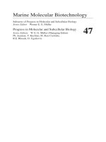
(FTIR) spectrum [Combes 2001]. Figure 2 shows the good correlation obtained
between HPO42- determined by chemical analysis] and by FTIR spectroscopy
using Gee et al. and Combes et al. methods, respectively. However, the curvefitting parameters (decomposition of the ν4PO4 band, see on figure 5) need to be
refined extensively to obtain a better correlation between these two methods, and
thus apply this tool to rapid investigations on biological apatites.
The carbonate content of apatites was determined using a CO2 coulometer
(UIC Inc., USA) that measures the CO2 released during sample dissolution in
acidic conditions (HClO4, 2M) and in a closed system. The CO2 released is
transferred into a photometric cell in a non-aqueous medium and titrated through
an acid-base reaction [Huffman 1977].
FTIR band relative intensity
Characterization of Apatites
15
28
26
24
22
20
18
16
14
5
10
15
20
25
Chemical Analysis
Figure 2. Correlation between HPO42- content in apatite determined by chemical analysis
and by FTIR spectroscopy [Combes 2001].
The determination of the amount of other ions taken up in PCA after ion
exchange (strontium, magnesium ions for example) was performed using atomic
absorption spectroscopy.
From the determination of the concentration of calcium, phosphate, carbonate
and foreign ions if present (Mg2+, Sr2+), we calculated atomic ratios such as Ca/P,
Ca/(P+C), or C/P in order to follow the evolution of the chemical composition of
synthetic and biological PCA during maturation and/or after ion exchange
processes (direct or inverse).
4.B. DIFFRACTION TECHNIQUES
The vast majority of the diffraction studies dealing with nanocrystalline
apatites and reported in the literature is based on X-ray diffraction and only few
electron diffraction data are available. This could be partly explained by the
strong tendency for apatite nanocrystals to agglomerate leading to broad diffuse
rings [Suvorova 1999]. Also, the way that electron beams affect these rather
unstable compounds has not yet been established. To our knowledge, no neutron
diffraction work has been dedicated so far to such poorly crystallized compounds.
The X-ray diffraction technique applied to the study of nanocrystalline apatite
specimens is often primarily used to determine their apatitic phase purity.
Although the presence of secondary crystalline phases such as pyrophosphates or
whitlockite can generally be distinguished from the sharpness of their
characteristic XRD patterns (within the detection limit of the equipment), the
detection of other phases such as octacalcium phosphate (OCP), whose diffraction
16
D. Eichert, C. Drouet, H. Sfihia et al.
pattern is rather similar to that of hydroxyapatite, or amorphous calcium
phosphate (ACP), generally requires special attention. The occurrence of a sharp
low-angle diffraction peak around d=18 Å can however betray the presence of
OCP for concentrations above the detection limit. A background halo in the range
27-40° (λCo = 1.78892 Å), and to a lesser extent 50-60°, is evidence of the
presence of ACP. A quantitative comparison with the XRD patterns obtained for
mixtures of known amounts of apatite and ACP can then be used for an estimate
of the ACP amount present in the specimen. Pattern fitting methods can also be
used for this evaluation. This was done for example by Rogers et al. who followed
the amounts of ACP and nanocrystalline apatite present in coatings formed after
immersion of a titanium substrate in simulated body fluid [Rogers 2005].
Beside phase purity evaluation, X-ray diffraction studies related to
nanocrystalline apatites have mostly been dedicated to the evaluation of average
crystal dimensions based on line broadening analysis [Arsenault 1988, Bonar
1983, Burnell 1980, Fisher 1987]. General findings indicate that the platelet-like
apatite nanocrystals exhibit an average length along the c-axis in the range 200400 Å for a thickness of about 20-80 Å. The width of a diffraction peak is indeed
dependent on the size of the crystallites constituting the sample, following a
1/cosθ mathematical law (where θ is the diffraction angle) such as the Scherrer
formula [Scherrer, 1918]:
Lhkl =
Kλ
β size cos θ hkl
(eq. 4)
where Lhkl is the average crystallite size perpendicular to the plane (hkl), λ is the
X-ray wavelength, K is a constant close to unity dependent on the particle shape,
and βsize is the line broadening due to the size effect and θhkl is the diffraction
angle corresponding to the (hkl) plane.
However, two other factors also contribute to the overall line broadening: the
existence of strain within the sample, giving a broadening effect following a tgθ
law, and the instrument-related broadening effect. The latter can generally be
evaluated from the XRD pattern of a well-crystallized reference sample such as
stoichiometric hydroxyapatite (HA), for which size and strain broadening effects
are considered negligible. The line broadening due to the sample itself (βsample)
can then be reached from the peak width observed after elimination of this
instrumental contribution (βinstr). However, this process depends on the
geometrical shape of the peak. Gaussian, Lorentzian (Cauchy), or a convolution of
the two, are mathematical functions generally used for fitting X-ray diffraction