Hệ thống xương khớp trên gia cầm
Bạn đang xem bản rút gọn của tài liệu. Xem và tải ngay bản đầy đủ của tài liệu tại đây (3.4 MB, 13 trang )
1
Pathology
Pathology
of
of
the
the
locomotor
locomotor
system
system
Skeletal system
Pathology
Pathology
of
of
the
the
bones
bones
• Transformation of the vertebrae and
bones
• Osteopathies
– abnormalities of the osteogenesis
• Disturbances of the mineralization
• Regressive changes of the bones
Transformation of the vertebrae
Transformation of the vertebrae
and bones
and bones
• Spondylolisthesis
• Tibiarotation
• Twisted leg syndrome
• Spread leg syndrome
Transformation of the vertebrae
Transformation of the vertebrae
• Spondylolisthesis
– devolution of the vertebra
– and tilting at the same time
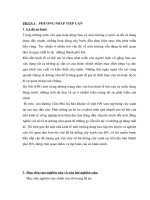
Anatomy
• functional anatomy
– The spinal column is parallel with the ground in a
newly hatched chick on the first day
– the physiological lordosis develops on the first
week
– and so does the position, pose typical for the
species
Photo: Dr. Dobos-Kovács, Mihály
Spondylolisthesis
Spondylolisthesis
(
(
devolution
devolution
of
of
the
the
vertebra
vertebra
)
)
• The chicks develop fastest in the first 3
weeks of age
– The pectoral muscles grow considerably
– later it is even more explicit
• the large pectoral musculature….
– pulls the 6. thoracic vertebra and moves it
from its normal position….
– The spinal column suffers refraction at the
level of the 6th vertebra….
2
Photo: Dr. Dobos-Kovács, Mihály
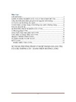
Spondylolisthesis
Spondylolisthesis
(
(
devolution
devolution
of
of
the
the
vertebra
vertebra
)
)
During walking
• from the direction of the hind legs
• power acts forward and downward
– On the previously tilted vertebra
• the back of the vertebra moves
– up and forward
• the front of the vertebra moves
– down and backwards
• the 6. thoracic vertebra moves downwards
– slips (devolution)
• and tilts from its original pose
Photo: Dr. Dobos-Kovács, Mihály
Photo: Dr. Dobos-Kovács, Mihály
Spondylolisthesis
Spondylolisthesis
(
(
devolution
devolution
of
of
the
the
vertebra
vertebra
)
)
• Typical lesion in large meat type chicken hybrids with
huge pectoral musculature
– affects smaller or larger part of the flock
– husbandry problems (keeping and nutrition factors) are also
involved
• The 6th vertebra which moved from its original
position…
– compresses the ventral part of the spinal cord
– Waller-degeneration occurs in the motoric nerve tracts
– weakness and paralysis develops in the hind legs
• consequences:
• the affected animals first limp, later are not able to
move
– no drinking (uricosis)
– exsiccosis
– starving to death
Photo: Dr. Dobos-Kovács, Mihály
3
Transformations
Transformations
of
of
the
the
spinal
spinal
column
column
• Lordosis
– the spinal column bends up
• Kyphosis
– the spinal column bends down
• Scoliosis
– the spinal column bends in S shape
Transformations
Transformations
of
of
the
the
spinal
spinal
column
column
• In case of spondylolisthesis
– and during spondylosis too
• pain occurs
– due to the compression of the spinal cord
• and due to the compression of the nerve tracts too
• The bird tries to reach the less painful pose
(position of the spinal column)
– in most cases immediately gets paralyzed
• The abnormal pose of the spinal column
– ossifies soon and gets fastened
– according to the abnormal, but less painful position!
Scoliosis
Photo: Dr. Dobos-Kovács, Mihály
Photo: Dr. Dobos-Kovács, Mihály
Transformation
Transformation
of
of
the
the
bones
bones
• Tibiarotation
– the tibiotarsal bone gets twisted
around its longitudinal axis
– usually turns laterally
• 45-90-180°
– the distal part of the join turns with
– and so does the lower part of the
leg
– The joint turns too
The tendon follows the joint, it
remains in the pit!!!
• Twisted leg syndrome
• Spread leg syndrome
Photo: Dr. Dobos-Kovács, Mihály
4
Photo: Dr. Dobos-Kovács, Mihály Photo: Dr. Dobos-Kovács, Mihály
Tibiarotation
Tibiarotation
• The cause is not known
– genetic predisposition?
– malnutrition?
– husbandry problems?
• Increasingly often seen in young poult
– according to field experiences
– malsecretion maldigestion and malabsorption
increases the possibility of the lesion
– in young age
• Rickets is the predisposing factor in guinea
fowl chicks for the tibiarotation
Twisted leg
Twisted leg
syndrome
syndrome
• The disease starts with the lateral or medial rotation
of the distal part of the tibiotarsal joint
– the leg follows the turning part of the joint with the tarsal
bone
– the distal part of the leg turns out or in together with the
tibiotarsal joint
– It looks like the joint was twisted
• Diagnosed in broiler chicken and turkey
Twisted leg
Twisted leg
syndrome
syndrome
• The pathogenesis is
not cleared yet
• The appearance of
the syndrome
increases with the
appearance of new
genetic lines in
broiler chicken and
turkey industry
• It highlights the
predisposition of the
modern hybrids
towards this
condition
5
Spread
Spread
leg
leg
syndrome
syndrome
• The legs are spread to the side or to the
back
– the legs turn from the coxofemoral joint
– it can occur uni- or bilateral
• Causes – not clear
– high humidity during hatching?
– according to observations it develops in
newly hatched birds on slippery floor
• In some cases the lesion develops only
in 2-3 weeks old birds
Photo: Dr. Dobos-Kovács, Mihály
Photo: Dr. Dobos-Kovács, Mihály
Osteopathies
Osteopathies
• Several different substances are needed for the
normal development of the bones
– In case some materials are not available in proper amount,
typical lesions develop in the bones
• Forms:
– Osteopathy due to lack of vitamin D3 (rickets)
– Osteopathy due to lack of calcium
• Rickets due to Ca deficiency
– Osteopathy due to lack of phosphorus
• rickets due to P deficiency
– Osteopathy due to lack of proteins
– Osteopathy due to lack of vitamins
– Osteopathy due to lack of minerals
– Osteopathy due to lack of viral infection
– Other osteopathies
Osteopathy due to lack of
Osteopathy due to lack of
vitamin D3 (rickets)
vitamin D3 (rickets)
• this is the original disease
– lack of sunshine – not enough vitamin D was produced in the skin
– vitamin D supply was not enough for the animal
• the original meaning of the disease:
– rickets is the incomplete development and ossification of the
growing bones
• incomplete development and ossification of the growing bones
due to lack of vitamin D
– there is enough Ca and P in the feed
– no vitamin D3, no transport proteins are produced which are
eccential for the Ca absorption from the gut
• In broader meaning:
– Besides the rachitis caused by lack of vitamin D
– We call rickets the osteopathies due to lack of Ca- and/or P!!!
• In case of rachitis lesions develop in all growing bones
• Most important to check
– The long bones, the ribs and the sternum
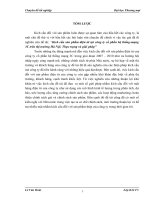
Rickets
Rickets
• In the long bones of birds
– enchondral ossification is
examined in the proximal
epiphysis
• The parts of the proximal
epiphysis:
– Epiphyseal cartilage
– Proliferation zone
• production of cartilage
• necrotizes and calcification
occurs
– Growth zone
• With the active bone marrow
• resorption of the calcified
cartilage
• new bone replaces the
cartilage
Photo: Dr. Dobos-Kovács, Mihály
6
Rickets
Rickets
due
due
to
to
lack
lack
of vitamin D
of vitamin D
or
or
Ca
Ca
• In the deep layers of the epiphyseal cartilage new
chondrocytes and cartilage is formed
– later the chondrocytes necrotize and the cartilage calcifies
– active bone marrow intrudes into that layer
– and it resorps the necrotized and calcified cartilage
• Production of new bone tissue
– At the beginning osteoid is formed
– This calcifies
– 1:9 - Ca-carbonate and Ca-phosphate
– + small amount of Mg-phosphate and calcium-fluoride
• In case of rickets due to lack of vitamin D3 in the long
bones
– epiphyseal cartilage is produced
– The other processes are slower or do not floolw the cartilage
prodution
• So the epiphyseal cartilage becomes thicker
– The cartilage extends due to the bodyweight
– The end becomes butt, and painful, the bone bends
Photos: Dr. Dobos-Kovács Mihály
Rickets
Rickets
due
due
to
to
lack
lack
of
of
Ca
Ca
Photos: Dr. Dobos-Kovács, Mihály
Photo: Dr. Dobos-Kovács, Mihály Photo: Dr. Dobos-Kovács, Mihály
7
Photo: Dr. Dobos-Kovács, Mihály Felvétel: Dr. Dobos-Kovács Mihály
The
The
bone
bone
can
can
be
be
broken
broken
without
without
sound
sound
• There is constant ossification and the depots
are used up
• The compacta of the bone becomes thinner
and the bone is spongy
– juvenile osteoporosis
Rickets
Rickets
due
due
to
to
lack
lack
of P
of P
Photo: Dr. Dobos-Kovács, Mihály
Osteopathies
Osteopathies
• Osteopathy due to lack of proteins
– Lack of amino acids
• Lysine
– hypoplastic type of bones are produced
• Osteopathy due to lack of vitamins
– Lack of vitamin C
– Lack of nicotinic acid
• vitamin B5, niacin
• rickets-like lesions
– Lack of vitamin A
8
Osteopathy due to lack of vitamins
Osteopathy due to lack of vitamin C
• the malfunction of the mesenchymal cells
• because of their fatty infiltration
– cartilage and osteoid production
– deceased production of the basic substance
• no problem with the calcification
Osteopathy due to lack of vitamin niacin
(rickets-like lesion)
• the lesions are similar like in case of osteopathy due
to lack of P
• in geese
Osteopathy due to lack of vitamin A
• the enchondral ossification is decreased
– In some case the skull and the vertebrae are also affected
Osteopathy due to lack of
Osteopathy due to lack of
minerals
minerals
• Osteopathy due to lack of manganese (Mn)
– perosis
• the Achilles-tendon slips from the sulcus to the side
• the distal end of the leg turns lateral
– appears in young chicks and poult
– rarely in pheasant and other birds
• Osteopathy due to lack of zinc (Zn)
– Zn is essential to the development of the
skeleton
• when Zn is missing, the lesions appear in the distal
epiphysis of the tibia
Perosis
Perosis
• Osteopathy due to lack of Manganese (Mn)
– this lesion is mainly seen in case inadequate
amount of manganese is available
– but lack of other substances can also induced
similar lesions
• pyridoxine (vitamin B6)
• biotin (vitamin B7 or H)
• nicotinic acid (vitamin B5 or niacin)
• folic acid /folacin/- (vitamin Bc or M)
• choline (vitamin B komplex)
– alone or in combination
• the manganese influences the enchondral
ossification
– the extracellular substance (the matrix) of the
cartilage is produced in decreased amount
Perosis
Perosis
• In a classic case the lateral part of sulcus
tendinis at the distal end of the tibiotarsal
bone grows slower
– the lateral part becomes shorter
– the Achilles-tendon can easily slip to the
lateral side
– the distal end of the leg turns out
• because of these processes
– the distal end of the tibiotarsal bone is more
smooth then normally
– the sulcus and the bone condyles are flat
Photo: Dr. Dobos-Kovács, Mihály Photo: Dr. Dobos-Kovács, Mihály
9
Photo: Dr. Dobos-Kovács, Mihály Photo: Dr. Dobos-Kovács, Mihály
Photo: Dr. Dobos-Kovács, Mihály
Osteopathy
Osteopathy
due
due
to
to
lack
lack
of
of
Zn
Zn
• The lateral epyphyseal cartilage grows and ossifies faster then
the medial
– The distal epyphisis of the tibia bends inward
– the Achilles-tendon slip to the middle from the sulcus tendinis
Photo: Dr. Dobos-Kovács, Mihály
Photo: Dr. Dobos-Kovács, Mihály Photo: Dr. Dobos-Kovács, Mihály
10
Photo: Dr. Dobos-Kovács, Mihály Photo: Dr. Dobos-Kovács, Mihály
Osteopthies
Osteopthies
caused
caused
by
by
viruses
viruses
• Short beak and dwarfism syndrome of mule duck
– SBDS
• Mulard = pekin duck ♀ x Muscovy duck ♂ hybrid,
„mule duck”
• Pathogenesis
– the mulard ducklings develop slowly and heterogenously
• from young age already
– Lack of development shows on the bones of the beak and
tarsus
• the beak becomes shorter then normally, the head is
deformed
• looks like the beak and head of goose
– the tongue grows normally, so it hangs out from the beak
– and dries out on the air
– the animals look smaller, underdeveloped
• because of the shortness of the tarsus
Photo: Dr. Palya, Vilmos
Photo: Dr. Palya, Vilmos
Other
Other
osteopathies
osteopathies
• Lack of salt
– the waterfowl are very sensitive
• especially duck
– In NaCl (table salt, especially natrium)
deficency rickets-like lesions develop
• similar to osteopathies due to lack of Ca and
vitamin D
• the proliferation zone of the epiphyseal
cartilage becomes thicker
• the osteoid tissue is not properly calcified,
and the matrix is less
11
Disturbances
Disturbances
of
of
mineralization
mineralization
• Osteoporosis
• Osteomalatia
• Osteodystrophia fibrosa
Osteoporosis
Osteoporosis
• develops in case the balance between the bone
remodelling (resorption and formation) splits and
the resorption exceeds formation
– faulty mineralization of the bones
– which cause the bones to become porous within, with a
lot of embedded hollow-like spaces
– the cortex of the bone becomes thinner
– osteoporosis often develops in the epiphysis of the bone
– more brittle bones, and get fractured easily
• appears in all ages
• in young age it is called juvenile osteoporosis
– in broilers the malsecretion, maldigestion, malabsorption
syndrome can lead to osteoporosis
– in poult the PEMS or other independent viral enteritides
– the nutrirent supply is not adequate in these cases
• proteins, vitamins, minerals
Osteoporosis
Osteoporosis
• In pullets or adult the quality and the
quantity iof the feed influences the
appearance of osteoporosis
– the most often seen causes are lack of protein,
vitamins A, B2 and C, lack of Cu, Mn, Zn
– In this case the osteoporosis effect
multitudinous birds and bones! – systemic,
generalized lesion
• Solitary osteoporosis
– partial (affects few bones only) osteoporosis
– In case of disuse (paralysis, mechanical trauma)
– atrophia ex inactivitate
– osteoporosis occurs on areas where the
remodelling is very intense
• f.e. epiphysis
Osteoporosis
Osteoporosis
In cage layers
• osteoporosis occurs usually at the end of the
egg laying season
– It is called „caged layer fatigue”
– involving bone brittleness, paralysis, and death
– the bone loss results in increased bone fragility
and susceptibility to fracture
• affected bones are the pelvis, vertebrae, sternum, ribs,
humerus and femur
• the fractures cause compression of the
nerves, resulting in innervation problems and
decreased loadability
– „caged layer paralysis”
Osteomalacia
Osteomalacia
• the softening of the bones caused by defective bone
mineralization secondary to inadequate amounts of available
phosphorus and calcium
– it develops in adult birds – in bones finished growing
• in case inadequate amounts of Ca and P are available
• The body resorps the wanted minerals from the already
formed bone to cover the needs
– f.e. Ca required for egg laying
• generalized bone condition in which there is inadequate
mineralization of the bone
• the compact and the spongy (trabecular) bones loose their
strength
• the bones of the adult bird are
– easily cut, sawed
– the spongy bone is more visible
– the bone bend
– With histopathology:
• broad, not calcified, elastic, soft osteoid can be observed
Osteodystrophia
Osteodystrophia
fibrosa
fibrosa
• In case the statics of the bones is disturbed
• the trabecules move from the normal position
• The organism tries to get back the balance
• It can occur in both growing or finished bones
– in different forms of rickets
– in case of osteomalacia, osteoporosis
• Appears in different bones
– the bones might enlarge
• The regular lamellar structure of the bone disappears, the
lamellae of the bone are thin and - in certain areas - dissolved
due to the vigorous osteolysis
• Pathogenesis
– Increased activity of the osteoclasts (resorption)
– The compact and the trabecular bone become porous
• thin, irregularly positioned spicules
– Angiofibroblast replaces the bone tissue
– proliferating osteoblasts are present in great number, and
osteoid tissue produced by them can be seen without
calcification
12
Regressive
Regressive
changes
changes
in
in
the
the
bones
bones
• Atrophy (atrophia ossium)
• Usuration of the bone
– lacunar resorption following constant pressure (soft tumor)
• Osteochondrosis
• Epiphysiolysis
• Dyschondroplasia
• Remnant of the embryonic cartilage
• Discoloration, pigmentation of the bones
– Melanosis
– Discoloration caused by accumulation of tetracycline
• Necrosis of the bone
– femoral head necrosis
Atrophy
Atrophy
• occurs in long bones following disuse of the leg
– the compact becomes thinner
– the trabecules become thinner and decrease in number too
– atrophy of the exposed muscles usually also seen
• It can be observed if chronic inflammation develops
in the hind legs (joints or tendon sheets)
– orthoreoviral arthritis/tenosynovitis
– lesions of the bones
– tibiarotation, lack of Zn or Mn
• It is painful
• the animal does not stand on this side
– atrophia ex inactivitate
Osteochondrosis
Osteochondrosis
• Regressive change in the bones
– Disturbance of the normal development of the bone
– different forms of the disease
– appears in fast growing broiler chickens
• Described in several bones
– In the cervical and thoracic vertebraa
– In the femur and in the antitrochanter
• The lesions are microscopic and occur between the
epiphyseal cartilage and the proliferation zone
– serous infiltration, degeneration and blockage of the blood
vessels (thrombosis) can be seen in the proliferation zone
• According the data in some broiler flock 50% of the
chickens is affected to some extent
– most of them show clinical signs too
• osteochondrosis often occur togetehr with some
other skeletal lesions
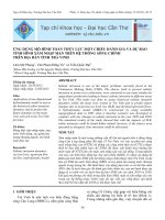
Epiphysiolysis
Epiphysiolysis
• Mainly in the proximal epiphysis of the femur
– loose structure with decreased strength
– in fast growing broiler chickens
• The two parts (epiphysis and diaphysis) of the bone
separate from each other
– at the proximal epiphysis
– due to a minor trauma (slightly in excess of normal physical
stimulus)
• normal movements (like jumping), handling
– between the two parts degenerative changes and necrosis can
be observed
• if the separation occurs in life, bleeding can be seen
– It can appear in carcasses following preparation for the
dissection
Epiphysiolysis
Epiphysiolysis
Photos:
Dr. Dobos-Kovács, Mihály
Dyschondroplasia
Dyschondroplasia
• In the growing long bones
– most often seen in the proximal epiphysis of the tibiotarsal
bone
– in the proximal and distal epiphysis of the femur
– in the tarsometatarsal bone and in the proximal epiphysis of
the humerus
• Affected: meat-type chicken, duck, turkey
• Characteristics in the long bones:
– in the growth zone of the epiphysis remnant cartilage is
observed without blood vessels
– the abnormal cartilage occurs circumscribed or in the whole
length of the bone
• The cartilage produced in the epiphysis
– does not contain blood vessels
– no calcification is seen
– It does not develop to osteoid and later bone tissue
• The bone are soft, weak, bend under the body weight
13
Photo: Dr. Dobos-Kovács, Mihály
Dyschondroplasia
Dyschondroplasia
• Causes
– Genetic factors
• fast growing hybrids
– 5-6 week-old chickens, from 11 weeks of age in turkey
– Malnutrition
• lack of Cu, vitamin C and D
– 1,25-dihydroxicholecalciferol -active metabolite
• fusarochromanon toxisosis, acidosis, inadequate Ca-P ratio
– Husbandry problems
• Light programs with constant lightning
• Special form:
– turkey’65 syndrome (TS-65 syndrome)
– usually follows Mycoplasma meleagridis infection
– in the proximal epiphysis of the tibiotarsal bone
– the epiphysis does not turn to osteoid, later bone
– tibia bends, forms typical „O” shape
Remnant
Remnant
of
of
embryonic
embryonic
cartilage
cartilage
• In long bones and vertebrae
– in newly hatched birds (ostrich,
emu, nandu, great bustard)
– in day-old gooslings aswell
– starting from the epiphysis trough
the metaphysis into the
diaphysis:
– bluish-gray cartilage replaces the
bone marrow
– The cartilage resembles the
normal embryonic cartilage
• this embryonic cartilage is
observed in ratites up to 8 weeks
of age
• In gooslings from the end of the
first week bone marrow is seen
instead of the embryonic cartilage
• This is not dyschondroplasia!
Photo: Dr. Dobos-Kovács, Mihály
Necrosis
Necrosis
of
of
the
the
bone
bone
• Due to circulatory disturbances
– The circulatory disturbances are caused by
• burn, frost bite, trauma, thrombosis,
inflammation
• Femoral head necrosis
– The head of the femur necrotizes due to lack
of enough blood (nutrient) supply
– this lesion is often seen in case of
malsecretion, maldigestion, malabsorption
syndrome
• often in broilers