comparative phototoxicity of nanoparticulate and bulk zno to a free-living
Bạn đang xem bản rút gọn của tài liệu. Xem và tải ngay bản đầy đủ của tài liệu tại đây (509.32 KB, 8 trang )
Comparative phototoxicity of nanoparticulate and bulk ZnO to a free-living
nematode Caenorhabditis elegans: The importance of illumination mode
and primary particle size
H. Ma
a
,
*
,
1
, N.J. Kabengi
b
, P.M. Bertsch
b
, J.M. Unrine
b
, T.C. Glenn
a
, P.L. Williams
a
a
Department of Environmental Health Science, College of Public Health, The University of Georgia, Athens, GA 30602, USA
b
Department of Plant and Soil Sciences, University of Kentucky, Lexington, KY 40546, USA
article info
Article history:
Received 17 November 2010
Received in revised form
14 March 2011
Accepted 16 March 2011
Keywords:
Nanoparticulate ZnO
Bulk ZnO
Phototoxicity
Reactive oxygen species (ROS)
Caenorhabditis elegans
abstract
The present study evaluated phototoxicity of nanoparticulate ZnO and bulk-ZnO under natural sunlight
(NSL) versus ambient artificial laboratory light (AALL) illumination to a free-living nematode Caeno-
rhabditis elegans. Phototoxicity of nano-ZnO and bulk-ZnO was largely dependent on illumination
method as 2-h exposure under NSL caused significantly greater mortality in C. elegans than under AALL.
This phototoxicity was closely related to photocatalytic reactive oxygen species (ROS) generation by the
ZnO particles as indicated by concomitant methylene blue photodegradation. Both materials caused
mortality in C. elegans under AALL during 24-h exposure although neither degraded methylene blue,
suggesting mechanisms of toxicity other than photocatalytic ROS generation were involved. Particle
dissolution of ZnO did not appear to play an important role in the toxicity observed in this study. Nano-
ZnO showed greater phototoxicity than bulk-ZnO despite their similar size of aggregates, suggesting
primary particle size is more important than aggregate size in determining phototoxicity.
Ó 2011 Elsevier Ltd. All rights reserved.
1. Introduction
Nanoparticulate metal oxides are being used within a great
variety of applications due to their novel optical, magnetic, and
electronic properties (Zhou et al., 20 06). With widespread use of
these manufactured nanoparticles, concerns about their potential
impact on the environment and human health have been raised
(Colvin, 2003). Although still in its infancy, toxicity studies on
manufactured metal oxide nanoparticles (such as TiO
2
, ZnO) are
expanding rapidly (Hall et al., 2009; Jiang et al., 2008; Brunet et al.,
20 09; Reeves et al., 2008; Franklin et al., 2007; Wang et al., 2009;
Ma et al., 2009).
Toxicity of manufactured nanoparticles may be attributed to
several different modes of action: chemical toxicity based on
chemical composition (e.g., release of toxic ions); surface catalyzed
reactions (e.g., formation of reactive oxygen species (ROS)); or
stress of stimuli caused by the surface, size, and shape of the
particles (Wang et al., 2009; Nel et al., 2006). Dissolution of
nanoparticles resulting in the release of toxic ions has been found
to play an important role in eliciting toxicity in both metal oxides
(Franklin et al., 2007; Wang et al., 2009; Ma et al., 2009) and
quantum dots (Priester et al., 2009; King-Heiden et al., 2009).
Generation of ROS by nanoparticles interacting with environmental
agents (e.g., UV) represents another important mode of action for
metal oxide nanoparticles with photocatalytic activities such as
TiO
2
or ZnO, as high concentration of ROS causes oxidative stress
and can eventually elicit toxicity in biological systems (Applerot
et al., 2009). A wealth of studies have demonstrated phototoxicity
of TiO
2
nanoparticles in a broad range of biological systems, from
bacteria (Brunet et al., 2009; Sunada et al., 2003; Adams et al.,
20 06) to mammalian cell lines (Sayes et al., 2006; Gopalan et al.,
20 09). The underlying mechanism of bactericidal activity of TiO
2
to Escherichia coli K-12 cells has been revealed to be associated with
lipid peroxidation induced by ROS generation under UV irradiation
(Maness et al., 1999). ZnO nanoparticles are similar to TiO
2
nano-
particles regarding their photodynamic and antibacterial proper-
ties (Daneshvar et al., 2007), and are receiving increasing
application in numerous areas such as electronics, rubber additives,
medicine, biosensors, personal-care products, etc However,
studies on phototoxicity of manufactured ZnO nanoparticles have
been very limited except for those reporting their antibacterial
effects (Jones et al., 2008). A recent study by Gopalan et al. (2009)
*
Corresponding author.
E-mail addresses: , (H. Ma).
1
Present address: Mid-Continent Ecology Division, United States Environmental
Protection Agency, Duluth, MN 55804, USA. Tel.: þ1 218 529 5071.
Contents lists available at ScienceDirect
Environmental Pollution
journal homepage: www.elsevier.com/locate/envpol
0269-7491/$ e see front matter Ó 2011 Elsevier Ltd. All rights reserved.
doi:10.1016/j.envpol.2011.03.013
Environmental Pollution 159 (2011) 1473e1480
found that genotoxicity of ZnO nanoparticles to human sperm and
lymphocytes was enhanced by UV irradiation. Given the increasing
applications of ZnO nanoparticles and its consequent release to the
environment either by intended disposal or accidental release, it is
essential to understand their potential phototoxicity to the natural
biota.
The objective of the current study is to evaluate phototoxicity of
nanoparticulate ZnO (nano-ZnO) and its bulk counterpart (bulk-
ZnO) to a free-living nematode Caenorhabditis elegans under natural
sunlight (NSL) versus ambient artificial laboratory light (AALL)
illumination. Aqueous ZnCl
2
was used as control. A representative
species of the nematode phylum which is of great ecological
significance, together with its thoroughly understood biology, short
life cycle, and ease of culture in the laboratory, C. elegans has served
as a good model for both terrestrial and aquatic receptors for eco-
toxicological studies for a wide range of environmental toxicants
(Leung et al., 2008). More recently, it has also been used for eco-
toxicological studies on manufactured nanoparticles (Ma et al.,
20 09; Wang et al., 2009). To help elucidate the possible mecha-
nism of the phototoxicity (i.e., localization of ROS toxicity), photo-
catalytic activity/ROS generation of the ZnO particles and lipid
peroxidation in the nematodes were also measured. The hypotheses
are that: (i) phototoxicity of nano-ZnO will be greater under NSL
than under AALL, and this phototoxicity will be positively correlated
with photocatalytic activity/ROS generation of the nanoparticles;
(ii) nano-ZnO will have greater phototoxicity than bulk-ZnO, as
smaller particles have a greater surface area per unit mass than
larger particles and thus may be more effective in generating ROS
(Applerot et al., 2009). Natural sunlight instead of arti ficial UV light
was used to activate the nanoparticles because exposure under NSL
is a more realistic scenario under which organisms might be
exposed to nanoparticles released to the environment.
2. Materials and methods
2.1. Nano-ZnO and bulk-ZnO sample preparation
Powdered nanoparticulate ZnO (NanoGard
Ò
zinc oxide) was purchased from Alfa
Aesar (Ward Hill, MA, USA) with a stated size of 40e100 nm. Stock suspensions of
nano-ZnO and bulk-ZnO (Mallinckrodt; Phillipsburg, NJ) (100 mg/l (80% Zn)) were
prepared by sonication for 2 h in an ultrasonic bath (Branson; Danbury, CT). Specific
surface areas (SSA) of both materials were determined by BrunauereEmmeteTeller
(BET) method and were found to be 17.0 and 4.2 m
2
/g for nano-ZnO and bulk-ZnO,
respectively. Reagent grade ZnCl
2
(Mallinckrodt; Phillipsburg, NJ) was used to make
ZnCl
2
stock solution. Test solutions were freshly diluted from the stock which was
sonicated for 30 min prior to dilution to ensure proper dispersion of the materials.Both
stock suspension and dilutions were made in K-medium (0.032 M KCl, 0.051 M NaCl,
pH 5.8e6.0) (Williams and Dusenbery, 1990), an aqueous medium used for C. elegans
based bioassays. The pH of the test solutions was measured using a pH meter.
2.2. Particle characterization
The nanoparticulate and bulk ZnO were characterized using a variety of
analytical techniques. Transmission electron microscopy (TEM, FEI/Philips Electron
Optics, Eindhoven, The Netherlands) was used to characterize particle morphology
and measure the primary particle size. Approximately 100 particles from four
representative images of each material were measured and average sizes of the
particles were reported. Dynamic light scattering (DLS) was performed using
a photo correlation spectrophotometer (Malvern Instruments, Worcestershire, UK)
to determine the d
h
(hydrodynamic diameter) of the particles or aggregates. To
analyze aggregates > 1
m
m in diameter, suspensions of nanoparticles were exam-
ined by differential interference contrast microscopy (DIC) using a motorized
microscope (Nikon Instruments; Melville, NY) equipped with a 40Â objective lens
and a cooled CCD monochrome camera. At least 100 aggregates were recorded for
each material and average diameters were calculated.
Dissolution of nano-ZnO or bulk-ZnO in suspensions was assessed by filtration
through regenerated cellulose membranes with a 3000 Da nominal molecular
weight cutoff (approximately 0.9 nm) using a centrifugal filtration device. Three ml
of nano-ZnO or bulk-ZnO solutions at 100 mg/l were added to the filter units and
centrifuged (Eppendorf 5810R; Westbury, NY) for 30 min at 3220 Â g. Filtrates were
collected and analyzed for Zn concentration using an inductively coupled plasma
mass spectrometer (Agilent Technologies, Santa Clara, CA). Recovery of Zn ions in the
filtrates was determined by filtering ZnCl
2
solutions of similar concentrations
through the device.
2.3. C. elegans toxicity assay
C. elegans (wild type N2) was obtained from Caenorhabditis Genetics Center
(Minneapolis, MN). The nematode culture maintenance and generation of age-
synchronized worms followed the description by Donkin and Williams (Donkin and
Williams, 1995). Toxicity test was conducted for three materials: nano-ZnO, bulk-
ZnO, and ZnCl
2
. For each material, exposure was conducted under three different
illumination conditions: NSL, AALL, and in dark. All tests were conducted in 24-well
tissue culture plates. Each test consisted of six concentrations of test substance (4, 8,
20, 40, 60, 80 mg/l Zn, corresponding to 5, 10, 25, 50, 75, 100 mg/l ZnO and 8, 17, 42,
84, 125, 167 mg/l ZnCl
2
) and a control, with three replicate wells for each concen-
tration. A 1.0 ml aliquot of test solution was added to each well which was subse-
quently loaded with 10 (Æ1) nematodes. Light intensity was measured using
a digital luxmeter (Precision Mastech, Kowloon, Hong Kong, China). For exposure
under NSL, the plates (without lids) were left under direct sunlight for 2 h on the
outside ledge of a window facing southwest in the laboratory on bright days
(25 Æ 1.5
C average temperature, UV index 4e5) in October in Athens, GA (33
57
0
19
00
N, 83
22
0
59
00
W). Temperature of the exposure solution was recorded before
and after exposure, and was found to increase by 2e3
C; and the water loss in the
exposure solution was estimated to be less than 5% after 2-h NSL exposure. The
average incident illuminance during the test period was 15,000 Æ 250 lux. Mortality
was monitored following the 2-h NSL exposure. The exposure was continued for an
extended 22 h under AALL by relocating the plates on a bench in the laboratory,
which does not receive any sunlight. Mortality was monitored again. This 2-h NSL
followed by 22-h AALL exposure allows for detection of possible synergistic toxicity
of the nanoparticles if different modes of action are involved under these two
irradiation conditions. For exposure under AALL, the plates (without lids) were
placed on a bench in the laboratory with room temperature 22 Æ 0.5
C. The ambient
laboratory lighting uses fluorescent lamps, and had an intensity of 525 Æ 25 lux
during the test period. For exposure in dark, the plates were covered by aluminum
foil and placed in a cabinet at ambient room temperature. Mortality was monitored
at 2 h and 24 h for exposure under AALL and in dark. The plates were observed under
a dissecting microscope and the nematodes were counted and scored as live or dead
following established protocol e nematodes that did not move in response to
a gentle touch by a metal wire were counted dead (Williams and Dusenbery, 1990).
2.4. Photocatalytic activity measurement
In parallel to C. elegans toxicity assay, photocatalytic activity of the three
materials was determined under different illumination methods by measuring
photodegradation of methylene blue (MB) in aqueous solution (Shen et al., 2008;
Jang et al., 2006). Degradation of MB by photocatalysts such as ZnO or TiO
2
involves generation of radicals such as O
À
2
and
OH (Houas et al., 2001; Lachheb et al.,
2002); therefore, degradation of the dye can be used as an indicator for photo-
catalytic activity/ROS generation of these materials. A series of concentrations of
each material were prepared in an identical manner as those for nematode bioassay;
one ml of 25 mg/l MB was added to each well. The plates were incubated for 30 min
to allow for equilibrium for the dye sorptionto particles. Prior to exposure, the plates
were gently shaken by hand for a few minutes to homogenize the solution. MB
concentrations were measured at the beginning, after 2 h and 24 h of exposure,
using a NanoDrop ND-1000 Spectrophotometer (NanoDrop Technologies, Wil-
mington, DE) at 665 nm. A negative control using a series of MB concentrations in
K-medium under AALL was also included.
2.5. Lipid peroxidation measurement
Lipid peroxidation in C. elegans after exposure to nano-ZnO and bulk-ZnO under
NSL or AALL was measured by the thiobarbituric acid assay (TBARS) for malon-
dialdehyde (MDA) using an OxiSelect TBARS Assay Kit (Cell Biolabs, San Diego, CA).
C. elegans exposure for lipid peroxidation measurement was similar to the toxicity
assay except that larger number of nematodes (w0.5 ml worm pellet) was used and
only one nano-ZnO or bulk-ZnO concentration (25 mg/l) was tested. After exposure,
nano-ZnO or bulk-ZnO solutions were removed and the worms were rinsed with M9
buffer for three times, then immediately frozen at À80
C until TBARS analysis.
TBARS analysis was performed following the manufacturer’s instructions. Fluoro-
metric measurement of MDA was obtained using a Bio-Tek Synergy 4 Hybrid Multi-
Mode Microplate Reader at 540 nm excitation and 590 nm emission. MDA content
was standardized by total nematode protein which was measured using bicincho-
ninic acid (BCA) assay (Thermo Scientific, Rockford, IL, USA).
2.6. Data analysis
All data reported were based on three independent experiments. Median lethal
concentrations (LC50s) and corresponding 95% CIs were calculated by Probit anal-
ysis using TOXSTAT software (WEST, Inc, 1994). One-way ANOVA was used to
compare the % MB degradation between nano-ZnO and bulk-ZnO, and lipid
H. Ma et al. / Environmental Pollution 159 (2011) 1473e14801474
peroxidation between NSL and AALL exposure. Correlation between C. elegans
mortality and % MB degradation was assessed using Pearson’s correlation
coefficients.
3. Results
3.1. Particle characterization
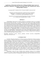
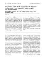
Significant particle aggregation was observed for both nano-
ZnO and bulk-ZnO (Fig. 1). A quantitative measurement of
approximately 100 particles indicated an average primary particle
size of 60 Æ 25 and 550 Æ 256 (Mean Æ SD, n ¼ 100) nm for nano-
ZnO and bulk-ZnO, respectively. The average diameters of rotation
for aggregates were 2.79 Æ 1.84 and 2.43 Æ 1.59 (Mean Æ SD,
n > 100)
m
m for nano-ZnO and bulk-ZnO, respectively. The size
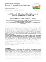
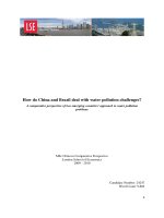
distribution was similar between the two materials (Fig. 2). The
working solutions of nano-ZnO and bulk-ZnO had pH values of
6.5e7.0. Dissolution of nano-ZnO and bulk-ZnO was estimated to be
7.1% and 4.8% respectively after correction for Zn ions recovery in
the filtration system.
3.2. Toxicity of nano-ZnO and bulk-ZnO
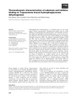
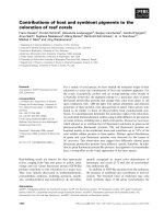
Concentration-dependent mortality in C. elegans was observed
after 2-h exposure to nano-ZnO or bulk-ZnO under NSL (Fig. 3 (A)
and (B)). At an identical concentration, nano-ZnO caused greater
mortality than bulk-ZnO. The 2-h LC50s for nano-ZnO and bulk-
ZnO were 38 (95% CI: 30, 45) and 65 (95% CI: 55, 78) mg/l of Zn,
respectively (Table 1). No mortality occurred after 2-h exposure to
ZnCl
2
under NSL. For both materials, an extended 22-h exposure
under AALL caused a significant increase in mortality compared to
the initial 2-h exposure under NSL (Fig. 3 (A) and (B)). With the 2-h
NSL and 22-h AALL exposure combined, the 24-h LC50s for nano-
ZnO and bulk-ZnO were 17 (95% CI: 10, 25) and 38 (95% CI: 29, 50)
mg/l of Zn, respectively (Table 1). Again, this extended exposure
under AALL did not cause mortality in C. elegans for ZnCl
2
.
Fig. 1. TEM images of nano-ZnO and bulk-ZnO in K-medium (pH ¼ 6.8): (A) 10 mg/l nano-ZnO, (B) 100 mg/l nano-ZnO, with an estimated particle size of 60 Æ 25 nm (n ¼ 100),
(C) 10 mg/l bulk-ZnO, (D) 100 mg/l bulk-ZnO, with an estimated particle size of 550 Æ 256 nm (n ¼ 100).
Fig. 2. Aggregate size distribution of nano-ZnO and bulk-ZnO suspensions using DIC
microscopy. The average diameters of rotation for these aggregates were 2.79 Æ 1.84
and 2.43 Æ 1.59 (Mean Æ SD, n > 100)
m
m for 100 mg/l nano-ZnO and bulk-ZnO,
respectively.
H. Ma et al. / Environmental Pollution 159 (2011) 1473e1480 1475
Neither nano-ZnO nor bulk-ZnO caused mortality in C. elegans
after 2-h exposure under AALL, but both induced mortality after 24-h
exposure (Fig. 3 (A) and (B)). For nano-ZnO, this 24-h toxicity under
AALL was lower than 2-h toxicity under NSL; however, there was no
significant difference between these two types of toxicity for bulk-
ZnO. LC50s could not be determined for the 24-h AALL exposure
because of insufficient mortality; however, an extrapolation from the
concentration-response curves suggested that the LC50 for nano-
ZnO was approximately 60e80 mg/l of Zn and that for bulk-ZnO was
even higher. No mortality was observed for nano-ZnO or bulk-ZnO
exposure in the dark, regardless of the duration of exposure.
Mortality incontrol nematodes was below 10% forall tests conducted.
3.3. Methylene blue degradation
Methylene blue degradation was used as an indicator of pho-
tocatalytic activity/ROS generation by the ZnO particles. ZnCl
2
did
not degrade MB, regardless of the illumination method or exposure
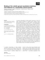
duration. Under 2-h NSL exposure, concentration-dependent MB
degradation was observed for both nano-ZnO (Fig. 4 (A)) and bulk-
ZnO (Fig. 4 (B)). The maximum MB degradation (%) by nano-ZnO
and bulk-ZnO were 67% and 45%, respectively (Table 1). A strong
positive correlation was found between C. elegans mortality and MB
degradation (%) for both nano-ZnO (r ¼ 0.94, p ¼ 0.002, n ¼ 7) and
bulk-ZnO (r ¼ 0.90, p ¼ 0.005, n ¼ 7). MB degradation did not occur
during the extended 22-h exposure under AALL (Table 1). Under
AALL, nano-ZnO and bulk-ZnO degraded MB by 7.5% and 5.2%,
respectively during 2-h exposure, and this degradation did not
increase as the exposure extended to 24 h (Table 1). No MB
degradation by nano-ZnO or bulk-ZnO was observed in the dark,
regardless of the duration of exposure.
3.4. Lipid peroxidation
Elevated lipid peroxidation in C. elegans exposed to 25 mg/l
nano-ZnO or bulk-ZnO compared to control was found under all
exposure conditions (Fig. 5). Significantly greater lipid peroxidation
caused by nano-ZnO than bulk-ZnO was only observed after 2-h
NSL exposure. Neither nano-ZnO nor bulk-ZnO caused statistically
different lipid peroxidation between 2-h NSL and 2-h AALL expo-
sure. Enhanced lipid peroxidation was also observed when expo-
sure was extended from 2 h to 24 h, under both NSL and AALL.
4. Discussion
Although several studies have investigated toxicity of ZnO
nanoparticles to a variety of environmentally relevant species
(Franklin et al., 2007; Wang et al., 2009; Ma et al., 2009; Reddy
et al., 2007; Brayner et al., 2010), few have considered their
phototoxicity (Adams et al., 2006). The current study found that
phototoxicity of nano-ZnO was substantially enhanced under NSL
illumination compared to AALL or in the dark using C. elegans as test
organism. Concurrent photodegradation of MB under NSL sug-
gested this phototoxicity is associated with photoactivation/ROS
generation of the nanoparticles. A strong positive correlation
(r ¼ 0.94, p ¼ 0.002, n ¼ 7) between C. elegans mortality and MB
degradation during 2-h NSL exposure suggested that photocatalytic
activity of the nanoparticles may be a predictor for phototoxicity.
Several studies have documented increased antibacterial activity of
ZnO nanoparticles under ambient laboratory light (Applerot et al.,
2009; Jones et al., 2008) or natural sunlight (Adams et al., 2006)
as compared to dark conditions, presumably due to photoactivation
of the nanoparticles. The present study, however, demonstrates
that photocatalytic ROS generation by ZnO nanoparticles under NSL
can cause mortality to a terrestrial/aquatic organism e the
nema-
tode C. elegans.
Phototoxicity of the ZnO nanoparticles is strictly dependent on
illumination mode. Nano-ZnO has band gap energy of 3.37 ev,
equivalent to a wavelength of 368 nm (Wang, 2004). Upon excita-
tion by light with a wavelength less than 368 nm, electronehole
pairs are generated on the ZnO surface. The holes in the valence
band can react with H
2
O or hydroxide ions adsorbed on the surface
to produce hydroxyl radicals, and the electrons in the conduction
band can reduce O
2
to produce superoxide ions (Maness et al.,
1999). Both of these radicals are extremely reactive with contact-
ing biological molecules. Photoactivation and the consequent
phototoxicity of nano-ZnO occurred under NSL but not AALL. The
spectrum of natural sunlight used in this study can be roughly
represented by a typical solar radiation spectrum (supplemental
Fig. 1(A)), although the dosimetry and spectrum of solar radiation
in a particular geographical location can be affected by a number of
factors, such as, geographical conditions, atmospheric variability,
weather conditions, etc. (Diamond et al., 2002). It is estimated that
approximately 8e9% of the total solar energy emitted from the sun
0 20406080100120
Nano-ZnO (mg/l)
Mortality (%)
0
20
40
60
80
100
120
0
20
40
60
80
100
120
0 20 40 60 80 100 120
Bulk-ZnO (m
g
/l)
Mortality (%)
2h NSL
2h AALL
2h NSL+22h AALL
24h AALL
A
B
Fig. 3. Toxicity of nano-ZnO (A) and bulk-ZnO (B) to C. elegans under different illumination method (NSL-natural sunlight, AALL-ambient artificial laboratory light).
Table 1
LC50s for nano-ZnO and bulk-ZnO and the corresponding maximum degradation of
methylene blue (MB) by the two materials under different illumination method and
exposure time.
2-h NSL 2-h AALL 2-h NSL+ 22-h AALL 24-h AALL
LC50 (%95 CI, mg/l Zn)
Nano-ZnO 38 (30,45) NA 17 (10, 25) 60e80
Bulk-ZnO 65 (55,78) NA 38 (29,50) >60e80
Maximum % MB degradation (MeanÆSEM, n¼3)
Nano-ZnO 66.9Æ4.5
*
7.5Æ3.0 70.3Æ5.3
*
9.8Æ4.2
Bulk-ZnO 44.6Æ6.1
*
5.2Æ1.1 44.9+4.7
*
6.4Æ3.3
NSL-natural sunlight, AALL-ambient artificial laboratory light.
*
p < 0.01.
H. Ma et al. / Environmental Pollution 159 (2011) 1473e14801476
falls in the UV region of the electromagnetic spectrum (6.3% UVA
(320e400 nm), 1.5% UVB (290e320 nm)) (Frederick, 1995). There-
fore, the natural sunlight could efficiently activate ZnO nano-
particles and produce ROS, as indicated by significant MB
degradation (70 Æ 4%), and consequently cause toxicity to the
nematodes. In contrast, ZnO nanoparticles were marginally acti-
vated under AALL, as suggested by a subtle MB degradation of
7 Æ 3%, and caused no mortality to the nematodes. The AALL used
typical “white cool” fluorescent lamps, and a typical light spectrum
of these lamps (supplemental Fig. 1(B)) contains negligible amount
of UV (<400 nm) light. It is suggested that UV exposure from sitting
under fluorescent lights for eight hours is equivalent to only one
minute of sun exposure (Lytle et al., 1993). Thus, the AALL cannot
activate the nanoparticles to cause toxicity. These findings have
important implication in risk assessment for photoactive nano-
particles. As most toxicity assays are conducted under standard
laboratory conditions, which do not perceive all risk factors;
exposure under natural sunlight conducted in this study represents
at least some range of potential exposure that may occur in the
environment.
It should be noted that the ZnO concentrations tested in
the present study are relatively high compared to those found in
the environment. Current estimates of ZnO concentrations in the
environment range from the low
m
g/l to a few hundred
m
g/l (Boxall
et al., 2007), and a more recent study by Gottschalk et al. (2009)
reported modeled nano-ZnO concentrations in the environment
to be approximately 10 ng/l in natural surface water and 1
m
g/l in
treated wastewater. However, as ZnO nanoparticles are being
widely used in a broad range of applications including personal-
care products such as cosmetics and sunscreens, it is expected that
their concentrations in the environment will increase continually
(Daughton and Ternes, 1999). Furthermore, results from these
relatively high concentrations serve as a foundation for further
investigation on phototoxicity of these nanoparticles at environ-
mental relevant concentrations using more sensitive endpoints at
cellular/subcellular and molecular levels.
Elevated lipid peroxidation after 2-h NSL exposure to both nano-
ZnO and bulk-ZnO confirmed that the phototoxicity is related to
ROS generation by the ZnO particles and oxidative stress in the
nematodes. Although the current study did not directly identify the
location of the phototoxicity in C. elegans, two different modes of
action may be proposed. First, the ROS generation and resulting
toxicity may occur at the surface of the nematodes. Surface acting
toxicity is not a new concept in ecotoxicology as certain toxicants
can be adsorbed to the exterior surface of the organism and elicit
toxicity (Handy et al., 2008a). This process has been implicated in
the toxicity of TiO
2
nanoparticles to trout (Federici et al., 2007). The
C. elegans cuticle has an evenly distributed net negative charge at
neutral pH (Himmelhoch and Zuckerman, 1983), which may
enhance particle aggregation on the surface of the animals via
electrostatic interactions (Wang et al., 2009). Particle aggregates
attached onto the nematode cuticle was observed during exposure.
The C. elegans cuticle is an extracellular matrix with a major
component of collagen. ROS such as superoxide radical anion or
hydroxyl radical has been reported to cause damage to calf skin
collagen by degrading the protein (Monboisse and Borel, 1992). It is
possible that intensive ROS generation by nano-ZnO under NSL
caused lethal toxicity to C. elegans through damaging the cuticle.
Another possibility is that nanoparticles were ingested by C. elegans
and the toxicity occurred internally. C. elegans unselectively ingest
bacteria and fine particles that are less than 5
m
m in size (Donkin
and Dusenbery, 1993), and its transparency and small body diam-
eter allow for adequate UV penetration (Coohill et al., 1988; Mills
and Hartman, 1998). A measure of oxidative stress (e.g., lipid per-
oxidation) within the nematodes may help to differentiate these
two modes of action. If ROS generation and phototoxicity occurred
internally, oxidative stress would be a good indicator of the
observed toxicity. Greater lipid peroxidation in the nematodes after
2-h NSL exposure to nano-ZnO was consistent with the greater
mortality caused by the nano-ZnO compared to bulk-ZnO, sug-
gesting that the internal-acting mechanism might have been
involved. However, lipid peroxidation alone is not sufficient to
differentiate the two possible modes of action as oxidative stress
within a multicellular organism may be mediated by a variety of
factors in addition to the photocatalytic ROS generation. Studies
0.000
0.050
0.100
0.150
0.200
0.250
Nano-ZnO (m
g
/l)
Methylene blue (abs)
0.000
0.050
0.100
0.150
0.200
0.250
0 20 40 60 80 100 120
020406080100120
Bulk-ZnO (mg/l)
Methylene blue (abs)
2h NSL
2h AALL
2h NSL+22h AALL
24h AALL
AB
Fig. 4. Methylene blue degradation by nano-ZnO (A) and bulk-ZnO (B) under different illumination methods (NSL-natural sunlight, AALL-ambient artificial laboratory light).
0
0.3
0.6
0.9
1.2
1.5
1.8
2.1
2.4
Cont 2h NSL 2h AALL 2h
NSL+22h
AALL
24h AALL
MDA (nmol/mg protein)
Nano
Bulk
∗
∗
∗
∗
∗
∗
∗
∗
Fig. 5. Lipid peroxidation in C. elegans after exposure to 50 mg/l nano-ZnO and bulk-
ZnO under NSL or AALL (NSL-natural sunlight, AALL-ambient artificial laboratory light)
(
*
p < 0.001, compared to control).
H. Ma et al. / Environmental Pollution 159 (2011) 1473e1480 1477
using more direct and specific in-situ and/or in vivo techniques for
ROS identification including dichlorofluorescein fluorescence assay
(H2DCFDA) ( Xie et al., 2006; Kim et al., 2009) and Aminophenyl
fluorescein assay (APF) (Setsukinai et al., 2003) are currently
undergoing to further understand the underlying mechanism of
the phototoxicity.
As phototoxicity of nano-ZnO associated with ROS generation
occurred in as quickly as 2 h under NSL, toxicity of nano-ZnO under
AALL was only observed after 24-h exposure. This 24-h toxicity
under AALL was lower than 2-h phototoxicity under NSL (Table 1),
and seemed to be mediated by mechanisms different from photo-
activation as no significant MB degradation occurred concurrently
(Table 1). Dissolution and release of ionic zinc from nanoparticulate
ZnO has been recognized as a major contributor to its toxicity to
freshwater alga (Franklin et al., 2007) and fish embryos (Zhu et al.,
20 08). Dissolution of nano-ZnO in the current study was estimated
to be 7.1% at the highest concentration tested, equivalent to 5.6 mg/l
of Zn. This amount of zinc does not seem to be able to cause
mortality in C. elegans, as aqueous ZnCl
2
at higher concentrations
did not cause mortality during 24-h exposure. Elevated lipid per-
oxidation in C. elegans exposed to nano-ZnO under AALL when the
exposure time was extended from 2 h to 24 h was consistent with
the dramatically increased toxicity (Fig. 3), suggesting that oxida-
tive stress is involved in this toxicity. This non-photocatalytic
toxicity of the nanosized ZnO was also observed during the
extended 22-h exposure under AALL following 2-h NSL exposure,
causing a synergistic effect in C. elegans mortality under the test
conditions when both NSL and AALL illumination were involved.
Zhu et al. (2009) reported that toxicity of nanosized ZnO on
developing zebrafish embryos and larvae under ambient laboratory
conditions was not solely a result of particle dissolution; and the
authors proposed that the nano-ZnO aggregates might have elicited
toxicity by increasing ROS formation and/or compromising the
cellular oxidative stress response upon interaction with the bio-
logical system. This particle-dependent, non-photocatalytic toxicity
of ZnO nanoparticles to aquatic organisms certainly warrants
further investigation.
Many studies on metal oxide particles such as TiO
2
and ZnO
have demonstrated that nanoparticles induce greater toxicity than
their larger counterparts at equivalent mass concentrations, using
test species from rats/mice to bacteria (Oberdörster et al., 2000;
Oberdöerster et al., 2005; Applerot et al., 2009; Jones et al.,
20 08). Findings from the present study were in good agreement
with these studies, that the nanosized ZnO had greater MB degra-
dation capability and phototoxicity compared to bulk-ZnO at the
same mass dose. The primary particle size of bulk-ZnO
(550 Æ 256 nm) was approximately 10 times that of nano-ZnO
(60 Æ 25 nm), and the latter had a specific surface area about four
times of the former. Several authors have suggested that total
surface area may be a more appropriate dose metric to describe
doseeresponse relationships than mass dose, especially when
evaluating poorly soluble particles (Oberdörster et al., 2000; Jiang
et al., 2008). Therefore, the methylene blue degradation and
C. elegans mortality after 2-h NSL exposure were also plotted
toward total surface area of the two materials (Fig. 6). Surprisingly,
MB degradation did not show significant difference between nano-
ZnO and bulk-ZnO when normalized to unit surface area, and the
bulk-ZnO seemed to be even more toxic than nano-ZnO. This
suggests that the greater photocatalytic ROS generation and
phototoxicity observed in nano-ZnO is a pure effect of surface area
rather than “nano-specific” effects (e.g., enhanced surface reac-
tivity). This is not surprising given that the size of nano-ZnO
particles (60 Æ 25 nm) used in this study is still rather large. It is
mostly at 10 nm and smaller that nano-specific effects to start to
appear for ZnO particles (Oberdöerster et al., 2005). Similarly,
a comprehensive study of the effect of particle size on ROS gener-
ation in anatase TiO
2
found that a sharp increase in ROS generation
per unit surface area occurs only for particles with a size range of
10e30 nm, and relatively constant ROS generation per unit surface
area are observed for particles below 10 nm and above 30 nm (Jiang
et al., 2008).
In addition to primary particle size, aggregation of nanoparticles
in aqueous medium may strongly impact their reactivity (Handy
et al., 2008b), nanoparticleebiological system interactions, and
toxicity (Grassian et al., 2007). Aggregate size has been found to be
a determining factor in the uptake and response of immortalized
brain microglia to nano-TiO
2
(Long et al., 2006) and in the
bioavailability of nanoparticles to plant roots, algae, and fungi
(Navarro et al., 2008). Nano-ZnO and bulk-ZnO formed similar-
sized aggregates (approximately 2 microns) in test solution in the
current study, yet the former exhibited greater phototoxicity than
the later at an identical mass concentration. Therefore, primary
particle size appears to be more important than aggregate size in
determining phototoxicity of ZnO particles. This may be related to
the greater accessible surface area of nanosized ZnO than its bulk
counterparts in terms of ROS generation.
0
20
40
60
80
100
120
Mortality (%)
Nano ZnO
Bulk ZnO
0.000
0.030
0.060
0.090
0.120
0.150
0.180
0.210
Methylene blue (abs.)
Nano ZnO
bulk ZnO
Total surface area (m /l)
2
Total surface area (m /l)
2
AB
Fig. 6. Toxicity of nano-ZnO and bulk-ZnO to C. elegans (A) and methylene blue degradation (B) by the two materials after 2-h NSL exposure when dose is expressed as total surface
area (NSL-natural sunlight, AALL-ambient artificial laboratory light).
H. Ma et al. / Environmental Pollution 159 (2011) 1473e14801478
5. Conclusions
This paper demonstrates that phototoxicity of nano-ZnO and
bulk-ZnO was dramatically enhanced under natural sunlight illu-
mination as compared to artificial laboratory light illumination.
This phototoxicity was well-correlated with photocatalytic ROS
generation of the ZnO particles, suggesting that photocatalytic
activity of such metal oxide nanoparticles may be a predictor of
their phototoxicity. This phototoxicity under natural sunlight has
great implications for the environmental risk assessment of such
metal oxide nanoparticles as most toxicity assays are conducted
under standard laboratory conditions which do not perceive all risk
factors, whereas nanoparticles spilled or disposed into the envi-
ronment will inevitably be exposed to sunlight.
This study also found toxicity of both nano-ZnO and bulk-ZnO
under ambient laboratory conditions, which only occurred in
a longer timeframe and was less in magnitude as compared to the
toxicity under natural sunlight. This toxicity was not related to
photocatalytic ROS generation. Dissolution of the ZnO particles to
ionic zinc did not seem to be a major contributor to the observed
toxicity either. This non-photocatalytic, particle-dependent toxicity
warrants further investigation. These findings suggest that toxicity
of nanoparticles may be mediated by multiple mechanisms or
modes of action, depending on the physicochemical properties of
the nanoparticles as well as exposure conditions. Toxicity resulting
from either mode of action should not be neglected during the risk
assessment of these nanoparticles.
Lastly, through comparing the phototoxicity of nano-ZnO and
bulk-ZnO by taking into account their primary particle size and
agglomerate size in the test medium, this study suggests that
primary particle size seems to be more important than aggregate
size in determining phototoxicity.
Acknowledgments
This work was supported by the United States Environmental
Protection Agency through Science to Achieve Results Grant
number 832530. The authors acknowledge Dr. Stephen Diamond
for his constructive suggestions for improving the manuscript. The
authors also acknowledge the Department of Physiology and
Pharmacology at The University of Georgia for use of the Bio-Tek
Synergy 4 microplate reader.
Appendix. Supplementary data
Supplementary data related to this article can be found online at
doi:10.1016/j.envpol.2011.03.013.
References
Adams, L.K., Lyon, D.Y., Alvarez, P.J.J., 2006. Comparative eco-toxicity of nanoscale
TiO
2
, SiO
2
, and ZnO water suspensions. Water Research 40 (19), 3527e3532.
Applerot, G., Lipovsky, A., Dror, R., Perkas, N., Nitzan, Y., Lubart, R., 2009. Enhanced
antibacterial activity of nanocrystalline ZnO due to increased ROS-mediated cell
injury. Advanced Functional Materials 19 (6), 842e852.
Boxall, A.C., Sinclair, C., Jones, A., Aitken, R., Jefferson, B., Watts, C., 2007. Current and
Future Predicted Environmental Exposure to Engineered Nanoparticles. UK
Central Science Laboratory, York.
Brayner, R., Dahoumane, S.A., é;pré;mian, C.Y., Djediat, C., Meyer, M., Couté, A.,
Fiévet, F., 2010. ZnO nanoparticles: synthesis, characterization, and ecotoxico-
logical studies. Langmuir 26 (9), 6522e6528.
Brunet, L., Lyon, D.Y., Hotze, E.M., Alvarez, P.J.J., Wiesner, M.R., 2009. Comparative
photoactivity and antibacterial properties of C
60
fullerenes and titanium dioxide
nanoparticles. Environmental Science and Technology 43 (12), 4355e4360.
Colvin, V.L., 2003. The potential environmental impact of engineered nano-
materials. Nature Biotechnology 21 (10), 1166e1170.
Coohill, T., Marshall, T., Schubert, W., Nelson, G., 1988. Ultraviolet mutagenesis of
radiation-sensitive (rad) mutants of the nematode Caenorhabditis elegans.
Mutation Research 209, 99e106.
Daneshvar, N., Rasoulifard, M.H., Khataee, A.R., Hosseinzadeh, F., 2007. Removal of
C.I. Acid Orange 7 from aqueous solution by UV irradiation in the presence of
ZnO nanopowder. Journal of Hazardous Materials 143 (1e2), 95e101 .
Daughton, C.G., Ternes, T.A., 1999. Pharmaceuticals and personal care products in
the environment: agents of subtle change? Environmental Health Perspectives
107, 907e938.
Diamond, S.A., Peterson, G.S., Tietge, J.E., Ankley, G.T., 2002. Assessment of the risk
of solar ultraviolet radiation to amphibians. III. Prediction of impacts in selected
northern midwestern wetlands. Environmental Science and Technology 36 (13),
2866e2874.
Donkin, S.G., Dusenbery, D.B., 1993. A soil toxicity test using the nematode Cae-
norhabditis elegans and an effective method of recovery. Archives of Environ-
mental Contamination and Toxicology 25, 145e151.
Donkin, S.G., Williams, P.L., 1995. Influence of developmental stage, salts and food
presence on various end points using Caenorhabditis elegans for aquatic toxicity
testing. Environmental Toxicology and Chemistry 14, 2139e2147 .
Federici, G., Shaw, B.J., Handy, R.D., 2007. Toxicity of titanium dioxide nanoparticles
to rainbow trout (Oncorhynchus mykiss): gill injury, oxidative stress, and other
physiological effects. Aquatic Toxicology 84 (4), 415e430.
Franklin, N.M., Rogers, N.J., Apte, S.C., Batley, G.E., Gadd, G.E., Casey, P.S., 2007.
Comparative toxicity of nanoparticulate ZnO, bulk ZnO, and ZnCl
2
to a fresh-
water microalga (Pseudokirchneriella subcapitata): the importance of particle
solubility. Environmental Science and Technology 41 (24), 8484e8490.
Frederick, J.E., 1995. Ultraviolet climatology. Photochemistry and Photobiology 61,
224e227.
Gopalan, R.C., Osman, I.F., Amani, A., De Matas, M., Anderson, D., 2009. The effect of
zinc oxide and titanium dioxide nanoparticles in the Comet assay with UVA
photoactivation of human sperm and lymphocytes. Nanotoxicology 3 (1),
33e39.
Gottschalk, F., Sonderer, T., Scholz, R.W., Nowack, B., 2009. Modeled environmental
concentrations of engineered nanomaterials (TiO
2
, ZnO, Ag, CNT, Fullerenes) for
different regions. Environmental Science and Technology 43 (24), 9216e9222.
Grassian, V.H., Adamcakova-Dodd, A., Pettibone, J.M., O’Shaughnessy, P.T.,
Thorne, P.S., 2007. Inflammatory response of mice to manufactured titanium
dioxide nanoparticles: comparison of size effects through different exposure
routes. Nanotoxicology 1 (3), 211e226.
Hall, S., Bradley, T., Moore, J.T., Kuykindall, T., Minella, L., 2009. Acute and chronic
toxicity of nano-scale TiO
2
particles to freshwater fish, cladocerans, and green
algae, and effects of organic and inorganic substrate on TiO
2
toxicity. Nano-
toxicology 3 (2), 91e97.
Handy, R., Owen, R., Valsami-Jones, E., 2008a. The ecotoxicology of nanoparticles
and nanomaterials: current status, knowledge gaps, challenges, and future
needs. Ecotoxicology 17 (5), 315e 325.
Handy, R., von der Kammer, F., Lead, J., Hassellöv, M., Owen, R., Crane, M., 2008b.
The ecotoxicology and chemistry of manufactured nanoparticles. Ecotoxicology
17 (4), 287e31 4.
Himmelhoch, S., Zuckerman, B.M., 1983. Caenorhabditis elegans: characters of
negatively charged groups on the cuticle and intestine. Experimental Parasi-
tology 55, 299e305.
Houas, A., Lachheb, H., Ksibi, M., Elaloui, E., Guillard, C., Herrmann, J.M., 2001.
Photocatalytic degradation pathway of methylene blue in water. Applied
Catalysis B 31 (2), 145e157.
Jang, Y., Simer, C., Ohm, T., 2006. Comparison of zinc oxide nanoparticles and its
nano-crystalline particles on the photocatalytic degradation of methylene blue.
Materials Research Bulletin 41, 67e77.
Jiang, J., Oberdörster, G., Elder, A., Gelein, R., Mercer, P., Biswas, P., 2008. Does nano-
particle activitydepend upon size andcrystal phase? Nanotoxicology2 (1), 33e42.
Jones, N., Ray, B., Ranjit, T.K., Manna, C.A., 2008. Antibacterial activity of ZnO
nanoparticle suspensions on a broad spectrum of microorganisms. FEMS
Microbiology Letters 279 (1), 71e76.
Kim, J., Park, Y., Choi, K., 2009. Phototoxicity and oxidative stress responses in
Daphnia magna under exposure to sulfathiazole and environmental level
ultraviolet B irradiation. Aquatic Toxicology 91 (1), 87e94.
King-Heiden, T.C., Wiecinski, P.N., Mangham, A.N., Metz, K.M., Nesbit, D.,
Pedersen, J.A., Hamers, R.J., Heideman, W., Richard, E., 2009. Quantum dot
nanotoxicity assessment using the zebrafish embryo. Environmental Science
and Technology 43 (5), 1605e1611 .
Lachheb, H., Puzenat, E., Houas, A., Ksibi, M., Elaloui, E., Guillard, C., Herrmann, J.M.,
2002. Photocatalytic degradation of various types of dyes (alizarin S, crocein
orange G, methyl red, congo red, methylene blue) in water by UV-irradiated
titania. Applied Catalysis B 39 (1), 75e90.
Leung, M.C.K., Williams, P.L., Benedetto, A., Au, C., Helmcke, K.J., Aschner, M.,
Meyer, J.N., 2008. Caenorhabditis elegans: an emerging model in biomedical and
environmental toxicology. Toxicological Sciences 106 (1), 5e28.
Long, T.C., Saleh, N., Tilton, R.D., Lowry, G.V., Veronesi, B., 2006. Titanium dioxide
(P25) produces reactive oxygen species in immortalized brain microglia (BV2):
implications for nanoparticle neurotoxicity. Environmental Science and Tech-
nology 40 (14), 4346e4352.
Lytle, C., Cyr, W., Beer, J., Miller, S., James, R., Landry, R., Jacobs, M.E.,
Kaczmarek, R.G., Sharkness, C.M., Gaylor, D., 1993. An estimation of squamous
cell carcinoma risk from ultraviolet radiation emitted by fluorescent lamps.
Photodermatology, Photoimmunology & Photomedicine 9 (6), 268e274.
Ma, H., Bertsch, P.M., Glenn, T.C., Kabengi, N.J., Williams, P.L., 2009. Toxicity of
manufactured zinc oxide nanoparticles in the nematode Caenorhabditis elegans.
Environmental Toxicology and Chemistry 28 (6), 1324e1330.
H. Ma et al. / Environmental Pollution 159 (2011) 1473e1480 1479
Maness, P C., Smolinski, S., Blake, D.M., Huang, Z., Wolfrum, E.J., Jacoby, W.A., 1999.
Bactericidal activity of photocatalytic TiO
2
reaction: toward an understanding of
its killing mechanism. Applied Environmental Microbiology 65 (9), 4094e4098.
Mills, D.K., Hartman, P.S., 1998. Lethal consequences of simulated solar radiation on
the nematode Caenorhabditis elegans in the presence and absence of photo-
sensitizers. Photochemistry and Photobiology 68 (6), 816e823.
Monboisse, J.C., Borel, J.P., 1992. Oxidative damage to collagen. EXS 62, 323e327.
Navarro, E., Baun, A., Behra, R., Hartmann, N., Filser, J., Miao, A.J., Quigg, A.,
Santschi, P., Sigg, L., 2008. Environmental behavior and ecotoxicity of engi-
neered nanoparticles to algae, plants, and fungi. Ecotoxicology 17 (5), 372e386.
Nel, A., Xia, T., Madler, L., Li, N., 2006. Toxic potential of materials at the nanolevel.
Science 311, 622e627.
Oberdöerster, G., Oberdöerster, E., Oberdöerster, J., 2005. Nanotoxicology: an
emerging discipline evolving from studies of ultrafine particles. Environmental
Health Perspectives 113 (7), 823e839.
Oberdörster, GF.J., Johnston, C., Gelein, R., Cox, C., Baggs, R., 2000. Acute pulmonary
effects of ultrafine particles in rats and mice. Research Report/ Health Effects
Institute 96, 5e74.
Priester, J.H., Stoimenov, P.K., Mielke, R.E., Webb, S.M., Ehrhardt, C., Zhang, J.P.,
Stucky, G.D., Holden, P.A., 2009. Effects of soluble cadmium salts versus CdSe
quantum dots on the growth of planktonic Pseudomonas aeruginosa. Environ-
mental Science and Technology 43 (7), 2589e2594.
Reddy, K.M., Feris, K., Bell, J., Wingett, D.G., Hanley, C., Punnoose, A., 2007. Selective
toxicity of zinc oxide nanoparticles to prokaryotic and eukaryotic systems.
Applied Physics Letters 90 (21), 213902.
Reeves, J.F., Davies, S.J., Dodd, N.J.F., Jha, A.N., 2008. Hydroxyl radicals (OH) are
associated with titanium dioxide (TiO
2
) nanoparticle-induced cytotoxicity and
oxidative DNA damage in fish cells. Mutation Research-Fundamental and
Molecular Mechanisms of Mutagenesis 640 (1e2), 113e122.
Sayes, C.M., Wahi, R., Kurian, P.A., Liu, Y., West, J.L., Ausman, K.D., Warheit, D.B.,
Colvin, V.L., 2006. Correlating nanoscale titania structure with toxicity: a cyto-
toxicity and inflammatory response study with human dermal fibroblasts and
human lung epithelial cells. Toxicological Science 92 (1), 174e185.
Setsukinai, K., Urano, Y., Kakinuma, K., Majima, H., Nagano, T., 2003. Develop-
ment of novel fluorescence probes that can reliably detect reactive oxygen
species and distinguish specific species. Journal of Biological Chemistry 278,
31 70e3175.
Shen, W., Li, Z., Wang, H., Liu, Y., Guo, Q., Zhang, Y., 2008. Photocatalytic degradation
for methylene blue using zinc oxide prepared by codeposition and sol-gel
methods. Journal of Hazardous Materials 152 (1), 172e175.
Sunada, K., Watanabe, T., Hashimoto, K., 2003. Studies on photokilling of bacteria on
TiO
2
thin film. Journal of Photochemistry and Photobiology A 156 (1e3),
227e233.
Wang, Z.L., 2004. Zinc oxide nanostructures: growth, properties, and applications.
Journal of Physics: Condensed Matter 16, R829eR858.
Wang, H., Wick, R.L., Xing, B., 2009. Toxicity of nanoparticulate and bulk ZnO, Al
2
O
3
and TiO
2
to the nematode Caenorhabditis elegans. Environmental Pollution 157
(4), 1171e1177.
Williams, P.L., Dusenbery, D.B., 1990. Aquatic toxicology testing using the nematode
Caenorhabditis elegans. Environmental Toxicology and Chemistry 9, 1285e1290.
Xie, F., Koziar, S.A., Lampi, M.A., Dixon, D.G., Norwood, W.P., Borgmann, U.,
Huang, X.D., Greenberg, B.M., 2006. Assessment of the toxicity of mixtures of
copper, 9,10-phenanthrenequinone, and phenanthrene to Daphnia magna:
evidence for a reactive oxygen mechanism. Environmental Toxicology and
Chemistry 25 (2), 613e622.
Zhou, L., Xu, J., Li, X., Wang, F., 2006. Metal oxide nanoparticles from inorganic
sources via a simple and general method. Materials Chemistry and Physics 97
(1), 137e142.
Zhu, X., Zhu, L., Duan, Z., Qi, R., Li, Y., Lang, Y., 2008. Comparative toxicity of
several metal oxide nanoparticle aqueous suspensions to Zebrafish (Danio
rerio) early developmental stage. Journal of Environmental Science and
Health, Part A: Toxic/Hazardous Substances and Environmental Engineering
43 (3), 278e284.
Zhu, X., Wang, J., Zhang, X., Chang, Y., Chen, Y., 2009. The impact of ZnO nano-
particle aggregates on the embryonic development of zebrafish (Danio rerio).
Nanotechnology 20 (19), 195103.
H. Ma et al. / Environmental Pollution 159 (2011) 1473e14801480