viral vectors for gene therapy
Bạn đang xem bản rút gọn của tài liệu. Xem và tải ngay bản đầy đủ của tài liệu tại đây (8.74 MB, 606 trang )
M E T H O D S I N M O L E C U L A R M E D I C I N E TM
Viral Vectors for
Gene Therapy
Methods and Protocols
Edited by
Curtis A. Machida
Humana Press
i
Viral Vectors for Gene Therapy
ii
METHODS IN MOLECULAR MEDICINE
TM
John M. Walker, SERIES EDITOR
77. Psychiatric Genetics: Methods and
Reviews, edited by Marion Leboyer and
Frank Bellivier, 2003
61. Melanoma Techniques and Protocols:
Molecular Diagnosis, Treatment, and
Monitoring, edited by Brian J. Nickoloff, 2001
76. Viral Vectors for Gene Therapy:
Methods and Protocols, edited by Curtis
A. Machida, 2003
60. Interleukin Protocols, edited by Luke A. J.
O’Neill and Andrew Bowie, 2001
75. Lung Cancer: Volume 2, Diagnostic and
Therapeutic Methods and Reviews, edited
by Barbara Driscoll, 2003
74. Lung Cancer: Volume 1, Molecular
Pathology Methods and Reviews, edited by
Barbara Driscoll, 2003
73. E. coli: Shiga Toxin Methods and
Protocols, edited by Dana Philpott and
Frank Ebel, 2003
72. Malaria Methods and Protocols, edited
by Denise L. Doolan, 2002
71. Hemophilus influenzae Protocols, edited
by Mark A. Herbert, E. Richard Moxon,
and Derek Hood, 2002
70. Cystic Fibrosis Methods and Protocols,
edited by William R. Skach, 2002
59. Molecular Pathology of the Prions, edited
by Harry F. Baker, 2001
58. Metastasis Research Protocols: Volume 2,
Cell Behavior In Vitro and In Vivo, edited by
Susan A. Brooks and Udo Schumacher, 2001
57. Metastasis Research Protocols: Volume 1,
Analysis of Cells and Tissues, edited by Susan
A. Brooks and Udo Schumacher, 2001
56. Human Airway Inflammation: Sampling
Techniques and Analytical Protocols, edited by
Duncan F. Rogers and Louise E. Donnelly, 2001
55. Hematologic Malignancies: Methods and
Protocols, edited by Guy B. Faguet, 2001
54. Mycobacterium tuberculosis Protocols, edited
by Tanya Parish and Neil G. Stoker, 2001
53. Renal Cancer: Methods and Protocols, edited
by Jack H. Mydlo, 2001
69. Gene Therapy Protocols, 2nd ed., edited
by Jeffrey R. Morgan, 2002
52. Atherosclerosis: Experimental Methods and
Protocols, edited by Angela F. Drew, 2001
68. Molecular Analysis of Cancer, edited by
Jacqueline Boultwood and Carrie Fidler, 2002
51. Angiotensin Protocols, edited by Donna H.
Wang, 2001
67. Meningococcal Disease: Methods and
Protocols, edited by Andrew J. Pollard
and Martin C. J. Maiden, 2001
50. Colorectal Cancer: Methods and Protocols,
edited by Steven M. Powell, 2001
66. Meningococcal Vaccines: Methods and
Protocols, edited by Andrew J. Pollard and
Martin C. J. Maiden, 2001
49. Molecular Pathology Protocols, edited by
Anthony A. Killeen, 2001
48. Antibiotic Resistance Methods and Protocols,
edited by Stephen H. Gillespie, 2001
65. Nonviral Vectors for Gene Therapy:
Methods and Protocols, edited by Mark A.
Findeis, 2001
47. Vision Research Protocols, edited by P.
Elizabeth Rakoczy, 2001
64. Dendritic Cell Protocols, edited by Stephen
P. Robinson and Andrew J. Stagg, 2001
46. Angiogenesis Protocols, edited by J.
Clifford Murray, 2001
63. Hematopoietic Stem Cell Protocols,
edited by Christopher A. Klug and Craig
T. Jordan, 2002
45. Hepatocellular Carcinoma: Methods and
Protocols, edited by Nagy A. Habib, 2000
62. Parkinson’s Disease: Methods and Protocols,
edited by M. Maral Mouradian, 2001
44. Asthma: Mechanisms and Protocols, edited by
K. Fan Chung and Ian Adcock, 2001
iii
METHODS IN MOLECULAR MEDICINE
Viral Vectors for
Gene Therapy
Methods and Protocols
Edited by
Curtis A. Machida
Department of Oral Molecular Biology, School of Dentistry
Oregon Health & Science University, Portland, OR
Humana Press
Totowa, New Jersey
TM
iv
© 2003 Humana Press Inc.
999 Riverview Drive, Suite 208
Totowa, New Jersey 07512
www.humanapress.com
All rights reserved. No part of this book may be reproduced, stored in a retrieval system, or transmitted in
any form or by any means, electronic, mechanical, photocopying, microfilming, recording, or otherwise
without written permission from the Publisher. Methods in Molecular Biology™ is a trademark of The
Humana Press Inc.
The content and opinions expressed in this book are the sole work of the authors and editors, who have
warranted due diligence in the creation and issuance of their work. The publisher, editors, and authors are
not responsible for errors or omissions or for any consequences arising from the information or opinions
presented in this book and make no warranty, express or implied, with respect to its contents.
This publication is printed on acid-free paper. ∞
ANSI Z39.48-1984 (American Standards Institute) Permanence of Paper for Printed Library Materials.
Cover design by Patricia F. Cleary.
Cover illustrations: Background-AAV5eGFP transduction of murine cerebellar neurons (green) contrasted
against GFAP positive (red) astrocytic processes. SM Hughes, JM Alisky and BL Davidson, University of
Iowa. Foreground-EGFP expression (green) in mouse tibialis muscle following co-infection with two transsplicing rAAV vectors which reconstitute an Epo-IRES-EGFP transgene. Previously unpublished image was
obtained from a study reported in Proc Natl Acad Sci USA (2000) 97: 6716 by Ziying Yan, Yulong Zhang,
Dongsheng Duan, and John F. Engelhardt.
For additional copies, pricing for bulk purchases, and/or information about other Humana titles, contact
Humana at the above address or at any of the following numbers: Tel.: 973-256-1699; Fax: 973-256-8341;
E-mail: ; or visit our Website: www.humanapress.com
Photocopy Authorization Policy:
Authorization to photocopy items for internal or personal use, or the internal or personal use of specific
clients, is granted by Humana Press Inc., provided that the base fee of US $10.00 per copy, plus US $00.25
per page, is paid directly to the Copyright Clearance Center at 222 Rosewood Drive, Danvers, MA 01923.
For those organizations that have been granted a photocopy license from the CCC, a separate system of
payment has been arranged and is acceptable to Humana Press Inc. The fee code for users of the Transactional
Reporting Service is [1-58829-019-0/03 $10.00 + $00.25].
Printed in the United States of America. 10 9 8 7 6 5 4 3 2 1
Library of Congress Cataloging in Publication Data
Viral vectors for gene therapy : methods and protocols / edited by Curtis A. Machida.
p. ; cm. -- (Methods in molecular medicine ; 76)
Includes bibliographical references and index.
ISBN 1-58829-019-0 (alk. paper)
1. Gene therapy–Laboratory manuals. 2. Genetic vectors–Laboratory manuals.
3. Transfection–Laboratory manuals. 4. Viral genetics–Laboratory manuals. I. Machida,
Curtis A. II. Series.
[DNLM: 1. Genetic Vectors. 2. Gene Therapy. 3. Gene Transfer Techniques. 4. Viruses. QH
442.2 V8129 2003]
RB155.8.V54 2003
616'.042–dc21
2002075944
v
Preface
Viral Vectors for Gene Therapy: Methods and Protocols consists of 30 chapters detailing the use of herpes viruses, adenoviruses, adeno-associated
viruses, simple and complex retroviruses, including lentiviruses, and other
virus systems for vector development and gene transfer. Chapter contributions provide perspective in the use of viral vectors for applications in
the brain and in the central nervous system. Viral Vectors for Gene Therapy:
Methods and Protocols contains step-by-step methods for successful replication of experimental procedures, and should prove useful for both
experienced investigators and newcomers in the field, including those
beginning graduate study or undergoing postdoctoral training. The
“Notes” section contained in each chapter provides valuable troubleshooting guides to help develop working protocols for your laboratory. With
Viral Vectors for Gene Therapy: Methods and Protocols, it has been my intent
to develop a comprehensive collection of modern molecular methods for
the construction, development, and use of viral vectors for gene transfer
and gene therapy.
I would like to thank the many chapter authors for their contributions.
They are all experts in various aspects of viral vectors, and I appreciate
their efforts and hard work in developing comprehensive chapters. As
editor, it has been a privilege to preview the development of Viral Vectors
for Gene Therapy: Methods and Protocols, and to acquire insight into the
various methodological approaches from the many different contributors. I would like to thank the series editor, Professor John Walker, for his
guidance and help in the development of this volume, and Thomas Lanigan, President of Humana Press. I would also like to thank Danielle
Mitrakul for her administrative assistance in the preparation of this volume. Danielle is deeply appreciated for her willingness to help and for
her tireless work. I would also like to acknowledge the support of my
laboratory members, Ying Bai and Philbert Kirigiti, and thank Dr. Tom
Shearer, Associate Dean for Research, for his support of my research program. Special thanks are extended to my wife Dr. Cindy Machida, and
my daughter, Cerina, for their support during the long hours involved in
v
vi
Preface
the compilation and editing of this volume. Their understanding of the
importance of this work and their support made the development of this
volume possible.
Curtis A. Machida
vii
Contents
Preface ................................................................................................. v
Contributors ......................................................................................... xi
1
2
3
4
5
6
7
Use of the Herpes Simplex Viral Genome to Construct
Gene Therapy Vectors
Edward A. Burton, Shaohua Huang, William F. Goins,
and Joseph C. Glorioso ..................................................... 1
Construction of Multiply Disabled Herpes Simplex Viral
Vectors for Gene Delivery to the Nervous System
Caroline E. Lilley and Robert S. Coffin .................................. 33
Improved HSV-1 Amplicon Packaging System Using
ICP27-Deleted, Oversized HSV-1 BAC DNA
Yoshinaga Saeki, Xandra O. Breakefield,
and E. Antonio Chiocca ................................................... 51
Herpes Simplex Amplicon Vectors
Charles J. Link, Nicholas N. Vahanian,
and Suming Wang ............................................................ 61
Strategies to Adapt Adenoviral Vectors for Targeted Delivery
Catherine R. O’Riordan, Antonius Song,
and Julia Lanciotti ............................................................ 89
Use of Recombinant Adenovirus for Gene Transfer
into the Rat Brain: Evaluation of Gene Transfer
Efficiency, Toxicity, and Inflammatory
and Immune Reactions
Andres Hurtado-Lorenzo, Anne David, Clare Thomas,
Maria G. Castro, and Pedro R. Lowenstein ................. 113
Generation of Adenovirus Vectors Devoid of All Virus Genes
by Recombination Between Inverted Repeats
Hartmut Stecher, Cheryl A. Carlson,
Dmitry M. Shayakhmetov, and André Lieber .............. 135
vii
viii
Contents
8
Packaging Cell Lines for Generating Replication-Defective
and Gutted Adenoviral Vectors
Jeffrey S. Chamberlain, Catherine Barjot,
and Jeannine Scott ......................................................... 153
Improving the Transcriptional Regulation of Genes
Delivered by Adenovirus Vectors
Semyon Rubinchik, Jan Woraratanadharm,
Jennifer Schepp, and Jian-yun Dong .......................... 167
Targeted Integration by Adeno-Associated Virus
Matthew D. Weitzman, Samuel M. Young, Jr.,
Toni Cathomen, and Richard Jude Samulski ............. 201
Development and Optimization of Adeno-Associated
Virus Vector Transfer into the Central Nervous System
Matthew J. During, Deborah Young, Kristin Baer,
Patricia Lawlor, and Matthias Klugmann .................... 221
A Method for Helper Virus-Free Production of
Adeno-Associated Virus Vectors
Roy F. Collaco and James P. Trempe .................................. 237
Novel Tools for Production and Purification of Recombinant
Adeno-Associated Viral Vectors
Julian D. Harris, Stuart G. Beattie,
and J. George Dickson .................................................. 255
Recombinant Adeno-Associated Viral Vector
Types 4 and 5: Preparation and Application
for CNS Gene Transfer
Beverly L. Davidson and John A. Chiorini .......................... 269
Trans-Splicing Vectors Expand the Packaging Limits
of Adeno-Associated Virus for Gene
Therapy Applications
Dongsheng Duan, Yongping Yue, Ziying Yan,
and John F. Engelhardt ................................................. 287
Generation of Retroviral Packaging and Producer
Cell Lines for Large-Scale Vector Production
with Improved Safety and Titer
Thomas W. Dubensky, Jr. and Sybille L. Sauter ................ 309
9
10
11
12
13
14
15
16
Contents
17
18
19
20
21
22
23
24
25
26
ix
An Ecdysone-Inducible Expression System for Use
with Retroviruses
Karen Morse and John Olsen ............................................... 331
In Vivo Infection of Mice by Replication-Competent
MLV-Based Retroviral Vectors
Estanislao Bachrach, Mogens Duch, Mireia Pelegrin,
Hanna Dreja, Finn Skou Pedersen,
and Marc Piechaczyk ...................................................... 343
Development of Simian Retroviral Vectors for
Gene Delivery
Biao Li and Curtis A. Machida .............................................. 353
Self-Inactivating Lentiviral Vectors and a Sensitive
Cre-loxP Reporter System
Lung-Ji Chang and Anne-Kathrin Zaiss .............................. 367
Lentiviral Vectors for Gene Transfer to the Central
Nervous System: Applications in Lysosomal
Storage Disease Animal Models
Deborah J. Watson and John H. Wolfe ................................ 383
A Highly Efficient Gene Delivery System Derived
from Feline Immunodeficiency Virus (FIV)
Sybille L. Sauter, Medhi Gasmi,
and Thomas W. Dubensky, Jr. ...................................... 405
A Multigene Lentiviral Vector System Based
on Differential Splicing
Yonghong Zhu and Vicente Planelles ................................. 433
Production of Trans-Lentiviral Vector
with Predictable Safety
John C. Kappes, Xiaoyun Wu,
and John K. Wakefield ................................................... 449
Human Immunodeficiency Virus Type 1-Based Vectors
for Gene Delivery to Human Hematopoietic Stem Cells
Ali Ramezani and Robert G. Hawley .................................... 467
Semliki Forest Viral Vectors for Gene Transfer
Jarmo Wahlfors and Richard A. Morgan ............................. 493
x
27
28
29
30
Contents
Semliki Forest Virus (SFV) Vectors in Neurobiology
and Gene Therapy
Kenneth Lundstrom and Markus U. Ehrengruber .............. 503
Semliki Forest Virus Vectors for Large-Scale Production
of Recombinant Proteins
Kenneth Lundstrom ............................................................... 525
Development of Foamy Virus Vectors
George Vassilopoulos, Neil C. Josephson,
and Grant Trobridge ....................................................... 545
Poxviral/Retroviral Chimeric Vectors Allow
Cytoplasmic Production of Transducing Defective
Retroviral Particles
Georg W. Holzer and Falko G. Falkner ............................... 565
Index ................................................................................................. 579
xi
Contributors
ESTANISLAO BACHRACH • Institut de Génétique Moléculaire, CNRS, Montpellier,
France
KRISTIN BAER • Department of Molecular Medicine & Pathology, Faculty of
Medical and Health Sciences, The University of Auckland, Auckland, New
Zealand
CATHERINE BARJOT • UMR INRA, Nantes Cedex 3, France
STUART G. BEATTIE • Division of Biochemistry, School of Biological Sciences,
Royal Holloway University of London, United Kingdom
XANDRA O. BREAKEFIELD • Molecular Neurogenetics Unit, Department of
Neurology, Massachusetts General Hospital, Harvard Medical School,
Charlestown, MA
EDWARD A. BURTON • Department of Molecular Genetics and Biochemistry,
School of Medicine, University of Pittsburgh, Pittsburgh, PA
CHERYL A. CARLSON • Division of Medical Genetics, University of Washington,
Seattle, WA
MARIA G. CASTRO • Gene Therapeutics Research Institute, Cedars-Sinai Medical
Center and Department of Medicine, University of California Los Angeles
(UCLA), Los Angeles, CA
TONI CATHOMEN • Salk Institute for Biological Studies, Laboratory of Genetics,
La Jolla, CA
JEFFREY S. CHAMBERLAIN • Department of Neurology, University of Washington
School of Medicine, Seattle, WA
LUNG-JI CHANG • Department of Molecular Genetics and Microbiology, Powell
Gene Therapy Center, Gainesville, FL
E. ANTONIO CHIOCCA • Molecular Neuro-Oncology Lab, Department of Neurosurgery,
Massachusetts General Hospital, Harvard Medical School, Charlestown, MA
JOHN A. CHIORINI • AAV Biology Unit, Gene Therapy and Therapeutics Branch,
NIDCR, National Institutes of Health, Bethesda, MD
ROBERT S. COFFIN • Department of Immunology and Molecular Pathology,
University College London, London, UK, and Biovex Ltd, Oxford, UK
ROY F. COLLACO • Department of Biochemistry and Molecular Biology, Medical
College of Ohio, Toledo, OH
xi
xii
Contributors
ANNE DAVID • Molecular Medicine and Gene Therapy Unit, University of
Manchester, Manchester, UK
BEVERLY L. DAVIDSON • Program in Gene Therapy, Departments of Internal
Medicine, Neurology, Physiology & Biophysics, and Center for Gene
Therapy of Cystic Fibrosis and Other Genetic Diseases, University of Iowa
College of Medicine, Iowa City, IA
J. GEORGE DICKSON • Division of Biochemistry, School of Biological Sciences,
Royal Holloway University of London, United Kingdom
JIAN-YUN DONG • Department of Microbiology and Immunology, Medical University
of South Carolina, Charleston, SC
HANNA DREJA • Institut de Génétique Moléculaire, CNRS, Montpellier, France
DONGSHENG DUAN • Department of Molecular Microbiology and Immunology,
School of Medicine, University of Missouri, Columbia, MO
THOMAS W. DUBENSKY, JR. • Vice President, Research, Cancer Vaccines, Cerus
Corporation, Concord, CA
MOGENS DUCH • Departments of Molecular and Structural Biology and Medical
Microbiology and Immunology, University of Aarhus, Denmark
MATTHEW J. DURING • CNS Gene Therapy Center, Department of Neurosurgery,
Jefferson Medical College, Philadelphia, PA; Department of Molecular Medicine
& Pathology, Faculty of Medical and Health Sciences, The University of Auckland,
Auckland, New Zealand
MARKUS U. EHRENGRUBER • Brain Research Institute, University of Zurich, Zurich,
Switzerland
JOHN F. ENGELHARDT • Departments of Anatomy & Cell Biology, Department of
Internal Medicine, and Center for Gene Therapy of Cystic Fibrosis and Other
Genetic Diseases, College of Medicine, The University of Iowa, Iowa City, IA
FALKO G. FALKNER • Baxter Bioscience, Austria
MEHDI GASMI • Manager, Vector Development, Ceregene, Inc., San Diego, CA
JOSEPH C. GLORIOSO • Department of Molecular Genetics and Biochemistry, School
of Medicine, University of Pittsburgh, Pittsburgh, PA
WILLIAM F. GOINS • Department of Molecular Genetics and Biochemistry, School
of Medicine, University of Pittsburgh, Pittsburgh, PA
JULIAN D. HARRIS • Division of Biochemistry, School of Biological Sciences, Royal
Holloway University of London, United Kingdom
ROBERT G. HAWLEY • Cell Therapy Research and Development, Jerome H. Holland
Laboratory for the Biomedical Sciences, American Red Cross, Rockville, MD and
Department of Anatomy and Cell Biology, The George Washington University
Medical Center, Washington, DC
Contributors
xiii
GEORG W. HOLZER • Baxter Bioscience, Austria
SHAOHUA HUANG • Department of Molecular Genetics and Biochemistry, School
of Medicine, University of Pittsburgh, Pittsburgh, PA
ANDRES HURTADO-LORENZO • Gene Therapeutics Research Institute, Cedars-Sinai
Medical Center, and Department of Medicine, University of California Los
Angeles (UCLA), Los Angeles, CA
NEIL C. JOSEPHSON • Division of Hematology, University of Washington, Seattle, WA
JOHN C. KAPPES • Departments of Medicine and Microbiology, University of
Alabama at Birmingham, Birmingham, AL
MATTHIAS KLUGMANN • Department of Molecular Medicine & Pathology, Faculty
of Medical and Health Sciences, The University of Auckland, Auckland, New
Zealand
JULIA LANCIOTTI • Genzyme Corporation, Framingham, MA
PATRICIA LAWLOR • Department of Molecular Medicine & Pathology, Faculty of
Medical and Health Sciences, The University of Auckland, Auckland, New
Zealand
BIAO LI • Center for Human Molecular Genetics, Munroe-Meyer Institute; and
Department of Cell Biology and Anatomy, University of Nebraska Medical
Center, Omaha, NE
ANDRÉ LIEBER • Division of Medical Genetics, University of Washington,
Seattle, WA
CAROLINE E. LILLEY • Department of Immunology and Molecular Pathology,
University College London, London, UK
CHARLES J. LINK • Stoddard Cancer Research Institute, Iowa Methodist Medical
Center, Des Moines, IA and Newlink Genetics Corporation, Ames, IA
PEDRO R. LOWENSTEIN • Gene Therapeutics Research Institute, Cedars-Sinai Medical
Center, and Department of Medicine, University of California Los Angeles (UCLA),
Los Angeles, CA
KENNETH LUNDSTROM • Regulon Inc./BioXtal, Epalinges, Switzerland
CURTIS A. MACHIDA • Department of Oral Molecular Biology, School of Dentistry,
Oregon Health & Science University, Portland, OR; Department of Biochemistry and
Molecular Biology, School of Medicine, Oregon Health & Science University, Portland, OR
RICHARD A. MORGAN • Surgery Branch, National Cancer Institute, Bethesda, MD
KAREN MORSE • Cystic Fibrosis/Pulmonary Medicine Department, University of
North Carolina at Chapel Hill, Chapel Hill, NC
JOHN OLSEN • Cystic Fibrosis/Pulmonary Medicine Department, University of
North Carolina at Chapel Hill, Chapel Hill, NC
xiv
Contributors
CATHERINE R. O’RIORDAN • Genzyme Corporation, Framingham, MA
FINN SKOU PEDERSEN • Departments of Molecular and Structural Biology and
Medical Microbiology and Immunology, University of Aarhus, Denmark
MIREIA PELEGRIN • Institut de Génétique Moléculaire, CNRS, Montpellier, France
MARC PIECHACZYK • Institut de Génétique Moléculaire, CNRS, Montpellier, France
VICENTE PLANELLES • Department of Pathology, University of Utah School of
Medicine, Salt Lake City, UT
ALI RAMEZANI • Department of Hematopoiesis, Jerome H. Holland Laboratory for
the Biomedical Sciences, American Red Cross, Rockville, MD
SEMYON RUBINCHIK • Department of Microbiology and Immunology, Medical
University of South Carolina, Charleston, SC
YOSHINAGA SAEKI • Molecular Neuro-Oncology Lab, Department of Neurosurgery,
Massachusetts General Hospital, Harvard Medical School, Charlestown, MA
RICHARD JUDE SAMULSKI • Department of Pharmacology and Gene Therapy Center,
University of North Carolina, Chapel Hill, NC
SYBILLE L. SAUTER • Director for Vaccines & Immunotherapy, GenStar Therapeutics
Inc., San Diego, CA
JENNIFER SCHEPP • Department of Microbiology and Immunology, Medical
University of South Carolina, Charleston, SC
JEANNINE SCOTT • Department of Neurology, University of Washington School of
Medicine, Seattle, WA
DMITRY M. SHAYAKHMETOV • Division of Medical Genetics, University of Washington,
Seattle, WA
ANTONIUS SONG • Genzyme Corporation, Framingham, MA
HARTMUT STECHER • Division of Medical Genetics, University of Washington,
Seattle, WA
CLARE THOMAS • Department of Pediatrics and Genetics, Stanford University,
Stanford, CA
JAMES P. TREMPE • Department of Biochemistry and Molecular Biology, Medical
College of Ohio, Toledo, OH
GRANT TROBRIDGE • Division of Hematology, University of Washington, Seattle, WA
NICHOLAS N. VAHANIAN • NewLink Genetics Corporation, Ames, IA
GEORGE VASSILOPOULOS • Division of Hematology, University of Washington,
Seattle, WA
JARMO WAHLFORS • A.I. Virtanen Institute for Molecular Sciences, University of
Kuopio, Kuopio, Finland
JOHN K. WAKEFIELD • Tranzyme Inc., Birmingham, AL
Contributors
xv
SUMING WANG • Stoddard Cancer Research Institute, Iowa Methodist Medical
Center, Des Moines, IA
DEBORAH J. WATSON • Department of Pathobiology and Center for Comparative
Medical Genetics, School of Veterinary Medicine, University of Pennsylvania
and Department of Neurology and Neuroscience Research, Children’s Hospital
of Philadelphia, Philadelphia, PA
MATTHEW D. WEITZMAN • Salk Institute for Biological Studies, Laboratory of
Genetics, La Jolla, CA
JOHN H. WOLFE • Department of Pathobiology and Center for Comparative Medical
Genetics, School of Veterinary Medicine, University of Pennsylvania and Department
of Neurology and Neuroscience Research, Children’s Hospital of Philadelphia,
Philadelphia, PA
JAN WORARATANADHARM • Department of Microbiology and Immunology, Medical
University of South Carolina, Charleston, SC
XIAOYUN WU • Department of Medicine, University of Alabama at Birmingham,
Birmingham, AL
ZIYING YAN • Department of Anatomy & Cell Biology and Center for Gene Therapy of
Cystic Fibrosis and Other Genetic Diseases, College of Medicine, The University of
Iowa, Iowa City, IA
DEBORAH YOUNG • Department of Molecular Medicine & Pathology, Faculty of
Medical and Health Sciences, The University of Auckland, Auckland, New
Zealand
S AMUEL M. Y OUNG, J R. • Salk Institute for Biological Studies, Molecular
Neurobiology Laboratories, La Jolla, CA
YONGPING YUE • Department of Molecular Microbiology and Immunology, School
of Medicine, University of Missouri, Columbia, MO
ANNE-KATHRIN ZAISS • Department of Molecular Genetics and Microbiology,
Powell Gene Therapy Center, Gainesville, FL
YONGHONG ZHU • Departments of Microbiology & Immunology and Medicine,
University of Rochester Cancer Center, Rochester, NY
HSV Genome
1
1
Use of the Herpes Simplex Viral Genome
to Construct Gene Therapy Vectors
Edward A. Burton, Shaohua Huang, William F. Goins,
and Joseph C. Glorioso
1. Introduction
1.1. Basic Biology of HSV-1
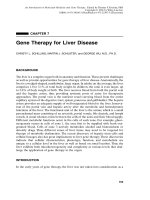
Herpes simplex virus (HSV) is an enveloped double-stranded DNA virus
(see Fig. 1A—
reviewed in ref. 1). The mature virion consists of the following
components:
1. A trilaminar lipid envelope, in which are embedded 10 viral glycoproteins—these
are responsible for several functions including receptor-mediated cellular entry
(2–5).
2. A matrix of proteins, the tegument, which form a layer between the envelope
and the underlying capsid. Functions of the tegument proteins include: induction
of viral gene expression (6–8); shutoff of host protein synthesis immediately
following infection (9–12); virion assembly functions.
3. An icosadeltahedral capsid, typical of the herpesvirus family (13,14).
4. A core of toroidal double-stranded DNA (dsDNA) (14–16).
Viral genes encode the majority of the proteins and glycoproteins of the
mature virion. The HSV genome consists of 152 kb of dsDNA arranged as long
and short unique segments (UL and US) flanked by repeated sequences (ab,
b′a′, ac, c′a′) (17–20). Eighty-four viral genes are encoded, and these may be
classified according to whether their expression is essential for viral replication
in a permissive tissue culture environment (see Fig. 1B). Nonessential genes
often encode functions that are important for specific virus-host interactions
in vivo, for example, immune evasion, replication in nondividing cells or
From: Methods in Molecular Medicine, vol. 76: Viral Vectors for Gene Therapy: Methods and Protocols
Edited by: C. A. Machida © Humana Press Inc., Totowa, NJ
1
2
2
Burton et al.
Fig. 1. A. Schematic depiction of a mature HSV virion illustrating the main components of the virus particle. B. The HSV
genome is organized into unique long and short segments (UL, US) flanked by repeated sequences. The 84 viral open reading
frames can be divided into genes that are essential for replication in a permissive tissue culture environment, and those that are
dispensable. The functions of the nonessential gene products are related to viral interactions with the host in vivo.
HSV Genome
3
shutdown of host protein synthesis. The importance of this observation is that
nonessential genes may be deleted in the generation of gene therapy vectors,
allowing the insertion of exogenous genetic material (21,22). In addition,
deletion of specific accessory genes may limit viral replication to certain
cellular subsets (23–27).
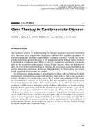
During lytic infection, viral genes are expressed in a tightly regulated,
interdependent temporal sequence (28, 29, reviewed in ref. 1) (see Fig. 2).
Transcription of the five immediate-early (IE) genes, ICP0, ICP4, ICP22,
ICP27, and ICP47 commences on viral DNA entry to the nucleus. Expression
of these genes is regulated by promoters that are responsive to VP16, a viral
structural protein that is transported to the host cell nucleus with the viral
DNA. VP16 is a potent trans-activator that associates with cellular transcription factors and binds to cognate motifs within the IE promoter sequences.
Expression of IE genes initiates a cascade of viral gene expression (see Fig. 2).
Transcription of early (E) genes, which primarily encode enzymes involved in
DNA replication, is followed by expression of late (L) genes mainly encoding
structural components of the virion (28–31, reviewed in ref. 1). Of the IE gene
products, only ICP4 and ICP27 are essential for expression of E and L genes,
and hence viral replication (32–34).
The life cycle of HSV-1 in vivo is illustrated in Fig. 3. Following primary
cutaneous or mucosal inoculation, the virus undergoes lytic replication in the
infected epithelia. Viral particles are released at the site of the primary lesion;
they may enter sensory neurons whose axon terminals innervate the affected
area. The nucleocapsid and tegument are carried by retrograde axonal transport
from the site of entry to the neuronal soma in the dorsal root ganglia or
trigeminal ganglia, where the viral genome and VP16 enter the nucleus (35–37).
At this point, one of two chains of events may ensue. First, the lytic replicative
cycle described above may take place. This pathway results in neuronal cell
death and egress of infectious particles. Alternatively, the viral DNA can
enter the latent state. During latency, the viral genome persists as a stable
episomal element, sometimes for the lifetime of the host (38). The DNA adopts
a chromatin-like structure; it is not extensively methylated (39,40). No IE, E,
or lytic L genes are expressed during latency, but a set of nontranslated RNA
species, the latency-associated transcripts (LATs), is produced and detectable
in the nuclei of latently infected neurons (41–45 and see later). At a timepoint that may be remote from the establishment of latency, alterations in
the host–virus interaction may cause “reactivation” of the viral infection. IE
genes are expressed and the lytic cascade of gene expression follows, resulting
in the production of mature virions. The nucleocapsid and glycoproteins are
transported by separate anterograde axonal transport pathways to the peripheral
nerve terminals, where they are assembled and released (46,47).
4
Burton et al.
Fig. 2. Diagrammatic illustration of the life cycle of wild-type HSV in vivo.
The processes regulating the establishment of and reactivation from latency
are not well understood. The LATs are a hallmark of HSV latency; the major
2.0-kb and 1.5-kb species are abundant, stable, lariat introns that arise by
splicing of a primary transcript (48–53). The functions of the LATs remain
unknown, although several putative roles have been suggested. These include:
efficient establishment of latency (54,55); effective reactivation from latency
(56–62); antisense regulation of IE gene transcripts (63–65); prevention of
apoptosis in infected neurons (66); expression of proteins that compensate for
the absence of IE gene expression during latency (67); and functions relating
to RNA-mediated catalysis (68). However, it is clear that the LAT genes are not
an absolute requirement for establishment, maintenance or reactivation from
latency (69–72). This has important implications for vector construction, as it
is possible to insert transgenes within the LAT loci, disrupting the LAT genes.
This allows use of the LAT cis-acting regulatory sequences, LAP1 (73–80)
HSV Genome
5
Fig. 3. Flowchart showing the tightly regulated cascade of gene expression that occurs
during lytic HSV infection. In order to proceed to viral DNA replication and expression
of structural viral proteins, the two IE genes ICP4 and ICP27 must be expressed. Absence
of either prevents the transcriptional program from progressing to the early phase,
resulting in an abortive infection that resembles latency in many respects.
and LAP2 (73, 80–82), to drive transgene expression (72,83,84), thus allowing
stable long-term expression of therapeutic genes (85–87).
1.2. Using HSV-1 to Make Gene Therapy Vectors
Various aspects of the basic biology of HSV-1 are attractive when considering the design of gene therapy vectors:
1. HSV has a broad host cell range; the cellular entry receptors HveA (88,89) and
HveC (90–93) are widely expressed cell surface proteins of unknown function.
2. HSV is highly infectious—it is possible to transduce 70% of a cell population
in vitro at a low multiplicity of infection (1.0), with a replication-defective
vector (21,94).
3. Nondividing cells may be efficiently transduced and made to express transgenes
(21,84).
6
Burton et al.
4. Of the 84 known viral genes, approximately half are nonessential for growth in
tissue culture. This means that multiple or very large therapeutic transgenes can
be accommodated, by replacing dispensable viral genes (22,95).
5. Recombinant replication-defective HSV-1 may readily be prepared to high titer
and purity without contamination from wild-type recombinants.
6. The latent behavior of the virus may be exploited for the stable long-term
expression of therapeutic transgenes in neurons (84,86,96–98).
7. The abortive gene expression cascade produced when a replication-defective
vector enters a cell results in a state that is similar to latency, the main difference
being that the virus cannot reactivate. This enables chronic transgene expression
in both neuronal and nonneuronal cells (87).
Broadly speaking, there are three ways that the HSV genome may be used to
generate nonpathogenic gene therapy vectors (see Fig. 4).
1.2.1. Conditionally Replicating Vectors
Deletion of some nonessential genes results in viruses that retain the ability
to replicate in vitro, but are compromised in vivo, in a context-dependent
manner (99). For example, deletion of the gene encoding ICP34.5 results in a
virus that may replicate in vitro, but not in neurons in vivo (25,26,100,101).
The virus, however, retains the ability to undergo lytic replication in rapidly
dividing cancer cells. ICP34.5 mutants have been used to treat patients with
brain tumors in phase I clinical trials, in the hope that the virus will destroy the
tumor cells and spare normal brain tissue (102,103). Although these mutants
appear nontoxic at present, it is not yet clear whether this therapeutic strategy
is efficacious.
1.2.2. Replication-Defective Vectors
Deletion of one or other of the essential IE genes (ICP4, ICP27) results in
a virus that cannot replicate (32,34,104–107), except in cells that complement
the null mutations by providing ICP4 or ICP27 in trans (32,105,108). In
appropriate complementing cell lines, the virus replicates similar to wild-type
virus. By using this method, it is possible to prepare high titer viral stocks
that are free from contaminating replication-competent viruses. In addition,
the genetic manipulation of these viruses is straightforward, exploiting the
recombinogenic properties of HSV-1 to introduce exogenous sequences by
homologous recombination (21, 22, and see later). In vivo, these viruses
undergo abortive cascades of lytic gene transcription, resulting in a state
that is very similar to latency. The genomes may persist for long periods
in neuronal and nonneuronal cells, but cannot reactivate in the absence of
the essential IE genes (87,109–111). These vectors may be further refined
HSV Genome
7
Fig. 4. Strategies for using the HSV genome to generate nonpathogenic gene therapy vectors.
7
8
Burton et al.
to prevent cytotoxicity resulting from nonessential IE gene expression (see
later).
1.2.3. Amplicons
The entire viral genome may be supplied in trans, generating particles that
contain very few viral gene sequences. In this instance, the desired transgene
cassette is placed in a plasmid containing the viral genomic packaging/cleavage
signals, in addition to both viral and bacterial origins of replication—an
“amplicon” plasmid (112–115). Defective HSV-like particles are generated
by double transfection of eukaryotic cells with i) the amplicon plasmid and
ii) a bacterial artificial chromosome containing the viral genome, but devoid
of packaging and eukaryotic replication signals (116–118). Concatermerized
plasmid DNA is packaged into disabled particles that contain HSV structural
proteins and surface glycoproteins. The HSV BAC is a recent advance on the
prior practice of using a series of cosmids or a helper virus to supply viral
functions. Although a perceived advantage of the amplicon system is that
no viral coding sequence is delivered, it has proven difficult in practice to a
produce pure preparation of vector with clinically useful yields.
Over the past decade, our laboratory has amassed considerable experience in
the generation, use and propagation of replication-defective vectors. Amplicons
and helper-dependent vectors are dealt with in other chapters in this volume.
The remainder of this chapter describes the replication-defective system and
provides protocols for its use.
1.3. Minimizing Toxicity from Replication-Defective Vectors
Blocking viral replication prevents toxicity associated with lytic wild-type
HSV infection. As E and L gene expression, and therefore replication, is
fully dependent upon the expression of IE genes, generation of replicationincompetent vectors can be accomplished by disruption of one or other essential
IE gene, ICP4 or ICP27. For example, an ICP4 null mutant is unable to
replicate in noncomplementing cells in culture (32). However, the IE gene
products, with the exception of ICP47, are all toxic to host cells (104,107,119).
Infection with an ICP4 null mutant results in extensive cell death in the absence
of viral replication (21,32,104,120). This is caused by overexpression of other
IE gene products, some of which are negatively regulated by ICP4 (32). To
prevent cytotoxicity, a series of vectors has been generated that are multiply
deleted for IE genes. Quintuple mutants, null for ICP0, ICP4, ICP22, ICP27,
and ICP47, have been produced, are entirely nontoxic to cells and the genomes
are able to persist for long periods of time (107). However, vectors grow poorly
in culture and express transgenes at very low levels in the absence of ICP0
(121–125). Retention of the gene encoding the trans-activator ICP0 allows
HSV Genome
9
efficient expression of viral genes and transgenes, and allows the virus to
be prepared to high titer. Recent work has shown that the post-translational
processing of ICP0 in neurons is different to that in glia (126). It appears
that, although ICP0 mRNA is efficiently expressed in both cell types, ICP0
undergoes proteolytic degradation in neurons. It might be predicted that a
vector carrying an intact ICP0 gene would not be toxic to neurons, but may
be advantageous for oncological applications, where ICP0 toxicity may be
desirable. Deletion of ICP47 restores the expression and priming of MHC
class I molecules to the surface of the cells (127–129). This may potentially
confer advantages in gene therapy of malignancy, although the utility of
this modification is unclear at present. For most other applications, where
immune evasion is desirable, triple mutants (ICP4–: ICP22–: ICP27–) have been
used. These vectors show minimal cytotoxicity in vitro and in vivo, are efficient
vehicles for transgene delivery and can be grown efficiently in cells that
complement the absence of ICP4 and ICP27 in trans (21,96,104). The construction of the prototype triple-mutant virus is illustrated in Fig. 5.
1.4. Inserting Transgenes into Replication-Defective Vectors
Insertion of transgenes into the replication-defective HSV vectors is achieved
by homologous recombination in eukaryotic cells in cell culture. The transgene
cassette is inserted into a shuttle plasmid that contains sequence from the
targeted viral locus. In the resulting shuttle vector, the transgene is flanked
either side by 1–2 kb of viral sequence. The plasmid DNA is linearized and
transfected into cells that complement the deleted IE genes from the defective
virus. The cells are cotransfected with viral genomic DNA. Plaques form as
viral genes are expressed and virions are generated. The recombination rate
between linearized plasmid and purified viral DNA ranges from 0.1% to 1%
of the plaques, when the calcium phosphate method is used for the transfection. Virus is prepared from the plaques, and the viral DNA screened for
recombinants.
There are two features that we have built into this system to simplify the
isolation of recombinant plaques:
1. The replication-defective vectors discussed above have been designed to express
reporter genes in certain important loci. Recombination of the transgenic cassette into these loci results in loss of reporter gene activity, which is readily
assayed. This allows rapid screening of plaques for putative reporter-negative
recombinants, which are then subjected to secondary screening by Southern
blot analysis (22,130).
2. The viral DNA may be cleaved at the site of the desired recombination event by
using rare 8-bp recognition site restriction endonucleases that are not present
elsewhere in the HSV vector genome (see Fig. 6). We have engineered unique